Venous Thrombosis:
Risk Factors and Management
Marc Righini, Henri Bounameaux
1Abstract
During the past 2 decades, the diagnostic and thera-peutic approach to deep vein thrombosis (DVT) has greatly evolved. First, the assessment of the clinical probability has gained wide acceptance. Second, novel noninvasive diagnostic tools such as venous compres-sion ultrasonography and D-dimer measurement have become available, drastically reducing the need for in-vasive tools such as phlebography. Third, new antico-agulant drugs, in particular low-molecular-weight heparins (LMWHs), have become available and have made DVT treatment a lot easier by allowing out-of-hospital management.
Several diagnostic algorithms, based on the as-sessment of clinical probability, D-dimer measurement
and venous compression ultrasonography, have shown to be safe in management studies. In addition to im-provements in diagnostic algorithms and anticoagu-lant treatment, compression therapy by elastic stock-ings to diminish the prevalence of the postthrombotic syndrome has been validated in prospective studies.
The dilemma of the need or no need for looking for and treating isolated calf DVT with anticoagulants remains a controversial issue, as do the optimal length and intensity of anticoagulation. In the near future, the emergence of several new, totally synthetic, orally active anticoagulant compounds, such as direct thrombin or factor Xa inhibitors that are presently be-ing tested in clinical studies, could profoundly change the therapeutic approach to DVT.
Tiefe Venenthrombose: Risikofaktoren und Management
ZusammenfassungIn den letzten 2 Jahrzehnten haben Diagnostik und Therapie der tiefen Venenthrombose (TVT) entschei-dende Veränderungen erfahren. Erstens hat die Beur-teilung der klinischen Wahrscheinlichkeit eine breite Akzeptanz gefunden. Zweitens haben sich nichtinva-sive Abklärungsmethoden wie Kompressionsechogra-phie und Bestimmung der D-Dimere durchgesetzt, was die Notwendigkeit einer invasiven Abklärung mit-tels Phlebographie drastisch reduziert hat. Drittens finden neue Formen der Antikoagulation, im Speziellen die niedermolekularen Heparine, breite Anwendung und erleichtern die Behandlung einer TVT, die zurzeit meist ambulant durchgeführt werden kann.
Zahlreiche diagnostische Algorithmen basieren auf klinischer Wahrscheinlichkeit, Bestimmung der
D-Dimere und Kompressionsechographie und haben sich in Studien als sicher erwiesen. Neben dem Fort-schritt der diagnostischen Algorithmen und der Anti-koagulation konnten prospektive Studien die Wich-tigkeit der Kompressionstherapie mittels elastischer Binden zeigen und eine Verminderung der Prävalenz des postthrombotischen Syndroms bestätigen.
Die Diskussion über die Notwendigkeit einer Ab-klärung und Behandlung der TVT des Unterschenkels bleibt kontrovers, ebenso die optimale Dauer und In-tensität der Antikoagulation. In absehbarer Zukunft könnten einige neue, völlig synthetische, oral aktive Antikoagulanzien, wie direkte Antithrombine oder Anti-Faktor Xa, welche kürzlich in klinischen Studien getestet wurden, die therapeutischen Ansätze bei der TVT grundsätzlich verändern. Schlüsselwörter: Tiefe Venenthrombose (TVT) · Klinische Wahr-scheinlichkeit · D-Dimere · Kompressionsultrasono-graphie · Antikoagulation Key Words:
Deep vein throm-bosis · Clinical prob-ability · D-dimer · Lower-limb com-pression ultra-sonography · Anti-coagulation 1 Division of
Angio-logy and Hemo-stasis, Depart-ment of Internal Medicine, Geneva University Hospi-tal and Faculty of Medicine, Geneva, Switzerland. Herz 2007;32:27–34 DOI 10.1007/ s00059-007-2931-1
Introduction
Venous thromboembolism (VTE) and its manifes-tations, including deep vein thrombosis (DVT) and pulmonary embolism (PE), pose a life-threaten-ing health problem for thousands of people each year. Overall, incidence of deep vein thrombosis is 1–2/1,000 persons/year. However, incidence raises exponentially from less than 5 cases/100,000 persons < 15 years of age to 500–1,000 cases/100,000 persons aged 80 years [1]. Approximately one third of
pa-tients with VTE manifest PE, whereas two thirds manifest DVT alone. Symptoms of DVT include swelling of the affected limb, tenderness, erythema, and pain, whereas PE presents as sudden breathless-ness with or without chest pain, or, less frequently, collapse with hemodynamic shock.
During the past 2 decades, the diagnostic and therapeutic approach to DVT has changed in several major ways. First, novel noninvasive diagnostic tools such as venous compression ultrasonography (CUS)
and D-dimer (DD) measurement have become avail-able, drastically reducing the need for invasive tools such as phlebography [2]. Second, new anticoagulant treatment, in particular low-molecular-weight hepa-rins (LMWHs), have become available and have ren-dered the treatment of DVT easier by allowing out-of-hospital treatment. Third, the index of clinical suspicion has progressively become lower, resulting in the fact that the vast majority (80% or even more) of patients with suspected VTE do not have the dis-ease. Fourth, noninvasive strategies have been vali-dated in large-scale outcome studies, and more atten-tion has been paid to their cost implicaatten-tions [3].
Risk Factors for Venous Thromboembolism
To improve survival, avoid recurrences, prevent com-plications and reduce health-care costs, the occurrence of VTE must be reduced. To reduce VTE incidence, persons at risk for VTE must first be identified [4], in order to target prevention. Risk factors for VTE include increasing age, surgery, trauma, hospital or nursing-home confinement, active neoplasia with or without concurrent chemotherapy, central vein catheterization or transvenous pacemaker, prior superficial vein throm-bosis, varicose veins, and neurologic disease with ex-tremity paresis; patients with chronic liver disease have a reduced risk [5]. Hospitalization and nursing-home residence together account for almost 60% of incident VTE events in the community. Thus, hospital confine-ment provides an important opportunity to significantly reduce VTE incidence [6]. Of note, hospitalization for medical illness and hospitalization for general surgery account for almost equal proportions of VTE, empha-sizing the need to provide prophylaxis to both of these risk groups. A recent epidemiologic study suggested that active cancer may account for almost 20% of inci-dent VTE events occurring in the community [4]. The risk appeared to be higher for patients with pancreatic cancer, lymphoma, malignant brain tumors, cancer of the liver, leukemia, and colorectal and other digestive cancers. Cancer patients receiving immunosuppressive or cytotoxic chemotherapy are even at higher risk for VTE [7]. Central venous catheter or transvenous pace-maker are well known risk factors for upper-limb DVT. However, systematic prophylaxis in these situations is not widely accepted [8, 9].Among women, additional risk factors for VTE include oral contraceptive use and hormone therapy, pregnancy and the postpartum period, and therapy with selective estrogen receptor modulators, such as raloxifene. First- and third-generation oral contra-ceptives convey higher risk than second-generation oral contraceptives. Other conditions associated with VTE include heparin-induced thrombocytopenia, myeloproliferative disorders, nephrotic syndrome,
paroxysmal nocturnal hemoglobinuria, thromboan-giitis obliterans (Buerger’s disease), thrombotic thrombocytopenic purpura, Behçet’s syndrome, sys-temic lupus erythematosus, inflammatory bowel dis-ease, homocystinuria, and possibly, hyperhomocys-teinemia. Recently, acute infections of the urinary or respiratory tract in the community setting have also been related with a transient and modest increase in the risk of VTE [10].
Thrombophilias are inherited or acquired abnor-malities associated with an increased risk of venous thromboembolic disease. Before 1993, a heritable cause of thrombophilia was detectable in a relatively small proportion of patients presenting with DVT or PE. Such abnormalities were confined to rare de-ficiencies like antithrombin, protein C or protein S. The recent discovery of other two other prothrom-botic mutations (the factor V Arg506Gln, or factor V Leiden mutation and the prothrombin G20210A mu-tation), quite prevalent (about 5% up to 30% in some part of Sweden) in Caucasians, has highlighted a ge-netic-mediated basis of VTE [11, 12].
Diagnosis of Deep Vein Thrombosis
Clinical Probability Assessment
Sensitivity and specificity of clinical symptoms and signs in suspected DVT are low when considered sin-gly. Nevertheless, clinicians can combine these find-ings effectively along with elements of personal and family history either implicitly (empirically) or by prediction rules or scores in order to classify patients according to their probability of having the disease, the so-called prior clinical probability [13].
In the setting of suspected DVT, the Wells’ score [14] has gained relatively wide acceptance in spite of its partial subjectivity (Table 1). It is probably not more accurate than the simple implicit evaluation [13]. However, it is easier to teach to junior physicians and allows discussing occasional disagreements among experienced clinicians on explicit grounds. Nevertheless, both the implicit assessment and the explicit Wells’ score allow usefully categorizing pa-tients in low, intermediate, or high clinical probability groups in which the prevalence of DVT is approxi-mately 5%, 20%, and 80%, respectively [15]. Most patients with suspected DVT have a low or interme-diate clinical probability of having the disease. These patients can usually be investigated by entirely nonin-vasive algorithms, which is an important advantage resulting from the evaluation of clinical probability.
DD Measurement
DD is a degradation product of cross-linked fibrin and its level increases in plasma of patients with acute
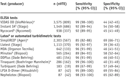
VTE. DD, when assayed by a quantitative enzyme-linked immunosorbent assay (ELISA) or by some automated turbidimetric assays, has been shown highly sensitive (> 98%) in acute DVT, usually at a cutoff value of 500 µg/l [16]. Hence, a DD level below this value reasonably rules out acute DVT, at least in patients with a low or intermediate prior clinical probability [16]. Table 2 summarizes the pooled anal-ysis of the published literature on most commercially available rapid DD tests. Tests vary with respect to several characteristics, including assay technique (ELISA, latex, or automated turbidimetric tests), specificity of monoclonal antibodies used, calibra-tors, and quantitative or semiquantitative results.
In summary, highly sensitive DD assays allow ex-clusion of DVT in outpatients with a low or intermedi-ate clinical probability, while whole-blood agglutina-tion tests only rule out the disease in patients with a low clinical probability [17]. Finally, DD is unlikely to be useful in very elderly patients or in hospitalized pa-tients with suspected VTE [18] because of diminishing specificity of the measurement with increasing age.
Venous CUS
In studies using phlebography as gold standard, low-er-limb venous CUS, an entirely noninvasive test, has a sensitivity of 97% (95% confidence interval [CI]: 96–98%) and a specificity of 98% for symptomatic proximal DVT [2]. It has become the cornerstone of DVT diagnosis in clinically suspected individuals [19, 20]. The single well-validated diagnostic criterion for DVT on CUS is absence of full compressibility of the deep vein when applying pressure through the ultra-sound probe. Intraluminal venous ultrasonography has been reported to have variable accuracy, and nei-ther changes in venous diameter during Valsalva ma-neuver nor assessment of Doppler flow have been found to improve diagnostic accuracy for DVT [2].
Proximal Versus Proximal and Distal CUS
The observation of a very low 3-month thromboem-bolic risk in patients with a negative CUS of the proxi-mal veins and with clinically suspected DVT (around 1% in management studies [13, 21–24]) questions the need for diagnosing so-called calf (or distal, infrapop-liteal) DVT, at least in non-high clinical probability patients. Indeed, in such patients, detecting clots in the posterior tibial or peroneal veins or even in calf muscle veins may be double-edged: on the one hand, the potential of reducing the 3-month thromboem-bolic risk is limited (because it is already quite low), and, on the other hand, the risk of false-positive find-ings and subsequent unnecessary anticoagulant treat-ment in patients who could be left untreated, is quitehigh. Nevertheless, recent large series suggest that complete examination of the leg deep vein system without any other exam is safe and effective in man-aging patients with clinically suspected DVT [25–27]. For example, Elias et al. examined 623 patients of whom 401 (64.4%) were declared without DVT and were followed up for 3 months with a low thrombo-embolic risk of 0.5% (95% CI: 0.1–1.8%). However, among the 204 patients with DVT, 92 (45%) were dis-tal and most of them would not have been treated (and submitted to the hazards of anticoagulant treat-ment) with a similarly low 3-month thromboembolic Items Points
Cancer + 1
Paralysis or recent immobilization + 1
Bedridden > 3 days, or surgery/trauma < 4 weeks + 1
Pain on palpation of the deep veins + 1
Edema of thigh and calf + 1
Pitting edema (symptomatic side only) + 1
Dilated superficial veins (symptomatic side only) + 1 Alternative diagnosis at least as likely as DVT – 2
Clinical probability
Low ≤ 0
Intermediate 1–2
High ≥ 3
Table 1. The Wells’
score for assess-ment of prior clinical probabili-ty of deep vein thrombosis (DVT). Tabelle 1. Kli-nische Wahr-scheinlichkeit ei-ner tiefen Ve-nenthrombose (DVT): Wells-Score.
Table 2. Performances of various D-dimer (DD) assays for diagnosing venous
thromboembolism (VTE). Results are almost identical for suspected deep vein thrombosis and for suspected pulmonary embolism. CI: confidence interval.
Tabelle 2. D-Dimer-(DD-)Bestimmung für die Diagnose der venösen
Thromboem-bolie (VTE). Die Resultate für den Verdacht auf tiefe Venenthrombose und Lun-genembolie sind annähernd identisch. CI: Konfidenzintervall.
Test (producer) n (nVTE) Sensitivity Specificity [% (95% CI)] [% (95% CI)] ELISA tests
VIDAS DD (bioMérieux)a 3,575 (809) 99 (98–100) 44 (42–45)
Instant IAb (Stago) 1,549 (686) 92 (89–94) 54 (50–58)
Nycocardb (Nycomed) 938 (337) 92 (89–95) 45 (41–49)
Latexb or automated turbidimetric tests
SimpliREDb (Agen)a 2,303 (567) 85 (82–88) 69 (66–71)
Liatest (Stago) 1,113 (370) 95 (92–97) 39 (36–43)
MDA (Organon Tecnika) 642 (133) 96 (91–99) 46 (41–51)
Minutexb (Biopool) 565 (264) 89 (84–93) 58 (52–64)
DDPlus (Dade Behring) 481 (154) 96 (91–99) 46 (40–51)
Tinaquant (Boehringer Mannheim)a 286 (162) 99 (96–100) 40 (31–49)
Turbiquant (Dade Behring) 183 (19) 89 (67–99) 57 (49–64)
LPIA D-Dimer (Mitsubishi) 87 (42) 95 (89–100) 69 (55–84)
Nephelotex (Biopool) 87 (42) 98 (93–100) 65 (62–89)
a outcome studies available b semiquantitative test
risk if only a proximal CUS had been performed [25]. Schellong et al. reported a similar 3-month thrombo-embolic risk of 0.3% in a series of 1,646 patients of whom 275 were positive for DVT but, again, 154 DVTs (56%) were distal, suggesting a risk of over-diagnosis and overtreatment [26]. Table 3 offers a comparison of the results obtained with the simpli-fied or complete lower-limb venous CUS.
Ascending Phlebography
Ascending phlebography is still considered the diag-nostic standard for diagnosing DVT but it is invasive, costly, and not devoid of risk. It consists in the injec-tion of iodinated contrast dye in a superficial foot vein with sequential radiograms of the leg in order to follow the dynamic course of the contrast in the veins. Tourniquets can be used to force the dye into the deep veins but their use is controversial. The docu-mentation is adequate when images are obtained in different views, and a filling defect surrounded by contrast is characteristic of a fresh thrombus.
Sequential Diagnostic Strategies for
Suspected DVT
Several noninvasive strategies have been reported to have a high sensitivity for diagnosing DVT [3]. Some rely on serial proximal CUS of proximal veins (com-mon femoral, superficial femoral, and popliteal veins). The rationale for repeating CUS after 1 week in pa-tients in whom it was initially negative is the detection of the rare proximal extension of the distal DVTs that were not searched for. Since the yield of repeat CUS after 1 week is very low [14, 21, 22], CUS has been
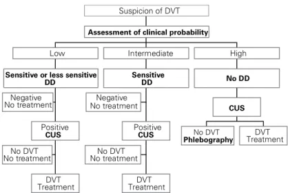
combined with a clinical prediction rule and/or DD measurement to lower the number of necessary repeat compression sonograms [14, 21, 22, 28, 29]. In another strategy, single CUS was restricted to patients with a DD concentration above a critical cutoff level (500 µg/ l), thereby avoiding about 30% of CUS exams [13]. All these strategies are associated with a low (about 2% or less) 3-month thromboembolic risk, which is similar to that observed in suspected patients left untreated fol-lowing a normal phlebogram [30]. In a randomized study, Wells et al. compared the clinical outcome in 530 patients who underwent CUS (control group) and 566 patients who underwent DD testing followed by CUS unless the DD test was negative and the patient was considered at low clinical probability [31]. The 3-month thromboembolic risk among patients in whom the initial diagnostic strategy had ruled out DVT was 0.4% in the DD group, compared with 1.4% in the control group. The difference did not reach sta-tistical significance but the use of DD resulted in a highly significant reduction in the use of CUS, since 39% of patients did not require ultrasound imaging. Overall, modern diagnostic approach to DVT is based upon evaluation of clinical probability, DD measure-ment and CUS. Figure 1 suggests a possible diagnostic algorithm for suspected DVT and the interested read-er may find a complete description of all diagnostic strategies used in the literature in a recent review [32].
Treatment of Deep Vein Thrombosis
Medical Treatment
The aims of treatment are to relieve symptoms, re-duce the risk of PE, and prevent postthrombotic syn-drome (PTS) and recurrent VTE. Anticoagulation is Table 3. Comparison of diagnostic strategies including proximal only or proximal and distal compression ultrasonography
(CUS) for diagnosing deep vein thrombosis (DVT; outcome studies).
Tabelle 3. Vergleich verschiedener diagnostischen Strategien mit ausschließlich proximaler oder kompletter (proximaler +
distaler) Kompressionsultrasonographie (CUS) für die Diagnose der tiefen Venenthrombose (DVT; pragmatische Studien).
Series Patients Prevalence Proportion Number of CUS 3-month thrombo-(n) of DVT (%) of distal performed per embolic risk
DVT (%) 100 patients (n) [% (95% CI)]a
Proximal CUS only
Cogo et al. [21] 1,702 24 – 176 0.7 (0.3–1.2)
Bernardi et al. [22] 946 28 – 109 0.4 (0–0.9)
Wells et al. [14] 593 16 – 128 0.6 (0.1–1.8)
Perrier et al. [13] 474 24 – 73 2.6 (0.2–4.9)
Kraaijenhagen et al. [24] 1,756 22 – 121 0.6 (0.1–1.6)
Proximal and distal CUS
Elias et al. [25] 623 36 45 100 0.5 (0.1–1.8)
Schellong et al. [26] 1,646 17 56 100 0.3 (0.1–0.8)
Stevens et al. [27] 445 14 31 100 0.8 (0.2–2.3)
VTE because UFH is much less expensive than LMWH, at least in some countries.
Thrombolysis and Invasive Treatment
Systematically administered or catheter-directed thrombolysis should not be routinely used in patients with lower-limb DVT and should be confined to pa-tients requiring limb salvage (phlegmasia coerulea dolens). Catheter extraction, fragmentation or surgi-cal thrombectomy should not be used in routine for the treatment of DVT [37]. However, these options may be discussed in young patients with massive iso-lated iliofemoral DVT at risk of limb gangrene secon-dary to massive, proximal venous occlusion.
Duration and Intensity of Anticoagulant
Long-Term Treatment
The duration of anticoagulant treatment following a venous thromboembolic episode has always been a matter of debate with recommended durations fol-lowing a first episode varying between 3 months and 1 year, depending upon the weight given to the re-spective risks of recurrence and bleeding induced by the treatment [38]. Since VTE is a recurrent disease, anticoagulation should ideally last forever. However, the risk of recurrence diminishes with time and the risk of bleeding, after an initial peak, is rather con-stant over time. Thus, there must be some time point the mainstay of treatment of DVT. The initial
treat-ment of acute DVT is based upon administration of LMWH or unfractionated heparin (UFH).
The Seventh ACCP (American College of Chest Physicians) Consensus on Antithrombotic and Thrombolytic Therapy recommends treating patients with a high clinical probability of DVT while awaiting the results of diagnostic tests (grade 1C+). However, as the prevalence of DVT in patients with an interme-diate clinical probability of DVT may be as high as 20–30%, a more appropriate option would be to be-gin anticoagulation while awaiting outcome of diag-nostic tests also in patients with an intermediate clini-cal probability of DVT. In most cases, warfarin can be started on the 1st day. LMWH or UFH should be continued for at least 5 days and until the Interna-tional Normalized Ratio (INR) is within the thera-peutic range (i.e., between 2 and 3) on at least two dosages at an interval of 24 h.
LMWH has become the standard of care for the initial treatment of DVT as it is as effective and safe as UFH but is more practical to use and has a more favorable side effects’ profile. Many available LMWHs may be used once or twice daily. Contrarily to UFH, laboratory monitoring is not mandatory in the vast majority of patients treated with LMWHs. Some ex-ceptions are patients with renal impairment, obesity and pregnancy. As LMWHs are excreted by the kid-ney, they should not be used in patients with a creati-nine clearance of < 30 ml/min. In these patients using UFH is recommended.
The predictable anticoagulant response of LMWHs allows out-of-hospital treatment in more than 80% of patients with acute DVT. Outpatient therapy offers reduced economic costs to patients and the health-care system as a result of decreased resource utilization. Once or twice daily dosing and fixed dosages combined with a well-planned program of patient education and professional support offer convenience to patients while enhancing successful adherence to home treatment. In 1996, the results of a meta-analysis of 20 randomized controlled studies comparing multiple clinical outcomes of LMWH and UFH [33] showed that treatment with LMWH had a more favorable benefit-to-risk ratio than treatment with UFH. More recently, two other meta-analyses showed that treatment with LMWH is at least as ef-fective as UFH in terms of risk of major bleeding, PE, and recurrent DVT [34, 35]. The safety and efficacy profiles of LMWHs, such as dalteparin, enoxaparin, nadroparin, logiparin and tinzaparin, are well estab-lished. Recently, Kearon et al. demonstrated that fixed-dose weight-adjusted subcutaneous UFH was as effective and safe as LMWH in patients with acute VTE and was suitable for outpatient treatment [36], a finding that might impact the management of acute
Negative No treatment No DVT No treatment DVT Treatment Positive CUS
Sensitive or less sensitive DD Low Negative No treatment No DVT No treatment DVT Treatment Positive CUS Sensitive DD Intermediate No DVT Phlebography TreatmentDVT CUS No DD High
Assessment of clinical probability
Suspicion of DVT
Figure 1. Diagnostic algorithm for clinically suspected deep vein thrombosis (DVT).
Sensitive D-dimer assay means at least 95% sensitivity for the presence of DVT. CUS: lower limb venous compression ultrasonography; DD: D-dimer measure-ment.
Abbildung 1. Diagnostischer Algorithmus bei klinischem Verdacht auf tiefe
Ve-nenthrombose (DVT). Ein sensitiver D-Dimer-Test bedeutet eine Sensitivität von mindestens 95% für die Anwesenheit einer DVT.
for which the bleeding risk exceeds the protective ef-fect of the treatment, which should then be stopped [37].
As already mentioned, there is no consensus for duration of anticoagulation for all situations clini-cians may be faced with. In practice, after an initial course of 3–12 months of conventional-intensity war-farin, the prolongation of anticoagulation treatment may be tailored on an individual basis. Some indica-tions for long-term anticoagulation are active neopla-sia, antiphospholipid syndrome with previous VTE and persistent antiphospholipid antibodies, and some congenital thrombophilias as severe antithrombin or protein C deficiency [37]. Maintaining long-term an-ticoagulation may be discussed as well in presence of two or more previous idiopathic VTEs, even if the evidence supporting this attitude are scarce. Obvi-ously, the necessity of maintaining long-term antico-agulation should be reassessed annually, in particular to weight the possibility of new clinical events associ-ated with an increased hemorrhagic risk, which could limit the benefit-risk ratio of long-term anticoagulant treatment. The option of prolonging the treatment with low-intensity warfarin (for example INR be-tween 1.5–2) may be useful at least in selected pa-tients at higher risk of bleeding [39, 40]. A shorter period of anticoagulation (3 months) is accepted for VTE occurring after a transient risk factor like sur-gery or trauma [37].
Compression Treatment of DVT in the Acute
and Chronic Phases
The PTS develops in 20–50% of patients after symp-tomatic DVT and is the most common long-term complication of DVT [41–43]. Patients with PTS ex-perience pain, heaviness, swelling, cramps, itching or tingling in the affected limb. Symptoms may be present in various combinations and may be persis-tent or intermitpersis-tent. Symptoms are usually aggra-vated by standing or walking and improve with rest-ing, leg elevation any lying down. In severe cases, skin changes and ulcers may develop. The frequency of PTS is likely to be reduced by preventing DVT with the use of effective thromboprophylaxis in high-risk patients and settings. However, once DVT has developed, the use of compression stockings for 2 years after DVT appears to reduce the incidence and severity of PTS.
To date, three trials have evaluated the effec-tiveness of long-term use of elastic stockings for the prevention of PTS after acute DVT. The first study by Brandjes et al. randomly allocated 194 patients to daily use of a 30- to 40-mmHg knee-length elastic compression stockings for at least 2 years or no stockings [44]. Use of elastic compression stockings
resulted in a decrease from 47% to 20% of mild/ moderate PTS and a decrease from 23% to 11% of severe PTS. A second study by Ginsberg et al. com-pared the effect of active stockings to sham stock-ings to prevent PTS after proximal DVT in patients who had venous valvular incompetence but no clini-cal PTS 1 year after the index event [45]. This study showed no benefit of elastic stockings as none (0%) of the 24 patients allocated to the active stockings developed PTS, compared with one (4.4%) of 23 pa-tients treated with sham stockings (p = 0.49). An-other trial by Prandoni et al. evaluated the effective-ness of 30- to 40-mmHg elastic stockings versus no stockings in 180 patients after a first symptomatic proximal DVT [46]. The 2-year cumulative inci-dence of PTS, evaluated by using the Villalta’s score was 25% in the stocking group compared with 49% in the control group (p = 0.011). Lastly, a meta-ana-lysis pooled these three studies and reported an overall 54% relative risk reduction in PTS with the use of stockings [47]. Even if the lack of blinding in the two positive studies reduces the confidence in the results as scores used for the evaluation of PTS are highly subjective and even if some questions such as the optimal length and strength of compres-sion remain unanswered, initiation of comprescompres-sion therapy has to be considered an important point in the care of acute DVT [48].
Treatment of Calf DVT
In spite of the reassuring data obtained from the out-come studies using proximal CUS (Table 3), recent consensus conferences, including that of the ACCP, still recommend to treat calf DVT with anticoagu-lants for a period of 3 months [37]. Nevertheless, opt-ing for a 3-month anticoagulant treatment in the presence of a distal DVT raises several problems. First, series based on serial ultrasonography indicate that only a small fraction of distal DVTs extends proximally in medical outpatients. Indeed, the rate of proximal DVTs detected by the repeated ultrasound varies from 0.9% to 5.7% [14, 21, 22, 24]. Second, the randomized DOTAVK study showed a similar safety of an anticoagulant treatment of 6 or 12 weeks for distal DVT, suggesting that a shorter period of anti-coagulation (6 weeks) would be safe [49]. Third, mus-cle vein thromboses (i.e., gemellar and soleal vein thrombosis) are probably less dangerous than throm-bosis of the deep distal veins (i.e., peroneal and tibial posterior veins). Macdonald et al. showed, in a pro-spective study where muscular thromboses were not treated but followed by ultrasonography, that only 3% of muscular thromboses extended in the popliteal vein [50]. This suggests that the vast majority of mus-cular vein thromboses need either no anticoagulation
or a shorter period of anticoagulation. Fourth, in studies using proximal and distal CUS, half of detect-ed thromboses were distal (Table 3) and a risk of overtreatment should not be neglected. Therefore, the 1A grade of recommendation delivered by the ACCP consensus conference suggesting a similar treatment for proximal DVTs and calf DVTs [37] seems not to be supported by the available evidence.
Conclusion and Perspectives
Venous thromboembolic disease encompasses a wide spectrum of conditions with various severity and prog-nostic implications. DVT is an important cause of mor-bidity and mortality, and as the awareness of this pa-thology has increased during the last 2 decades, the prevalence of the disease in suspected populations has dramatically decreased. Recent studies have shown that algorithms combining simple diagnostic tests may provide a safe diagnostic approach to suspected DVT, with a 3-month thromboembolic risk similar to or low-er than that reported aftlow-er a normal phlebography, the accepted gold standard. Assessment of clinical proba-bility, done empirically or by a prediction rule, has shown to be highly useful and allows identifying pa-tients with a low prevalence of the disease, who can be investigated by noninvasive diagnostic tools. Plasma DD measurements have been widely validated and al-low to rule out the disease with an acceptable safety in selected patients. CUS has become the cornerstone diagnostic test for DVT and its diagnostic performanc-es are excellent in the case of symptomatic proximal DVTs. Many algorithms combining in various order these last three tests have been validated, and may be safely used in clinical practice. In spite of these advanc-es, the field faces several challenges for the future. One challenge will consist in establishing which conditions need to be treated with anticoagulant drugs, a question that pertains essentially to calf or muscle vein throm-bosis. Its answer will have consequences for diagnostic algorithms.
As a matter of fact, LMWHs have considerably simplified the initial management of DVT and al-lowed out-of-hospital treatment. However, their por-cine origin is still a concern. Several new, totally syn-thetic, orally active anticoagulant compounds, such as direct thrombin or factor Xa inhibitors, are pres-ently being tested in clinical studies and could pro-foundly modify DVT treatment in the near future.
Interessenkonflikt: Es besteht kein Interessenkonflikt.
Die Autoren versichern, dass sie keine finanziellen oder persönlichen Beziehungen zu Dritten haben, deren Inter-essen das Manuskript positiv oder negativ beeinflusst haben könnten.
References
1. Oger E. Incidence of venous thromboembolism: a commu-nity-based study in Western France. EPI-GETBP Study Group. Groupe d’Etude de la Thrombose de Bretagne Occi-dentale. Thromb Haemost 2000;83:657–60.
2. Kearon C, Ginsberg JS, Hirsh J. The role of venous ultraso-nography in the diagnosis of suspected deep venous thrombosis and pulmonary embolism. Ann Intern Med 1998;129:1044–9.
3. Perrier A, Bounameaux H. Cost-effective diagnosis of deep vein thrombosis and pulmonary embolism. Thromb Hae-most 2001;86:475–87.
4. Heit JA. Venous thromboembolism: disease burden, out-comes and risk factors. J Thromb Haemost 2005;3:1611–7. 5. Michota F. Venous thromboembolism: epidemiology,
charac-teristics, and consequences. Clin Cornerstone 2005;7:8–15. 6. White RH. The epidemiology of venous thromboembolism.
Circulation 2003;107:I4–8.
7. Mousa SA. Anti-thrombotics in thrombosis and cancer. Fu-ture Oncol 2005;1:395–403.
8. Baarslag HJ, Koopman MM, Hutten BA, et al. Long-term follow-up of patients with suspected deep vein thrombosis of the upper extremity: survival, risk factors and post-throm-botic syndrome. Eur J Intern Med 2004;15:503–7.
9. Agnelli G, Verso M. Is antithrombotic prophylaxis required in cancer patients with central venous catheters? No. J Thromb Haemost 2006;4:14–5.
10. Smeeth L, Cook C, Thomas S, et al. Risk of deep vein throm-bosis and pulmonary embolism after acute infection in a community setting. Lancet 2006;367:1075–9.
11. Bauer KA. Management of thrombophilia. J Thromb Hae-most 2003;1:1429–34.
12. Rosendaal FR, Bovill EG. Heritability of clotting factors and the revival of the prothrombotic state. Lancet 2002;359:638–9. 13. Perrier A, Desmarais S, Miron MJ, et al. Non-invasive
diag-nosis of venous thromboembolism in outpatients. Lancet 1999;353:190–5.
14. Wells PS, Anderson DR, Bormanis J, et al. Value of assess-ment of pretest probability of deep-vein thrombosis in clinical management. Lancet 1997;350:1795–8.
15. Righini M, Le Gal G, Perrier A, et al. Effect of age on the as-sessment of clinical probability of pulmonary embolism by prediction rules. J Thromb Haemost 2004;2:1206–8. 16. Stein PD, Hull RD, Patel KC, et al. D-dimer for the exclusion
of acute venous thrombosis and pulmonary embolism: a systematic review. Ann Intern Med 2004;140:589–602. 17. Ginsberg JS, Wells PS, Kearon C, et al. Sensitivity and specificity
of a rapid whole-blood assay for D-dimer in the diagnosis of pulmonary embolism. Ann Intern Med 1998;129:1006–11. 18. Righini M, Goehring C, Bounameaux H, et al. Effects of age
on the performance of common diagnostic tests for pul-monary embolism. Am J Med 2000;109:357–61.
19. Kearon C, Julian JA, Newman TE, et al. Noninvasive diagno-sis of deep venous thrombodiagno-sis. McMaster Diagnostic Imag-ing Practice Guidelines Initiative. Ann Intern Med 1998; 128:663–77.
20. Keeling DM, Mackie IJ, Moody A, et al. The diagnosis of deep vein thrombosis in symptomatic outpatients and the po-tential for clinical assessment and D-dimer assays to re-duce the need for diagnostic imaging. Br J Haematol 2004; 124:15–25.
21. Cogo A, Lensing AW, Koopman MM, et al. Compression ul-trasonography for diagnostic management of patients with clinically suspected deep vein thrombosis: prospec-tive cohort study. BMJ 1998;316:17–20.
22. Bernardi E, Prandoni P, Lensing AW, et al. D-dimer testing as an adjunct to ultrasonography in patients with clinically
suspected deep vein thrombosis: prospective cohort study. The Multicentre Italian D-dimer Ultrasound Study Investi-gators Group. BMJ 1998;317:1037–40.
23. Wells PS, Hirsh J, Anderson DR, et al. Accuracy of clinical assess-ment of deep-vein thrombosis. Lancet 1995;345:1326–30. 24. Kraaijenhagen RA, Piovella F, Bernardi E, et al. Simplification
of the diagnostic management of suspected deep vein thrombosis. Arch Intern Med 2002;162:907–11.
25. Elias A, Mallard L, Elias M, et al. A single complete ultra-sound investigation of the venous network for the diag-nostic management of patients with a clinically suspected first episode of deep venous thrombosis of the lower limbs. Thromb Haemost 2003;89:221–7.
26. Schellong SM, Schwarz T, Halbritter K, et al. Complete com-pression ultrasonography of the leg veins as a single test for the diagnosis of deep vein thrombosis. Thromb Hae-most 2003;89:228–34.
27. Stevens SM, Elliott CG, Chan KJ, et al. Withholding antico-agulation after a negative result on duplex ultrasonogra-phy for suspected symptomatic deep venous thrombosis. Ann Intern Med 2004;140:985–91.
28. Bounameaux H, Perrier A. Compression ultrasonography for diagnosing deep vein thrombosis. Repeat testing is un-justified. BMJ 1998;316:1534–5.
29. Tick LW, Ton E, van Voorthuizen T, et al. Practical diagnostic management of patients with clinically suspected deep vein thrombosis by clinical probability test, compression ultraso-nography, and D-dimer test. Am J Med 2002;113:630–5. 30. Hull R, Hirsh J, Sackett DL, et al. Clinical validity of a negative
venogram in patients with clinically suspected venous thrombosis. Circulation 1981;64:622–5.
31. Wells PS, Anderson DR, Rodger M, et al. Evaluation of D-di-mer in the diagnosis of suspected deep-vein thrombosis. N Engl J Med 2003;349:1227–35.
32. Goodacre S, Stevenson M, Wailoo A, et al. How should we diagnose suspected deep-vein thrombosis? Q J Med 2006; 99:377–88.
33. Leizorovicz A. Comparison of the efficacy and safety of low molecular weight heparins and unfractionated heparin in the initial treatment of deep venous thrombosis. An up-dated meta-analysis. Drugs 1996;52:Suppl 7:30–7. 34. Gould MK, Dembitzer AD, Doyle RL, et al.
Low-molecu-lar-weight heparins compared with unfractionated hepa-rin for treatment of acute deep venous thrombosis. A me-ta-analysis of randomized, controlled trials. Ann Intern Med 1999;130:800–9.
35. Siragusa S, Cosmi B, Piovella F, et al. Low-molecular-weight heparins and unfractionated heparin in the treatment of patients with acute venous thromboembolism: results of a meta-analysis. Am J Med 1996;100:269–77.
36. Kearon C, Ginsberg JS, Julian JA, et al. Comparison of fixed-dose weight-adjusted unfractionated heparin and
low-molecular-weight heparin for acute treatment of ve-nous thromboembolism. JAMA 2006;296:935–42. 37. Buller HR, Agnelli G, Hull RD, et al. Antithrombotic therapy
for venous thromboembolic disease: the Seventh ACCP Conference on Antithrombotic and Thrombolytic Therapy. Chest 2004;126:Suppl:401S–28S.
38. Agnelli G, Prandoni P, Santamaria MG, et al. Three months versus one year of oral anticoagulant therapy for idiopathic deep venous thrombosis. Warfarin Optimal Duration Ital-ian Trial Investigators. N Engl J Med 2001;345:165–9. 39. Ridker PM, Goldhaber SZ, Glynn RJ. Low-intensity versus
conventional-intensity warfarin for prevention of recur-rent venous thromboembolism. N Engl J Med 2003;349: 2164–7, author reply 2164–7.
40. Ridker PM, Goldhaber SZ, Danielson E, et al. Long-term, low-in-tensity warfarin therapy for the prevention of recurrent ve-nous thromboembolism. N Engl J Med 2003;348:1425–34. 41. Prandoni P, Lensing AW, Cogo A, et al. The long-term clinical
course of acute deep venous thrombosis. Ann Intern Med 1996;125:1–7.
42. Kahn SR, Ginsberg JS. Relationship between deep venous thrombosis and the postthrombotic syndrome. Arch Intern Med 2004;164:17–26.
43. Schulman S, Lindmarker P, Holmstrom M, et al. Post-throm-botic syndrome, recurrence, and death 10 years after the first episode of venous thromboembolism treated with warfarin for 6 weeks or 6 months. J Thromb Haemost 2006; 4:734–42.
44. Brandjes DP, Buller HR, Heijboer H, et al. Randomised trial of effect of compression stockings in patients with symptom-atic proximal-vein thrombosis. Lancet 1997;349:759–62. 45. Ginsberg JS, Hirsh J, Julian J, et al. Prevention and treatment
of postphlebitic syndrome: results of a 3-part study. Arch Intern Med 2001;161:2105–9.
46. Prandoni P, Lensing AW, Prins MH, et al. Below-knee elastic compression stockings to prevent the post-thrombotic syndrome: a randomized, controlled trial. Ann Intern Med 2004;141:249–56.
47. Kolbach DN, Sandbrink MW, Neumann HA, et al. Compres-sion therapy for treating stage I and II (Widmer) post-throm-botic syndrome. Cochrane Database Syst Rev 2003;4: CD004177.
48. Kahn SR. The post-thrombotic syndrome: progress and pit-falls. Br J Haematol 2006;134:357–65.
49. Pinede L, Ninet J, Duhaut P, et al. Comparison of 3 and 6 months of oral anticoagulant therapy after a first episode of proximal deep vein thrombosis or pulmonary embolism and comparison of 6 and 12 weeks of therapy after isolated calf deep vein thrombosis. Circulation 2001;103:2453–60. 50. Macdonald PS, Kahn SR, Miller N, et al. Short-term natural
history of isolated gastrocnemius and soleal vein thrombo-sis. J Vasc Surg 2003;37:523–7.
Address for Correspondence Marc Righini, MD Division of Angiology and Hemostasis Geneva University Hospital 24, rue Micheli-du-Crest 1211 Geneva 14 Switzerland Phone (+41/22) 372-9294, Fax -9299 e-mail: Marc.Righini@ hcuge.ch