© 1998 Society for In Vitro Biology 1071-2690/98 $05.00 + 0.00
AN I M P R O V E D M E T H O D F O R ISOLATION OF MICROVASCULAR E N D O T H E L I A L CELLS
F R O M NORMAL A N D I N F L A M E D H U M A N LUNG
JIN NING LOU, ~ NABIL MILl, CHRISTINE DECRIND, YVES DONATI, SYLVIE KOSSODO, ANASTASE SPILIOPOULOS, BARA RICOU, PETER M. SUTER, DENIS R. MOREL, PHILIPPE MOREL, AND GEORGES E. GRAU
Division of Anesthesiological Investigations (J. N. L., N. M., C. D., E D, S. K., B. R., P. M. S., D. R. M., G. E. G.), Division of Surgical Intensive Care, Clinics of Cardiovascular and Thoracic Surgery (A. S.), and Division of Investigative Surgery (J. N. L., P. M.), Department
of Surgery, University Hospital and University Medical Center, CH-1211 Geneva 4, Switzerland (Received 22 April 1997; accepted 20 January 1998)
SUMMARY
Microvascular endothelial cells (MVEC), which differ from large vessel endothelial ceils, have been isolated successfully from lungs of various species, including man. However, contamination by nonendothelial cells remains a major problem in spite of several technical improvements. In view of the organ specificity of MVEC, endothelial cells should be derived from the tissue involved in the diseases one wishes to study. Therefore, to investigate some of the immunopathological mechanisms leading to acute respiratory distress syndrome (ARDS), we have attempted to isolate lung MVEC from patients undergoing thoracic surgery for lung carcinoma and patients dying of ARDS. The method described here includes four main steps: (1) full digestion of pulmonary tissue with trypsin and collagenase, (2) aggregation of MVEC induced by human plasma, (3) Percoll density centrifugation, and (4) selection and transfer of MVEC after local digestion with trypsin/EDTA under light microscopy. Normal and ARDS-derived lung MVEC purified by this technique presented contact inhibition (i.e., grew in monolayer), and expressed classical endothelial markers, including yon Willebrand factor (vWF), platelet endothelial cell adhesion molecule I(PECAM-1, CD31), and transcripts for the angiotensin converting enzyme (ACE). The cells also formed capillarylike structures, took up high levels of acetylated low-density lipoprotein (Ac-LDL), and exhibited ELAM-1 indu- cibility in response to TNF. Contaminant cells, such as fibroblasts, smooth muscle cells, or pericytes, were easily recognized on the basis of morphology and were eliminated by selection of plasma-aggregated cells under light microscopy. The technique presented here allows one to study the specific involvement and contribution of pulmonary endothelium in various lung diseases.
Key words: lung microvascular endothelial cell; vWF; CD31; LDL; ACE; ELAM-1.
INTRODUCTION
Since the endothelial cell culture was developed in 1973 (19), the awareness has grown that these cells play active roles in homeostasis and pathology. In particula, microvascular endothelial cells (MVEC) have been shown to have more physiological and pathological sig- nificance than large vessel endothelial cells (LVEC) [for review see Scott and Bicknell (29)]. In physiological conditions, MVEC are cru- cial in modulating metabolisms and tissue functions, while in patho- logical conditions, they are central to the process of inflammation (31). MVEC have been successfully isolated from almost all organs and tissues (29). MVEC differ from LVEC by various morphological and functional variables (14). Furthermore, endothelial cells from arterial origin are different from those of venous origin (1). These data suggest that LVEC may not be adequate for the study of patho- logical events occurring in microvessels. Moreover, MVEC derived from various organs also differ in some characteristic (2). Indeed, MVEC derived from different areas of the microcirculation exhibit differential adhesive properties for granulocyte (23).
~To whom correspondence should be addressed at Division of Investigative Surgery, Department of Surgery, University Hospital, University of Geneva, 24, rue Micheli-du-crest 1211, Geneva 14, Switzerland.
In view of the organ specificity of MVEC, endothelial cells should be derived from the tissue involved in the diseases one wishes to study (29). Therefore, to further investigate the pathophysiology of acute respiratory distress syndrome (ARDS), a clinical condition dur- ing which lung MVEC express increased levels of cell adhesion mol- ecules and thereby mediate the sequestration of polymorphonuclear leukocytes in microvessels (35), we attempted to isolate lung MVEC from patients who died of this syndrome.
MVEC have been successfully isolated from bovine (11), sheep (25), rat (33), mouse (2), rabbit (36), and human (16) lungs, but isolation and culture of these MVEC seems more laborious than from other organs. Although more than 25% of MVEC of the body are found in the lung, the presence of about 40 other cell types in this organ renders the isolation of pure MVEC difficult. The contamina- tion by nonendothelial cells, such as fibroblasts, pericytes, and smooth muscle cells remains a major problem in lung MVEC culture. Several techniques have been established to purify MVEC, including the use of selective culture rnedimn (13), treatment with trypsin (27), subcellular cloning (25), weeding of nonendothelial cells by manual manipulation (24), labeling endothelial cells with fluorescent probes and subsequent cell sorting by fluorescence-activated cell sorter (3),
5 3 0 LOU ET AL.
A
B
O
D
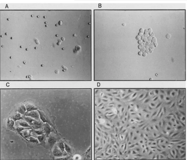
FIG. 1. Four steps of lung microvascular endothelial cell (MVEC) isolation. A, Cell suspension after digestion with trypsin and collagenase; B, induction of MVEC aggregates by plasma; C, formation of MVEC colony after a 3-d culture; D, purified lung MVEC grown to confluence.
and sorting using magnetic beads conjugated with Ulex europaeus agglutinin-1 (UEA-1) (14). However, it was noted that adherent b e a d s are phagocytosed by cells and thereby interfere with cell functions (8). Moreover, the antibody conjugated on magnetic beads seriously affects the phenotypic analysis, as d i s c u s s e d below.
Here, we describe a method allowing a highly pure MVEC popu- lation to be obtained from human lung, either in normal or diseased conditions. These purified lung MVEC were identified by three typ- ical features of endothelial cells: expression of yon Willebrand factor (vWF), platelet endothelial cell adhesion molecule I(PECAM-1, CD31), and transcripts for the angiotensin converting enzyme (ACE). The cells also formed capillalylike structures, took up high levels of acetylated low-density lipoprotein (Ac-LDL), and exhibited endothe- lial cell leukocyte Adhesion Molecule 1 (ELAM-1) inducibility in response to tumor necrosis factor alpha (TNF).
MATERIALS AND METHODS Isolation of MVEC From Human Lung
Human lung tissue samples (20-30 g) were collected from AnDS patients who died in the surgical intensive care unit of our hospital. Informed consent
for postmortem biopsy of the lung was obtained from the next of kin. Control human lung tissues were obtained from patients undergoing thoracic surgery. Samples were collected from macroscopically and microscopically normal tissue. The study protocol was examined and approved by the institutional Ethical Committee for Research in Humans (15). Peripheral lung parenchyma was aseptically removed and stored in Dulbecco's modified Eagle's medium (DMEM) containing 0.1% ethylene diaminetetraacetic acid (EDTA) at 4 ° C and kept up to 6 h prior to processing. The tissue was washed with DMEM and the pleuron was carefully dissected from the underlying tissue with scis- sors to preclude contamination by mesothelial cells. The peripheral lung tissue devoid of large vessels was dissected, finely minced into 3 X 3 mm pieces and washed with DMEM on a sterile 20-~m metal mesh to remove blood cells. The tissue was then digested for 20-30 rain at 37 ° C in 0.1% trypsin (Sigma Chemical Co., St. Louis, MO, USA) containing 0.1% EDTA (1 ml/g tissue) pH 7.2, followed by 0.2% collagenase (Sigma II type) pH 7.4, for another 20 min at 37 ° C. The digested solution was filtered through 100- ~m nylon mesh to discard large fragments of connective tissue. The filtrate was centrifuged at 500 × g for 5 rain at 4 ° C and the pellet was resuspended in 5 ml DMEM containing 20% human plasma and incubated for 5 min to induce the aggregation of MVEC. The cell suspension was overlaid on 20 ml of 20% Percoll and centrifuged at 1500 × g for 15 min at 4 ° C. The pellet was collected and washed twice in DMEM by centrifugation at 500 X g for 5 min. The cells were resuspended in DMEM containing 2 nrM L-glutamine, 100 U/ml penicillin, 100 tag/ml streptomycin, 30% fetal calf serum (FCS),
A
B
A
B
C
C
FIG. 2. Morphology of lung MVEC. A, Microvilli ( × 10 000), B, capillar- ylike tube formation ( × 400); C, Weibel-Palade bodies (X 28 000).
40 U/ml hepm'in, and 100 gg/ml endothelial cell growth supplement (Sigma). The cells were plated onto T25 flasks precoated with 2% gelatin (Sigma) and cultured at 37 ° C in a 5% COs atmosphere.
Purification of Human Lung MVEC
The cells were cultured for 4B h, washed with DMEM to remove nonad- herent cells, and fresh growth medium was added. After 1 wk, MVEC grew
FIG. 3. Immunoeytochemieal staining for endothelial markers. A, von Willebrand factor; B, CD31; C, acetylated low-density lipoprotein uptake on normal lung microvascular endothelial cells.
out as typical "cobblestone" colonies and exhibited the characteristics of monolayer growth and contact inhibition. These "cobblestone" colonies were marked under an inverted microscope in a laminar flow hood. The nonen- dothelial cells around the colonies were weeded by manual manipulation, as described (24). The medium was removed, the flask was washed with DMEM,
5 3 2 LOU ET AL. e~ E - 1 e - q ~ to N O R M A L 1 NORMAL 2 NO RMAL 3 N O R M A L 4 NORMAL 5
f\,alk
A R D S I A R D S 2A
A R D S 3 u i ~ Ill= IB-" I 0 " A R D S 4 A R D S 5Z1
~ 1"0 ~ 1"043 ° 10' 102 103 104 Log f l u o r e s e n c s intensity normal 110 10 10 104
Fl.2-Hei,qht normal 2~1'o ~ lb' lb2~b~ 1'o4
FL2-Height P; normal 3 F 1 2 2 - H e i g h t -=_ ~ A R D S 1 FI_2.-H eight ~ ; ~ 1 ARDS 2 ° I 0 0 '~'0 ~ i"0 ~ 1"0~ i'04 FL2-Height ~' ' ARDS 3~ o
~
o
F L 2 * H e i g h t ~ normal 4 ~ . ARDS 4 U J . o ° to' lo = 10 3 1"o" 13 0 10' 10 2 10 ~ 1'0 4 F I - 2 - H e i g h t FL2-Height g~. normal 5 ee Fl_2-Height 1'0 4 SMC~1o 1'o To~ lb ~
F L , ' 2 - H e i ~ l h t mt-~" ARDS 5 FL2-Height PJ HUVEC o~
\
,
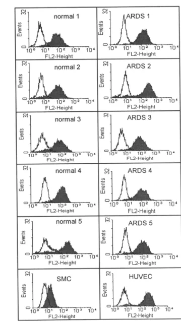
FL2-HeightFIG. 4. Profiles of yon Willebrand factor (vWF) (A) and acetylated low-density lipoprotein (Ac-LDL) (B). Flow cytometric analysis of lung microvascular endothelial cells (MVEC) from control patients and patients who died of acute respiratory distress syndrome (ARDS). (A). Analysis ofvWF expression. The open histograms represent the fluorescence with second step only, and the shaded histograms indicate the specific staining with anti-human vWF antibody. Human umbilical vein endothelial ceils (HUVEC) and human fibroblasts (MRC) used respectively as positive and negative controls. (B). Analysis of Ac-LDL uptake. The open histograms represent the lung MVEC alone, and the shaded histograms indicate uptake of Ac-LDL after 4 h incubation with Ac-LDL. HUVEC and human smooth muscle cells (SMC) used respectively as positive and negative controls.
and a drop of trypsin/EDTA was added onto the marked colonies with a micropipette. Under an inverted microscope, the digested cells were carefully transferred to a new flask with a micropipette. Purified MVEC were cultured until cells reached confluence, then passaged after trypsin/EDTA treatment at a rate dependent on their growth.
Characterization of Human Lung MVEC
1. Morphology. Purified lung MVEC were seeded on a chamber slide (Nune, Inc., Naperville, IL) and grown to confluence. To check their ability to form capillarylike structures in vitro, the cells were cultured in chamber slides coated with 3% gelatin. After 3 d of culture, the formation of capillar- ylike structm'es was observed under an inverted phase contrast microscope.
2. Traltsmission electron microscopy. Cells were seeded in a gelatin-coated cell culture insert with 0.45 #m pore size (Falcon, Meylan Cedex, France) and grown to confluence. The cells were washed with DMEM and then fixed with 2.5% glutaraldehyde for 30 min at 4 ° C, washed with 0.1 M phosphate- buffered saline (PBS) pH 7.2, and postfixed with 1% osmium tetroxide for 20 rain. The cells were dehydrated with different concentrations of ethanol and embedded in Epon 812. Selected areas of monolayer cell cultures were stained with uranyl acetate and lead citrate, and examined in a Philips EM 400 electron microscope.
3. Expression of endothelial markers: vWF and PECAM-1. Lung MVEC (5 X 10³ cells in 0.5 ml) were plated in a chamber slide and grown to subcon- fluence. Human umbilical vein endothelial cells (HUVEC), purified as de-
TABLE ]
ACE
272 bp
SUMMARY OF DIFFERENCES BETWEEN MICROVASCULAR ENDOTHELIAL CELLS (MVEC), LARGE VESSEL ENDOTHELIAL CELLS (LVEC), AND POSSIBLE CONTAMINANT CELLS, SUCH AS
FABROBLASTS AND SMOOTH MUSCLE CELLS (SMC) °
MVEC LVEC fibrobiasts SMC
1 2 3 4 5 6 7 8 9 1 0
FIG. 5. Expression of angiotensin converting enzyme (ACE) transcripts in normal and acute respiratory distress syndrome (ARDS)-derived lung mi- crovascular endothelial ceils (MVEC) Reverse transcription/polymerase chain reaction (RT/PCR) analysis. Lane 1: a DNA ladder. Lanes 2-5: normal lung MVEC. Lanes 6-9: ARDS-derived lung MVEC. Last lane: THP-1 cells used as negative control.
cobblestone/
Morphology spindle cobblestone spindle spindle
Growth characteristic m o n o l a y e r m o n o l a y e r polylayer polylayer
Capillarylike structures yes yes no no
Microvilli + + + - - -
Weibel-Palade Bodies + + + + - -
vWF + + + + - -
CD31 + / - + - -
Ac-LDL uptake high high low low
ACE + + + + + - ELAM-1 inducibility + + - -
0.3
0.2. N o r m a I - M V E C
0.1
0
0
30
100
300
1000
3000
E
e,,,, ¢'4 0'~GI
0
hTNF (U/ml)
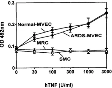
FIG. 6. Inducibility of ELAM-1 on lung microvascular endothelial cells (MVEC). Normal or acute respiratory distress syndrome (ARDS)-derived lung MVEC were stinmlated for 4 h with recombinant human TNF (hTNF). Inten- sity of staining was determined by a cell-based enzyme-linked immunosor- bent assay (ELISA). Negative controls consisted of human lung fibroblasts (MRC) and smooth muscle cells (SMC).
scribed (19), and human fibroblasts (MRC, a kind gift from Dr. M. Pepper, University of Geneva Medical School), were used as positive and negative controls, respectively. The cells were fixed with methanol at - 2 0 ° C for 10 rain, washed three times with PBS containing 1% bovine serum albumin (BSA), and preincubated with PBS containing 5% FCS, 0.05% Tween 20 for 20 rain at 37 ° C to block nonspecific binding. The cells were stained with 1/ 20 diluted rabbit antiserum to human vWF (Sigma) or 1/100 diluted mono- clonal antibody to human PECAM-1 (9Gll, from British Biotechnology) for 45 min at room temperature. Cells were washed three times with PBS/BSA and incubated with 1/100 dilnted fluorescein isothiocyanate (F1TC)-conju- gated goat anti-rabbit IgG (Sigma) or 1/100 diluted FITC-conjugated goat anti-mouse IgG antibody (Sigma) for 30 min at room temperature. After wash- ing and mounting, the staining was observed under a fluorescence microscope. Alternatively, for flow cytometry, single-cell suspensions were prepared by trypsinization, fixed 5 min with - 2 0 ° C methanol, and then incubated with PBS containing 5% FCS and 0.05% Tween 20 for 10 min to block nonspecifie binding. Cells were washed, stained as above at room tem- perature, and analyzed with a FACSean (Beeton-Dickinson, San Jose, CA).
4. Uptake ofacetylated LDL. Uptake was demonstrated with the fluorescent probe l'-dioctadeeyl-3,3,3',3'-tetramethyl-indoearbncyanide perehlorate
a vWF = yon Willebrand factor; Ac-LDL = acetylated low-density lipo- protein; ACE = angiotensin converting enzyme; ELAM-1 = endothelial cell leukocyte adhesion molecule 1; + = positive; - = negative.
conjugated to Ac-LDL (Dil-Ac-LDL) (Paesel + Lorei, Frankfurt, Germany). The ceils were incubated with 10 gg/ml DiI-Ac-LDL in DMEM containing 10% FCS, for 4 h at 37 ° C. The medium was removed and ceils were incu- bated for 10 min in fresh medium. For fluorescence microscopy, ceils were fixed with 10% buffered formalin, washed, mounted, and then observed under fluorescence microscopy. For flow cytometry, after 4 h of incubation with Ac- LDL, single-cell suspensions were prepared by trypsinization. HUVEC and human smooth muscle cells (SMC, kind gift from Dr. G. Gabbiani, University of Geneva Medical School) were used as positive and negative controls, re- spectively. The fluorescence intensity was analyzed with a FACScan.
5. Expression of angiotensin converting enzyme. The expression of ACE mRNA was studied by reverse transcription/polymerase chain reaction (RT/ PCR) as described (10). Briefly, total RNA was isolated from 1 X ]06 cells by the single guanidinium thiocyanate-phenol-chloroform mixture extraction method (5). RNA was incubated for 30 rain at 37 ° C in 40 mM Tris-HC1 pH 7.5, 10 mM NaCI, 6 mM MgC12, and 2.5 units of RQ1 DNAse (Promega Corp., Madison, WI, USA) to remove any contaminating genomic DNA from the preparations. After phenol°chloroform extraction and ethanol precipitation, pellets were resuspended in water. Synthesis of the first strand of cDNA was performed according to the instructions delivered with the cDNA Synthesis Kit (Boehringer Mannheim, Germany), using random primers and AMV re- verse transcriptase (10 units/sample) in a final volume of 20 pl. After 1 h incubation at 42 ° C, samples were heat inactivated and kept frozen ( - 2 0 ° C) until use. Two gl of cDNA were then amplified in a 25 gl reaction mixture containing buffer, deoxynucleotide triphosphates (dNTP), and 2.5 units of ampliTaq (Perkin Elmer Cetus, Emeryville, CA). Samples were overlaid with mineral oil and amplified at 94 ° C for 5 min, 60 ° C for 1 min, and 72 ° C for 30 s followed by 35 cycles at 94 ° C for 30 s, 60 ° C for 30 s, and 72 ° C for 1 min, 30 s. PCR was carried out in an automated DNA Thermal Cycler (Perkin Elmer Cetus), in the presence of 0.2 p~M of each primer. The following oli- gonucleotides were used:
GAPDH 1,5'-TGAAGGTCGGTGTGAACGGATTTGG-3' GAPDH 2,5'-ACGACATACTCAGCACCAGCATCAC-3' ACE 1,5'-GATGTGGCCATCACATFCGTCAGA-3' ACE 2,5'-GCTGCAGAAGAACATGCAAA-3'
One half of the reaction was electrophoresed on 1.5% agarose gels containing ethidium bromide and the appropriate bands were visualized under UV light. 6. Inducibility of ELAM-1. A cell-based enzyme-linked immunosorbent assay (ELISA) was used to detect and quantitate TNF-inducible ELAM-1 expression. Briefly, purified MVEC were seeded in 96-well plates and cul- tured to confluence. Cells were then stimulated with different doses of re- combinant human TNF in DMEM containing 10% FCS for 4 h at 37 ° C. Cells were washed with DMEM and fixed with - 20 ° C methanol for 10 rain. After fixation, PBS containing 5% FCS and 0.05% Tweeu 20 was added for 20 rain to block nonspecific binding. Cells were incubated with monoclonal antibody
534 LOU ET AL. to human ELAM-1 (28.109.76 from Janssen Biochemica, Beerse, Belgium), diluted in PBS/BSA (final concentration 10 gg/ml) for 45 rain at room tem- perature, washed with PBS/BSA, and then incubated with 1/1000 diluted peroxidase-conjugated anti-mouse IgG (Sigma) for 30 rain at room tempera- ture. Cells were further incubated with diluted substrate, tetrameth- ylbenzidine (TMB, from Medgenix, Fleurus, Belgium) for 30 rain, stopped with 2 N
H2S04,
and optical density was read in an automatic ELISA reader at 492 nm.RESULTS
The four steps of lung MVEC purification are shown in Fig. 1 A- D. The cell aggregation induced by human plasma was demonstrated to be vWF positive by immunofluorescence (data not shown). After Percoll centrifugation, the aggregated cells were found in the pellet. When cells adhered to the plastic well, the aggregated MVEC pre- sented a cobblestonelike morphology and exhibited the characteris- tics of monolayer growth and contact inhibition. Lung MVEC from both types of tissues formed classical capillarylike "tubular" struc- tures when cultured on 3% gelatin-coated slides. Transmission elec- tron microscopy showed the presence of Weibel-Palade bodies in the cytoplasm and abundant microvilli on MVEC plasma membrane (Fig.
2 A-C). Although lung MVEC are positive for vWF, the immunoflu- orescence staining pattern was less granular, because they have fewer Weibel-Palade bodies than HUVEC (9,17,21). Mesothelial cells can also exhibit cell surface microvilli, but we found the microvilli and Weibel-Palade bodies within the same cells. The isolated ceils were stained with cytokeratin 8, a marker of mesothelial ceils by immu- nocytochemistry. The results indicated that less than 1% of the cells were positive in both normal and in ARDS lung MVEC. The isolated cells expressed vWF and CD31, as shown by immunofluorescence staining (Fig. 3 A and B), and took up large amounts of Ac-LDL (Fig. 3 C). The staining for vWF and uptake of Ac-LDL were also studied by flow cytometry, to analyze the intensities of fluorescence (Fig. 4 A and B). By RT/PCR, all purified MVEC from normal and ARDS lung showed the expression of ACE mRNA and its absence in the monocytic THP-1 cell line (a kind gift of Dr. J. Pugin, University of Geneva Medical School), used as negative control (Fig. 5). Moreover, both normal and ARDS-derived lung MVEC exhibited a clear in- ducibility of ELAM-1 after a 4-h in vitro stimulation with recombi- nant human TNF, in a dose-dependent manner, as shown by cell- based ELISA (Fig. 6). In contrast, both MRC and SMC remained negative for ELAM-1 induction.
DISCUSSION
In the present study, we report a method to isolate and purify human lung MVEC, from either normal or inflamed lung tissue, that leads to cells expressing classical morphological and functional markers of endothelial cells, including monolayer growth, expression of vWF, CD31, ACE, high Ac-LDL uptake, and, most importantly, ELAM-1 inducibility in response to TNE This method consists of four main steps:
1. Pulmonary tissue was digested fully with trypsin and collage- nase to release MVEC from vascular walls. We found that trypsin is capable of digesting perivaseular connective tissues while collage- nase is beneficial to separate intercellular junctions. Using trypsin/ eollagenase digestion sequentially can result in higher yield in MVEC from mierovessels.
2. Aggregation of MVEC was induced by human plasma. In usual conditions, plasma does not induce MVEC aggregation, but it did
after trypsin/collagenase digestion. The aggregated cells were dem- onstrated to be vWF positive by immunocytoehemical staining. The mechanism involved in MVEC aggregation is not known. A probable explanation is that trypsin/collagenase may induce the expression of a glycoprotein IIb/IIIa--like molecule (26) on the surface of MVEC acting as a fibrinogen receptor, or the adherence of blood platelets to isolated MVEC. That MVEC aggregation may be induced by the fibrinogen contained in plasma.
3. The aggregated MVEC were collected after 20% Pereoll density eentrifugation.
4. Because endothelial colonies were formed quickly after aggre- gated MVEC adhered to plastic wells, these colonies were easily selected and transferred after local digestion with trypsin/EDTA un- der light microscopy. Lung MVEC were successfully isolated from normal human lung using magnetic beads conjugated with anti-en- dothelial antibody. Using anti-CD31-conjugated magnetic beads, we obtained pure MVEC, but the anti-CD31 antibody on the bound beads affected the phenotypie analysis of purified MVEC by flow cytometry. Indeed, FITC-conjugated second antibody bound to both antibodies to CD31 and to other surface molecules, leading to false positive results (data not shown).
The appearance of MVEC as "cobblestone" or "spindle-shaped" may depend on the culture medium and the origin of MVEC (arte- riole, capillary, posteapillary venule, or venules). It has been shown that MVEC from arteriolar and capillary origin exhibit typical cob- blestonelike morphology, while MVEC from postcapilla1"y venules are spindle-shaped (30). We selected the cell colonies with cobblestone- like morphology because they can be easily recognized from fibro- blasts and SMC that present spindle-shaped morphology. MVEC from normal lungs consistently presented the "cobblestone" pattern, while those purified from lungs of ARDS patients often change morphology to "spindle-shaped" after cell transfer. This morphological change may imply that MVEC derived from ARDS patients were activated
in vivo, because these MVEC exhibited significant phenotypic change (15). Also, it has been shown that upon activation, such as stimulation by TNK endothelial cells can shift from the cobblestone- to the spindle-shape morphology (28). Contamination by fibroblasts or SMC is frequently seen in lung MVEC cultures. Unlike MVEC, both fibroblasts and SMC exhibit overlay growth (i.e., without contact inhibition), express neither vWF nor CD31, and take up only low levels of Ae-LDL (34). Although a contamination by fibroblasts or SMC is easily recognized on the basis of morphology and other char- acteristics (summarized in Table 1), their eradication from MVEC cultures is laborious or impossible. Therefore, selection of pure en- dothelial colonies is critical at the purification step. The induction of aggregation of lung MVEC by human plasma makes this selection easier.
Mesothelial cell contamination can be troublesome when lung MVEC are isolated from small animals such as mice or rats, because in these cases it is difficult to remove the pleura completely. In the case of human lung, nevertheless, this mesothelial cell contamination can be minimized by carefld dissection of the visceral pleura prior to proceeding with the isolation procedure. Mesothelial cells present the same morphology as MVEC, by light microscopy, and can take up Dil-Ac-LDL (22,32). Mesothelial cells have also been found to express ACE and vWF (6) but, unlike MVEC, they fail to express CD31, to form capillarylike structures and to express ELAM-1 in response to TNF (20). Staining for cytokeratin 8, a marker of meso- thelial cells, indicated that less than 1% of the ceils are positive in
MVEC isolated from either normal or ARDS lung (data not shown). Finally, contamination by pericytes c a n sometimes occur in MVEC cuhures. These cells do not express endothelial markers, and are easily detached by strong washings with a P a s t e u r pipette, as they are less adherent to plastic t h a n MVEC (our u n p u b l i s h e d observa- tion).
To rule out a contamination of MVEC cultures by LVEC is not easy b e c a u s e both cell types express the same endothelial markers and morphology. Avoiding contamination of LVEC is also important in isolation of MVEC. Lung MVEC derived from ARDS patients exhibited a m a r k e d ICAM-1 upregulation, while the pulmonary vein endothelial cells isolated from the same patients did not (our u n p u b - lished data). Several differences exist between MVEC and LVEC, such as density of Weibel-Palade bodies, prostagtandin metabolism, and ACE activity (4,12,18,21), but some specific markers of MVEC would be necessary (Table 1). The a b u n d a n c e of microvilli have b e e n found on brain MVEC (30), but not LVEC such as HUVEC. This surface feature was also demonstrated on lung MVEC, but not on pulmonary artery endothelial cells (7). Moreover, mierovilli were also found in microvessels in tissue sections of brain and lung (our un- p u b l i s h e d data). T h e s e data indicated that cell surface microvilli are a feature of MYEC. Because large vessels are not found in the pe- ripheral lung p a r e n c h y m a (i.e., u n d e r the visceral pleura), we se- lected these areas for the MVEC isolation in order to avoid contam- ination by LVEC. In conclusion, by allowing the study of the relevant cell from the relevant tissue, this method may be useful for investi- gations dedicated to various immunopathological reactions occurring in the lung.
ACKNOWLEDGMENTS
This work is supported by grants No. 32.37802.93 (to N. M., D. R. M., G. E. G.) and No. 32.043637.95 (to B. R., G. E. G., D. R. M., and P. M. S.) from the Swiss National Science Foundation. G. E. G. was supported by the Clo~tta Foundation, Zurich, Switzerland. We thank Dr. J. Pugin, Dr. G. Gabbiani (Dept. of Pathology, University of Geneva), and Dr. M. Pepper (Dept. of Mor- phology, University of Geneva) for their kind gifts of HUVEC, TH-1 cell line, SMC, and MRC.
REFERENCES
1. Beekhuizen, H.; Vanfurth, R. Growth characteristics of cultured human microvascular venous and arterial and microvascular endothelial cells. J. Vasc. Res. 31:230-239; 1994.
2. Belloni, P. N.; Carney, D. H.; Nicolson, G. L. Organ-derived microvessel endothelial cells exhibit differential responsiveness to thrombin and other growth factors. Microvas. Res. 43:20-45; 1992.
3. Carlay, W. C. M.; Niedbala, J.; Gerritsen, M. E. Isolation, cultivation and partial characterization of microvascular endothelium derived from human lung. Am. J. Respir. Cell Mol. Biol. 7:620-630; 1992. 4. Charo, I.; Karasek, S.; Davision, M. A., et al. Prostaglandins I2 is not a
major metabolite of arachidonic acid in cultured endothelial cells from human foreskin microvessels. J. Clin. Invest. 74:914-919; 1984. 5. Chomczynski, P.; Sacchi, N. Single-step method of RNA isolation by acid guanidinium thiocyanate-phenol-chlorofornl extraction. Anal. Biochem. 162:156-159; 1987.
6. Chung-Welch, N.; Patton, W. E; Ameia Yen-Patton, G. P., et al. Pheno- typic comparison between mesothelial and microvascular endothelial cell lineage using conventional endothelial cell markers, cytoskeletal protein markers, and in vitro assays of angiogenic potential. Differ- entiation 42:44-53; 1989.
7. Chung-Welch, N.; Shepro, D.; Dunham, B., et al. Prostacyclin and pros- taglandin E2 secretion by bovine pulmonary microvascular endothe- lial ceils are altered by changes in culture conditions. J. Cell. Physiol. 135:224-234; 1988.
8. Danielseu, H.; Funderud, S.; Nustad, K., et al. The interaction between cell-surface antigens and antibodies bound to monodisperse polymer particles in normal and malignant cells. Scaud. J. Immunol. 24:179- 187; 1986.
9. Davison, P. M.; Bensch, K.; Karasek, M. A. Isolation and growth of en- dothelial cells from the microvessels of newborn human foreskin in cell culture. J. Invest. Dermatol. 75:316-321; 1980.
10. de Kossodo, S.; Grau, G. E. Profiles of cytokine production in relation with susceptibility to experimental cerebral malaria. J. Immunol.
151:4811--4820; 1993.
11. Delvecchio, P. J.; Siflingerbimboim, A.; Bclloni, P. N., et al. Culture and characterization of pulmonary microvaseular endothelial cells. In Vitro Cell. Dev. Biol. 28A:711-715; 1992.
12. Gerritsen, M. E.; Printz, M. P. Site of prostaglaudin synthesis in bovine heart and isolated bovine coronary microvessels. Circ. Res. 5:1152- 1163; 1981.
13. Gitlin, J. D.; D'Amore, R A. Culture of retinal capillary cells using se- lective growth media. Microvas. Res. 26:74-80; 1983.
14. Grau, G. E.; Lou, J. TNF in vascular pathology: importance of platelet- endothelium interactions. Res. Immunol. 144:355-363; 1993. 15. Grau, G. E.; Mill, N.; Lou, J. N., et al. Phenotypic and functional analysis
of pulmonary mierovascular endothelial cells from patients with acute respiratory distress syndrome. Lab. Invest. 74:761-770; 1996. 16. Hewett, P. W.; Murray, J. C. Human lung mierovessel endothelial cells--
isolation, culture, and characterization. Microvas. Res. 46:89-102; 1993.
17. Hewett, P. W.; Murray, J. C.; Price, E. A., et al. Isolation and character- ization of microvessel endothelial cells from human mammary adipose tissue. In Vitro Cell. Dev. Biol. 29A:325-331; 1993.
18. Hviid, L.; Elhassan, L M.; Dodoo, D., et al. Differential t-cell expression of LFA-1 in residents from Africa and Denmark--description of the phenomenon and its possible basis. Immunol. Lett. 39:147-151; 1994.
19. Jaffe, E. A.; Nachman, R. L.; Becker, C. G. Culture of human endothelial cells derived from umbilical veins: identification by morphologic cri- teria. J. Clin. Invest. 52:2745-2758; 1973.
20. Jonjic, N.; Pert, G.; Bernasconi, S., et al. Expression of adhesion mole- cules and chemotactic cytokines in cultured human mesothelial cells. J. Exp. Med. 176:1165-1174; 1989.
21. Kern, P. A.; Knedler, A.; Eckel, R. H. Isolation and the culture of the microvascular endothelium from adipose tissue. J. Clin. Invest. 71:1822-1829; 1983.
22. Latron, Y.; Alessi, M. C.; George, E, et al. Characterization of epithelial cells from human omentum: comparison with endothelial cells from umbilical veins. Thromb. Haemostasis 66:361-367; 1991.
23. Ley, K.; Gaehtgens, P.; Spanelborowski, K. Differential adhesion of gran- ulocytes to five distinct phenotypes of cultured microvascular endo- thelial cells. Microvas. Res. 43:119-133; 1992.
24. Marks, R. M.; Czerniecki, M.; Penny, R. Human dermal microvascular endothehal cells: an improved method for tissue culture and a de- scription of some singular properties in culture. In Vitro Cell. Dev. Biol. 21:627-635; 1985.
25. Meyrick, B.; Hoover, R.; Jones, M. R., et al. In vitro effects of endotoxin on bovine and sheep lung microvascular and pulmonary artery en- dothelial cells. J. Cell. Physiol. 138:165-174; 1989.
26. Newman, P. J.; Kawai, Y.; Montgomery, R. R., et al. Synthesis by cultured human umbilical vein endothelial ceils of two proteins structurally and immunologically related to platelet membrane glycoproteins IIb and IIIa. J. Cell Biol. 103:81-86; 1986.
27. Rupnick, M. A.; Carey, A.; Williams, S. K. Phenotypic diversity in cul- tured cerebral microvascular endothelial cells. In Vitro Cell. Dev. Biol. 24:435-444; 1988.
28. Sato, N.; Goto, T.; Haranaka, K., et al. Actions of tumor necrosis factor on cultured vascular endothelial ceils: morphologic modulation, growth inhibition, and cytotoxicity. J. Natl. Cancer Inst. 76:1113- 1121; 1986.
29. Scott, E A. E.; Bicknell, R. The isolation and culture of microvascular endothelium. J. Cell Sci. 105:269-273; 1993.
30. Spanel-Borowski, K.; Van der Bosch, J. Different phenotypes of cultured microvessel endothelial ceils obtained from the bovine corpus luteum. Cell Tissue Res. 261:35M~7; 1990.
5 3 6 LOU ET AL. 31. Swerlick, R. A.; Lawley, T. J. Role of microvascular endothelial ceils in
inflammation. J. Invest. Dermatol. 100:Sl11-Sl15; 1993.
32. Van Hinsbergh, V. W. M.; Kooistra, T.; Scheffer, M. A., et al. Character- ization and fibrinolytic properties of human omental tissue mesothe- lial cells: comparison with endothelial cells. Blood 75:1490-1497; 1990.
33. Visne~, G. A.; Chesrown, S. E.; Monnier, J., et al. Regulation of manganese superoxide dismutase--il-1 and mf induction in pulmonary artery and microvascular endothelial cells. Biochem. Biophys. Res. Com- mun. 188:453462; 1992.
34. Voyta, J. C.; Via, D. E; Butterfield, C. E., et al. Identification and isolation of endothelial ceils based on their increased uptake of acetylated-low density lipoprotein. J. Cell Biol. 99:2034-2040; 1984.
35. Wautier, J. L.; Setiadi, H.; Vilette, D., et al. Leukocyte adhesion to en- dothelial cells. Biorheology 27:425-432; 1990.
36. Woodley, N.; Barclay', J. K. Cultured endothelial cells from distinct vas- cular areas show differential responses to agonists. Can. J. Physiol. Pharmacol. 72:1007-1012; 1994.