Pediatr Radiol (2006) 36: 991–996 DOI 10.1007/s00247-006-0248-5
C A S E R E P O RT
Thierry A. G. M. Huisman . Marianne van der Hoef . Ulrich V. Willi . Rita Gobet . Robert L. Lebowitz
Pre- and postnatal imaging of a girl with a cloacal variant
Received: 29 March 2006 / Revised: 25 April 2006 / Accepted: 25 April 2006 / Published online: 20 July 2006 # Springer-Verlag 2006
Abstract We describe the prenatal MR findings in a 29-week fetus with a cloacal variant (urogenital sinus and anterior placed anus) in combination with an enlarged clitoris and urethral duplication and correlate them with postnatal imaging. Fetal MR imaging permits the diagnosis and characterization of cloacal and urogenital sinus malformations in utero. This information may guide pre-, peri- and postnatal management.
Keywords Cloacal variant . Urogenital sinus . Urethral duplication . Clitoromegaly . Fetus . MRI
Introduction
Cloacal malformation and urogenital sinus include a spectrum of complex, congenital malformations with different constellations and degrees of persisting fusion of parts of the urinary, genital and intestinal tracts [1,2]. Cloacal malformations and variants occur exclusively in phenotypic females; incidence is estimated to be 1 in 40,000–50,000 newborns [1]. Many coexisting anomalies involving the spine, pelvis, uterus and kidneys have been described [1, 3]. Infrequently, virilization of the external genitalia is seen [4–8]. Because of the high risk of secondary urogenital infections and sepsis with renal
damage, prompt and precise anatomical and functional diagnostic work-up is essential in these children in order to guide treatment [1–3].
This report is of a girl with a cloacal variant (urogenital sinus and an anterior anus) in combination with an enlarged clitoris and duplicated urethra. The purpose of this report is to present the prenatal MR features and to correlate these findings with postnatal US and genitography studies.
Case report
A 39-year-old pregnant woman was referred to our hospital for fetal MRI because of suspected fetal meconium peritonitis. Prenatal US in the 27th and 29th weeks of gestation had revealed an enlarging cystic abdominal mass (diameter 5.4 cm), ascites and bilateral hydronephrosis. With the exception of bilateral postaxial hexadactyly of the hands and a single umbilical artery, no additional fetal pathology was seen. The pregnancy was otherwise unremarkable; the mother was healthy. Prenatal karyotyp-ing showed a normal 46XX karyotype.
Fetal MRI was performed in the 29th week of gestation according to standard departmental imaging protocols. Multiplanar, T2-weighted (T2-W), single-shot fast spin-echo and T1-weighted (T1-W) gradient-spin-echo sequences were acquired. No maternal or fetal sedation was administered. The entire fetus plus the placenta and the maternal uterus were studied.
A well-circumscribed, sharply marginated, fluid-filled retrovesical‘cyst’ (diameter 5 cm) was confirmed by MRI (Fig. 1). The cyst had a beak-like lower margin pointing toward the pelvis. A second, smaller‘cyst’ (diameter 1.2 cm) communicated with the larger ‘cyst’. The large ‘cyst’ displaced the partially filled urinary bladder anteriorly. A moderate degree of ascites was seen, as well as bilateral hydronephrosis. The bowel was of normal calibre and appeared partially filled with T1-hyperintense meconium. No additional abdominal lesions or‘cysts’ were seen. There were no signs of peritoneal thickening or calcifications that would support the presumed diagnosis of meconium
peri-T. A. G. M. Huisman (*) . M. van der Hoef . U. V. Willi Department of Diagnostic Imaging,
University Children’s Hospital Zurich, Steinwiesstr. 75, 8032 Zurich, Switzerland e-mail: [email protected] Tel.: +41-44-2667110 Fax: +41-44-2667158 R. Gobet
Department of Urology, University Children’s Hospital Zurich, Zurich, Switzerland
R. L. Lebowitz
Department of Radiology,
Children’s Hospital Boston and Harvard Medical School, Boston, MA, USA
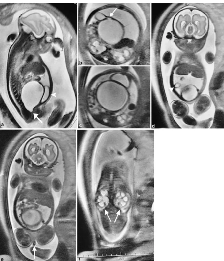
Fig. 1 Fetal MRI. a Sagittal T2-W image shows a distended fluid-filled‘cyst’ with a beak-like lower margin pointing into the pelvis (arrow). The urinary bladder is displaced anteriorly (arrowhead). Ascites is seen superior to the uterus and bladder. b, c Axial T2-W images show the cervix (arrow) connecting the fluid-filled uterus to a markedly distended fluid-filled vagina (b). The bladder and ascitic
fluid are anterior to the urometrocolpos (c). There is bilateral hydronephrosis. d–f Coronal T2-W images. The distended fluid-filled vagina as well as the uterus (arrowhead) is seen. A penis-like structure is identified between the legs (arrow). T2-hyperintense ascites reaches the liver and spleen; both kidneys are hydronephrotic (arrows)
tonitis. MRI revealed a penis-like structure, raising doubts about the 46 XX karyotype of the fetus. Based on the fetal MRI findings a cloacal malformation with urometrocolpos and obstructive hydronephrosis was postulated. Ascites was believed to have resulted from vaginouteroperitoneal reflux of urine through the fallopian tubes.
A caesarean section was performed in the 33rd week of gestation because of premature labour. The child had an uneventful primary adaptation (Apgar 4/9/9, umbilical cord pH 7.27). The placenta was unremarkable. On inspection, significant abdominal distension was seen in combination with the external manifestations of the suspected complex urogenital malformation (Fig.2). A narrow vaginal introi-tus from which urine flowed, plus an anteriorly placed anus was seen. She voided urine from a urethral meatus at the tip of an enlarged clitoris.
The child was transferred to The Children’s Hospital on the 7th day of life for further evaluation and treatment of the urogenital malformation. She underwent vaginoscopy, biochemical analysis, repeat karyotyping, and ‘syndrome evaluation’. An abdominal radiograph (Fig. 3) showed marked abdominal distension with displacement of bowel loops into the upper abdomen. The diaphragm was elevated. The spine and pelvic bones were normal. Sonography revealed a fluid-filled distended vagina (350 ml) (Fig. 4) plus bilateral moderate hydronephrosis. Spinal ultrasonography showed no spinal cord tethering. Genitography with simultaneous insertion of three cathe-ters into the urethral meatus at the tip of the enlarged clitoris, the vaginal introitus and the rectum showed the precise anatomy of this complex urogenital malformation and an intact urethral sphincter (Fig.5). Based on the work of Jaramillo et al. [1], the malformation was classified as a cloacal variant characterized by the combination of a urogenital sinus and an anterior anus. In addition, there was a second urethra at the tip of a markedly enlarged clitoris. Subsequently, the distended vagina was percutaneously drained by a pigtail catheter; 250 ml urine was evacuated. Follow-up ultrasonography showed a collapsed vagina (Fig.4b). The hydronephrosis resolved. Cystovaginoscopy showed that the urogenital sinus was 1.5 cm in length; and that the urethra opened into the ventral wall of the vagina. Biochemical analysis revealed elevated testosterone and gonadotrophin levels, indicating ovarian insufficiency, which normalized spontaneously on follow-up. The Guthrie test excluded adrenogenital syndrome. The adre-nal glands were normal on abdomiadre-nal ultrasonography. Repeat chromosome analysis confirmed 46 XX karyotype. Postaxial hexadactyly was well known within the family. The child’s father and sister, as well as several other family members, also had postaxial polydactyly. There was no congenital heart disease. Therefore McKusick-Kaufman syndrome was believed to be very unlikely.
At the age of 7 months the malformation was surgically corrected by means of total urogenital sinus mobilization (TUM), a technique initially described by Pena [9] in 1997 and modified by Ludwikowski et al. [10] in 1999 and Jenak et al. [11] in 2001. The child recovered well, without functional deficits.
Discussion
Cloacal malformation and urogenital sinus represent a spectrum of congenital urogenital malformations that are believed to result from a failure of normal development or
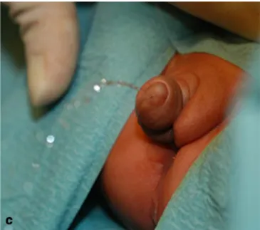
Fig. 2 Clinical photographs demonstrate a marked abdominal distension, b an anterior anus, and (c) urine exiting from the tip of the very enlarged clitoris
incomplete fusion of the urorectal septum with the cloacal membrane during the 4th to 6th week of gestation [1,2,4,
5]. A wide spectrum of resulting malformations have been described with different combinations and degrees of persisting fusion of parts of the urinary, genital and intestinal tracts.
Coexisting findings are frequent and include both congenital malformations such as anomalies of the spine, pelvis, uterus and kidneys (e.g. hemivertebrae, short or absent sacrum, tethered cord, duplication of the genital tract, renal ectopia) as well as acquired problems such as renal damage due to urinary tract obstruction or recurrent pyelonephritis from reflux of infected urine [1,3].
Infrequently there is virilization of the external genitalia with enlargement of the clitoris. This rare combination was first described in 1956 by Broster [12]. The exact aetiology of the virilization has not been determined but is believed to be secondary to a fundamental derangement of urogen-ital development [4, 5, 7]. All reported children have a normal 46XX genotype and unremarkable adrenal, meta-bolic and endocrine status after birth. In addition, the virilization has always been limited to the genitalia. McMullin and Hutson [4] hypothesized that early derange-ment of urogenital developderange-ment, well before sexual differentiation, may prevent the genitalia from acquiring their normal androgen-dependent growth control mecha-nisms. The exact incidence of virilization of the external
Fig. 4 Sagittal US. a During puncture of the distended fluid-filled vagina. b Follow-up imaging reveals the collapsed vagina; the uterus is identified by its normal zonal anatomy (arrow)
Fig. 2 (continued)
Fig. 3 Abdominal radiograph. Bowel loops are displaced into the upper abdomen by the lower abdominal/pelvic mass. No spinal or pelvic osseous malformations are seen
genitalia in association with cloacal or urogenital sinus malformations is unknown. Only a few cases have been described in the literature [4–8].
An early and complete anatomical and functional work-up is essential [3,9]. Imaging should delineate the exact urogenital anatomy and should also rule out additional anomalies or pathology. Surgical correction of cloacal or urogenital malformations is usually possible.
Fetal MRI is more precise than prenatal ultrasonography in the evaluation of this complex urogenital malformation. Our experience supports a recent report by Hayashi et al. [13] who stated that fetal MRI is a useful complementary tool for assessing fetal urogenital anomalies when ultraso-nography is inconclusive. The beak-like lower margin of the fluid-filled‘cyst’ pointing in the direction of the pelvis was a urometrocolpos. Identification of the fluid-filled, dilated uterus and vagina strengthened the diagnosis of a complex urogenital malformation. Ascites was believed to have resulted from reflux of urine within the vagina via the uterus into the peritoneal cavity. The homogeneous T2-hyperintense signal of the uterine fluid and ascites was highly suggestive of urine. MRI did not reveal any haemorrhagic components and/or calcifications. In addi-tion, W MR sequences confirmed the presence of T1-hyperintense meconium within non-distended bowel loops. No T1-hyperintense meconium was seen in the peritoneal
cavity. In addition, bilateral hydronephrosis resulting from the urometrocolpos is well known in complex urogenital malformations [1], but is not associated with meconium peritonitis. Meconium peritonitis was consequently re-garded as very unlikely. Fetal MRI was especially advantageous compared to prenatal ultrasonography be-cause of its greater spatial resolution, the high T2-contrast between fluid and solid tissue, the large field-of-view and the fact that fetal anatomy is not obscured by maternal bowel gas or pelvic bony structures. The large field-of-view facilitates evaluation of complex anatomical anoma-lies. Identification of a penis-like structure on fetal MRI was confusing and the 46XX karyotype was therefore questioned. The postnatal physical examination showed that it was an enlarged clitoris, not a penis.
Postnatally, genitography and vaginoscopy and/or cys-toscopy should be performed to confirm and refine the diagnosis. Additional anomalies or complications should be searched for by appropriate imaging studies including conventional radiography of the spine and pelvis, US of the upper urinary tract and MRI of the spinal cord.
In conclusion, high spatial resolution and high image contrast allow fetal MRI to identify cloacal or urogenital malformations in utero. A fluid-filled ‘cyst’ within the lower abdomen, with a beak-like lower margin pointing in the direction of the lower pelvis is suggestive of a uro- or hydrometrocolpos. A cloacal or urogenital anomaly should therefore be considered. T1-W MRI sequences may be helpful to rule out meconium peritonitis. Prenatal fetal MRI can be performed without maternal or fetal sedation. The ultrafast acquisition times ‘picture-freeze’ the fetus in utero. Prenatal fetal MRI may also be used as an alternative to postnatal MRI avoiding the need for neonatal sedation.
References
1. Jaramillo D, Lebowitz RL, Hendren WH (1990) The cloacal malformation: radiologic findings and imaging recommenda-tions. Radiology 177:441–448
2. Allen TD, Husmann DA (1990) Cloacal anomalies and other urorectal septal defects in female patients; a spectrum of anatomical abnormalities. J Urol 145:1034–1039
3. Hendren WH (1992) Cloacal malformations: experience with 105 cases. J Pediatr Surg 27:890–901
4. McMullin ND, Hutson JM (1991) Female pseudohermaphro-ditism in children with cloacal anomalies. Pediatr Surg Int 6:56–59
5. Chadha R, Kothari SK, Tanwar US, et al (2001) Female pseudohermaphroditism associated with cloacal anomalies: faulty differentiation in the caudal developmental field. J Pediatr Surg 36:E9
6. Karlin G, Brock W, Rich M, et al (1989) Persistent cloaca and phallic urethra. J Urol 142:1056–1059
7. Chatterjee SK, Chakravorti A, Haqaue J (1994) Clitoromegaly, duplex urethra, and dysplastic vagina. Pediatr Surg Int 9:444– 447
8. Escobar LF, Weaver DD, Bixler D, et al (1987) Urorectal septum malformation sequence. Am J Dis Child 141:1021– 1024
9. Pena A (1997) Total urogenital sinus mobilization– an easier way to repair cloacas. J Pediatr Surg 32:263–267
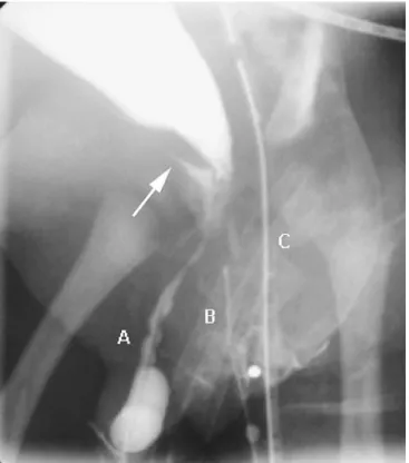
Fig. 5 Retrograde study with simultaneous injection through one catheter through the duplicated phallic urethra (A), one into the urogenital sinus (B) and one into the rectum (C). Contrast medium injection into catheter A opacifies the distal end of the ‘normal’ urethra up to the level of the urethral sphincter (arrow) suggesting that there will be good urinary continence. Injection of catheter B fills the distended vagina
10. Ludwikowski B, Oesch Hayward I, Gonzalez R (1999) Total urogenital sinus mobilization: expanded applications. BJU Int 83:820–822
11. Jenak R, Ludwikowski B, Gonzalez R (2001) Total urogenital sinus mobilization: a modified perineal approach for feminizing genitoplasty and urogenital sinus repair. J Urol 165:2347–2349
12. Broster L (1956) A form of intersexuality. Br Med J 1:149–151 13. Hayashi S, Sago H, Kashima K, et al (2005) Prenatal diagnosis of fetal hydrometrocolpos secondary to a cloacal anomaly by magnetic resonance imaging. Ultrasound Obstet Gynecol 26:577–579