Signature region within the 16S rDNA sequences of
Aeromonas popo¤i
A. Demarta *, M. Tonolla, A.-P. Caminada, N. Ruggeri, R. Peduzzi
Istituto Cantonale Batteriosierologico, Laboratory of Microbial Ecology, University of Geneva, Via Ospedale 6, 6904 Lugano, Switzerland Received 23 December 1998; accepted 8 January 1999
Abstract
To identify a group of eight Aeromonas strains of our collection showing ribotyping patterns similar to those described for the species Aeromonas popoffii, 16S rRNA gene sequence analysis was performed. Results were in agreement with the DNA binding values, and allowed the identification of a `signature region' differentiating the A. popoffii strains from all other members of the genus Aeromonas. z 1999 Federation of European Microbiological Societies. Published by Elsevier Science B.V. All rights reserved.
Keywords: Aeromonas popo¤i; 16S rDNA; Signature region
1. Introduction
The genus Aeromonas comprises bacteria that are autochthonous inhabitants of aquatic environments [1], and have been increasingly recognised as patho-genic for both animals and human beings [2,3].
Currently, 16 species of Aeromonas are identi¢ed on the basis of DNA^DNA hybridisation, but many of the taxa remain di¤cult to distinguish from one another through phenotypic tests because of the lim-ited number of discriminating characteristics [4].
Besides DNA^DNA hybridisation, several ap-proaches have been proposed in order to di¡erenti-ate between aeromonad hybridisation groups (HGs) including ribotyping and multilocus enzyme electro-phoresis [5], restriction fragment length
polymor-phism (RFLP) [6], and ampli¢ed fragment length polymorphism (AFLP) [7]. The complete 16S rDNA sequences of members of the genus Aeromo-nas were published [8^10], and subsequently used to design PCR-primers and hybridisation probes for the speci¢c detection of some species based on 16S rDNA regions which show enough variability to dis-criminate, with few exceptions, between species [11^ 13].
Recently, Huys and coworkers [14] have described the new species Aeromonas popo¤i. Strains belong-ing to this species were isolated from Flemish drink-ing water production plants and Scottish drinkdrink-ing water supplies. In our laboratory, we performed ri-botyping of 286 Aeromonas strains isolated from children su¡ering from diarrhoea, as well as from their domestic environments, including drinking and pipe system waters. By clustering the ribopro-¢les, we found a group of eight strains of
environ-* Corresponding author. Tel.: +41 (91) 923-2522; Fax: +41 (91) 922-0993; E-mail: antonella.demarta@ti.ch
mental origin which could not be classed with certi-tude in any Aeromonas species.
The aim of this work was to classify our unidenti-¢ed strains by comparing their low-molecular-eight bands patterns with those of reference strains of A. popo¤i and by 16S rDNA sequencing, methods which have both been demonstrated to be valuable for the characterisation of Aeromonas genospecies [13,15].
2. Materials and methods 2.1. Bacterial strains
The strains of our collection (Table 1) were col-lected in the period between August 1992 and Sep-tember 1993 from drinking water samples and a fountain swab taken during a study on the di¡usion of Aeromonas strains in domestic environments of diarrhoeic children in our region (Canton Ticino, Switzerland).
Water samples were ¢ltered through 0.45-Wm ni-trocellulose membranes and incubated on m-Aero-monas Selective Agar (Biolife, Milan, Italy) with 10 mg l31 ampicillin at 30³C for 24 h. Columbia
Agar Base with 5% sheep erythrocytes supplemented with 10 mg l31 ampicillin was used for the direct
plating of the swabs, for plating after enrichment in alkaline Peptone Water at pH 8.6 (incubated at room temperature for 24 h), and for subculturing colonies showing the typical characteristics of the genus after growing on m-Aeromonas Selective Agar. The strains were further analysed using classi-cal tests to con¢rm the genus Aeromonas: Gram stain, oxidase, catalase, oxidation and fermentation of glucose, resistance to the vibriostatic compound 0/129.
Seven strains from the LMG collection (Table 1) were also analysed, and the following reference strains were included for the ribotyping: A. hydro-phila ATCC 7966 (HG 1), A. bestiarum CDC 9533-76 (HG 2), and A. hydrophila A909 (HG 3).
The strains used in ¢lter hybridisation assays were the following: A. popo¤i strains LMG 17541T, LMG 17542, LMG 17543, F498B and F479E; A. hydrophila ATCC 7966 (HG 1); A. bestiarum CDC 9533-76 (HG 2); A. salmonicida subsp. salmonicida
ATCC 33658 (HG 3); A. caviae ATCC 15468 (HG 4); A. media ATCC 33907 (HG 5B); A. eucrenophila NCMB 74 (HG 6); A. sobria CIP 7433 (HG 7); A. veronii ATCC 35624 (HG 8/10); A. jandaei ATCC 49568 (HG 9); A. encheleia ATCC 35941 (HG 11); A. schubertii ATCC 43700 (HG 12); A. trota ATCC 49657 (HG 13); A. ichthiosmia DSM 6393; A. ente-ropelogenes DSM 6394; A. allosaccharophila CECT 4199; A. encheleia DSM 11577.
2.2. Ribosomal DNA gene restriction fragment analysis
Puri¢ed genomic DNA samples were prepared ac-cording to the method described by Ausubel and coworkers [16] and successively digested with restric-tion endonuclease SmaI. Fragments were separated by electrophoresis on a 0.8% agarose gel in TBE bu¡er 1U(Tris-Borate-EDTA, pH 8.0) at 25 V for 14 h. The fragments were transferred to a positively charged nylon membrane as described in Maniatis et al. [17], ¢xed to the membrane for 30 min at room temperature and for 15 min at 80³C.
Plasmid pKK3535 [18], labelled with digoxigenin (DIG DNA labelling Kit, Boehringer Mannheim), was used as probe. Prehybridisation, hybridisation (both at 68³C), and the immunological detection of the fragments were performed according to the pro-tocol of the DIG Nucleic Acid Detection Kit (Boehr-inger Mannheim).
Reconstructed images of the visualised ribotyping band patterns in the low-molecular-weight zone (0.8^ 4.0 kb) were scanned with Sharp JX-330 densitoscan-ner. Transmission image data were stored in TIFF ¢les and were further processed by using the Gel-Compare software, version 3.1 (applied Maths, Kortrijk, Belgium). Levels of similarity between ribotyping pro¢les were calculated by using the band-matching Dice coe¤cient, and cluster analysis was performed by using the unweighted pair group method with arithmetic averages.
2.3. Sequencing methods
Genomic DNA was isolated as described above. The 16S rDNA was selectively ampli¢ed by PCR using the forward primer corresponding to position 8^26 of Escherichia coli rDNA (5P-
AGAGTTT-GATCATGGCTCA-3P), and the reverse primer cor-responding to the complement of position 1411^1391 (5P- GTGTGACGGGCGGTGTGTA-3P). From 0.1 to 0.75 Wg of the extracted DNA was mixed with 1 U of Taq DNA polymerase (Boerhinger Mannheim), 10UPCR Bu¡er (Boerhinger Mannheim), 10 mM of each dNTP (Boerhinger Mannheim), and 0.5 WM of each primer in a ¢nal volume of 50 Wl and ampli-¢ed by PCR using the GeneAmp PCR System 2400 thermal cycler (Perkin-Elmer). After an initial denat-uration at 94³C for 5 min, the mixture was subjected to 35 cycles, with each cycle consisting of denatura-tion for 30 s at 94³C, primer annealing at 52³C for 30 s, and chain extension at 72³C for 1 min (¢nal extension 7 min).
The PCR products were puri¢ed with the QIA-quick PCR Puri¢cation Kit (Qiagen), and prepared for sequencing employing the ABI PRISM dRhod-amine Terminator Cycle Sequencing Ready Reaction Kit (Perkin-Elmer), according to the respective man-ufacturer's instructions. Excess terminators were re-moved by spin-column puri¢cation (Centri-Sep spin columns, Princeton Separations) before preparing and loading the samples onto the ABI PRISM 310 Genetic Analyzer (Perkin-Elmer) as speci¢ed on the manufacturer's protocol.
Sequencing primers were the following (numbers in brackets indicate the relative position on E. coli rDNA): forward primers,
5P-GCTGGTCTGAGAG-GATGA-3P (289^334), 5P-GATAAGTTAGATGTG-AA-3P (592^624), 5P-CAAGCGGTGGAGCATGT-G-3P (936^953); and reverse primers, 5P-GCAC-CTGTGTTCTGATTCC-3P (510^492), 5P-CTCTA-CAAGACTCTAGCTGG-3P (896^877), 5P-GCTGA-TCATCCTCTCAGAC-3P (1250^1232).
2.4. A. popo¤i probe design, labelling and hybridisation conditions
From the 16S rDNA sequences of the A. popo¤i strains, the oligonucleotide probe 5P-GTTGCTGGRTATTAGCCAA-3P (corresponding to the complement of position 477^459 of E. coli rDNA) was designed, and purchased from Pharma-cia Biotech. The A. popo¤i oligonucleotide was la-belled with £uorescein-11-dUTP by using the ECL 3P-end labelling kit, and detection was carried out as indicate in the manufacturer's protocol (Amersham). The stringency of the hybridisation was achieved in the post-hybridisation washes using as stringency wash bu¡er 0.2USSC, 0.1% (w/v) SDS at 44³C. 2.5. Filter hybridisation assays
Four microlitres of puri¢ed nucleic acid [16] were dot-blotted on a nylon membrane, ¢xed and hybrid-ised with the labelled A. popo¤i probe following the conditions described above.
Table 1
A. popo¤i strains used in the study
Straina Source of isolation Geographical origin
LMG 17541T Drinking water production plant Oelegem (Belgium)
LMG 17542 Drinking water production plant De Blankaart (Belgium)
LMG 17543 Drinking water production plant Snellegem (Belgium)
LMG 17544 Drinking water production plant Eeklo (Belgium)
LMG 17545 Drinking water production plant Snellegem (Belgium)
LMG 17546 Drinking water service reservoir Udny Station (Scotland)
LMG 17547 Drinking water treatment plant Turri¡ (Scotland)
F479E Fountain swab Vezio (Switzerland)
F498B Tap water Cadro (Switzerland)
F533E Tap water Pregassona (Switzerland)
F539A Tap water Viganello (Switzerland)
F548B Tap water Viganello (Switzerland)
F548C Tap water Viganello (Switzerland)
F600B Tap water Viganello (Switzerland)
F600C Tap water Viganello (Switzerland)
aLMG, Culture Collection of the Laboratorium voor Microbiologie Gent, Universiteit Gent, Ghent, Belgium; F, culture collection of the
For Southern blot assays, 10 Wl of the 16S rDNA samples obtained by PCR were electrophoresed on 0.8% agarose gel and blotted onto a nylon mem-brane following the same procedure as for ribotyp-ing. Hybridisation of the fragments with the labelled A. popo¤i probe and detection were performed at the conditions described. The autoradiography ¢lm (Hyper¢lm-ECL; Amersham) was exposed for 3 h. 3. Results and discussion
In 1997, Huys et al. [14] proposed the new species A. popo¤i to accommodate mesophilic Aeromonas strains recovered from Flemish and Scottish drinking water production plants and reservoirs. The strains showed AFLP ¢ngerprints constituting a genotypic cluster unidenti¢ed against their AFLP-based identi-¢cation library, unique phenotypic pro¢les and ribo-typing patterns, and DNA binding values to other Aeromonas spp. comprised between 22 and 63%.
In order to verify if eight strains of our collection, isolated from drinking water of our region, could belong to the species A. popo¤i, we performed the ribotyping of our strains together with those of the
LMG collection, and the reference strains of the hy-bridisation groups which were indicated by Huys et al. as the most closely related (HG 1, HG 2 and HG 3).
Clustering analysis of ribotyping patterns gener-ated from the strains analysed (Fig. 1) clearly dem-onstrate that our strains are closely related to the A. popo¤i reference strains. The strains formed a group that was distinct from the related taxa HG 1, HG 2 and HG 3. As already found by Huys et al. [14], no bands were present in the molecular weight region between 0.8 and 1.6 kb, whereas the band at 1.8 kb was present in all A. popo¤ strains, except LMG 17542. The A. popo¤i strains shared at least three bands with the reference strains of HG 1, HG 2 and HG 3 analysed. Besides the two bands in positions 0.8 and 1.6, described by Huys et al. [14], we found an additional band at 3.7 kb.
The 16S rDNA sequences (from position 8 to 1411 in the E. coli sequence) of all of our strains, as well as of those of strains LMG 17541T, LMG 17542,
LMG 17543 and LMG 17546, were determined by PCR-DNA sequencing.
The sequences of all the strains were nearly iden-tical, and consisted of a continuous stretch of 1407
Fig. 1. Clustering analysis (unweighted pair-group method using arithmetic averages) of ribotyping patterns of 15 A. popo¤i strains, and type and/or reference strains for Aeromonas HG 1, HG 2 and HG 3. Similarities are expressed as the band-matching Dice coe¤cient (SD). Positions of the markers are indicated by arrows.
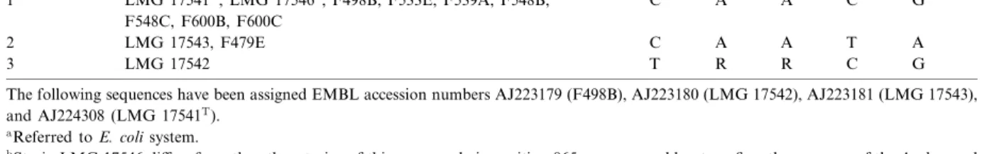
nucleotides representing more than 90% of the total 16S rRNA molecule. Although among the 12 strains analysed we found variations in ¢ve nucleotide posi-tions, allowing for clustering the strains into three groups (Table 2), the analysis of the 12 A. popo¤i sequences showed that they formed a distinct genetic line within the genus Aeromonas (Fig. 2).
The relationship between Aeromonas species de-rived by 16S rDNA sequencing generally correlates well with the results of DNA^DNA reassociation [8,9], although several discrepancies between the two approaches have been described [10]. Our study demonstrated that in the case of A. popo¤i, the 16S rDNA sequencing results agree with the DNA bind-ing values. The phylogenetic tree constructed on the basis of 1407 bp placed the new species in the group formed by A. bestiarum (HG 2), A. salmonicida (HG
3), A. eucrenophila (HG 6), A. encheleia (HG 11), and A. sobria (HG 7), which are all mainly from environmental origin.
The A. popo¤i 16S rDNA sequences di¡ered from those of other aeromonads at least in seven nucleo-tides. The sequence in position 154^167 (Table 3) was identical to that determined for A. hydrophila HG 1, A. bestiarum HG 2, A. salmonicida subsp. salmonicida HG 3, A. caviae HG 4, A. media HG 5B, A. sobria HG 7, A. encheleia HG 11, A. trota HG 13, A. enteropelogenes, and A. allosaccharophila, whereas the sequence in position 457^476 allowed the di¡erentiation of all the A. popo¤i strains from the other species. In fact, the nine A. popo¤i strains showed a T instead of G or A in position 459 and a G instead of C or T in position 473 (Table 3).
An oligonucleotide probe was thus designed and
Fig. 2. Phylogenetic interrelationships in the genus Aeromonas. The tree was obtained using the Jukes^Cantor distance and the Neighbor-joining method [19] to compare 16S rDNA sequences of 1407 bp derived from our own or from published data (see Table 3). Numbers indicate the bootstrap con¢dence level (100 replications).
used in ¢lter hybridisation assays in order to recog-nise this variable region. These methods of bacterial identi¢cation do not require the sequencing of frag-ments and therefore represent less expensive and sim-pler tools approachable by most laboratories for the identi¢cation of bacterial species. The probe
permit-ted the speci¢c identi¢cation of strains belonging to the species A. popo¤i in dot blot (data not shown) and Southern blot of 16S rDNA fragments tests (Fig. 3).
In conclusion, the species A. popo¤i, which was proposed by Huys et al. [14] on the basis of AFLP,
Fig. 3. Southern blot assay. (A) PCR performed with genomic DNA of Aeromonas strains and universal primers for 16S rRNA genes (see Section 2). (B) Southern blot hybridisation of the ampli¢ed 16S rRNA genes using labelled A. popo¤i oligonucleotide as probe. Lane 1, negative control (no DNA); lane 2, A. popo¤i LMG 17541T; lane 3, A. popo¤i LMG 17542; lane 4, A. popo¤i LMG 17543, lane 5,
A. popo¤i F498B; lane 6, A. popo¤i F479E; lane 7, A. eucrenophila NCMB 74 (HG 6); lane 8, A. media ATCC 33907 (HG 5B); lane 9, A. schubertii ATCC 43700 (HG 12); lane 10, A. veronii ATCC 35624 (HG 8/10); lane 11, A. hydrophila ATCC 7966 (HG 1); lane 12, A. caviae ATCC 15468 (HG 4); lane 13, negative control (no DNA); lane 14, negative control (no DNA); lane 15, A. sobria CIP 7433 (HG 7); lane 16, A. bestiarum CDC 9533-76 (HG 2); lane 17, A. salmonicida subsp. salmonicida ATCC 33658 (HG 3); lane 18, A. jandaei ATCC 49568 (HG 9); lane 19, A. encheleia ATCC 35941 (HG 11); lane 20, A. trota ATCC 49657 (HG 13); lane 21, A. ichthiosmia DSM 6393; lane 22, A. enteropelogenes DSM 6394; lane 23, A. allosaccharophila CECT 4199; lane 24, A. encheleia DSM 11577; lane 25, nega-tive control (no DNA).
Table 2
Base di¡erences in the 16S rDNA sequences among the A. popo¤i strains analysed
Group Strain Positionsa
469 559 782 1011 1018
1 LMG 17541T, LMG 17546b, F498B, F533E, F539A, F548B,
F548C, F600B, F600C C A A C G
2 LMG 17543, F479E C A A T A
3 LMG 17542 T R R C G
The following sequences have been assigned EMBL accession numbers AJ223179 (F498B), AJ223180 (LMG 17542), AJ223181 (LMG 17543), and AJ224308 (LMG 17541T).
aReferred to E. coli system.
bStrain LMG 17546 di¡ers from the other strains of this group only in position 865 were we could not con¢rm the presence of the A observed
ribotyping and fatty acid pro¢les, phenotypic char-acteristics, and DNA^DNA hybridisation data, could also be identi¢ed by a unique primary struc-ture of the 16S rRNA genes, and by a `speci¢c sig-nature' in the variable region comprised between po-sitions 457 and 476, which is the most highly diagnostic region for delineating Aeromonas geno-species. This sequence could also be recognised in ¢lter hybridisation assays, showing that this zone represents e¡ectively a signature region for this spe-cies.
Acknowledgments
We are indebted to M. Altwegg, who provided us with the reference strain of A. hydrophila HG 3, and the strains LMG of A. popo¤i. We would like to thank him as well as M. Aeschbacher for their crit-ical review of this manuscript.
References
[1] Holmes, P., Niccolls, L.M. and Sartory, D.P. (1996) The ecol-ogy of mesophilic Aeromonas in the aquatic environment. In: The Genus Aeromonas (Austin, B., Altwegg, M., Gosling, P.J. and Joseph S., Eds.), pp. 127^150. John Wiley and Sons, Chi-chester.
[2] Santos, Y., Toranzo, A.E., Barja, J.L., Nieto, T.P. and Villa, T.G. (1988) Virulence properties and enterotoxin production of Aeromonas strains isolated from ¢sh. Infect. Immun. 56, 3285^3293.
[3] Goèmez Campdera, J., Munìoz, P., Loèpez Prieto, F., Rodr|èguez Fernaèndez, R., Robles, M., Rodr|èguez Creixems, M. and Bou-za Santiago, E. (1996) Gastroenteritis due to Aeromonas in pediatrics. An. Esp. Pediatr. 44, 548^552.
[4] Carnahan, A.M. and Altwegg, M. (1996) Taxonomy. In: The Genus Aeromonas (Austin, B., Altwegg, M., Gosling, P.J. and Joseph S., Eds.), pp. 1^38. John Wiley and Sons, Chichester. [5] Tonolla, M., Demarta, A. and Peduzzi, R. (1991) Multilocus genetic relationships between clinical and environmental Aero-monas strains. FEMS Microbiol. Lett. 81, 193^200. [6] Borrell, N., Acinas, S.G., Figueras, M.-J. and
Mart|ènez-Mur-cia, A.J. (1997) Identi¢cation of Aeromonas clinical isolates by Table 3
16S rDNA sequences of Aeromonas strains at two variable regions corresponding to positions 154^167 and 457^476 of the E. coli 16S rDNA sequence
Taxon DNA group Straina Region 1 (position 154^167) Region 2 (position 457^476)
A. hydrophilab 1 ATCC 7966 AGTTGGAAACGACTGCT TGATGCCTAATACGTATCAA
A. bestiarumc 2 CDC 9533-76 AGTTGGAAACGACTGCT TGGCGCCTAATACGTGTCAA
A. salmonicida subsp. salmonicidab 3 ATCC 33658 AGTTGGAAACGACTGCT TGGCGCCTAATACGTGTCAA
A. caviaeb 4 ATCC 15467 AGTTGGAAACGACTGCT CAGTAGCTAATATCTGCTGG
A. mediab 5B ATCC 33907 AGTTGGAAACGACTGCT TGATGCCTAATACGCATCAG
A. eucrenophilab 6 ATCC 23309 AGTTGGAAACGGCTGCT TGATGCCTAATACGCATCAG
A. sobriab 7 ATCC 43979 AGTTGGAAACGACTGCT TGGCAGCTAATATCTGTCAG
A. veronii biogroup veroniib 8/10 ATCC 35624 TACTGGAAACGGTAGCT TGGTAGCTAATAACTGCCAG
A. jandaeib 9 ATCC 49568 TACTGGAAACGGTAGCT CAGTAGCTAATATCTGCTGG
A. encheleiab 11 ATCC 35941 AGTTGGAAACGACTGCT TGGTCGCTAATAACGGCCAA
A. schubertiib 12 ATCC 43700 TACTGGAAACGGTAGCT TGGTGGTTAATACCTGCCAG
A. trotab 13 ATCC 49657 AGTTGGAAACGACTGCT CAGTAGCTAATATCTGCTGG
A. enteropelogenesd NDf DSM 6394 AGTTGGAAACGACTGCT CAGTAGCTAATATCTGCTGG
A. ichthiosmiad ND DSM 6393 TACTGGAAACGGTAGCT TGGTAGCTAATAACTGCCAG
A. allosaccharophilae ND CECT 4199 AGTTGGAAACGACTGCT TGGTAGCGAATAACTGCCAG
A. popo¤ic ND LMG 17541T AGTTGGAAACGACTGCT TGTTGGCTAATACCCAGCAA
aA, Institute of Medical Microbiology, Zuërich, Switzerland,; ATCC, American Type Culture Collection, Rockville, MD; CDC, Centers for
Disease Control, United States Public Health Service, Atlanta, GA, USA; CECT, Coleccioèn Espanìola de Cultivos Tipo, Universidad de Valeència, Valeència, Spain; DSM, Deutsche Sammlung von Mikroorganismen und Zellkulturen GmbH, Braunschweig, Germany; LMG, Culture Collection of the Laboratorium voor Microbiologie Gent, Universiteit Gent, Ghent, Belgium.
bData from [8]. cOur data. dData from [10]. eData from [9]. fND, not de¢ned.
restriction fragment length polymorphism of PCR-ampli¢ed 16S rRNA genes. Int. J. Syst. Bacteriol. 35, 1671^1674. [7] Huys, G., Coopman, R., Janssen, P. and Kersters, K. (1996)
High-resolution genotypic analysis of the genus Aeromonas by AFLP ¢ngerprinting. Int. J. Syst. Bacteriol. 46, 572^580. [8] Mart|ènez-Murcia, A.J., Benlloch, S. and Collins, M.D. (1992)
Phylogenetic interrelationships of members of the genera Aeromonas and Plesiomonas as determined by 16S ribosomal DNA sequencing: lack of congruence with results of DNA^ DNA hybridizations. Int. J. Syst. Bacteriol. 42, 412^421. [9] Mart|ènez-Murcia, A.J., Esteve, C., Garay, E. and Collins,
M.D. (1992) Aeromonas allosaccharophila sp. nov., a new mes-ophilic member of the genus Aeromonas. FEMS Microbiol. Lett. 91, 199^206.
[10] Collins, M.D., Mart|ènez-Murcia, A.J. and Cai, J. (1993) Aero-monas enteropelogenes and AeroAero-monas ichthiosmia are identi-cal to Aeromonas trota and Aeromonas veronii, respectively, as revealed by small-subunit rRNA sequence analysis. Int. J. Syst. Bacteriol. 43, 855^856.
[11] Ash, C., Mart|ènez-Murcia, A.J. and Collins, M.D. (1993) Identi¢cation of Aeromonas schubertii and Aeromonas jandaei by using a polymerase chain reaction-probe test. FEMS Mi-crobiol. Lett. 108, 151^156.
[12] Ash, C., Mart|ènez-Murcia, A.J. and Collins, M.D. (1993) Mo-lecular identi¢cation of Aeromonas sobria by using a polymer-ase chain reaction-probe test. Med. Microbiol. Lett. 2, 80^ 86.
[13] Dorsch, M., Ashbolt, N.J., Cox, P.T. and Goodman, A.E. (1994) Rapid identi¢cation of Aeromonas species using 16S rDNA targeted oligonucleotide primers: a molecular ap-proach based on screening of environmental isolates. J. Appl. Bacteriol. 77, 722^726.
[14] Huys, G., Kaëmpfer, P., Altwegg, M., Kersters, I., Lamb, A., Coopman, R., Luëthy-Hottenstein, J., Vancanneyt, M., Jans-sen, P. and Kersters, K. (1997) Aeromonas popo¤i sp. nov., a mesophilic bacterium isolated from drinking water production plants and reservoirs. Int. J. Syst. Bacteriol. 47, 1165^1171. [15] Martinetti Lucchini, G. and Altwegg, M. (1992) rRNA gene
restriction patterns as taxonomic tools for the genus Aeromo-nas. Int. J. Syst. Bacteriol. 42, 384^389.
[16] Ausubel, F.M., Brent, R., Kingston, R.E., Moore, D.D., Seid-man, J.G., Smith, J.A. and Struhl, K. (1989) Current Proto-cols in Molecular Biology. Green^Wiley Interscience, New York.
[17] Maniatis, T., Fritsch, E.F. and Sambrook, J. (1982) Molecular Cloning. A Laboratory Manual. Cold Spring Harbor Labo-ratory, Cold Spring Harbor, NY.
[18] Brosius, J., Ullrich, A., Raker, M.A., Gray, A., Dull, T.J., Gutell, R.R. and Noller, H.F. (1981) Construction and ¢ne mapping of recombinant plasmids containing the rrnB riboso-mal operon of E. coli. Plasmid 6, 112^118.
[19] Kumar, S., Tamura, K. and Nei, M. (1993) MEGA: Molec-ular Evolutionary Genetics Analysis. Version 1.02. Pennsylva-nia State University, University Park.

![Fig. 2. Phylogenetic interrelationships in the genus Aeromonas. The tree was obtained using the Jukes^Cantor distance and the Neighbor- Neighbor-joining method [19] to compare 16S rDNA sequences of 1407 bp derived from our own or from published data (see T](https://thumb-eu.123doks.com/thumbv2/123doknet/14903666.654912/5.816.154.672.108.598/phylogenetic-interrelationships-aeromonas-obtained-neighbor-neighbor-sequences-published.webp)