Impaired modulation of quadriceps tendon jerk
reflex during spastic gait: differences between
spinal and cerebral lesions
Michael Faist,
1Matthias Ertel,
1Wiltrud Berger
1and Volker Dietz
21Department of Clinical Neurology and Neurophysiology, Correspondence to: Dr Michael Faist, Department of
University of Freiburg, Germany and2Swiss Paraplegic Clinical Neurology and Neurophysiology, University of
Centre, University Hospital Balgrist, Zu¨rich, Switzerland Freiburg, Breisacherstr. 64, D-79106-Freiburg, Germany E-mail: [email protected]
Summary
In healthy subjects, functionally appropriate modulation of short latency leg muscle reflexes occurs during gait. This modulation has been ascribed, in part, to changes in presynaptic inhibition of Ia afferents. The changes in modulation of quadriceps tendon jerk reflexes during gait of healthy subjects were compared with those of hemi-or paraparetic spastic patients. The spasticity was due to unilateral cerebral infarction or traumatic spinal cord injury, respectively. The modulation of the quadriceps femoris tendon jerk reflex at 16 phases of the step cycle was studied. The reflex responses obtained during treadmill walking were compared with control values obtained during gait-mimicking standing postures with corresponding levels of voluntary muscle contraction and knee angles. In healthy subjects the size of the reflexes was profoundly modulated and was generally depressed
Keywords: tendon-tap reflexes; gait; spasticity; hemiparesis; paraparesis Abbreviation: MI5 modulation index
Introduction
During normal human gait short latency leg muscle reflexes (H-reflex and stretch/tendon jerk reflex) have been shown to be profoundly modulated throughout the step cycle (Capaday and Stein, 1986, 1987; Crenna and Frigo, 1987; Llewellyn et al., 1987; Dietz et al., 1990a, b; Yang et al., 1991a; Sinkjaer et al., 1996b; van de Crommert et al., 1996). However, this modulation is not merely a reflection of the changes in motor neuron excitability associated with the strength of the electromyographic (EMG) activity. Part of the overall effect of reflex modulation may be due to changes in presynaptic inhibition of Ia afferents (Morin et al., 1982; Capaday and Stein, 1986; Dietz et al., 1990a, b; for review,see Stein, 1995). Indeed, recent studies have suggested that there are rhythmic changes in presynaptic inhibition of Ia terminals projecting onto the soleus muscle during gait (Faist et al., 1996a).
© Oxford University Press 1999
throughout the step cycle. In patients with spinal lesion the reflex depression during gait was almost removed and was associated with weak or no modulation during the step cycle. In patients with cerebral lesion there was less depression of the reflex size associated with a reduced reflex modulation on the affected side compared with healthy subjects. On the ‘unaffected’ side of these patients reflex modulation was similar to that of healthy subjects, but the reflex size during gait was not significantly different from standing control values. These observations suggest that the mechanisms responsible for the depression of reflex size and the modulation normally seen during gait in healthy subjects are impaired to different extents in spasticity of spinal or cerebral origin, possibly due to the unilateral preservation of fibre tracts in hemiparesis.
As the modulation of reflexes is considered to be of functional importance during normal locomotion, defective reflex behaviour may contribute to gait disorders. In fact, impaired modulation of soleus short latency reflexes has been described in patients with spastic gait resulting from multiple sclerosis or spinal cord injury (Sinkjaer et al., 1995, 1996a). It was concluded that this impairment may contribute to the functional deficit of spastic patients and that a deficient spinal processing of segmental and/or supraspinal input onto the motor neuron pool might be responsible for the dysregulation (Yang et al., 1991b). However, for the resting muscle there is increasing evidence indicating that the mechanisms underlying the impaired reflex behaviour in spasticity depend on the site of the lesion. For example, in patients with spinal cord injury (Faist et al., 1994) or multiple sclerosis (Nielsen et al., 1995), presynaptic inhibition of Ia afferents is reduced
568 M. Faist et al.
compared with healthy subjects, whereas it is unchanged following ischaemic stroke (Faist et al., 1994). Recurrent inhibition in the resting muscle is unchanged following cerebral lesions (Katz and Pierrot-Deseilligny, 1982), but increased following spinal lesions (Shefner et al., 1992) and decreased in patients with amyotrophic lateral sclerosis (Raynor and Shefner, 1994). In addition, autogenic Ib inhibition in the resting muscle has been found to be reduced in patients with hemiparesis (Delwaide and Oliver, 1988), but seems to be unchanged after spinal lesion (Downes et al., 1995). These findings may represent pathophysiological differences between spasticity of cerebral or spinal origin, which may also result in different therapeutic approaches being considered. In this context it seems noteworthy that the patients described in the various studies presented similar symptoms of spasticity and that there was no clear correlation between the impairment assessed by the clinical examination and any of the spinal reflex mechanisms described above.
The above studies were performed under resting conditions. Therefore, it remains unclear as to how far the reflex behaviour is defective during movement. As it is only the spastic movement disorder that hampers the patient, it should itself be the focus of any therapeutic treatment. The aim of the present study was to investigate differences in the modulation of the quadriceps tendon jerk reflex during gait in a relatively homogeneous population of spastic patients. Comparison was made between patients with spasticity resulting from spinal cord (paraparesis) or unilateral ischaemic cerebral (hemiparesis) lesions. Some of the findings presented here have been published in abstract form (Faist et al., 1996b).
Patients and methods
Patients
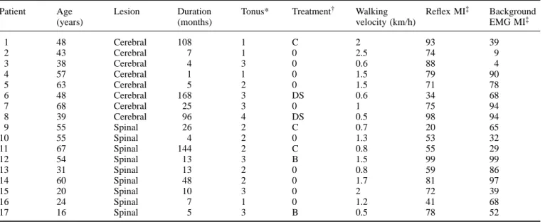
The ethical committees of the Universities of Freiburg and Zu¨rich approved the protocol and the patients gave informed consent before recordings were taken from 12 healthy subjects aged 19–34 years (mean6 SD: 24.9 6 4.1) and 28 spastic patients aged 16–68 years (48.36 14.6). All patients were diagnosed as having moderate spasticity due either to incomplete traumatic lesion of the cervical or thoracic spinal cord (n5 12) or unilateral cerebral stroke in the area of the middle cerebral artery (n 5 16). All patients presented exaggerated stretch reflexes and/or increased muscle tone as assessed by the Ashworth scale (Ashworth, 1964). The main clinical parameters of the 17 patients (eight with hemiparesis and nine with paraparesis), who were studied with the full protocol, are shown in Table 1.
Experimental paradigm
Each patient was required to walk, with the aid of a handrail, on a treadmill for at least 2–3 min at a speed of at least 0.5 km/h. Only 17 patients were able to perform the full
experimental protocol during standing and gait. Control experiments were also performed in 12 healthy subjects walking at a speed of 1.5 km/h (approximately the average speed of the patients).
Quadriceps tendon jerk reflexes were recorded during level split-belt treadmill locomotion at a comfortable pace for the patients (mean: 1.28 km/h, range 0.5–2.0 km/h). Tendon jerk reflexes were evoked by a 90 g hammer with an arc radius of 11 cm (see Dietz et al., 1990a). The hammer was driven by a revolving field magnetic motor (power 120 W) fixed to the anterior part of the calf (total weight 950 g). The hammer was accelerated by the motor up to a constant angular velocity of 800o/s. A potentiometer located at the motor’s axis of
rotation indicated the movement of the hammer and also indicated the time of impact of the hammer on the tendon. Force measuring platforms under each belt of the treadmill were connected to a circuit which triggered the hammer motor at a given delay after heel contact. At least 10 tendon reflexes were elicited during 16 different phases of the step cycle with a 4 s inter-stimulus interval. As a control the subjects produced various levels of controlled tonic quadriceps activity at different knee joint angles during standing. The change in activity was achieved by altering the body loading on the leg under examination. Visual feedback of the ongoing EMG activity enabled the subjects to keep the quadriceps EMG activity constant. The position thus adopted corresponded to those of the various phases of the step cycle.
Depending on the patients’ walking ability 6–16 different control conditions of standing, each including 10 tendon reflexes, were recorded. These control trials were interspersed with gait trials. Extended periods of rest were allowed between trials in order to avoid fatigue and to enable most of the patients to perform the full experimental protocol. In all patients both legs were examined on the same day. In the healthy control group only the right leg was examined.
Data recording
The EMG activity of the quadriceps was recorded by surface electrodes placed 3 cm apart over the belly of the rectus femoris muscle about 15–20 cm above the patella. EMG activity was also obtained from the biceps femoris, the tibialis anterior and the soleus. A sampling rate of 5 kHz was used and signals were filtered from 3 Hz to 1 kHz. Knee and hip joint movements were recorded using potentiometers as described earlier (Dietz et al., 1990a). Reflex responses were assessed as rectified and averaged EMG activity triggered by the tendon tap. To quantify the net reflex response, the corresponding rectified and averaged background EMG recorded during the step cycles without stimuli was subtracted. The reflex EMG signal was integrated over 25 ms after a latency of 26–33 ms following hammer impact. The duration of the EMG responses was typically ~25 ms. The latency and waveform of the EMG responses obtained
Table 1 Clinical and neurological features
Patient Age Lesion Duration Tonus* Treatment† Walking Reflex MI‡ Background
(years) (months) velocity (km/h) EMG MI‡
1 48 Cerebral 108 1 C 2 93 39 2 43 Cerebral 7 1 0 2.5 74 9 3 38 Cerebral 4 3 0 0.6 88 4 4 57 Cerebral 1 1 0 1.5 79 90 5 63 Cerebral 5 2 0 1.5 71 78 6 48 Cerebral 168 3 DS 0.6 34 68 7 68 Cerebral 25 3 0 1 75 94 8 39 Cerebral 96 4 DS 0.5 98 94 9 55 Spinal 26 2 C 0.7 20 65 10 55 Spinal 4 2 0 1.3 53 32 11 67 Spinal 144 2 C 0.8 55 29 12 54 Spinal 13 3 B 1.5 99 99 13 31 Spinal 13 2 0 0.8 59 86 14 60 Spinal 48 2 0 1.7 81 97 15 20 Spinal 10 3 0 2 72 39 16 24 Spinal 7 1 0 1.2 41 68 17 16 Spinal 5 3 B 0.5 78 52
*The degree of spasticity as assessed by Ashworth scale.†05 none, C 5 Clonazepam, DS 5 Dantrolene Sodium, B 5 Baclofen; ‡MI5 Modulation Index
indicated that they were indeed typical short latency reflexes evoked by the tendon jerk.
Assessment of reflex modulation during the step
cycle
In order to allow inter-individual comparison of reflex modulation during walking, the reflexes were normalized to the mean reflex response obtained in all 16 phases of the step cycle. For the assessment of the influence of EMG background activity on reflex modulation, the reflexes elicited during walking were compared with those elicited during the control trials, i.e. the reflex responses obtained at a similar level of EMG activity and knee joint angle during standing. For each phase of the step cycle the closest control condition was selected for comparison. Only differences of ,20% in the rectified background EMG activity of the quadriceps and ,10o for the knee joint angle were considered to be
comparable.
For each of the 16 phases of the step cycle the mean reflex size was calculated for each of the three groups of subjects (i.e. healthy subjects and patients with either paraparesis or hemiparesis). Statistical significance for the modulation in terms of the time-course of changes in reflex size during the step cycle and the reflex depression during gait compared with standing were evaluated by repeated measurements analysis of variance. After adjustment of the data for their skewness by adding a constant and calculating the logarithms we first investigated differences between subject groups and two effects (time-course and gait versus standing) within subject groups. For simplification of the model, the 16 phases were reduced to stance phase (mean of the first eight phases) and swing phase (mean of the last five phases) and the transition from stance to swing was omitted. Secondly, we
tested for an interaction between the time-course of the reflex changes and the reflex depression during gait compared with standing control. Thirdly, we carried out a three-way interaction performed to test the differences in time-course and reflex depression among groups of subjects. In the main model healthy subjects, paraparetic patients and the affected side of hemiparetic patients were included. As separate questions the affected and the unaffected side of hemiparetic patients were compared in a paired model and the unaffected side was also compared with healthy subjects.
To illustrate the degree of modulation during the step cycle in individual subjects the modulation index (MI) (according to Yang et al., 1991a) was calculated using the formula of Sinkjaer et al. (1996b):
MI 5 (max. reflex – min. reflex) * 100 / max. reflex where max. reflex and min. reflex were the maximum and minimum mean reflex size obtained during the stance and swing phases, respectively. The modulation index for the background EMG activity given by the integral of the quadriceps EMG activity in the corresponding 16 phases of the step cycle was calculated for each subject. Mean values and standard errors of the mean (SEM) were calculated for each of the three populations (i.e. healthy subjects and patients with either paraparesis or hemiparesis). Analysis of variance was performed and the significance of the difference between results obtained in the different populations was examined using the Scheffe´ test.
Results
Reflex changes during the step cycle of gait
Healthy subjects
Figure 1A and B show recordings taken from a healthy subject. Figure 1A displays the net reflex quadriceps EMG
570 M. Faist et al.
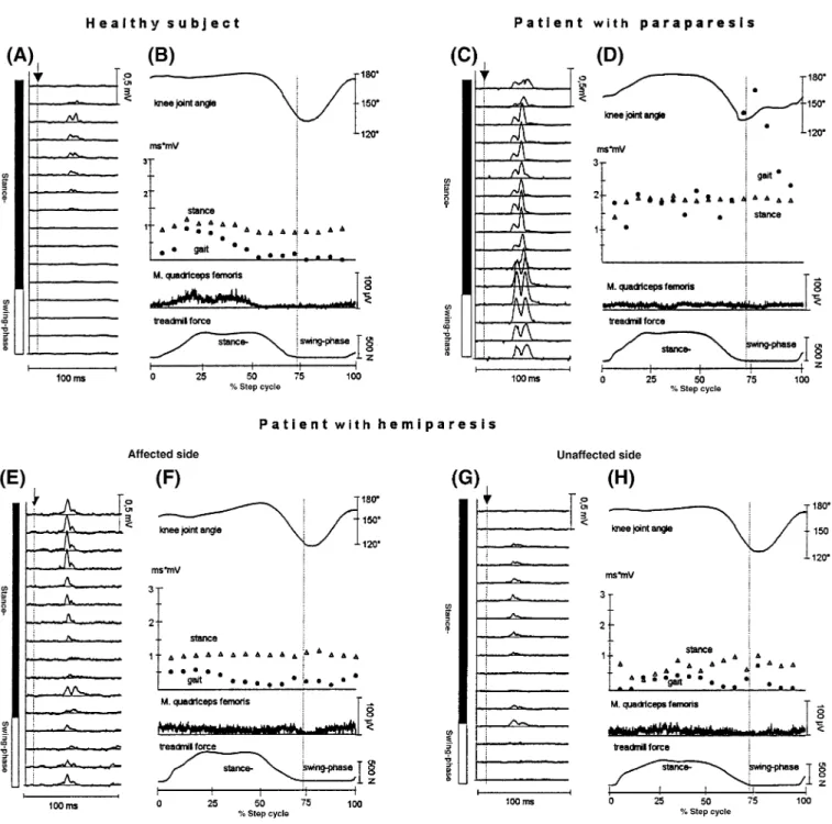
Fig. 1 Individual examples of reflex modulation during gait. A, Rectified and averaged (n5 10) net reflex responses, i.e. with
background EMG subtracted at 16 phases throughout the step cycle. The arrow and the vertical line indicate the time of hammer impact and the filled and open vertical bars the stance and swing phases, respectively. B, Quantitative values of the quadriceps tendon reflexes during the step cycle from an individual healthy subject (same subject as in A). The percentage of the step cycle at the time of the hammer impact was calculated from heel contact. Circles represent the EMG integral of the reflex responses obtained during gait, triangles represent the control reflexes obtained at a similar level of EMG activity during standing. The top trace shows the knee joint angle, the two bottom traces show the rectified EMG of the quadriceps and the treadmill force applied by the subject.
C and D, E and F, and G and H, The corresponding data to that described in A and B of a patient with paraparesis, the affected side of
a patient with hemiparesis and the unaffected side of the same hemiparetic patient, respectively.
activity (i.e. after subtraction of background EMG activity) at different times during the step cycle. Figure 1B illustrates the quantitative values of the reflex during gait and during the corresponding gait-mimicking standing control together with the biomechanical parameters (knee joint angle and treadmill force) as well as the rectified and averaged
quadriceps background EMG activity over one step cycle. It is evident that there was modulation of both the net quadriceps reflex response and backgound EMG activity during the step cycle. Little or no reflex activity was observed during the late stance phase or at any time during the swing phase. These effects are seen more clearly in Fig. 2A and B which
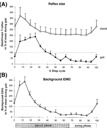
Fig. 2 Quantified values of the normalized quadriceps tendon
reflex during gait and standing. (A) Modulation of the tendon jerk reflex during gait and standing (control) at corresponding knee angles and voluntary EMG activity. To allow intersubject comparison, the reflex integrals were expressed as the percentage of the mean reflex observed during gait. Each symbol represents the mean of 12 healthy subjects. Vertical bars indicate one SEM. (B) Modulation of the integrated background quadriceps EMG activity during the step cycle. The background EMG values were normalized to the mean EMG value obtained during gait. Vertical bars indicate one SEM of the stance phase.
summarize the results obtained from all 12 healthy subjects. The size of the reflex responses was significantly reduced during the whole step cycle during gait compared with those recorded under gait-mimicking control conditions (Fig. 2A). These differences were especially so during the late stance and swing phases. Although the reflex modulation was similar in all subjects, a large inter-individual variability was observed as indicated by the large SEM.
Patients with spinal lesions
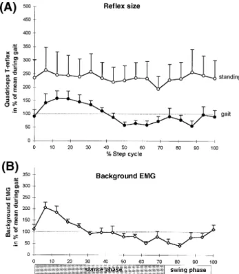
In patients with paraparesis, the modulation of both the reflex response and the background EMG during the step cycle was markedly reduced compared with healthy subjects. A typical example of the reflex modulation of one patient is shown in Fig. 1C and D. There was a large reflex response at all phases of the step cycle with the greatest responses being in the early and late stance phase and also during the swing phase. Figure 3 summarizes the results obtained from all nine patients with paraparesis. It is clear from Fig. 3A that there was no depression of the reflex response during the late stance or swing phases compared with the gait-mimicking
Fig. 3 Quantified values obtained from the group of patients with
paraparesis (n5 9). For details see legend to Fig. 2.
controls (cf. Fig. 2A for healthy subjects). Furthermore, compared with healthy subjects the background EMG (Fig. 3B) was less well modulated during the step cycle (cf. Figs 2B and 3B). Essentially similar results were obtained for both legs in patients with paraparesis. The mean data shown in Fig. 3 was taken from the more affected leg of the patients. If both legs were similarly affected then the data from the right leg was taken.
Patients with cerebral lesion—affected side
Figure 4 shows the results obtained from the affected leg of all eight patients with hemiparesis. Results from one subject are presented in Fig. 1E and F. It is evident that there was some modulation of the reflex response at different phases of the step cycle (Fig. 1F). Furthermore, compared with healthy subjects the reflexes in the swing phase were less depressed with respect to the gait-mimicking control (cf. Fig. 2A and Fig. 4A). However, compared with paraparetic patients a reflex depression was present with respect to the control condition (cf. Fig. 3A and Fig. 4A). There was still some modulation of background EMG activity in some hemiparetic patients (Fig. 4B).
Patients with cerebral lesion—unaffected side
Summarized data from the unaffected leg of hemiparetic patients is shown in Fig. 5. Individual recordings from one such patient are shown in Fig. 1G and H. The reflex
572 M. Faist et al.
Fig. 4 Quantified values obtained on the affected side from the
group of patients with hemiparesis (n5 8). For details see legend to Fig. 2.
modulation pattern in the unaffected leg was similar to that observed in healthy subjects (cf. Figs 5A and 2A). Both the reflex during gait (Fig. 5A) and the background EMG (Fig. 5B) were well modulated. However, compared with healthy subjects, there was no clear reflex depression with respect to gait-mimicking control during the stance phase and a more variable depression during the swing phase.
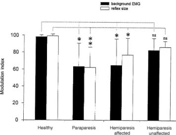
Modulation index
Figure 6 shows the modulation index of the reflex size (open columns) and background EMG (filled columns) for each subject group. The mean reflex modulation index and quadriceps background EMG activity (6 SD) for healthy subjects during gait was 99.26 2.1% (range 93–100%) and 98.16 2.2% (range 93–100%), respectively. In paraparetic patients the corresponding modulation index values were 62.2 6 23.7% (range 20–99%) and 63.3 6 27.2% (range 31–99%). The difference between the patients with paraparesis and the healthy control group was significant for both the reflex (P , 0.01) and the background EMG modulation index (P , 0.05). For the affected leg of the hemiparetic patients the reflex modulation index was 76.86 19.7% (range 34–98%; P , 0.05 compared with healthy subjects) and the background EMG modulation index was 64.86 30.8% (range 9–94%; P , 0.05 compared with healthy subjects). The reflex and background EMG modulation index values for the unaffected leg were 86.1 6 7% (range 77–
Fig. 5 Quantified values obtained on the unaffected side from the
group of patients with hemiparesis (n5 8). For details see legend to Fig. 2.
97%) and 82.56 14% (range 58–100%), respectively. These values were not statistically different from those of the healthy subjects (Fig. 6). It is important to note that there was no significant difference in the modulation index for either the reflex or background EMG data between the affected and unaffected legs of hemiparetic patients. There was also no significant difference in modulation index between the paraparetic patients and the affected legs of hemiparetic patients.
Time-course and depression of reflexes during
the step cycle
In the analysis of variance we found a significant (P , 0.0001) interaction between the time-course (reflex size modulation of the stance versus the swing phase of the step cycle) and the three groups of subjects (healthy, paraparetic, affected side of hemiparetic subjects). This indicates that there is a different reflex modulation in the stance and swing phases among the groups of subjects. The reflex depression during gait compared with standing control was significant as an overall effect across groups (P , 0.0001). The interaction between time-course and reflex depression, however, was not significant (P5 0.095), i.e. some depression was present to some extent in all subject groups, e.g. also during the stance phase in paraparetic patients. The three way interaction was significant (P, 0.0001) which indicates differences in the time-course of modulation and the reflex depression among the different subject groups and reflects
Fig. 6 Modulation indices in the different subject groups. Mean
values for each population are displayed. Open columns represent the modulation index for the reflex, closed columns the
modulation index for the background EMG. Vertical bars indicate SEM. Significance levels were calculated using the Scheffe´ test (*P, 0.05; **P , 0.01; ns 5 not significant).
the lack of reflex depression during the swing phase of the paraparetic subjects. The direct comparison between each of the three subject groups revealed significant differences for healthy versus paraparetic subjects (P , 0.0001), healthy versus hemiparetic subjects (P, 0.05) and paraparetic versus hemiparetic subjects (P, 0.01).
As a separate question the unaffected side of hemiparetic patients was compared with the affected side in a paired design. A significant difference was present between the two sides of hemiparetic patients as a general effect (P, 0.05) and for the three-way interaction (P, 0.05) while the two-way interactions were not significant. Finally, the unaffected side was compared with healthy control subjects. The interaction of the time-course and the two groups was significant (P, 0.01). The reflex depression was significant as an overall effect across both groups (P, 0.0001) but the interaction of depression and groups and also the three-way interaction only showed a trend (P5 0.069 and P 5 0.056, respectively).
Reflex size and background activity: differences
between patients with cerebral and spinal
lesions
Figure 7 shows relative (part A) and absolute (part B) values of reflex size during an entire step cycle (as an average of the reflex size in the 16 phases of the step cycle) in the three groups of subjects. When the reflex size during gait was related to the standing condition (Fig. 7A) the relative reflex size was largest in the group of patients with a spinal lesion. A significant reflex depression was seen in both the group of healthy subjects (P , 0.001) and on the affected leg of
Fig. 7 Quantified values of the mean reflex size during a step
cycle (i.e. average of reflex size over the 16 phases of the step cycle) of the three groups of subjects. In A the reflex size during gait is normalized to the value of reflex size obtained in the standing condition. In B the absolute values of the integrated background EMG (closed columns) and reflex size during gait (open columns) and standing (hatched columns) are displayed for the three groups of subjects. Vertical bars indicate one standard deviation. Significance levels were calculated by the Scheffe´ test (**P, 0.01; ***P , 0.001).
the patients with a cerebral lesion (P , 0.01). A more detailed analysis is given in Fig. 7B where the absolute (integrated) EMG values for the entire step cycle are presented.
Compared with healthy subjects, the reflex size was largest in patients with a spinal lesion (gait P, 0.01; standing P , 0.05) but not significantly different from both legs of patients with hemiparesis. Compared with patients with a spinal lesion, the reflex size in patients with a cerebral lesion was significantly smaller on the affected side (gait P , 0.05; standing P, 0.05) and the unaffected side (gait P , 0.01; reflex P, 0.05), although the background EMG did not differ significantly between the groups. There was no difference in the reflex size during gait or standing between both sides of the hemiparetic patients. Corresponding to the results shown
574 M. Faist et al.
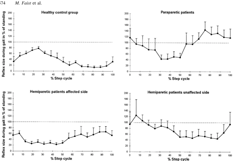
Fig. 8 Gait reflex in relation to the standing control for the different groups of subjects throughout the step cycle. Each symbol
represents the mean of all subjects in the respective group in the respective phase of the step cycle. Vertical bars indicate one SEM.
in Fig. 7A, a significant difference between gait and control reflex was present only in healthy subjects and the affected side of patients with hemiparesis.
Reflex size during gait with respect to control
values
Figure 8 illustrates for the subject groups the mean rectus femoris tendon jerk reflexes obtained during the 16 phases of gait expressed as a percentage of the gait-mimicking control value during standing. As in Fig. 7A, a reflex depression was present for healthy subjects and the hemiparetic patients throughout the step cycle. In the paraparetic patients a lack of depression during the swing phase was most remarkable.
Background activity in other major leg muscles
during gait
In the present study for biceps femoris, tibialis anterior and soleus muscles, no relationship between background EMG activity and rectus femoris reflex size was seen during gait. The background EMG activity of the biceps femoris in healthy subjects showed maximal activity in the late swing and early stance phases. In the paraparetic patients and on
the affected side of hemiparetic patients biceps femoris EMG was largest during late swing and the first half of stance phase. A less pronounced modulation was seen on the unaffected side. The strongest modulation of background EMG occurred in the patient groups—similar to healthy subjects—in the tibialis anterior during swing and in the soleus in the midstance.
Discussion
The aim of this study was to evaluate the changes in behaviour of quadriceps short latency reflexes during spastic gait. While recent studies have investigated the changes in soleus/ gastrocnemius reflex behaviour in spastic patients (Yang et al., 1991a; Sinkjaer et al., 1995, 1996b), the present study, to our knowledge, is the first to investigate quadriceps reflex modulation in patients with either spinal or supraspinal lesions. Two questions were addressed. (i) Are there differences between spasticity of spinal or cerebral origin in the modulation or depression of short latency reflexes during locomotion? This possibility has been extrapolated from resting conditions (see Faist et al., 1994). (ii) Is the reflex modulation on the clinically ‘unaffected’ side in patients with hemiparesis different from that in healthy subjects? This has
been proposed previously based upon arm reflexes studied under passive conditions (Thilmann et al., 1990).
The main results obtained in this study were as follows. (i) Differences in the depression of the short latency tendon jerk reflex were revealed between patients with paraparesis and the affected leg in patients with hemiparesis. (ii) Reflex depression and modulation were maximally reduced in patients with paraparesis compared with healthy subjects. (iii) There was a reduced reflex modulation on the affected side of patients with hemiparesis compared to healthy subjects. (iv) An impaired reflex depression can be assumed on the clinically unaffected side of patients with hemiparesis.
Validity of the comparison
The differences in reflex modulation pattern during the step cycle between the groups of subjects were not due to an altered motor neuronal excitability because the time-course of the background EMG activity was changed in a similar fashion in the spastic legs of patients with either paraparesis or hemiparesis (see Figs 3 and 4). To allow comparison of reflex size between different subjects the reflex size was normalized to the mean reflex during gait. To assess the influence of background EMG activity all subjects were tested at corresponding levels of muscle contraction during standing. It was evident that the alteration of the reflex modulation pattern and the reflex size were related to the site of the lesion (i.e. spinal or supraspinal, see Fig. 8).
It was possible that biomechanical changes during the different step phases contributed to the modulation of the reflex size observed. In an attempt to negate these effects a number of conditions were imposed. The impact of the hammer at the tendon occurred at a constant angular velocity in all step phases (see Dietz et al., 1990b). Differences in background EMG activity and thus differences in the tension of the tendon were taken into account by the control condition. The different knee joint angles were also comparable between gait and standing. Nevertheless, it was not possible to quantify force or tension exerted by tendons in this control condition. Therefore, we are aware that this control is less than ideal, although it represents the best possible approach to perform these types of experiments in spastic patients.
The asymmetric gait pattern of patients with hemiparesis compared with the patients with paraparesis could also have been responsible for some of the differences observed. Furthermore, changes in the gamma drive to muscle spindles as well as changes in presynaptic inhibition of group Ia afferents or other spinal mechanisms (see below) may also have accounted for the differences. Although the mechanical conditions should have been similar in all three groups of subjects, especially during the swing phase, the strongest differences in reflex responses were observed during swing. In spite of the fact that the afferent stimulus of the H-reflex bypasses the muscle spindles, the quadriceps (Dietz et al., 1990a) and soleus (Llewllyn et al., 1987; M. Faist and W. Berger, unpublished observations) tendon tap reflexes in
healthy subjects were modulated in a similar way during gait as were the H-reflexes (quadriceps: Dietz et al., 1990b; soleus: Faist et al., 1996a, their Fig. 5B). Although some differences were reported between the modulation of soleus stretch reflexes (Sinkjaer et al., 1996a) and H-reflexes (Capaday and Stein, 1986) regarding the depression during gait, all described a maximum in the late stance phase. Furthermore, not all studies showed a depression in all phases of the step cycle for the H-reflex of the soleus (cf. Faist et al., 1996a, their Fig. 5B) or the quadriceps (Dietz et al., 1990b). This indicates that there is no dramatic change in the gamma drive to the respective muscle spindles in healthy subjects during locomotion. Data available for the ankle joint in spinal lesion-induced spasticity showed that the modulation of both soleus H-reflexes (Yang et al., 1991a) and stretch reflexes (Sinkjaer et al., 1996b) is severely disturbed during locomotion, i.e. the modulation index is reduced and the maximum reflex is not found during midstance. For hemiparetic patients soleus H-reflexes and tendon tap reflexes are modulated in parallel during gait in a similar fashion as in healthy subjects (M. Faist and W. Berger, unpublished observations). This would argue against the assumption that an altered gamma drive is responsible for the changes observed in patients with paraparesis and hemiparesis. Finally, it could be argued that the reduced modulation found in the paraparetic patients might be due to a ceiling effect, i.e. that a saturation of reflex amplitude occurred with the consequence that the reflex became more or less insensitive to any modulatory effect during gait. This is unlikely for the following reasons. (i) Reflexes in paraparetic patients were smaller during the stance phase than in the swing phase which indicates that the reflexes during the stance phase were not yet maximal and thus still susceptible to modulation. (ii) Although the reflex size was smaller in hemiparetic compared with paraparetic patients, the modulation index was reduced in both patient groups.
Differences between patients with paraparesis
and hemiparesis
The present results for quadriceps tendon tap reflex modulation in patients with paraparesis agree with previous studies for the ankle joint which have suggested that modulation of both the soleus H and stretch reflexes during gait is reduced (Yang et al., 1991a; Sinkjaer et al., 1995, 1996b). It is therefore conceivable that for paraparetic patients a similarly impaired quadriceps H-reflex modulation may be present. To the best of our knowledge only one study published for hemiparetic patients has investigated soleus H-reflexes during gait in a small sample of three patients after head injury. A partly preserved modulation was present at least in the one subject whose data were presented separately (Yang et al., 1991a, their Fig. 6). However, due to the small number of subjects, no significant differences were found compared with spinal cord injured patients.
576 M. Faist et al.
In the present study, the clinical signs of spasticity were similar in the two patient groups, but the reflex modulation and size clearly differed compared with control conditions during standing. In contrast to paraparetic patients there was a partly preserved reflex depression on the affected side of hemiparetic patients while on the unaffected side no significant reflex depression with respect to standing was seen, although the direct statistical comparison only showed a trend. This observation that also the clinically unaffected side of patients with hemiparesis may not be ‘normal’ fits with the behaviour of elbow and ankle joint stretch reflexes in passive muscles of hemiparetic patients (Thilmann et al., 1990; Thilmann and Fellows, 1991).
There exists little correlation between impaired stretch reflex mechanisms and the clinical symptoms of spasticity (see O’Dwyer et al., 1996) or spastic movement disorder (Dietz et al., 1981, 1994, 1995). To date only reciprocal Ia inhibition was shown to be decreased in spasticity of both spinal and cerebral origin (Artieda et al., 1991; Crone et al., 1994; Boorman et al., 1996). This decrease has been correlated with clinical signs of spasticity in patients with hemiparesis (Okuma and Lee, 1996). In contrast to these more recent data an increase of reciprocal inhibition from ankle dorsi- to plantarflexors (Boorman et al., 1991) and from plantar- to dorsiflexors (Ashby and Wiens, 1989) was found in patients with incomplete spinal lesion.
Depending on the site of the lesion, there are disturbances of various other spinal reflex mechanisms which may contribute to the hyperexcitability of short latency stretch reflexes (see Introduction). However, in neither of the latter investigations was a correlation found between the impairment of spinal reflex mechanisms and clinical signs of spasticity. Furthermore, the present observations indicate that pathophysiological differences in reflex behaviour between spasticity of spinal or cerebral origin are not reflected in the clinical examination. The difference in the reflex behaviour between patients with spinal and cerebral lesions found in this study indicates that some common features of spasticity, such as increased muscle tone, may develop independently from hyperactive short latency stretch reflexes. A similar suggestion has been proposed from studies on the response of spastic passive (O’Dwyer et al., 1996) and active muscles (Dietz et al., 1981; Berger et al., 1984, 1988; Ibrahim et al., 1993) to stretch. If disturbances in spinal reflex mechanisms do have a causal relationship to the spastic movement disorder then their differential impairment could reflect pathophysiological differences between spasticity of spinal or cerebral origin. This, in turn, could have important consequences for the therapeutic approach that is chosen (see Functional considerations).
Mechanisms contributing to the differences in
reflex modulation
Part of the net modulation of short latency reflexes during the step cycle has been attributed to rhythmic changes of
presynaptic inhibition of Ia afferents (see Introduction). As this study investigated tendon jerk reflexes the contribution from presynaptic inhibition or, alternatively, of gamma-motor neuron activity to the observed reflex modulation cannot be determined. The present investigation included a gait-mimicking standing control condition not included in previous studies. A rough matching of the strength of the EMG activity and leg geometry could be achieved by using such a control condition. However, one has to admit that the tonic quadriceps activity during standing is physiologically different from that during the step cycle even with the same level of EMG activity. The quadriceps tendon reflex was significantly smaller during gait of healthy subjects than during the control condition. A similar depression during gait compared with standing has been reported for the soleus and the vastus medialis muscle H-reflexes (Capaday and Stein, 1986; Brooke et al., 1991). This supports the hypothesis that in healthy subjects, changes in presynaptic inhibition contribute to the net modulation of the tendon reflex during gait.
Compared with healthy subjects reflex depression was almost removed in patients with spinal cord lesion during gait, in relation to standing. In the latter patients the net reflex size (absolute values) was also strongly enhanced during locomotion and during the control condition, while it was less enhanced in relation to the background EMG on the affected side and not significantly changed on the unaffected side in patients with cerebral lesions, compared with healthy subjects. These findings are compatible with the hypothesis that in patients with paraparesis, presynaptic inhibition of quadriceps Ia afferents is disturbed during gait, an effect also observed in soleus Ia afferents (Yang et al., 1991a). A similar differential effect of presynaptic inhibition between patients with paraparesis and hemiparesis has been described under resting conditions (Faist et al., 1994). In the present study the average age in the patient group was higher than in the control group. As presynaptic inhibition might increase with human ageing (Morita et al., 1995) one would expect a decrease in reflex size in the patient group. However, reflexes were rather enhanced especially in the paraparetic patients.
Alternative explanations for the impaired reflex behaviour might be as follows. (i) Descending tract lesions might exert a differential effect on low- and high-threshold motor neurons, thus producing a compression of the range of functional thresholds in the motor neuron pool and thereby an increase in the input–output relationship (‘recruitment gain’) of the reflex (Kernell and Hultborn, 1990). As a result the same Ia afferent input would excite more motor neurons than in healthy subjects and thus increase the reflex in spastic patients. However, in this case one would expect that only the reflex level is enhanced, but that the rhythmic reflex modulation during the step cycle remains preserved which was not the case in the paraparetic patients. (ii) Homo-synaptic post-activation depression is probably related to a reduced transmitter release (Hultborn et al., 1996), which is decreased in spasticity (Nielsen and Hultborn, 1993). As the
time course of this depression is in the range of 10 s it should not alter the reflex modulation in a muscle which is rhythmically active during gait. More serious problems might occur if a resting condition is compared with voluntary contraction. (iii) Widespread disturbances of other spinal interneuronal mechanisms might occur secondary to a defective control of Renshaw cell activity (Mazzochio and Rossi, 1997). At present no data are available on changes in recurrent inhibition contributing to reflex changes in spastic gait. In the resting muscle recurrent inhibition was estimated to be unchanged or increased following cerebral lesions (Katz and Pierrot-Deseilligny, 1982). During postural or voluntary contractions in most patients a defective supraspinal control of Renshaw cell excitability was found which was interpreted as causing difficulties in grading the strength of a muscular contraction and regulating reciprocal Ia inhibition according to the requirements of a voluntary contraction. (iv) The lack of reflex depression during the swing phase might be due to a decrease in reciprocal inhibition especially in the ankle muscles. To our knowledge no experimental data are available concerning the effect of reciprocal inhibition on the modulation of tendon jerk reflexes during gait. In healthy subjects the biceps femoris is most active at the end of the swing and in the early stance phase. In all patient groups the background activity of the biceps femoris was also largest at the end of the swing and early stance so that the lack of reflex depression during the entire swing phase cannot be explained exclusively by reduced antagonistic activity. (v) Finally, group II inhibition was found to be decreased in spinal spasticity (Eriksson et al., 1996) and could contribute to the reflex changes in spastic patients.
It cannot be decided from our experiments which one or which combination of mechanisms established above is responsible for our observations. Irrespective of the mechanisms contributing to the reflex changes, the difference between patients with paraparesis and hemiparesis observed here may be due to compensation by novel ipsilateral motor pathways with abnormal branching on brainstem/spinal level as is suggested in hemiplegic cerebral palsy (Carr et al., 1993). Another explanation is bypassing the lesioned pyramidal tract by polysynaptic corticoreticulospinal connections. This view is supported by the observation that suprathreshold stimulation of the affected hemisphere in hemiplegic patients could evoke a bilateral response (Fries et al., 1991). The extent to which peripheral or central inputs are responsible for the modulation of group Ia input during locomotion has not yet been determined (see Yang et al., 1991b; Yang and Whelan, 1993; Misiaszek et al., 1995; Stein, 1995). Alternatively, the differences may be due to a change in proprioceptive feedback mechanisms. As described recently, rhythmic peripheral feedback is induced by locomotion which may contribute to the rhythmic reflex modulation seen in healthy subjects (Fung and Barbeau, 1994; Misiaszek et al., 1995). Theoretically, afferent information arising from the unaffected side of patients with hemiparesis may be sufficient to preserve part of the rhythmic reflex modulation. As both
sides are affected in paraparetic patients the reflex modulation would be expected to be more severely disturbed.
Functional considerations
The modulation of short latency reflexes of the triceps surae is considered to be of importance during gait in intact humans (Capaday and Stein, 1986, 1987; Crenna and Frigo, 1987). Quadriceps reflex modulation has been interpreted as being necessary to allow the yielding of the knee during the stance phase (Dietz et al., 1990a, b). In addition, a reflex depression of extensor stretch reflexes during swing is functionally appropriate, as passive stretch of the quadriceps and the triceps surae muscles without eliciting a reflex response is necessary for adequate foot clearance during the swing phase of gait. In spasticity of spinal origin this mechanism is defective and stretch reflexes are elicited during this phase of the step cycle. This may contribute to the spastic movement disorder of such patients. The action of most antispastic drugs (e.g. Baclofen and Clonazepam) is directed towards reducing the reflex excitability in spastic patients (cf. Dietz and Young, 1996). However, in the light of the present experiments this approach would be pointless in patients with hemiparesis as they show a largely preserved reflex depression during gait and no functional benefit would be expected by additional depression of reflex activity.
Acknowledgements
The authors wish to thank Mrs U. Ro¨mmelt and Mr F. Pfister for their excellent technical assistance, Professor E. Pierrot-Deseilligny and Dr I. Gibson for valuable editorial help and Professor J. Schulte-Mo¨nting, R. Rossner and Th. Erni for statistical analysis. This work was supported by grants from Deutsche Forschungsgemeinschaft (Be 936/4–1), Bundesministerium fu¨r Bildung und Forschung (01KL9402), the Swiss National Foundation (Grant No. 31–53526.98) and from the Schweizer Bankgesellschaft on behalf of a client.
References
Artieda J, Quesada P, Obeso JA. Reciprocal inhibition between forearm muscles in spastic hemiplegia. Neurology 1991; 41: 286–9. Ashby P, Wiens M. Reciprocal inhibition following lesions of the spinal cord in man. J Physiol (Lond) 1989; 414: 145–57.
Ashworth B. Preliminary trial of carisoprodol in multiple sclerosis. Practitioner 1964; 192: 540–2.
Berger W, Horstmann G, Dietz V. Tension development and muscle activation in the leg during gait in spastic hemi-paresis: independence of muscle hypertonia and exaggerated stretch reflexes. J Neurol Neurosurg Psychiatry 1984; 47: 1029–33.
Berger W, Horstmann G, Dietz V. Spastic paresis: impaired spinal reflexes and intact motor programs. J Neurol Neurosurg Psychiatry 1988; 51: 568–71.
578 M. Faist et al.
Boorman G, Hulliger M, Lee RG, Tako K, Tanaka R. Reciprocal Ia inhibition in patients with spinal spasticity. Neurosci Lett 1991; 127: 57–60.
Boorman GI, Lee RG, Becker WJ, Windhorst UR. Impaired ‘natural reciprocal inhibition’ in patients with spasticity due to incomplete spinal cord injury. Electroencephalogr Clin Neurophysiol 1996; 101: 84–92.
Brooke JD, Collins DF, Boucher S, McIlroy WE. Modulation of human short latency reflexes between standing and walking. Brain Res 1991; 548: 172–8.
Capaday C, Stein RB. Amplitude modulation of the soleus H-reflex in the human during walking and standing. J Neurosci 1986; 6: 1308–13.
Capaday C, Stein RB. Difference in the amplitude of the human soleus H reflex during walking and running. J Physiol (Lond) 1987; 392: 513–22.
Carr LJ, Harrison LM, Evans AL, Stephens JA. Patterns of central motor reorganization in hemiplegic cerebral palsy. Brain 1993; 116: 1223–47.
Crenna P, Frigo C. Excitability of the soleus H-reflex arc during walking and stepping in man. Exp Brain Res 1987; 66: 49–60. Crommert HW van de, Faist M, Berger W, Duysens J. Biceps femoris tendon jerk reflexes are enhanced at the end of the swing phase in humans. Brain Res 1996; 734: 341–4.
Crone C, Nielsen J, Petersen N, Ballegaard M, Hultborn H. Disynaptic reciprocal inhibition of ankle extensors in spastic patients. Brain 1994; 117: 1161–8.
Delwaide PJ, Oliver E. Short-latency autogenic inhibition (IB inhibition) in human spasticity. J Neurol Neurosurg Psychiatry 1988; 51: 1546–50.
Dietz V, Young RR. The syndrome of spastic paresis. In: Brandt T, Caplan LR, Dichgans J, Diener HC, Kennard C, editors. Neurological disorders. Course and Treatment. San Diego: Academic Press; 1996. p. 861–71.
Dietz V, Quintern J, Berger W. Electrophysiological studies of gait in spasticity and rigidity: evidence that altered mechanical properties of muscle contribute to hypertonia. Brain 1981; 104: 431–49. Dietz V, Discher M, Faist M, Trippel M. Amplitude modulation of the human quadriceps tendon jerk reflex during gait. Exp Brain Res 1990a; 82: 211–3.
Dietz V, Faist M, Pierrot-Deseilligny E. Amplitude modulation of the quadriceps H-reflex in the human during the early stance phase of gait. Exp Brain Res 1990b; 79: 221–4.
Dietz V, Colombo G, Jensen L. Locomotor activity in spinal man. Lancet 1994; 344: 1260–3.
Dietz V, Colombo G, Jensen L, Baumgartner L. Locomotor capacity of spinal cord in paraplegic patients [see comments]. Ann Neurol 1995; 37: 574–82. Comment in: Ann Neurol 1995; 37: 555–6. Downes L, Ashby P, Bugaresti J. Reflex effects from Golgi tendon organ (Ib) afferents are unchanged after spinal cord lesions in humans. Neurology 1995; 45: 1720–4.
Eriksson J, Olausson B, Jankowska E. Antispastic effects of L-dopa. Exp Brain Res 1996; 111: 296–304.
Faist M, Mazevet D, Dietz V, Pierrot-Deseilligny E. A quantitative assessment of presynaptic inhibition of Ia afferents in spastics: differences in hemiplegics and paraplegics. Brain 1994; 117: 1449–55.
Faist M, Dietz V, Pierrot-Deseilligny E. Modulation, probably presynaptic in origin, of monosynaptic Ia excitation during human gait. Exp Brain Res 1996a; 109: 441–9.
Faist M, Ertel M, Berger W. Modulation of the quadriceps tendon jerk reflex during gait in hemispastic patients [abstract]. Electroencephalogr Clin Neurophysiol 1996b; 99: 367.
Fries W, Danek A, Witt TN. Motor responses after transcranial electrical stimulation of cerebral hemispheres with a degenerated pyramidal tract. Ann Neurol 1991; 29: 646–50.
Fung J, Barbeau H. Effects of conditioning cutaneomuscular stimulation on the soleus H-reflex in normal and spastic paretic subjects during walking and standing. J Neurophysiol 1994; 72: 2090–104.
Hultborn H, Illert M, Nielsen J, Paul A, Ballegaard M, Wiese H. On the mechanism of the post-activation depression of the H-reflex in human subjects. Exp Brain Res 1996; 108: 450–62.
Ibrahim IK, Berger W, Trippel M, Dietz V. Stretch-induced electromyographic activity and torque in spastic elbow muscles. Differential modulation of reflex activity in passive and active motor tasks. Brain 1993; 116: 971–89.
Katz R, Pierrot-Deseilligny E. Recurrent inhibition of alpha-motor neurons in patients with upper motor neuron lesions. Brain 1982; 105: 103–24.
Kernell D, Hultborn H. Synaptic effects on recruitment gain: a mechanism of importance for the input-output relations of motor neurone pools? Brain Res 1990; 507: 176–9.
Llewellyn M, Prochazka A, Vincent S. Transmission of human tendon jerk reflexes during stance and gait. J Physiol (Lond) 1987; 382: 82P.
Mazzocchio R, Rossi A. Involvement of spinal recurrent inhibition in spasticity. Further insight into the regulation of Renshaw cell activity. Brain 1997; 120: 991–1003.
Misiaszek JE, Barclay JK, Brooke JD. Inhibition of canine H reflexes during locomotor-like rotation about the knee arises from muscle mechanoreceptors in quadriceps. J Neurophysiol 1995; 73: 2499–506.
Morin C, Katz R, Mazie`res L, Pierrot-Deseilligny E. Comparison of soleus H reflex facilitation at the onset of soleus contractions produced voluntarily and during the stance phase of human gait. Neurosci Lett 1982; 33: 47–53.
Morita H, Shindo M, Yanagawa S, Yoshida T, Momoi H, Yanagisawa N. Progressive decrease in heteronymous monosynaptic Ia facilitation with human ageing. Exp Brain Res 1995; 104: 167–70.
Nielsen J, Hultborn H. Regulated properties of notoneurons and primary afferents: new aspects on possible spinal mechanisms underlying spasticity. In: Thilman AF, Burke DJ, Rymer WZ, editors. Spasticity: mechanisms and management. Berlin: Springer Verlag; 1993. p.177–92.
Nielsen J, Petersen N, Crone C. Changes in transmission across synapses of Ia afferents in spastic patients. Brain 1995; 118: 995–1004.
O’Dwyer NJ, Ada LM, Neilson PD. Spasticity and muscle contracture following stroke. Brain 1996; 119: 1737–49.
Okuma Y, Lee RG. Reciprocal inhibition in hemiplegia: correlation with clinical features and recovery. Can J Neurol Sci 1996; 23: 15–23. Raynor EM, Shefner JM. Recurrent inhibition is decreased in patients with amyotrophic lateral sclerosis. Neurology 1994; 44: 2148–53.
Shefner JM, Berman SA, Sarkarati M, Young RR. Recurrent inhibition is increased in patients with spinal cord injury. Neurology 1992; 42: 2162–8.
Sinkjaer T, Toft E, Hansen HJ. H-reflex modulation during gait in multiple sclerosis patients with spasticity. Acta Neurol Scand 1995; 91: 239–46.
Sinkjaer T, Andersen JB, Larsen B. Soleus stretch reflex modulation during gait in humans. J Neurophysiol 1996a; 76: 1112–20.
Sinkjaer T, Andersen JB, Nielsen JF. Impaired stretch reflex and joint torque modulation during spastic gait in multiple scleorsis patients. J Neurol 1996b; 243: 566–74.
Stein RB. Presynaptic inhibition in humans. [Review]. Prog Neurobiol 1995; 47: 533–44.
Thilmann AF, Fellows SJ. The time-course of bilateral changes in the reflex excitability of relaxed triceps surae muscle in human hemiparetic spasticity. J Neurol 1991; 238: 293–8.
Thilmann AF, Fellows SJ, Garms E. Pathological stretch reflexes on the ‘good’ side of hemiparetic patients. J Neurol Neurosurg Psychiatry 1990; 53: 208–14.
Yang JF, Whelan PJ. Neural mechanisms that contribute to cyclical modulation of the soleus H-reflex in walking in humans. Exp Brain Res 1993; 95: 547–56.
Yang JF, Fung J, Edamura M, Blunt R, Stein RB, Barbeau H. H-reflex modulation during walking in spastic paretic subjects. Can J Neurol Sci 1991a; 18: 443–52.
Yang JF, Stein RB, James KB. Contribution of peripheral afferents to the activation of the soleus muscle during walking in humans. Exp Brain Res 1991b; 87: 679–87.