Annals of Oncology 24: 1378–1384, 2013 doi:10.1093/annonc/mds646 Published online 31 January 2013
Large genomic aberrations detected by SNP array are
independent prognosticators of a shorter time to
first
treatment in chronic lymphocytic leukemia patients
with normal FISH
M. Mian
1,2, A. Rinaldi
1, A. A. Mensah
1, D. Rossi
3, M. Ladetto
4, F. Forconi
5,6, R. Marasca
7, M. Uhr
8,
G. Stussi
8, I. Kwee
1,9,10, F. Cavalli
8, G. Gaidano
3, E. Zucca
8& F. Bertoni
1,8*
1
Lymphoma and Genomics Research Program, IOR Institute of Oncology Research, Bellinzona, Switzerland;2
Division of Hematology & CBMT, Hospital of Bolzano, Bolzano;3
Division of Hematology, Department of Translational Medicine, Amedeo Avogadro University of Eastern Piedmont, Novara;4
Division of Hematology, Department of Experimental Medicine and Oncology, University of Turin, Turin, Italy;5
Cancer Sciences Unit, CRUK Clinical Centre, University of Southampton, Southampton, UK;6
Division of Hematology, University of Siena, Siena;7
Division of Hematology, Department of Oncology and Hematology, University of Modena and Reggio Emilia, Modena, Italy;8
Lymphoma Unit and Hematology, IOSI Oncology Institute of Southern Switzerland, Bellinzona;9IDSIA Dalle Molle Institute for Artificial
Intelligence, Manno;10
SIB Swiss Institute of Bioinformatics, Lausanne, Switzerland
Received 21 July 2012; revised 25 October 2012; accepted 3 December 2012
Background:Genomic complexity can predict the clinical course of patients affected by chronic lymphocytic leukemia (CLL) with a normal FISH. However, large studies are still lacking. Here, we analyzed a large series of CLL patients and also carried out the so far largest comparison of FISH versus single-nucleotide polymorphism (SNP) array in this disease.
Patients and methods:SNP-array data were derived from a previously reported dataset.
Results:Seventy-seven of 329 CLL patients (23%) presented with a normal FISH. At least one large (>5 Mb) genomic aberration was detected by SNP array in 17 of 77 patients (22%); thisfinding significantly affected TTT. There was no correlation with the presence of TP53 mutations. In multivariate analysis, including age, Binet stage, IGHV genes mutational status and large genomic lesion, the latter three factors emerged as independent prognosticators. The concordance between FISH and SNP array varied between 84 and 97%, depending on the specific genomic locus investigated.
Conclusions:SNP array detected additional large genomic aberrations not covered by the standard FISH panel predicting the outcome of CLL patients.
Key words: affymetrix, chronic lymphocytic leukemia, FISH, microarray, prognosis, TP53
introduction
The clinical course of patients affected by chronic lymphocytic leukemia (CLL) is very heterogeneous and ranges from an indolent and chronic disease to a rapidly progressing leukemia or lymphoma necessitating aggressive treatment [1–3]. Several parameters, such as staging, lymphocyte doubling time, CD38, ZAP70, the mutational status of the variable region of the immunoglobulin heavy chain (IGHV) and the presence of specific genomic aberrations, are often used to stratify patients based upon their expected clinical outcome [2–5]. A prognostic model based on genomic aberrations (del 13q14.3, del 11q22.3,
del 17p13.1, trisomy 12) detected byfluorescent in situ hybridization (FISH) is also largely used [3,6].
In the last few years, high density, single-nucleotide polymorphism (SNP) genomic arrays (SNP arrays) proved to be efficient techniques to identify genetic aberrations in malignant cells [7]. The major advantage of this approach in comparison to FISH is that the whole genome can be scanned for genomic aberrations at once and not only at a limited number of regions. Moreover, recent data have shown that the prediction of the outcome of CLL patients might be improved by the use of SNP arrays. The detection of individual genetic aberrations other than the ones detected by the standard FISH panel [8], or of an overall increased genomic complexity [9,10] or of the capability to measure the size of the genomic
aberrations [11–13], all appear to have clinical implications, better defining the clinical outcome of CLL patients. Very recently, by carrying out cytogenetic analysis with DSP30/IL2
*Correspondence to: Dr F. Bertoni, Lymphoma and Genomics Research Program, IOR Institute of Oncology Research, IOSI Oncology Institute of Southern Switzerland, via Vincenzo Vela 6, 6500 Bellinzona, Switzerland. Tel: 8200-367; Fax: +41-91-8200-397; E-mail: frbertoni@mac.com
© The Author 2013. Published by Oxford University Press on behalf of the European Society for Medical Oncology. All rights reserved. For permissions, please email: journals.permissions@oup.com.
stimulation of CLL cells, Rigolin et al. [14] have shown that the presence of an abnormal karyotype correlates with a shorter time tofirst treatment (TTT) and shorter overall survival (OS). Although, in the last few years several investigators have described the application of microarrays in CLL [8,10,15–22], only a few of them have reported a detailed comparison with FISH, including the clinical implication of using a SNP array instead of the FISH panel to predict the outcome at diagnosis. In order to evaluate whether SNP array can better define the outcome of CLL patients with a normal FISH and can replace FISH, we analyzed a large group of CLL patients, characterized with both SNP array and the standard FISH panel.
materials and methods
tumor panelCLL samples from a previously published series were investigated [8]. All samples were collected at diagnosis and had an immunophenotypic score of >3 according to Matutes et al. [23]. Patients provided informed consent in accordance with local institutional review board requirements and the Declaration of Helsinki. The study was approved by the Ethical Committee of Bellinzona. CLL diagnosis and management were based on the National Cancer Institute working group criteria [3]. Cases of monoclonal B-cell lymphocytosis were discarded. Baseline clinical and molecular characteristics have been collected as previously described [8]. The FISH panel interrogated deletions at 13q14.3, 11q22, 17p13 and trisomy 12 [8].
SNP arrays
Copy-number data, obtained from high-molecular-weight genomic DNA of CLL patients using the Genome-Wide Human SNP Array Version 6.0 (Affymetrix, Santa Clara, CA), were derived from a previously reported dataset [8]. The bioinformatics pipeline used for the identification of
copy-number alterations is described in detail by Rinaldi et al. [8]. In addition, raw and smoothed copy-number values of each sample were plotted for quality assessment. Copy-number aberrations >5 Mb were identified by filtering on segment size and confirmed by visual inspection of the plotted genomic profiles. The cut-off was stabilized according to a previously published experience [24]. Validation of 13q14.3 losses was carried out using real-time PCR, as previously described [25]. TP53 gene mutations were evaluated as previously described [26].
statistical analysis
TTT was measured from the date of diagnosis untilfirst treatment or, for untreated patients, to last follow-up. OS was measured from the date of diagnosis to the last follow-up or death by any cause. The cumulative probability of OS and TTT were plotted as curves according to the Kaplan– Meier [27] method. A log-rank test was used to investigate the impact on OS and TTT of categorical variables. Cox’s proportional hazard model [28] was applied to evaluate the impact on OS and TTT of known clinical and biological risk factors as well as the large genomic aberrations identified by SNP array. A P-value of <0.05 was considered as statistically significant. Statistical analyses were carried out with the SPSS v.17.0.1 (SPSS, Chicago, IL) and with Stata/SE v.12.1 (StataCorp, College Station, TX).
results
patient characteristics
The main patient characteristics are summarized in Table1. The median age at the time of diagnosis was 66 years (range
27–94). The majority of the patients (60%) were male and only a few presented with advanced disease (28%). Unfavorable prognostic factors, namely unmutated IGHV-genes, CD38 >30%, ZAP 70 >20%, beta2-microglobulin above the upper normal value (UNV), lymphocyte doubling time <12 months were registered in 39%, 36%, 39%, 61% and 18%, respectively.
genomic lesions affect the outcome of patients with a‘normal’ FISH
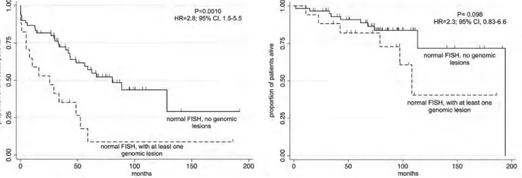
We investigated whether the presence of large genomic aberrations (>5 megabases; Mb; Figure1), not covered by the probes used in the FISH panel, would affect the outcome in patients with a‘normal’ FISH, who are known to have an intermediate outcome [6]. Seventy-seven of the 329 (23%) did not present any lesion by FISH. Seventeen (21%) of these patients with a normal FISH had additional DNA gains or losses in the SNP array affecting different genomic regions (Table2). No TP53 mutations were detected in 13 of 17 cases with genomic lesions that were analyzed for mutations. The presence of at least one large genomic aberration determined a significantly shorter TTT [P = 0.0010; hazard ratio (HR) 2.8; 95% CI 1.5–5.5] (P = 0.001), but not OS (P = 0.098; HR 2.3; 95% CI 0.83–6.6) (Figure2).
In multivariate analysis, including age >60 years, advanced Binet stage disease, unmutated IGHV genes and the presence of at least one large genomic lesion different from those described by Döhner et al. [29], the latter three factors emerged as independent prognosticators for TTT with a significance of P < 0.001, P < 0.001 and P = 0.036, respectively. None of the examined variables maintained their significance in the same model according to OS (data not shown). In particular, the presence of at least one large genomic aberration identified patients with a shorter TTT among those with mutated IGHV genes (P = 0.0002; HR 5.0; 95% CI 2.0–12.4) (Figure3, left panel) and with early stage disease (P = 0.005; HR 3.22; 95% CI 1.36–7.62) (Figure3, right panel).
SNP array and FISH give overall similar results
We compared the copy-number estimate obtained with genome-wide DNA SNP array with the standard FISH, for the
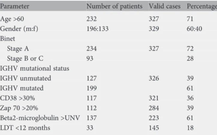
Table 1. Clinical parameters assessed at the time of diagnosis
Parameter Number of patients Valid cases Percentage
Age >60 232 327 71
Gender (m:f) 196:133 329 60:40 Binet
Stage A 234 327 72
Stage B or C 93 28
IGHV mutational status
IGHV unmutated 127 326 39 IGHV mutated 199 61 CD38 >30% 117 321 36 Zap 70 >20% 112 284 39 Beta2-microglobulin >UNV 137 223 61 LDT <12 months 33 145 18
m, male; f, female; UNV, upper normal value; LDT, lymphocyte doubling time.
Annals of Oncology
original articles
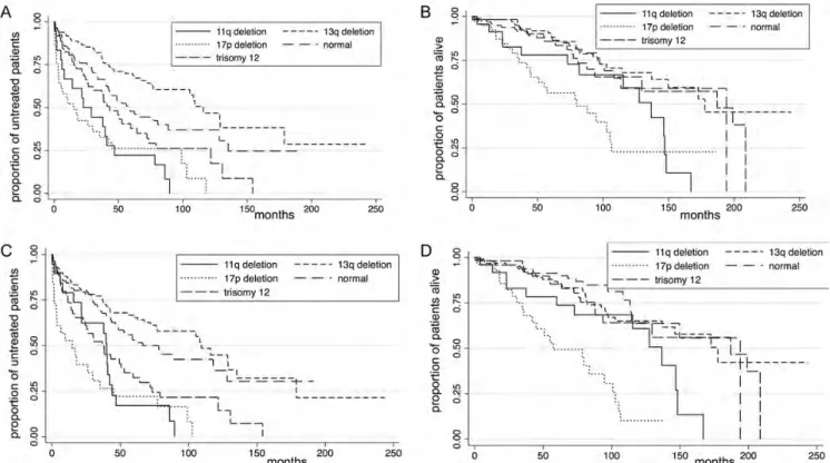
four loci investigated by the latter technique, in 329 CLL patients. Since FISH is considered the gold standard for the detection of prognostically relevant genomic aberrations in CLL samples, cases with a negative SNP array and a positive FISH were considered as false negative. As a whole, the percentage of SNP array‘false negative’ was relatively low, affecting only between 2% and 8% of the whole CLL series (Figure3). The highest concordance between FISH and SNP-array results was registered in samples with trisomy 12 (97%) followed by del 11q (96%), del 17p (95%) and del 13q (84%) (supplementary Figure S1A, available at Annals of Oncology online).
Among the 18 cases with 17p loss by FISH, all the eight samples with a loss detected by FISH in over 40% of the nuclei were also detected up by SNP array while half of the 10 cases with less than 40% of nuclei carrying the lesion were classified as normal by genomic profiling (supplementary Figure S1B, available at Annals of Oncology online).
Twenty-eight cases among those with del13q14.3, four among those with trisomy 12, six among those with del 11q22.3 and two among those with del 17p13.1 were not identified by FISH but by SNP array. Some of the observed discordant results, especially those regarding del 13q14.3 and trisomy 12, were likely due to different samples which might
Figure 1. Examples of three genomic profiles with genomic aberrations detected by SNP array in CLL patients with a normal FISH. Cases 08-515 presented multiple lesions on chromosomes 4q, 6p, 6q, 8q, 9p, 12q, 13q, 14q, 17q, 22q. Case 09-414 presented a single gain at 3q. Cases 08-560 presented a single-interstitial deletion at 4q. Black, raw copy-number values; red, smoothed copy-number values. X-axis, genomic mapping; Y-axis, log2 copy-number values.
Table 2. Large genomic aberrations spanning >5 Mb in patients with a normal FISH
Region Cytoband Type Candidate cancer genes Cases
chr1:225,493,922-235,530,710 1q42.12–q42.3 Loss – 2 chr3:145,475,764-176,735,018 3q24–q26.32 Gain – 2 chr6:30,403,481-39,295,352 6p21.33–p21.2 Gain TNF 2 chr6:87,049,197-125,406,247 6q14.3–q22.31 Loss PRDM1 1 chr6:136,647,604-142,392,181 6q23.3–q24.1 Loss TNFAIP3 1 chr7:125,307,489-141,917,586 7q31.33–q34 Loss – 2
chr8:106,322,626-146,131,926 8q23.1–qter Gain MYC 1
chr8:182,272-38,003,609 8p23.3–p11.23 Loss 1 chr9:21,302,665-32,558,663 9p21.3–p21.1 Loss CDKN2A 1 chr10:64,476,057-123,550,977 10q21.2–q26.13 Loss PTEN 1 chr14:70,726,012-79,783,628 14q24.2–q31.1 Loss – 2 chr18:51,454,691-77,917,301 18q21.2–q23 Gain BCL2, NFATC1 2 chr19:37,187,908-59,107,865 19q13.12–q13.43 Gain SPIB 1
have been examined by SNP array and by FISH or even due to false-positive FISH results. Real-time PCR carried out on a series of samples discordant in terms of the 13q14.3 status largely confirmed the SNP-array results (supplementary Figure S1C, available at Annals of Oncology online).
To ensure that the small discordances between the two detection methods did not influence the prognostic value of the assay, we applied the FISH-based prognostic model developed by Döhner et al. [29] to our cohort according to each method (Figure4). The Kaplan–Meier curves were comparable between FISH and SNP array, both for OS as for TTT and the log-rank test was highly significant. The results were evaluated using a Cox multivariate regression model adjusted for Binet stage, age at the time of diagnosis >60 years, IGHV mutational status and the information regarding the aberrations identified either by FISH or by SNP array. In both the models and for both OS and TTT, the clinical
characteristics and del17p emerged as independent prognosticators.
discussion
FISH is a powerful tool to predict the clinical course of patients affected by CLL [2, 3, 6]. However, this technique has some limitations and the SNP array provides additional prognostic information [8–13] and could represent an interesting comparison or, possibly, even an alternative technique to be implemented in the initial workup of CLL patients. In this article, in the so far largest comparison of FISH versus SNP array in CLL, we showed that SNP array was able to better define the prognosis of patients with a ‘normal’ FISH. Although SNP array provided results largely overlapping with FISH, it had the drawback of a reduced sensitivity in cases with aberrations present in only a low percentage of neoplastic cells, which can be easily seen by FISH.
An important benefit of SNP array over FISH is the ability to detect chromosomal aberrations not investigated by the standard FISH panel or even not targeted by any commercially available probes, but with a clinical relevance. In the present
Figure 3. TTT according to the presence of at least one genomic lesion detected by SNP array in CLL patients with a normal FISH, and with mutated IGHV (left panel) or in Binet stage A (right panel).
Figure 2. TTT (left panel) and OS (right panel) according to the presence of at least one genomic lesion detected by SNP array in CLL patients with a normal FISH.
Annals of Oncology
original articles
study, large genomic aberrations spanning >5 Mb were detected by SNP array in 20% of the patients with a‘normal’ FISH, which is in the absence of any of the four main genomic aberrations targeted by the probes included in the standard FISH panel (deletions at 13q14.3, 11q22, 17p13 and trisomy 12). Very importantly, the presence of at least one genomic lesion exclusively detected by SNP array did not appear to be associated with the presence of a mutated TP53 gene and determined a significantly shorter TTT, as also confirmed by multivariate analysis. Presumably due to the relatively indolent disease course, no statistically significant difference was noted in OS. With this regard, some limitations of our study have to be mentioned. Although based upon a very large series of cases, the number of patients actually bearing a normal FISH was relatively limited. Also, we have not validated thefindings in an independent series, which would be ideally represented by a series of samples obtained from CLL patients enrolled in a prospective clinical trial.
Of note, the presence of such large aberrations in patients with a normal FISH was able to identify patients with a more aggressive clinical course among those with mutated IGHV genes. Interestingly, it has just been reported that also conventional karyotyping, when carried out using DSP30/IL2 stimulation, is able to detect high-risk patients among this class of individuals classified as intermediate risk by FISH [14]. Thesefindings are in agreement with the prognostic role of genomic complexity in CLL, in which additional genomic aberrations would represent a clonal evolution possibly leading to a shorter survival [9,10,14].
TP53 gene resulted unmutated in cases with a normal FISH indicating that the phenotype we observed was unlikely due to
TP53 inactivation. Only integration with large-scale mutational screening programs will reveal the relationship between these lesions and the presence of the recently reported somatic mutations targeting, among others, BIRC3, SF3B1, NOTCH1 and MYD88 [30–32].
The additional genomic lesions we detected did not occur at specific sites but were found across the whole genome. Nevertheless, some of the genes located in these regions, namely TNF, PRDM1 and TNFAIP3, MYC, CDKN2A, PTEN, BCL2 and NFATC1, are known or seem to play a role in lymphoid neoplasms, including CLL. Other lesions did not contain known candidate cancer genes (7q loss, 3q gain, 14q loss), but were recurrent and already previously reported [20]. It is possible that some of these regions have a yet unknown role in the pathogenesis of CLL, but their individually low frequency makes it difficult to identify a minimal common region and define candidate genes (as well as choosing appropriate FISH probes).
The comparison between SNP array and FISH revealed that SNP array was able to detect the four prognostically relevant FISH loci in nearly all cases with an overall concordance of 93%, in line with recent publications [10,20,21,33]. In our series, the rate of samples called as normal by SNP array in the presence of a lesion detected by FISH (false negative) for 11q21 and 17p13 losses was nearly two-fold higher than for trisomy 12 or del 13q14.3. This result was expected, since these two lesions are known to be often subclonal, possibly present even in a very low percentage of tumor cells [34,35]. In this study, the higher sensitivity of FISH was clearly evident, when looking at the cases with 17p1.3 loss defined by FISH: while all cases with a percentage of >40% positive nuclei were detected
Figure 4. OS and TTT in 329 CLL patients according to the Döhner prognostic model by FISH (A and B; P < 0.001) or by SNP array (C and D; P < 0.001).
by both FISH and SNP array, half of the cases with <40% of nuclei carrying the loss in FISH were negative by genomic profiling. In line with this, other authors have reported that array based-karyotyping misclassified samples with a low percentage of neoplastic cells carrying a genomic aberration (up to 30%–40% of tumor cells in the sample) [15,16,20,21]. This makes now impossible to fully replace the use of the standard FISH panel with SNP array in all the patients. On the other hand, it is of clinical importance that, although having a high sensitivity, FISH cannot identify all the CLL patients with a direct genetic inactivation of TP53: a percentage of patients have somatic mutations inactivating TP53 in the absence of 17p loss, and these cases have an outcome overlapping the ones with 17p deletion [26,36]. Thus, the two techniques could be complementary in identifying poor-risk patients bearing a TP53 inactivation.
In line with previously published data [37], also false-positive FISH false-positive results were observed, mainly for the 13q14 region and the lesions detected by SNP array only and not by FISH were confirmed by real-time PCR.
Over the past decade, there have been important advances in the treatment of CLL [2], but the FISH-based prognostic model developed >10 years ago [6] is still largely applied. In order to validate the representativeness of our results in a clinical setting, we applied this model to our cohort using data generated by FISH or by SNP array. The Kaplan–Meier curves were comparable between FISH and SNP array, both for OS and for TTT, and the log-rank test was highly significant, also confirmed in multivariate analysis.
As the time and effort needed for both FISH and SNP array, as well their costs, are nowadays becoming less markedly different, a workflow could be suggested in which CLL samples would befirst analyzed with SNP array. Due to the lower sensitivity of SNP array, in the absence of losses affecting the TP53 gene locus, an interphase FISH test, investigating only this specific locus, would be mandatory, since the inactivation of TP53 can have important clinical consequences when starting treatment [2,3,38]. Since DNA samples are routinely obtained for the assessment of the IGHV mutational status [2,
3], but also, more and more, for the investigation of TP53 gene somatic mutations [26,36,38], SNP array would not require an additional blood sampling from the patients. As currently done for FISH, genomic profiling could be repeated during the follow-up, to identify the occurrence of new lesions, which could change patients’ outcome. However, a prospective evaluation of this approach and its possible integration with the information derived from the mutational status of other genes besides TP53 (for example, BIRC3, SF3B1, NOTCH1) will be necessary. Moreover, it is important to stress that the current guidelines indicate the evaluation of chromosomal abnormalities at diagnosis or before treatment as
mandatory only in the context of clinical trials and as ‘advisable’ in the general practice [2,3,38]. The testing for TP53 inactivation (by deletions or somatic mutations) can now be seen as the only genetic diagnostic assay that might affect the choice of the treatment, although there are still no data derived from randomized trials, demonstrating the superiority of a specific regimen for patients with inactivated TP53 [4,38].
In conclusion, here, the identification of large genomic lesions detected by SNP array represented an independent prognostic factor in CLL patients with a normal FISH. SNP array was also able to identify the most important known prognostically relevant genomic aberrations of CLL. A validation in prospective trials is needed.
funding
This study was supported by Helmut Horten Foundation, Lugano, Switzerland; San Salvatore Foundation, Lugano, Switzerland; Nelia et Amadeo Barletta Foundation, Lausanne, Switzerland; Computational life science/Ticino in rete [no grant numbers]. MM was a recipient of fellowship from Alto Adige Bolzano-AIL Onlus.
disclosure
The authors have declared no conflicts of interest.
references
1. Müller-Hermelink HK, Montserrat E, Catovsky D et al. Chronic lymphocytic leukemia/small lymphocytic lymphoma. In Swerdlow SH, Campo E, Harris NL, Jaffe ES, Pileri A, Stein H, Thiele J, Vardiman JW (eds), World Health Organization Classification of Tumours. Pathology and Genetics of Tumours of Haematopoietic and Lymphoid Tissues. Lyon: IARC Press 2008; 180–182. 2. Eichhorst B, Dreyling M, Robak T et al. Chronic lymphocytic leukemia: ESMO
Clinical Practice Guidelines for diagnosis, treatment and follow-up. Annals of Oncology 2011; 22: vi50–vi54.
3. Hallek M, Cheson BD, Catovsky D et al. Guidelines for the diagnosis and treatment of chronic lymphocytic leukemia: a report from the International Workshop on Chronic Lymphocytic Leukemia (IWCLL) updating the National Cancer Institute—Working Group (NCI-WG) 1996 guidelines. Blood 2008; 111: 5446–5456.
4. Zenz T, Gribben JG, Hallek M et al. Risk categories and refractory CLL in the era of chemoimmunotherapy. Blood 2012; 119: 4101–4107.
5. Cramer P, Hallek M. Prognostic factors in chronic lymphocytic leukemia—what do we need to know? Nat Rev Clin Oncol 2011; 8: 38–47.
6. Döhner H, Stilgenbauer S, Benner A et al. Genomic aberrations and survival in chronic lymphocytic leukemia. N Engl J Med 2000; 343: 1910–1916. 7. Maciejewski JP, Tiu RV, O’Keefe C. Application of array-based whole genome
scanning technologies as a cytogenetic tool in haematological malignancies. Br J Haematol 2009; 146: 479–488.
8. Rinaldi A, Mian M, Kwee I et al. Genome-wide DNA profiling better defines the prognosis of chronic lymphocytic leukaemia. Br J Haematol 2011; 154: 590–599. 9. Stilgenbauer S, Sander S, Bullinger L et al. Clonal evolution in chronic
lymphocytic leukemia: acquisition of high-risk genomic aberrations associated with unmutated VH, resistance to therapy, and short survival. Haematologica 2007; 92: 1242–1245.
10. Ouillette P, Collins R, Shakhan S et al. Acquired genomic copy number aberrations and survival in chronic lymphocytic leukemia. Blood 2011; 118: 3051–3061.
11. Mian M, Rinaldi A, Mensah AA et al. Del(13q14.3) length matters: an integrated analysis of genomic,fluorescence in situ hybridization and clinical data in 169 chronic lymphocytic leukaemia patients with 13q deletion alone or a normal karyotype. Hematol Oncol 2012; 30: 46–49.
12. Ouillette P, Collins R, Shakhan S et al. The prognostic significance of various 13q14 deletions in chronic lymphocytic leukemia. Clin Cancer Res 2011; 17: 6778–6790. 13. Parker H, Rose-Zerilli MJ, Parker A et al. 13q deletion anatomy and disease
progression in patients with chronic lymphocytic leukemia. Leukemia 2011; 25: 489–497.
14. Rigolin GM, Cibien F, Martinelli S et al. Chromosome aberrations detected by conventional karyotyping using novel mitogens in chronic lymphocytic leukemia
Annals of Oncology
original articles
with“normal” FISH: correlations with clinicobiologic parameters. Blood 2012; 119: 2310–2313.
15. Gunn SR, Mohammed MS, Gorre ME et al. Whole-genome scanning by array comparative genomic hybridization as a clinical tool for risk assessment in chronic lymphocytic leukemia. J Mol Diagn 2008; 10: 442–451.
16. Patel A, Kang SH, Lennon PA et al. Validation of a targeted DNA microarray for the clinical evaluation of recurrent abnormalities in chronic lymphocytic leukemia. Am J Hematol 2008; 83: 540–546.
17. Schwaenen C, Nessling M, Wessendorf S et al. Automated array-based genomic profiling in chronic lymphocytic leukemia: development of a clinical tool and discovery of recurrent genomic alterations. Proc Natl Acad Sci USA 2004; 101: 1039–1044.
18. Pfeifer D, Pantic M, Skatulla I et al. Genome-wide analysis of DNA copy number changes and LOH in CLL using high-density SNP arrays. Blood 2007; 109: 1202–1210.
19. Lehmann S, Ogawa S, Raynaud SD et al. Molecular allelokaryotyping of early-stage, untreated chronic lymphocytic leukemia. Cancer 2008; 112: 1296–1305.
20. Hagenkord JM, Monzon FA, Kash SF et al. Array-based karyotyping for prognostic assessment in chronic lymphocytic leukemia: performance comparison of Affymetrix 10K2.0, 250K Nsp, and SNP6.0 arrays. J Mol Diagn 2010; 12: 184–196.
21. Zhang L, Znoyko I, Costa LJ et al. Clonal diversity analysis using SNP microarray: a new prognostic tool for chronic lymphocytic leukemia. Cancer Genet 2011; 204: 654–665.
22. Brown JR, Hanna M, Tesar B et al. Integrative genomic analysis implicates gain of PIK3CA at 3q26 and MYC at 8q24 in chronic lymphocytic leukemia. Clin Cancer Res 2012; 18: 3791–3802.
23. Matutes E, Owusu-Ankomah K, Morilla R et al. The immunological profile of B-cell disorders and proposal of a scoring system for the diagnosis of CLL. Leukemia 1994; 8: 1640–1645.
24. Gunnarsson R, Isaksson A, Mansouri M et al. Large but not small copy-number alterations correlate to high-risk genomic aberrations and survival in chronic lymphocytic leukemia: a high-resolution genomic screening of newly diagnosed patients. Leukemia 2010; 24: 211–215.
25. Mian M, Scandurra M, Chigrinova E et al. Clinical and molecular characterization of diffuse large B-cell lymphomas with 13q14.3 deletion. Ann Oncol 2012; 23: 729–735.
26. Rossi D, Cerri M, Deambrogi C et al. The prognostic value of TP53 mutations in chronic lymphocytic leukemia is independent of Del17p13: implications for overall survival and chemorefractoriness. Clin Cancer Res 2009; 15: 995–1004. 27. Meier ELKaP. Nonparametric estimation from incomplete observations. J Am Stat
Assoc 1958; 53: 25.
28. Cox DR. Regression Models and Life-Tables. J R Stat Soc 1972; 34: 34. 29. Döhner H, Stilgenbauer S, Fischer K et al. Cytogenetic and molecular cytogenetic
analysis of B cell chronic lymphocytic leukemia: specific chromosome aberrations identify prognostic subgroups of patients and point to loci of candidate genes. Leukemia 1997; 11(Suppl 2): S19–S24.
30. Fabbri G, Rasi S, Rossi D et al. Analysis of the chronic lymphocytic leukemia coding genome: role of NOTCH1 mutational activation. J Exp Med 2011; 208: 1389–1401.
31. Wang L, Lawrence MS, Wan Y et al. SF3B1 and other novel cancer genes in chronic lymphocytic leukemia. N Engl J Med 2011; 365: 2497–2506. 32. Puente XS, Pinyol M, Quesada V et al. Whole-genome sequencing identifies
recurrent mutations in chronic lymphocytic leukaemia. Nature 2011; 475: 101–105.
33. Edelmann J, Holzmann K, Miller F et al. High-resolution genomic profiling of chronic lymphocytic leukemia reveals new recurrent genomic alterations. Blood 2012; 120: 4783–4794.
34. Tam CS, Shanafelt TD, Wierda WG et al. De novo deletion 17p13.1 chronic lymphocytic leukemia shows significant clinical heterogeneity: the M. D. Anderson and Mayo Clinic experience. Blood 2009; 114: 957–964. 35. Döhner H, Stilgenbauer S, James MR et al. 11q deletions identify a new subset
of B-cell chronic lymphocytic leukemia characterized by extensive nodal involvement and inferior prognosis. Blood 1997; 89: 2516–2522. 36. Zenz T, Krober A, Scherer K et al. Monoallelic TP53 inactivation is associated
with poor prognosis in chronic lymphocytic leukemia: results from a detailed genetic characterization with long-term follow-up. Blood 2008; 112: 3322–3329.
37. Smoley SA, Van Dyke DL, Kay NE et al. Standardization offluorescence in situ hybridization studies on chronic lymphocytic leukemia (CLL) blood and marrow cells by the CLL Research Consortium. Cancer Genet Cytogenet 2010; 203: 141–148.
38. Pospisilova S, Gonzalez D, Malcikova J et al. ERIC recommendations on TP53 mutation analysis in chronic lymphocytic leukemia. Leukemia 2012; 26: 1458–1461.