Printed in Great Britain
Amino acid sequence, haem-iron
co-ordination geometry and functional
properties of mitochondrial and
bacterial c-type cytochromes
HANS SENN AND KURT WUTHRICH
Institut fur Molekularbiologie und Biophysik, Eidgenossische Technische Hochschule, ETH-Honggerberg, CHSogj Zurich, Switzerland
1. I N T R O D U C T I O N 112 2. SURVEY OF T H E C O - O R D I N A T I O N GEOMETRY OF T H E
TWO H A E M - I R O N AXIAL L I G A N D S H I S T I D I N E AND M E T H I O N I N E I N FERROCYTOCHROMES C 1 1 4
(a) The axial methionine 114 ib) The axial histidine 116
(c) Co-ordination geometry in ferricytochrom.es c 116
3. A M I N O ACID SEQUENCE AND S T E R E O - S E L E C T I V E L I G A N D B I N D I N G TO T H E H A E M - I R O N I L 8
(a) The axial methionine 118 (Jb) The axial histidine 119
4. C O R R E L A T I O N B E T W E E N T H E C O O R D I N A T I O N G E O
-METRY OF THE AXIAL METHIONINE AND OTHER PRO-PERTIES RELATED TO CYTOCHROME C FUNCTION I2O (a) Axial methionine co-ordination geometry and electronic
haem c structure 120
(b) Axial methionine co-ordination geometry and redox potential 122
(c) Axial methionine co-ordination and enzymatic activity 124
5. P H Y L O G E N E S I S O F H A E M I R O N C O O R D I N A T I O N G E O
-METRY AND HAEM C ELECTRONIC STRUCTURE 126
6. A C K N O W L E D G E M E N T S 128 7. R E F E R E N C E S 129
QRB 18
https://doi.org/10.1017/S0033583500005151
Cambridge Core terms of use, available at https:/www.cambridge.org/core/terms.
112 H. SENN AND K. WUTHRICH
i. INTRODUCTION
Cytochromes are found in all biological oxidation systems which involve transport of reducing equivalents through organized chains of membrane bound intermediates, regardless of the ultimate oxidant (Keilin, 1966; Bartsch, 1978; Meyer & Kamen, 1982). Thus, cyto-chromes are present not only in the aerobic mitochondrial and bac-terial respiratory chain, but are also found in much more diversified procariotic systems, including all varieties of facultative anaerobes (nitrate and nitrite reducers), obligate anaerobes (sulphate reducers and phototrophic sulphur bacteria), facultative photoheterotrophes (phototrophic non-sulphur purple bacteria), and the photoautotrophic cyanobacteria (blue-green algae). Among the different types of cytochromes occurring in the cell, the soluble c-type cytochromes ('class I', Meyer & Kamen, 1982) are the most abundant and best characterized group of proteins (Bartsch, 1978; Meyer & Kamen, 1982; Dickerson & Timkovitch, 1975; Lemberg & Barrett, 1973; Salemme, 1977; Ferguson-Miller, Brautigan & Margoliash, 1979). The amino acid sequences of more than 80 mitochrondrial and close to 40 bacterial cytochromes c are known (Meyer & Kamen, 1982; Dickerson & Timkovitch, 1975; Schwartz & Dayhoff, 1976; Dayhoff & Barker, 1978).
A search for the biochemical rational behind the persistent occur-rence of histidine and methionine as axial ligands of the haem-iron has been our motivation for systematic studies of correlations between the active site conformation and the primary structure, the electronic structure of haem c and functional properties, such as the redox potential or the reactivity with cytochrome oxidases and reductases from different species. The omnipresence of soluble c-type cyto-chromes in nature has already stimulated many comparative struc-tural (Dickerson & Timkovitch, 1975; Timkovitch, 1979; Moore
et al. 1982), evolutionary (Meyer & Kamen, 1982; Dickerson,
19800,6,c) and functional studies (Ferguson-Miller et al. 1979; Errede & Kamen, 1978; Yamanaka & Okunuki, 1968; Sutin, 1977). Thereby the characterization of the protein surface responsible for the interaction with physiological redox-partners has been the target of numerous recent biochemical investigations (Ferguson-Miller
et al. 1979; Rieder & Bosshard, 1980; Waldmeyer et al. 1982; Kraut,
1981). In contrast, there have been few comparative studies of the structure of the active centre of cytochromes c. This centre lies in the interior of the protein and consists of the haem group and two axial
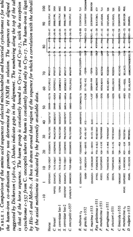
TABL E I . Comparison of the amino acid sequences of selected mitochondrial and bacterial cytochromes cfor which the haem-iron co-ordination geometry was determined by 1 H NMR in solution. The sequences are aligned as suggested by Dickerson (ig8oa, c). Dashes indicate gaps in the alignment. The numbering along the top of the table is that of the horse sequence. The haem group is covalently bound to Cys-14 and Cys-17, with the exception of cytochrome c-557 from C . oncopelt i where the haem is covalently linked only to Cys-ij. The haem-iron is ligated by His-18 and Met-80. A box is drawn around that region of the sequence for which a correlation with the chirality of the axial methionine is indicated by the presently available data. -1 0 1 1 0 2 0 3 0 4 0 5 0 6 0 7 0 Hors e GDVEKGKK I FVQK-CAQCH T VEKGGKHKT G PNLHGLFGR K TGQAPGFTY T DANKN—KGIT U KEETLMEYL E NPKKYI P G-I C. krusei PAPFE Q GSAKKGAT L FKTR-CAQCH T IEAGGPHKV G PNLHGIFSR H SGQAEGYSY T OANKR—AGVE U AEPTMSDYL E NPKKYI P G-T S. cerevisiae Iso-1 TEFK A GSAKKGAT L FKTR-CLQCH T VEKGGPHKV G PNLHGIFGR H SGQAEGYSY T DANIK—KNVL U DENNMSEYL T NPKKYI P G-T S. cerevisiae Iso-2 AKESTGFK P GSAKKGAT L FKTR-CQQCH T IEEGGPNKV G PNLHGIFGR H SGQVKGYSY T DAMN—KNVL U DEOSHSEYL T NPKKYI P G-I C. onccpelti c-55 7 GPKAREPLP P GDAAKGEK I FKGR-AAOCH T GAKGGANGV G PNLFGIVNR H SGTVEGFAY S KANAD—SGVV W TPEVLDVYL E NPKKFM P G-T 8 0 9 0 10 0 R. rubrum cs E. gracilh c-5 c-5 2 5 . maxima c-5 c-5 3 Rps. gelatinosa c-5 5 1 P. mendocina c-5 5 1 P. aeruginosa c-5 5 1 P. stutzeri c-551 C. limicola c-5 c-5 5 P. mendocina c 5 D. vulgaris c-5 c-5 3 E GDAAAGEK V SKK—CLACH T FDQGGANKV G PNLFGVFEN T AAHKDNYAY S ESYTEMKAKGLT U TEANLAAYV K DPKAFVLEKSGDPK--AK ! GGAD V F-ADNCSTCH V NG---GNVI S AG-KVL-SK T AI E EYLD G GY-TKE-AI E -YQVR N I GDVAAGAS V F-SANCAACH M GG—-RNVI V AN-KTL-SK S DLA K YLKG F DDDAVA-AV A -YQVT N GKNJ A ATPAE L ATKAGCAVCH Q PTA—K-GL G PSYQEIAKK Y K GQA G APALMAERV R KGSVGI F GK l ASGEE L FKSKPCGACH S VGA-K-LV G PALKDVAAK N A GVD G AADVLAGHI K NGSTGV U GA * EDPEV L FKNKGCVACH A IDT--K-MV G PAYKDVAAK F A GQA G AEAELAQRI K NGSQGVH — -GP I QDGEA L FKSKPCAACH S IDA--K-LV G PAFKEVAAK Y A GQD G AADLLAGHI K NGSQGV W GP I YOAAAGKA T YDAS-CAMCH K TG M MG A PKVGDKA A UAP H IAKGKNVMV A NSIKGYK G TK C AASA G GGARSADD I I-AKHCNACH G AG V LG A PKIGDTA A UKE R AD—HQGG L DGILAKI S GIN > ADGAA L Y--KSCIGCH S AD G GK A NNTNAVK G KYS D EELKALADY H KAAHGSAKPVKGQGAEEL Y H IF/i-IKK-K T EREDLIAYL K KATN E H AF C 5-LKK-A K DRNDLVTYH L EAS K H AF C i-LKK-E K ORNDLITYL K KAC E H AF * S-LKK-E K DRNDLITYH T KAA K « SF/3-IKK-P Q ERADLIAYL E NL K M TF I M — PAW I H—PGF I MTPTPPA! -H—PPN I H—PPN A -M — PPN P H---PAKGPN I M— I N — KGY , -LTK-D D IVLS — E D IRLS—P L IS--D A •-VT--E E -VS-D D -VT-E E IPKLTD A -C S 'AbGSY—G G EIENVIAYL K TL K EIVAVTDYV Y TQAGGAUAN V QIEOVAAYV V DQAEKG U DLKLVIDWI L KT P EAKTLAEUV L TL K EAQTLAKWV L SQ K EAKILAEWI L SQ K QVGNAVAYM V GQS K DDELREAIQ K MSG L ERKAMSK L https://doi.org/10.1017/S0033583500005151
Cambridge Core terms of use, available at https:/www.cambridge.org/core/terms.
114 H. SENN AND K. WUTHRICH
ligands of the haem-iron, i.e. histidine and methionine (Dickerson & Timkovitch, 1975; Wuthrich, 1969). In all known cytochrome c sequences the axial histidine and methionine (His 18 and Met 80 in Table 1) are the only strictly conserved amino-acid residues in the primary structure (Meyer & Kamen, 1982; Schwartz & Dayhoff,
1976; Dayhoff & Barker, 1978) besides Cys 17.
The present paper surveys the available data on the haem-iron co-ordination geometry in class I cytochromes (Senn, 1983; Senn & Wuthrich, 1983 a, b, c; Senn, Keller & Wuthrich, 1980; Senn, Eugster & Wuthrich, 1983 a; Senn et al. 1983ft; Senn, Billiter & Wuthrich, 1984 a; Senn, Bohme & Wuthrich, 19846; Senn, Cusanovich & Wuthrich, 1984c; Keller, Picot & Wuthrich, 1979, 1980; Ulrich, Krogmann & Markley, 1982; Salemme et al. 1973; Takano & Dickerson, 19810,6; Matsuura, Takano & Dickerson, 1982; Tim-kovich 1979) and investigates possible correlations with the amino acid sequence and functional properties. The proteins studied (Table 1) were selected from a broad range of eucaryotic and bacterial organisms so that the investigations could be extended to phylo-genetic information on the haem-iron co-ordination geometry and the haem c electronic structure.
2. SURVEY OF THE C O - O R D I N A T I O N GEOMETRY OF THE TWO H A E M - I R O N AXIAL L I G A N D S H I S T I D I N E AND M E T H I O N I N E I N FERROCYTOCHROMES C
(a) The axial methionine
Four different types of methionine co-ordination geometries in the c-type cytochromes investigated (Table 1) have been characterized in solution, and two of these have also been observed in cytochrome c crystal structures (Fig. 1). In cytochromes c from mammalian species (horse and tuna) (Senn et al. 1980, 1984a; Takano & Dickerson, 1981 a), and from yeast (5. cerevisiae, C. krusei) (Senn et al. 1983 a), cytochrome c-557 from C.oncopelti (Keller et al. 1979) cytochromes c-552 and c-553 from E. gracilis, S. platensis and 5. maxima (Keller, Schejter & Wuthrich, 1980; Senn et al. 19846, Ulrich et al. 1981) and cytochrome c2 from Rhodospirillum rubrum
(Senn & Wuthrich, 19836; Salemme et al. 1973) the axial methionine has R chirality at the iron-bound sulphur and the methionine side-chain is extended, with the Ca carbon near the /?-meso position of haem c and outside the porphyrin ring skeleton (Fig. 1 A). In the cytochromes c-551 from P. aeruginosa (Senn et al. 1980, 1984c;
B
Fig. 1. Conformation of the axial methionine in selected mitochondrial and bacterial c-type cytochromes. The view is perpendicular to the heme plane. The methyl and methylene groups are represented by equivalent spheres M and L, with the exception of C/Ha and C H , of methionine in the structures A and B,
where these methylene protons were stereospecifically assigned (Senn et at. 19840). (A) Mitochondrial ferrocytochromes c (S. cerevisiae Iso-i and lso-2,
C. krusei, C. oncopelti, horse), R. Rubrum cytochrome c2, S. platensis
ferrocyto-chrome c-553 and E.gracilis ferrocytoferrocyto-chrome c-552. (B) Ferrocytoferrocyto-chromes c-551 of P. aeruginosa, P. mendocina, P. stutzeri and Rps. gelatinosa. (C) P. mendocina ferrocytochrome c5. CaH is not shown (see text). (D) D. vulgaris and D.
desul-furicans ferrocytochromes c-553. In structure D the meso-positions and the /?
positions of haem c are identified by a-S, and by i-8, respectively. The pyrrole rings are numbered I-IV.
Matsuura et al. 1982), P. mendocina (Senn & Wiithrich, 19836),
P. stutzeri (Senn & Wiithrich, 19836) and Rps. gelatinosa (Senn &
Wiithrich, 1983 a) the axial methionine has S chirality at the iron-bound sulphur and the methionine side chain is bent, so that C^H2
is closer to the e-methyl group than CyH2. O*H is near pyrrole ring
III and outside the porphyrin ring skeleton (Fig. 1B). In cytochrome c6 from P. mendocina the axial methionine has S chirality, the
methionine side chain is extended and C^H, is located above the
https://doi.org/10.1017/S0033583500005151
Cambridge Core terms of use, available at https:/www.cambridge.org/core/terms.
I l 6 H. SENN AND K. WUTHRICH
pyrrole ring III (Fig. i C). CaH is not shown in Fig. i C, but observations on its chemical shift indicate that it is near the haem-plane and within the confines of the porphyrin ring (Senn & Wiithrich, 1983c). The structure for the ferrocytochromes c-553 from Desulfo-vibrio vulgaris and DesulfoDesulfo-vibrio desulfuricans (Fig. 1D) is most closely related to that found in cytochromes c-551 (Fig. iB) (Senn et al. 19836). It coincides with the latter in the S chirality at the iron-bound sulphur and the bent conformation of the S-C1'H2-C'?H2
fragment. It differs from the cytochrome c-551 structure by a clockwise rotation by approx. 450 of the methionine about the iron-sulphur bond. Furthermore, it is so far a unique feature of the two ferrocytochromes c-553 investigated that the methionine Cr-C^ bond is directed away from haem-plane (Fig. 1D). In all other cytochromes c (Fig. 1A-C) both methylene groups of the axial methionine adopt an orientation in which one proton points towards and the other points away from the haem-plane.
(b) The axial histidine
The same spatial arrangement of the axial histidine prevails in the conformations of all cytochromes c investigated so far in solution and in single crystals (Timkovitch, 1979) (Table 1): the imidazole ring plane is oriented approximately along a line through the meso-protons a and y and is roughly perpendicular to the haem-plane (see Fig. 1 D for haem nomenclature).
(c) Co-ordination geometry in ferricytochromes c
The chirality of the axial methionine binding to the haem-iron in the ferric state of the proteins is accessible for investigation by X-ray methods and CD-spectroscopy (Senn et al. 1980). With the exception of the two Desulfovibrio cytochromes c-553 (Senn et al. 19836) all cytochromes c of Table 1 show identical chirality at the axial methionine-sulphur in the oxidized and reduced state (Table 2). In Desulfovibrio cytochromes c-553 different methionine chirality was observed in the two oxidation states of the same protein, i.e. in the reduced ferro- and in the oxidized ferri-form.
T A B L E 2. Chemical shifts of the haem ring methyl 1H NMR lines in the ferric state, chirality of the axial methionine and oxidation-reduction potentials of the cytochromes c in Table 1. In the first column the numbers indicate the resonance positions of the ring methyls at 35 °C (see Fig. 1D for nomenclature used) with respect to the chemical shift scale at the
bottom of the table. The third column lists the chirality of the axial methionine sulphur in the oxidized and reduced state of the proteins, R or S (Keller et al. 1980; Senn & Wuthrich j ^ j a , b, c; Senn et al.
IQ8O, ig83a,b, ig84a,b,c). The fourth column lists literature data on the oxidation-reduction potentials in mV (Sugimura et al. 1968; Lemberg & Barrett 1973; Bartsch 1978; Bertrand et al. 1982; Meyer & Kamen 1982).
Ring methyl chemical shifts
8 8 5 8 50 40 8 3 8 3 8 3 8 3 8 3 8 3 3 5 1 5 5 5 3 1 3 8 30 ppm 5 8 1 8 1 8 1 8 5 1 3 5 i 2 0 5 1 5 1 5 1 5 1 5 1 5 1 1 3 3 3 3 1 0 Species Horse c C. krusei c S. cerevisiae c Iso-i S. cerevisiae c Iso-2 C. oncopelti c-557 R. rubrum c2 E. gracilis c-552* R. gelatinosa c-551 P. mendocina c-551 P. aeruginosa c-551 P. stutzeri c-551 C. limicola c-555 P. mendocina c5 D. vulgaris c-553t Met. Ox. R R R R R R R S s s
s
-t
s R chirality Red. R R R R R R R Ss
ss
-t
s
s
£" (mV) 260 260 260 260 255 320 325 280 ~ 200 285 280 145 320 0• Identical features were observed for S. platensis cytochrome c-553 (a cyanobacterium), for which only the sequence of the N-terminal 44 residues is known (Senn et al. 19846).
•(• In C. limicola cytochrome c-555 t n e co-ordination geometry of the axial methionine was not determined (Senn et al. 1984c).
X Identical heme co-ordination and oxidation-reduction potential prevail for D. desulf-uricans, of which the amino acid sequence is not known (Senn et al. 19836).
https://doi.org/10.1017/S0033583500005151
Cambridge Core terms of use, available at https:/www.cambridge.org/core/terms.
IL8 H. SENN AND K. WUTHRICH
3. AMINO ACID SEQUENCE AND STEREO-SELECTIVE LIGAND BINDING TO THE HAEM-IRON
(a) The axial methionine
The stereospecificity of the axial methionine binding to the haem-iron cannot be correlated with the overall primary structure homology. For example E. gracilis cytochrome c-552 has 13 sequence positions in common with horse cytochrome c and 19 positions in common with
P. mendocina cytochrome c-551 (Dickerson, 1980c). Nevertheless,
the axial methionine sulphur atoms in both horse cytochrome c and
E. gracilis cytochrome c-552 exhibit R chirality, whereas the sulphur
atom in cytochrome c-551 exhibits S chirality. Among the mitochon-drial and bacterial cytochromes c with identical stereospecificity of the methionine binding to the haem-iron (Table 2) amino acid sequence homology as low as 20% is observed. (Table 1, e.g.
E. gracilis cytochrome c-552 and horse cytochrome c). From these
data, we conclude that only local sequence segments are responsible for the stereoselective-methionine binding to the haem-iron.
One such segment was found in the immediate vicinity of the axial methionine (boxed region, Table 1).
All cytochromes c with S-chirality at the sulphur atom contain several prolines around the axial Met 80 (Table 1). The sequential order of these proline residues appears to be correlated with the stereospecific methionine binding. Proteins having two prolines in consecutive positions following Met 80 in the boxed region (Table 1) have S-chirality. However, if only one proline occurs after Met 80 in the primary structure, an R-chiral attachment of the methionine to the haem-iron is observed (e.g. E. gracilis and 5. maxima cyto-chrome c-552, Table 1). This correlation does not apply for
De-sulfovibrio cytochromes c-553 (Table 1), which show an S-chiral
axial methionine in the reduced and an R-chiral methionine in the oxidized form of the protein.
The aromatic amino acid at position 82 in the primary structure (Table 1) is observed in all cytochromes c with R-chiral axial methionine (M80) but is missing in the S-chiral co-ordination types. In recent model studies of the interaction of palladium with S-methyl-cysteinyl peptides, local changes in the peptide sequence have been shown to affect the diastereomeric ratio of the two Pd-complexes formed (Kozlowski et al. 1983). From single crystal studies on the tertiary structure of mitochondrial cytochromes c and
R. rubrum cytochrome c2, proteins with R-chiral methionine, it is
known that the lone pair sp3-orbital of the axial methionine sulphur is involved in an H-bond to tyrosine 67 O^ (Table 1) (Takano & Dickerson, 1981 a; Salemme et al. 1973). In the tertiary structure of
P. aeruginosa cytochrome c-551, a protein with S-chiral methionine,
Nj of Asn in position 64 (corresponds to position 82 in horse cytochrome c Table 1) takes on the function as H-donor to the axial methionine sulphur lone pair orbital (Matsuura et al. 1982). The homologous position to residue Asn 64, however, is Phe 82 and not Tyr 67 in mitochondrial and R. rubrum cytochrome c2. This results
from the different spatial orientations of the axial methionine sulphur lone pair orbital in the two classes of proteins (Fig. 1A and B). The residues in position 67 and 82 of the numeration used in Table 1 are conserved in most cytochromes c. In S. maxima cytochrome c-553 and E. gracilis cytochrome c-552, both proteins with R-chiral meth-ionine, no amino acid with H-donor capacity homologous to Tyr 67 is found in the homologous sequence alignments of Table 1. Whether the alignments have to be corrected for these two proteins remains an open question as long as their tertiary structures are unknown.
The two remaining structural types, P. mendocina cytochrome c5
(Fig. 1 C) and Desulfovibrio ferrocytochromes c-553 (Fig. i D ) have S-chiral methionine attachment, but otherwise completely different methionine conformations than Pseudomonas cytochromes c-551 (Fig. 1 B). The H-donor amino acid to the axial sulphur atom can therefore not be localized in the primary sequence from a consideration of homology to the primary structure of Pseudomonas cytochromes
(b) The axial histidine
The same spatial arrangement of the axial histidine as observed by high-resolution NMR techniques in solution (Senn, 1983; Senn & Wuthrich igS2a,b,c; Senn et al. 1980, 19830,6, 19846,^ has also been observed in the crystalline state (Timkovitch 1979). An important feature in determining the axial histidine orientation appears to be the presence of an H-bond between the NtH of the axial imidazole
ring and the C = O group of Pro 30 (Takano & Dickerson, 1981a; Salemme et al. 1973; Matsuura et al. 1982) (Table 1). From the observation that the protein sequences of all mitochondrial and most of the bacterial c-type cytochromes show a conserved proline at position 30 (Table 1) (Bartsch 1978; Schwartz & DayhofT, 1976;
https://doi.org/10.1017/S0033583500005151
Cambridge Core terms of use, available at https:/www.cambridge.org/core/terms.
I2O H. SENN AND K. WUTHRICH
Dayhoff & Barker, 1978; Dickerson, 1980 a), we infer that this homologous position assists in conservation of the spatial orientation of the axial histidine.
4. C O R R E L A T I O N BETWEEN THE C O - O R D I N A T I O N GEOMETRY OF THE AXIAL M E T H I O N I N E AND OTHER P R O P E R T I E S
RELATED TO CYTOCHROME C F U N C T I O N
(a) Axial methionine co-ordination geometryand electronic haem c structure
Earlier work on the unpaired electron spin density distribution in the porphyrin ring of cytochromes c indicated a clear correlation between the asymmetry of the haem c electronic structure and the presence of a methionine ligand at the sixth co-ordination position of the haem-iron (Wuthrich, 1970, 1971). Later, the unpaired spin density could be assigned to individual pyrrole rings of haem c (Redfield & Gupta, 1971; Keller & Wuthrich, 1978a).
In the present study we have further investigated correlations between conformational properties of the axial ligand sphere and the haem c electronic structure. Table 2 reveals common traits as well as differences between the cytochromes c investigated. The chemical shifts of the individually assigned haem-ring methyl resonances reflect the delocalization of the unpaired electron of the low-spin ferric iron in the haem-plane (Wuthrich, 1970, 1976). In all species, there are two methyl groups attached to opposite pyrrole rings (see Fig. 1 D for nomenclature used) which experience large hyperfine shifts, wheras the other two ring methyls are shifted to a lesser extent. The large hyperfine shifts indicate that up to 3 % of the unpaired electron spin density is localized in the n orbital of the /?-ring carbon to which the methyl is attached (Fig. 1 D). The small hyperfine shifts correspond to an unpaired electron spin density of less than 0-5 % in the corresponding /?-carbon n orbital (Wuthrich, 1976). (Cyto-chrome c-555 m Table 2 is omitted from this discussion because its methionine structure has not been determined (see Senn et al. 1984 c for a detailed discussion of this protein).)
The two classes of haem c electronic structure observed in chromes c are schematically shown in Fig. 2. In Pseudomonas cyto-chromes c-551 a nd in Rps. gelatinosa cytochrome c-551 the high spin density is at positions 1 and 5 on the pyrrole rings I and III (Fig. 2), whereas in all other cytochromes c investigated methyls 3 and 8 experience a larger shift, which manifests high spin density on the
Horse cytochrome c (14) H3C-CH ^ H3 C- H,C-/ V CH / / \ ,'N~ CH Fe - ^ C H A
Y
CH,1 CH, COO" CH, \ CHA
HI v T C H , 1 CH, coo; CH3 1 -CH,Q 0 / P. aeruginosa cytochrome c-551 (14) H3C —CH ^ CH3 i \ CH A.HaC^T Y»7
/ \ ' ^
CH Fe CH>N'' VX.
"'C-^CH«Y X
CH, 0 * CH, C H , ^ CH, ^ COO" COC CH, 1 Q s £cH, r (17)Fig. 2. Schematic representation of the electronic structure of haem c in horse ferricytochrome c and P. aeruginosa ferricytochrome c-551. The shaded pyrrole rings are those where high electron spin density (3-5 %) is observed on the f! carbon atoms. The thick broken line indicates the protein surface. In both species the edge of the pyrrole ring II is accessible on the surface. Because of extensive deletions in the polypeptide chain of P. aeruginosa cytochrome c-551 (Table 1) the edge of pyrrole ring III is also accessible on the protein surface of this species (Matsuura et al. 1982).
pyrrole rings II and IV. There is a strict correlation between the haem c electronic structure and the co-ordination geometry of the axial methionine (Table 2, Fig. 1). A likely explanation is suggested by the direct correspondance with the orientation of the lone pair electrons of the ligand methionine sulphur atom. The interaction of the sp3 lone-pair electrons of the methionine sulphur with the dxz and dyz
orbitals of the iron modifies the relative energies of the molecular orbital involving dxz and dyz, which results in a marked effect on the
distribution of the unpaired electron spin density of the low spin ferric iron between these two orbitals (Senn et al. 1980; Shulman, Glarum & Karplus, 1971). In the structures of Fig. 1A and B the lone pair is directed at the pyrrole nitrogens IV and I, respectively. As long as the change in methionine conformation is restricted to a transition from R chirality to S chirality at the sulphur atom, the ensuing variation of the electronic structure consists of a rotation of the principle axes of the electronic g-tensor by approx. 900 about an axis perpendicular to the haem-plane (Senn et al. 1980; Keller et al. 1980;
https://doi.org/10.1017/S0033583500005151
Cambridge Core terms of use, available at https:/www.cambridge.org/core/terms.
122 H. SENN AND K. WUTHRICH
Keller & Wuthrich, 19786). Concomitant with this rotation is a shift in the location of high spin density on the peripheral pyrrole /^-carbon atoms, from the pyrrole rings II and IV in structure A to the pyrroles I and III in structure B (Figs. 1 and 2). In ferricytochrome c5 of
P. mendocina the lone pair orbital of the axial methionine sulphur is
oriented along a line through the nitrogen atoms of the pyrrole rings I1 and IV (Fig. 1C). This coincides with the situation in mitochondrial cytochromes c (Fig. 1 A), except that the lone pair points in opposite directions in these two cases. Ferricytochrome c5 has, therefore, S
chirality at the axial methionine sulphur, but electron spin delocal-ization of the type observed, for example, in horse cytochrome c (Table 2, Fig. 2).
In the two investigated Desulfovibrio ferricytochromes c-553, the assymetry of the spin density distribution is less pronounced than in mammalian ferricytochromes c (Table 2) (Senn et al. 19836). Since the detailed conformation of the methionine in the oxidized protein has not been determined, it is then of interest that this would be expected in a structure differing from that in Fig. 1 D only by a change of the chirality at the iron-bound sulphur from S to R (Senn et al.
19836). The lone pair of the methionine sulphur atom would then not be directed straight at a pyrrole nitrogen, but would point in a direction somewhere between the pyrrole nitrogen IV and the meso-proton 8. The molecular orbitals derived from the dxz and dyz
atomic orbitals of the iron would thus both contain some admixture of the sulphur lone-pair orbital, hence quite similar hyperfine shifts for the four-ring methyl resonances would be anticipated. In all cytochromes c investigated the imidazole ring plane is oriented along a line through the meso-protons a and y and is roughly perpendicular to the haem-plane. The interaction of the 77-system of the imidazole ring with the dxz- and <fy2-orbitals of the haem-iron is symmetric in
this orientation and does not change the relative energies of those haem-molecular orbitals which arise from admixture with iron atomic orbitals. It is therefore not surprising that an almost symmetric unpaired electron spin distribution has been observed in Azido-ferricytochrome c and cyanoferri-cytochrome c (Wuthrich, 1969; Gupta & Redfield, 1970), where the second axial ligand does not impose a pronounced asymmetry.
(6) Axial methionine co-ordination geometry and redox potential The redox potential is the fundamental thermodynamic property of an electron transfer protein. It provides the basis to locate the protein
within an electron transport sequence. In order to understand mechanistic aspects of electron transfer reactions in proteins the structural basis for the control of the redox-potential has to be elucidated (Marcus, 1956; Hopfield, 1974; Jortner, 1976; Sutin,
1977; DeVault, 1980).
The redox potentials of the cytochromes c investigated vary over a wide range from approx. 350 mV for photosynthetic bacterial cytochromes c (Goldkorn & Scheijter, 1976; Yamanaka, Fukumori & Wada, 1978; Bohme et al. 1980) to approx. o mV for Desulfovibrio cytochromes c-553 (Table 2) (Bertrand et al. 1982). However, in eucariotic cytochromes c the heat and entropy of reaction corres-ponding to the redox couples have been highly conserved during phylogenetic evolution (Margalit & Schejter, 1973; Dickerson & Timkovitch, 1975; Pettigrew, Aviram & Schejter, 1975) and the redox potentials observed for mitochrondrial cytochromes c are all close to 260 mV (Table 2).
Several theories and hypotheses have been proposed to explain the structural bases responsible for the control of the redox properties in c-type cytochromes. These include the asymmetric distribution of electron density over the haem (Redfield & Gupta, 1971), variations in the hydrophobic environment of haem c (Kassner, 1972; 1973), different degrees of exposure of the haem edge to solvent (Stellwagen, 1978), differences in the length of the iron-sulphur bond (Moore & Williams, 1977), differences in the orientation of the axial histidine with respect to the haem-plane (Korszun et al. 1982), changes in the H-bond geometry of the axial histidine (His 18 N j H - P ^ o CO) (Valentine et al. 1979) and differences associated with the charge on the haem propionates (Moore, 1983). Experimental observations (Kassner, 1972; Mashiko et al. 1981) and theoretical considerations (Kassner, 1973) have shown that the high redox potentials observed for cytochromes c, relative to model haem compounds with identical axial ligands in aqueous solution, are mainly due to the hydrophobic environment of haem c in the interior of the protein. However, the variations in redox potentials between different species (Table 2) cannot be rationalized with any of these hypotheses. The available data on c-type cytochrome structures do not conclusively support any of the proposed theories and are, in most cases, even contradictory (Korszun & Salemme, 1977; Fiechtner & Kassner, 1978; Pettigrew
et al. 1978; Mashiko et al. 1981; Takano & Dickerson, 1981a;
Korszun et al. 1982). Experimental results from the comparative structural studies of the active site conformation in c-type cytochromes
https://doi.org/10.1017/S0033583500005151
Cambridge Core terms of use, available at https:/www.cambridge.org/core/terms.
1 2 4 H. SENN AND K. WUTHRICH
indicate a possible control mechanism for the redox properties in extreme low redox potential Desulfovibrio cytochromes c-553. In the
Desulfovibrio vulgar is and D. desulfuricans cytochromes c-553 a
different chirality at the axial methionine sulphur was observed in the ferri- and fero-state of the haem-iron (Table 2) (Senn et al. 19836). This intriguing correlation between low redox potential and electron transfer-coupled change in the haem-iron co-ordination geometry could explain the difference in redox potential observed in
Desulfo-vibrio cytochromes c-553. From the difference in redox potential of
approx. 250 mV between Desulfovibrio cytochromes c-553 a n (l t n e other c-type cytochromes c in Table 2 which show no chirality change in the ligand sphere upon reduction, the free energy needed for this conformational change can be estimated to be approx. 5 kcal/mol, i.e. this compares to about twice the free energy of a H-bond in a protein. The cause of the rearrangement of methionine conformation upon valency change of the haem-iron is unknown, but might be triggered by a charge effect on internal hydrogen bonds of the haem and its axial ligands. The methionine conformations observed in the two redox states are equilibrium states which result after the electron transfer has occurred. However, the scheme of redox potential control described above may also suggest a general mechanism for facilitated electron transfer in vivo. External forces such as strong interactions with the oxidase or the reductase might change the cytochrome c conformation. If the oxidized molecule, for example, were forced to adopt a ligand conformation similar to the reduced form, it would become more prone to accept an electron. The apparent free energy of electron transfer would be lowered as a result of this conformation change. Experimental support for the potential of c-type cytochromes to adopt such intermediate structures comes from single crystal X-ray studies (Takano & Dickerson, 19810,6; Matsuura et al. 1982), chemical modification (Ferguson-Miller et al. 1979; Osheroff et al. 1979, 1980; Koppenol & Margoliash, 1982) and NMR studies (Moore et al. 1982; Senn et al. 1983 a, 19846; Keller & Wiithrich, 1981). These studies also show that the biologically interacting surface of the globular molecule lies close to the axial methionine and is conformationally rather flexible.
(c) Axial methionine co-ordination and enzymatic activity
The surface topology and charge distribution in cytochromes c have been recognized as important structural determinants for the function of the molecule within its specific enzyme system (Errede & Kamen,
T A B L E 3. Comparison of cytochrome c reaction rates with
mito-chondrial oxidase and reductase and with Pseudomonas oxidase. The rel. reaction rates are taken from published data (Horio, 1958;
Yamanaka & Okunuki, 1968; Errede & Kamen, 1978; Meyer & Kamen, 1982) Cytochromes Mitochondrial horse C. oncopelti C. krusei Bacterial photosynthetic E. gracilis S. maxima R. rubrum c-551 type P. aeruginosa P. stutzeri Rps. gelatinosa C6-type Pseudomonas
Relative reaction rate in Mitochondrial Oxidase 100 220 70 005 005 o-o O"O5 0 0 — Reductase 100 98 3 2 69 2 0 — % with Pseudomonas Oxidase* 2-5 S° 8-5 22 IOO 82 5 Also named as cytochrome cd-nitrit reductase (Meyer & Kamen, 1982).
1978; Ferguson-Miller et al. 1979). Differences in the enzymatic activity of various chemically modified horse cytochromes could be quantitatively related to changes in the orientation of the electric dipole moments (Ferguson-Miller et al. 1979; Koppenol & Margol-iash, 1982). However, differences in the reactivity between various eucaryotic cytochromes c or between mitochondrial and bacterial cytochromes c (Yamanaka & Okunuki, 1968; Errede & Kamen, 1978; Ferguson-Miller et al. 1979) are presently not understood on a structural basis. If we compare known relative enzymatic activities (Table 3) with structural features of the active site (Table 2), the following observations can be made:
All Pseudomonas cytochromes c-551 have high unpaired electron spin density on pyrrole rings II and IV of haem c (Table 2, Fig. 2) and show no enzymatic crossreactivity with the mitochondrial enzyme system, but high reactivity with its own oxidase.
All the other cytochromes c in Table 3 possess a horse type haem c
https://doi.org/10.1017/S0033583500005151
Cambridge Core terms of use, available at https:/www.cambridge.org/core/terms.
126 H. SENN AND K. WUTHRICH
electronic structure (Fig. 2) and react poorly with the Pseudomonas cytochrome c oxidase.
There is no direct correlation of the enzymatic reactivity with the chirality of the axial methionine. This is clearly manifested in
Pseudomonas mendocina cytochrome c5, which does not react with
Pseudomonas oxydase (Horio, 1958; Meyer & Kamen, 1982).
Cyto-chrome c8 has S-chiral methionine attachment to the haem-iron, as in
all the cytochromes c-551, but its haem c electronic structure is of horse type due to the different orientation of its axial methionine (Fig. 1).
The asymmetric haem c electronic structure has previously been implicated in hypotheses concerning structure-function relationships in c-type cytochromes (Wuthrich, 1969; Redfield & Gupta, 1971). Based on the observation made above, a mechanism for facilitated electron transfer can be imagined. When cytochrome c binds to the enzyme, the binding interaction may be propagated from the surface to the axial methionine, which leads to an adjustment of its local conformation. The haem c electronic structure is thereby tuned to an optimal state in the productive complex. Experimental support for this hypothesis comes from enzymatic studies with various primate cytochromes c where a correlation has been found between productive complex formation and transition properties of the axial methionine sensitive absorption band at 695 nm (Osheroff et al. 1979). Further, the above hypothesis on the fine control of cytochrome c function are also based on the observation that the cytochrome c molecular structure is more flexible on the methionine side of the haem c (Keller & Wuthrich, 1981; Moore & Williams, 1980; Moore et al. 1982; Senn
et al. 1983a, 19846; Takano & Dickerson 19816).
5. P H Y L O G E N E S I S OF H A E M - I R O N C O - O R D I N A T I O N GEOMETRY AND HAEM C E L E C T R O N I C STRUCTURE
Cytochromes c have been used extensively to examine relations of evolutionary variations in the protein structure to the phylogeny of the species (Dickerson & Timkovitch, 1975; Schwartz & Dayhoff 1976; Dayhoff & Barker, 1978; Meyer & Kamen, 1982; Dickerson, 1980 a). The evolutionary trees constructed from similarities in primary and tertiary structures assume a common ancestor molecule for bacterial and mitochondrial cytochromes c (Schwartz & Dayhoff, 1976; Dayhoff & Barker, 1978; Dickerson, 1980a, b, c). However, the degree of divergence in the primary structure of cytochromes c is so
extensive that it has been argued that sequence variability has reached a limit which makes the constructions of such evolutionary trees questionable (Ambler et al. 1979 a; Ambler, Meyer & Kamen, 19796; Meyer & Kamen, 1982).
The available data show that in eucariotic cytochromes c the delocalization of the unpaired electronic spin in the haem-plane as well as the orientation of the electronic g-tensor have been conserved: the three yeast cytochromes c (S. cerevisiae, C. krusei), the protozoa cytochrome c-557 (C. oncopelti) and the mammalian cytochrome c have almost identical chemical shifts for the individual methyl and thiomethyl resonances of haem c (Table 2 (Keller & Wiithrich, i978a;Keller et al. 1979; Senn et al. 1983 a)). For tuna and turkey cytochromes c the same observation has also been made (Wiithrich, 1971; Moore & Williams, 1980). The invariant haem c electronic structure in the eucariotic proteins is clearly related to the strictly conserved haem-iron co-ordination geometry of the two axial ligands methionine and histidine (see Section 2). Thus, besides the electric dipole vector (Ferguson-Miller et al. 1979; Koppenol & Margoliash, 1982), the electronic and magnetic properties of the active centre have been conserved in eucariotic cytochromes c - even though approx. 50 % of the amino acids differ in the amino acid sequences (Schwartz & Dayhoff, 1976; Dayhoff& Barker, 1978) during phylogenesis.
The bacterial cytochromes c for which data on the haem-iron co-ordination are available originate from aerobic, facultative anaer-obic, strictly anaerobic and photosynthetic bacteria. These organisms show no close phylogenetic relationship to each other. In Tables 1 and 2 photosynthetic cytochromes are represented by C. limicola cytochrome c-555 (a green sulphur bacterium of the family
Chloro-biacea), cytochrome c2 from Rhodospirillum rubrum (a purple
nonsul-phur bacterium of the family Rhodospirillaceae), cytochrome c-553 from Spirulina platensis (a cyanobacterium) and chloroplast cyto-chrome c-552 from Euglena gracilis. All these proteins function in the phototrophic electron transport system. The specific function of cytochrome c-5 51 from the photosynthetic bacteria Rhodopseudomonas
gelatinosa is unknown (Senn & Wiithrich, 1983 a). The available data
show that among the photosynthetic proteins, cytochrome c-552 from
E. gracilis and cytochrome c-553 from S. platensis are most closely
homologous to each other not only in the primary structure (Meyer & Kamen, 1982) but also in the co-ordination geometry and haem c electronic structure (Table 2, Fig. 1) (Senn et al. 19846). This is in agreement with the close evolutionary relationship of the
photo-https://doi.org/10.1017/S0033583500005151
Cambridge Core terms of use, available at https:/www.cambridge.org/core/terms.
128 H. SENN AND K. WUTHRICH
synthetic apparatus in the blue-green algae and in eucariotic algae, as expressed in the endosymbiotic theory (Marguilis, 1970). In contrast, the haem c electronic structure of C. limicola cytochrome c-555 is most similar to P. mendocina cytochrome c5 (Senn & Wiithrich,
1983 c; Senn et al. 1984c) which possesses a unique axial methionine structure (Fig. 1).
Phylogenetically, it is interesting that non-sulphur purple bacteria, cyanobacteria and chloroplasts contain c-type cytochromes with co-ordination geometries and haem c electronic structure closely homologous to mitochondrial cytochromes.
Denitrifying Pseudomonas bacteria contain cytochromes c-551 which exhibit a complete change in the active site conformation and haem c electronic structure when compared to eucaryotic species (Fig. 1 & 2, Table 2). They function as electron donors to the terminal oxidase, which in turn reduces molecular oxygen to water in aerobically grown cells and nitrite to N2O in cells grown anaerobically on a
nitrate medium (Meyer & Kamen, 1982).
The strictly anaerobic sulphate-reducing bacteria from Desulfovibrio species are adopted to a low redox potential environment. From the primary structure their cytochromes c-553 show some homology to eucariotic cytochromes c (Bruschi & LeGall, 1972). In the presence of formate, cytochrome c-553 c a n D e reduced by the formate de-hydrogenase of the same organism (Yagi, 1979). Among the species studied Desulfovibrio cytochromes c-553 are unique in the haem-iron co-ordination in the reduced form (Fig. 1) and by the fact that different methionine chirality prevails in the two oxidation states (Senn et al. 19836).
A fourth type of co-ordination geometry for the axial methionine has been observed in P. mendocina cytochrome c5 (Fig. 1 D). Since
the amino acid sequence of cytochrome c5 shows only very distant
similarity to those of Chlorobium limicola cytochrome c-555 (Meyer & Kamen, 1982), and the functional role of cytochrome c5 is still
unknown, further study is required before an assessment can be made of how this unique cytochrome relates to inferences made here.
6. A C K N O W L E D G E M E N T S
Financial support: was obtained from the Schweizerischer National-fonds (projects 3.528.79 and 3.284.82) and through a special grant of the Eidgenossische Technische Hochschule (ETHZ) Zurich. We thank Mr R. Marani for the careful preparation of the manuscript.
7. R E F E R E N C E S
AMBLER R. P., DANIEL, M., HERMOSO, J., MEYER, T. E., BARTSCH, R. G.
& KAMEN M. D. (1979a). Cytochrome c2 sequence variation among
the recognized species of purple non-sulphur photosynthetic bacteria. Nature, Lond. 278, 659-660.
AMBLER, R. P., MEYER, T. E. & KAMEN, M. D. (19796). Anomalies in
amino acid sequence of small cytochromes c and cytochromes c' from two species of purple photosynthetic bacteria. Nature, Lond. 278, 661-662.
BARTSCH, R. G. (1978). Cytochromes. In The Photosynthetic Bacteria (ed. R. K. Clayton and W. R. Sistrom), pp. 249-279. New York: Plenum Press.
BERTRAND, P., BRUSCHI, M., DENIS, M., GAYDA, J. P. & MANCA, F. (1982). Cytochrome c-553 from Desulfovibrio vulgaris: Potentiometric characterization by optical and EPR studies. Biochem. biophys. Res. Contmun. 106, 756-760.
BOHME, H., BRUTSCH, S., WEITHMANN, G. & BOGER, P. (1980). Isolation and characterization of soluble cytochrome c-553 a nd membrane-bound cytochrome f-553 from thylakoids of the green algae Scenedesmus acutus. Biochim. biophys. Ada 590, 248-260.
BRUSCHI, M. & LEGALL, J. (1972). C-type cytochromes of Desulfovibrio vulgaris. The primary structure of cytochrome c-553. Biochim. biophys. Acta 271, 48-60.
DAYHOFF, M. O. & BARKER, W. C. (1978). Cytochromes. In Atlas of Protein Sequence and Structure, vol. 5, Suppl. 3 (ed. M. O. Dayhoff), pp. 29—44. Washington: Natl. Biomed. Res. Found.
DEVAULT, D. (1980). Quantum mechanical tunnelling in biological
systems. Q. Rev. Biophys. 13, 387-564.
DICKERSON, R. E. (1980a). Cytochrome c and the evolution of energy metabolism. Scient. Am. 242 (3), 99-112.
DICKERSON, R. E. (19806). Evolution and gene transfer in purple photo-synthetic bacteria. Nature, Lond. 283 210—212.
DICKERSON, R. E. (1980c). The cytochromes c. An Exercise in Scientific Serendipity. In Evolution of Protein Structure and Function, vol. 21 (ed. D. S. Sigman & M. A. B. Blazier). New York: Academic Press.
DICKERSON, R. E. & TIMKOVICH, R. (1975). Cytochromes c. in The
Enzymes, vol. xi (ed. P. D. Boyer), pp. 397-547. New York: Academic Press.
ERREDE, B. & KAMEN, M. D. (1978). Comparative kinetic studies of Cytochromes c in reactions with mitochondrial Cytochrome c Oxidase and Reductase. Biochemistry 17, 1015-1027.
FERGUSON-MILLER, S., BRAUTIGAN, D. L. & MARGOLIASH, E. (1979). The
electron transfer function of Cytochrome c. In The Porphyrins, vol. vii. (ed. D. Dolphin)., pp. 149-240. New York: Academic Press.
FIECHTNER, M. D. & KASSNER, R. J. (1978). Axial ligation and heme environment in cytochrome c-555 from Prosthecochloris aestuarii.
https://doi.org/10.1017/S0033583500005151
Cambridge Core terms of use, available at https:/www.cambridge.org/core/terms.
130 H. SENN AND K. WUTHRICH
Investigation by absorption and solvent perturbation difference spec-troscopy. Biochemistry 17, 1028-1031.
GOLDKORN, T. & SCHEJTER, A. (1976). The redox potential of cytochrome c-552 from Euglenagracilis: A thermodynamic study. Archs Biochem. Biophys. 177,
39-45-GUPTA, R. K. & REDFIELD, A. G. (1970). NMR double resonance study of azidoferricytochrome c. Biochem. biophys. Res. Commun. 41,
273-281.
HOPFIELD, J. J. (1974). Electron transfer between biological molecules by thermally activated tunnelling. Proc. natn. Acad. Sci. U.S.A. 71, 3640-3644.
HORIO, T. (1958). Terminal oxidation system in bacteria. J. Biochem., Tokyo 45, 267-279.
JORTNER, J. (1976). Temperature dependent activation energy for electron transfer between biological molecules. J. chem. Phys. 64, 4860—4867.
KASSNER, R. J. (1972). Effects of non-polar environments on the redox potentials of heme complexes. Proc. natn. Acad. Sci. U.S.A. 69, 2263—2267.
KASSNER, R. J. (1973). A theoretical model for the effects of local nonpolar heme environments on the redox potentials in cytochromes. J. Am. chem. Soc. 95, 2674-2677.
KEILIN, D. (1966). The History of Cell Respiration and Cytochrome. Cambridge University Press.
KELLER, R. M., PICOT, D. & WUTHRICH, K. (1979). Individual assignments of the heme resonances in the 360 MHz ' H - N M R spectra of cyto-chrome c-557 from Crithidia oncopelti. Biochim. biophys. Ada 580, 259-^65.
KELLF.- R. M., SCHEJTER, A. & WUTHRICH, K. (1980). 'H-HMR studies of the coordination geometry at the heme iron and the electronic structure of the heme group in cytochrome c-552 from Euglena gracilis. Biochim. biophys. Acta 626, 15-22.
KELLER, R. M. & WUTHRICH, K. (1978a). Assignment of the heme c resonances in the 360 MHz *H NMR spectra of cytochrome c. Biochim. biophys. Acta 533, 195-208.
KELLER, R. M. & WUTHRICH, K. (19786). Evolutionary change of the heme c electronic structure: Ferricytochrome c-551 from Pseudomonas aeruginosa and horse heart ferricytochrome c. Biochem. biophys. Res. Commun. 83, 1132-1139.
KELLER, R. M. & WUTHRICH, K. (1981). 'H-NMR studies of structural homologies between the heme environments in horse cytochrome c and in cytochrome c-552 from Euglena gracilis. Biochim. biophys. Acta 668, 307-320.
KOPPENOL, W. H. & MARGOLIASH, E. (1982). The assymetric distribution of charges on the surface of horse cytochrome c. Functional impli-cations. J. biol. Chem. 257, 4426-4437.
KORSZUN, Z. R., MOFFAT, K., FRANK, K. & CUSANOVICH, M. A. (1982). Extended X-ray absorption fine structure studies of cytochromes c:
Structural aspects of oxidation-reduction. Biochemistry 21, 2253-2258.
KORSZUN, Z. R. & SALEMME, F. R. (1977). Structure of cytochrome c-555
of Chlorobium thiosulfatophilum: primitive low-potential cytochrome c. Proc. natn. Acad. Sci. U.S.A. 74, 5244-5247.
KOZLOWSKI,H.,DECOCK-LE-REVEREND,B.,DELARUELLE, J. L.,LOUCHEUX,
C. & ACCIAN, B. (1983). NMR and CD studies of sulfur chirality center in Pd(II) complexes with iS-benzyl-cysteine and glycyl-5-benzyl-L-cysteine. Inorg. chim. Acta 78, 31-35.
KRAUT, J. (1981). Molecular geometry of cytochrome c and its peroxidase: a model for biological electron transfer. Biochem. Soc. Trans. 9, 197-204.
LEMBERG, R. & BARRETT, J. (1973). Cytochromes. New York: Academic Press.
MARCUS, R. A. (1956). On the theory of oxidation-reduction reactions involving electron transfer. J. chem. Phys. 24, 966-978.
MARGALIT, R. & SCHEJTER, A. (1973). Cytochrome c: a thermodynamic study of the relationships among oxidation state, ion-binding and structural parameters. I. The effects of temperature, pH and electro-static media on the standard redox potential of cytochrome c. Eur. J. Biochem. 32, 492-499.
MARGUILIS, L. (1970). Origin of Eukaryotic Cells. New Haven: Yale University Press.
MASHIKO, T., REED, C. A., HALLER, K. J., KASTNER, M. E. & SCHEIDT,
W. R. (1981). Thioether ligation in iron-porphyrin complexes: Models for cytochrome c. J. Am. chem. Soc. 103, 5758-5767.
MATSUURA, Y., TAKANO, T. & DICKERSON, R. E. (1982). Structure of cytochrome c-551 from Pseudomonas aeruginosa refined at 1-5 A reso-lution and comparison of the two redox forms. J. molec. Biol. 156, 389-409.
MEYER, T. E. & KAMEN, M. D. (1982). New perspectives on c-type cytochromes. Adv. Protein Chem. 35, 105-212.
MOORE, G. R. (1983). Control of redox properties of cytochrome c by special electrostatic interactions. FEBS Lett. 161, 171-175.
MOORE, G. R., HUANG, Z. X., ELEY, C. G. S., BARKER, H. A., WILLIAMS,
G., ROBINSON, M. N. & WILLIAMS, R. J. P. (1982). Electron transfer in biology, the function of cytochrome c. Faraday Discuss, chem. Soc. 74, 311-329.
MOORE, G. R. & WILLIAMS, R. J. P. (1977). Structural Basis for the variation in redox potential of cytochromes. FEBS Lett. 79, 229-232.
MOORE, G. R. & WILLIAMS, R. J. P. (1980). The solution structures of tuna and horse cytochromes c. Eur. J. Biochem. 103, 533-541.
OSHEROFF, N., BORDEN, D., KOPPENOL, W. H. & MARGOLIASH, E. (1979). The evolutionary control of cytochrome c function. In Cytochrome Oxidase (ed. T. E. King), pp. 385-397. Amsterdam: Elsevier, North Holland Biomedical Press.
OSHEROFF, N . , BORDEN, D . , KOPPENOL, W . H . & MARGOLIASH, E. (1980).
https://doi.org/10.1017/S0033583500005151
Cambridge Core terms of use, available at https:/www.cambridge.org/core/terms.
132 H. SENN AND K. WUTHRICH
Electrostatic interactions in cytochrome c. The role of interactions between residues 13 and 90 and residues 79 and 97 in stabilizing the heme crevice structure. J. biol. Chem. 255, 1689-1697.
PETTIGREW, G. W., AVIRAM, I. & SCHEJTER, A. (1975). Physicochemical
properties of two atypical cytochromes c, Crithidia cytochromes c-557 and Euglena cytochrome c-558. Biochem. J. 149, 155-167.
PETTIGREW, G. W., BARTSCH, R. G., MEYER, T. E. & KAMEN, M. D.
(1978). Redox potentials of the photosynthetic bacterial cytochromes c2 and the structural basis for variability. Biochim. biophys. Ada. 503,
509-523.
REDFIELD, A. G. & GUPTA, R. K. (1971). Pulsed NMR study of the structure of cytochromes c. Cold Spring Harb. Symp. quant. Biol. 36, 541-550.
RIEDER, R. & BOSSHARD, H. R. (1980). Comparison of the binding sites on cytochrome c for cytochrome c oxidase, cytochrome bcx and
cyto-chrome Cj. J. biol. Chem. 255, 4732-4739.
SALEMME, F. R. (1977). Structure and Function of Cytochromes. A. Rev. Biochem. 46, 299—329.
SALEMME, F. R., FREER, S. T., XUONG, N. H., ALDEN, R. A. & KRAUT, J.
(!973)- The structure of oxidized cytochrome c2 of Rhodospirillum
rubrum. J. biol. Chem., 248, 3910-3921.
SCHWARTZ, R. M. & DAYHOFF, M. O. (1976). Cytochromes. In Adas of Protein Sequence and Structure, vol. 5, Suppl. 2 (ed. M. O. Dayhoff), pp. 25-50. Washington: Natl. Biomed. Res. Found.
SENN, H. (1983). Zusammenhange zwischen Aminosauresequenz, Haem-Eisen-Koordinationsgeometrie und funktionellen Eigenschaften in Cytochromen c: ' H - N M R Studien. Ph.D. Thesis, Nr. 7314, ETH-Zurich.
SENN, H., BILLETER, M. & WUTHRICH, K. (1984a). The spatial structure of the axially bound methionine in solution conformations of horse ferrocytochrome c and Pseudomonas aeruginosa ferrocytochrome c- 5 51 by 'H-NMR. Eur. Biophys. J. 11, 3-15.
SENN, H., BOHME, H. & WUTHRICH, K. (19846). Studies of the solution conformation of Spirulina platensis cytochrome c-553 by 'H-nuclear magnetic resonance and circular dichroism. Biochim. biophys. Ada 789,
311-323-SENN, H., CUSANOVICH, M. & WUTHRICH, K. (1984c). ' H - N M R assign-ments for the heme group and electronic structure in Chlorobium thiosulfatophilum cytochrome c-555. Biochim. biophys. Ada 785,
46-53-SENN, H., EUGSTER, A. & WUTHRICH, K. (1983 a). Determination of the coordination geometry at the heme-iron in three cytochromes c from Saccharomyces cerevisiae and from Candida krusei based on individual 'H-NMR assignments for heme c and the axially coordinated amino acids. Biochim. biophys. Ada 743, 58-68.
SENN, H., GUERLESQUIN, F., BRUSCHI, M. & WUTHRICH, K. (19836). Coordination of the heme-iron in the low-potential cytochromes c-553
from Desulfovibrio vulgaris and Desulfovibrio desulfuriccms. Different chirality of the axially bound methionine in the oxidized and reduced states. Biochim. biophys. Ada 748, 194-204.
SENN, H., KELLER, R. M. & WUTHRICH, K. (1980). Different chirality of the axial methionine in homologous cytochromes c determined by 'H-NMR and CD spectroscopy. Biochern. biophys. Res. Commun 92, 1362-1369.
SENN, H. & WUTHRICH, K. (1983 a). Individual 'H-NMR assignments for the heme groups and the axially bound amino acids and determination of the coordination geometry at the heme-iron in a mixture of two isocytochromes c-551 from Rhodopseudomonas gelatinosa. Biochim. biophys. Ada 743, 69-81.
SENN, H. & WUTHRICH, K. (19836). Conformation of the axially bound ligands of the heme-iron and electronic structure of heme c in the cytochromes c-551 from Pseudomonas mendocina and Pseudomonas stutzeri and in cytochrome c2 from Rhodospirillum rubrum. Biochim.
biophys. Ada 746, 48-60.
SENN, H. & WUTHRICH, K. (1983 c). A new spatial structure for the axial methionine observed in cytochrome c5 from Pseudomonas mendocina.
Correlations with the electronic structure of heme c. Biochim. biophys. Ada 747, 16-25.
SHULMAN, R. G., GLARUM, S. H. & KARPLUS, M. (1971). Electronic
structure of cyanide complexes of hemes and heme proteins. J. Molec. Biol. 57, 93-115.
STELLWAGEN, E. (1978). Haem exposure as the determinate of oxidation-reduction potential of haem proteins. Nature, Lond. 275, 73-74.
SUGIMURA, Y., TODA, F., MURATA, T. & YAKUSHIJI, E. (1968). Studies on algal cytochromes. In Structure and function of cytochromes (ed. K. Okunuki, I. Sekuzu and M. D. Kamen), pp. 452-458. Baltimore, Maryland: University Park Press.
SUTIN, N. (1977). Electron transfer reactions of cytochrome c. Bio-inorganic Chemistry. Adv. Chem. Ser. 16, 156-172.
TAKANO, T. & DICKERSON, R. E. (1981a). Conformation change of cytochrome c. I. Ferrocytochrome c structure refined at 1-5 A resolution. J. Molec. Biol. 153, 79-94.
TAKANCVT. & DICKERSON, R. E. (1981&). Conformation change of cyto-chrome c. II. Ferricytocyto-chrome c refinement at i-8 A and comparison with the ferrocytochrome structure. J. Molec. Biol. 153, 95-115. TIMKOVICH, R. (1979). Cytochromes c. In The Porphyrins, vol. vn (ed.
D. Dolphin), pp. 241-294. New York: Academic Press.
ULRICH, E. L., KROGMANN, D.W. & MARKLEY, J. L. (1982). Structure and heme environment of Ferrocytochrome c 553 from 'H-NMR studies. J. biol. Chem. 257, 9356-9364.
VALENTINE, I. S., SHERIDAN, R. P., ALTEN, L. C. & KAHN, P. C. (1979). Coupling between oxidation state and hydrogen bond conformation in heme proteins. Proc. natn. Acad. Sci. U.S.A. 76, 1009-1013.
WALDMEYER, B., BECHTOLD, R., BOSSHARD, H. R. & POULOS, T. L. (1982).
https://doi.org/10.1017/S0033583500005151
Cambridge Core terms of use, available at https:/www.cambridge.org/core/terms.
134 H- SENN AND K. WUTHRICH
The cy tochrome c peroxidase. Cytochrome c electron transfer complex. J. biol. Chem. 257, 6073-6075.
WUTHRICH, K. (1969). High-resolution proton nuclear magnetic resonance spectroscopy of cytochrome c. Proc. natn. Acad. Sci. U.S.A. 63,
1071-1078.
WUTHRICH, K. (1970). Structural studies of hemes and hemoproteins by nuclear magnetic resonance spectroscopy. In Structure and Bonding, vol. VIII, pp. 53-121 (ed. P. Hemmerich, C. K. Jorgensen, J. B. Neilands, R. S. Nyholm, D. Reinen, R. J. P. Williams) Springer-Verlag, Berlin, Heidelberg, New York.
WUTHRICH, K. (1971). High resolution Proton NMR studies of the
coordination of the heme iron in cytochrome c. In Probes of Structure and Function of Macromolecules and Membranes, vol. 11: Probes of Enzymes and hemoproteins (ed. B. Chance, T. Yonetani and A. S. Mildvan) pp. 465-486. New York: Academic Press.
WUTHRICH, K. (1976). NMR in Biological Research: Peptides and Proteins. Amsterdam: North Holland.
YAGI, T. (1979). Purification and properties of cytochrome c-553, an electron acceptor for formate dehydrogenase of Desulfovibrio vulgaris, miyazaki. Biochim. biophys. Ada. 548, 96-105.
YAMANAKA, T. & OKUNUKI, K. (1968). Comparative studies on reactivities of cytochrome c with cytochrome oxidases. In Structure and Function of Cytochromes (ed. K. Okunuki, I. Sekutu and M. D. Kamen), pp. 360-403. Baltimore, Maryland: University Park Press.
YAMANAKA, T., FUKUMORI, Y. & WADA, K. (1978). Cytochrome c-553 derived from the blue-green algae Spirulina platensis. Plant and Cell Physiol. 19, 117-126.