Outcome of aortic surgery in patients with Loeys
–Dietz syndrome
primarily treated as having Marfan syndrome
†
Florian S. Schoenhoff
a, Christoph Mueller
a, Martin Czerny
a, Gabor Matyas
a,b, Alexander Kadner
a,
Juerg Schmidli
aand Thierry Carrel
a,*
a
Department of Cardiovascular Surgery, University Hospital Berne, Berne, Switzerland
b Center for Cardiovascular Genetics and Gene Diagnostics, Zurich, Switzerland
* Corresponding author. Department of Cardiovascular Surgery, University Hospital Berne, Freiburgstrasse, 3010 Berne, Switzerland. Tel: 31-6322111; fax: +41-31-6324443; e-mail: thierry.carrel@insel.ch (T. Carrel).
Received 17 September 2013; received in revised form 17 November 2013; accepted 28 November 2013
Abstract
OBJECTIVES: Loeys–Dietz syndrome (LDS) is characterized by acute aortic dissection (AAD) at aortic diameters below thresholds for inter-vention in patients with Marfan syndrome (MFS). The aim was to evaluate the outcome of LDS patients primarily treated as having MFS. METHODS: We analysed 68 consecutive patients who underwent surgery between 1995 and 2007 under the assumption of having MFS before retrospectively being screened for LDS when genetic testing became available. These patients were followed up until 2013, and underwent a total of 115 aortic surgeries.
RESULTS: Genetic testing was performed in 76% of the patients. Sixty per cent of these patients were positive forFBN1 mutations
asso-ciated with MFS, 20% had noFBN1 mutation and 17% harboured TGFBR1/2 mutations associated with LDS. Mean follow-up was 12.7 ± 7
years. All-cause 30-day, 6-month and 1-year mortality rates were 2.9, 4.4 and 7.3%, respectively. Interestingly, initial presentation with
AAD did not differ between LDS and MFS (33 vs 37%,P = 0.48) nor did long-term mortality compared with MFS patients (11 vs 16%,
P = 1.0) or within MFS subgroups (FBN1 positive 13%, P = 1.0; FBN1 negative 10%, P = 1.0; not tested 25%, P = 0.62). There was no difference
in the need for secondary total arch replacement between LDS and MFS patients (11 vs 14%,P = 1.0), nor within MFS subgroups (FBN1
positive 16%,P = 1.0; FBN1 negative 10%, P = 1.0; not tested 13%, P = 1.0). Total aortic replacement became necessary in 22% of LDS com-pared with 12% of MFS patients (P = 0.6) and did not differ significantly between MFS subgroups.
CONCLUSIONS: Although early surgical intervention in LDS is warranted to avoid AAD, the current data suggest that once the diseased segment is repaired, there seems to be no additional burden in terms of mortality or reoperation rate compared with that in MFS patients,
with or without confirmed FBN1 mutation.
Keywords:Aortic surgery• Loeys–Dietz syndrome • Marfan syndrome • Acute aortic dissection • Connective tissue disease
INTRODUCTION
In 2006, Bart Loeys and Hal Dietz [1] described a subset of patients seen in their Marfan clinics who exhibited certain phenotypic fea-tures characteristic of Marfan syndrome (MFS) but showed signi fi-cant overlap with other connective tissue diseases such as the
vascular form of Ehlers–Danlos syndrome (vEDS). These patients
demonstrated features such as chest deformities, scoliosis and aortic root aneurysm consistent with MFS, but also different yet characteristic features, namely bifid uvula and hypertelorism. Loeys and Dietz were able to identify the causative mutation, a defect in the gene encoding for transforming growth factorβ receptors such
asTGFBR1 and TGFBR2. Subsequently, these patients have been
classified as having Loeys–Dietz syndrome (LDS) [2].
Unfortunately, the cardiovascular component in these patients was reported to be very aggressive and in the initial reports from
the Baltimore group [3], patients frequently dissect well below the accepted surgical threshold of 5 cm for patients with MFS. Furthermore, Williamset al. reported cases of infants and children presenting with acute aortic dissection (AAD). Therefore, the 2010 AHA guidelines recommend prophylactic aortic root replacement in patients with LDS if the external diameter exceeds 4.2 cm [4]. In a subsequent publication, the Baltimore group differentiated between patients presenting with more severe cranio-facial ab-normalities termed LDS type I and those with mild abab-normalities termed LDS type II. The group recommends surgery for LDS type I
patients with a z-score of≥3 and z-score of ≥4 for LDS type II
patients [5]. LDS has to be actively ruled out when evaluating a patient for MFS and we now routinely perform genetic testing to confirm the diagnosis [6].
When reviewing the available data and discussing our institu-tional strategy to treat patients with newly diagnosed LDS, we had
to realize that we —and others—must have treated a significant
number of LDS patients in the past before it was described in 2006 and routine genetic testing became available in the following
†Presented at the 27th Annual Meeting of the European Association for Cardio-Thoracic Surgery, Vienna, Austria, 5–9 October 2013.
© The Author 2014. Published by Oxford University Press on behalf of the European Association for Cardio-Thoracic Surgery. All rights reserved.
European Journal of Cardio-Thoracic Surgery 46 (2014) 444–449
ORIGINAL ARTICLE
years. Owing to the phenotypic overlap with MFS, we hypothe-sized that a certain percentage of patients treated in our centre as having MFS indeed had LDS.
AIM
The aim of the current study was to evaluate the outcome of LDS patients primarily treated as having MFS before genetic testing became available in 2007.
PATIENTS AND METHODS
Data from 68 consecutive patients who underwent surgery between 1995 and 2007 under the assumption of having MFS before retrospectively being screened for LDS when genetic testing became available were retrospectively analysed. These patients were followed up until 2013 and underwent a total of 115 aortic surgeries. Patients have been followed up in our MFS clinics 3, 6 and 12 months after surgery and then depending on the findings. Patients were evaluated using electrocardiogramm-gated computed tomography (CT) angiography to plan surgery as a follow-up in patients with dissections and in those undergoing surgery on an emergent basis. In benign cases or after uneventful elective surgery, magnetic resonance imaging was performed to reduce cumulative radiation exposure. Furthermore, a phone interview was conducted according to a standardized question-naire that was sent to the patients in advance. Individual informed consent was obtained, and patients were asked if we would be allowed to contact their primary care provider regarding recent developments, changes in medication or CT scans that have been performed outside our institution. By this, a 99% completeness of follow-up was achieved. This study was approved by the institu-tional review board and individual informed consent from the patient or, in case of minors, the parent or the legal guardian was obtained.
DIAGNOSTIC CRITERIA FOR MARFAN
SYNDROME
Patients were evaluated for MFS by employing the current Ghent criteria, respectively the Berlin nosology until 1996 [7]. From 1996 to 2010, the Ghent criteria used a system of major and minor cri-teria for each organ system [8]. In 2010, the revised Ghent criteria have been published and the cardiovascular aspect of MFS has been highlighted [6].
Obviously, all LDS patients falsely classified as having MFS ful-filled at least one major criterion because all patients underwent aortic surgery for aneurysm or dissection. Furthermore, the phenotype in the LDS patients included other manifestation such as arachnodactylia, pectus deformities and scoliosis consistent with the clinical diagnosis of MFS. Interestingly, the presence of a bifid uvula or hypertelorism has not been noted in any patients at the time of admission.
GENETIC TESTING
Total DNA was extracted by the QIAamp DNA blood kit (Qiagen) or using the chemagic Magnetic Separation Module I (Chemagen)
from peripheral blood leucocytes or cells cultured from aneurys-mal aortic walls. Mutation analysis in all coding exons andflanking
intronic sequences of the genesFBN1, TGFBR1 and TGFBR2 was
performed as described elsewhere [9,10]. The detected sequence
variants were verified by repeated sequencing on newly amplified
PCR products. For the detection of large deletions/insertions,
multiplex ligation-dependent probe amplification (MLPA) was
performed using total DNA (referred to us or extracted from blood, tissue orfibroblast samples) and the appropriate MLPA kit
forFBN1, TGFBR1 and TGFBR2 (MRC-Holland) according to the
manufacturer’s instructions. MLPA fragments were separated by
capillary electrophoresis on an ABI PRISM 3100 Genetic Analyzer (Applied Biosystems). Each MLPA signal was normalized and com-pared with the corresponding peak area obtained in control DNA samples. Deviations of >30% were suspected as alterations and verified by repeated MLPA analysis.
STATISTICAL ANALYSIS
Values are given as mean ± SD, when appropriate. In addition to descriptive statistics, data underwent a Kaplan–Meier survival ana-lysis, with either reoperation or death as an event, followed by a log-rank test to compare the event risk for LDS and MFS patients. Analysis was performed with the SPSS software, version 15.0 (SPSS, Inc., Chicago, IL, USA).
INDICATION FOR SURGERY AND SURGICAL
TECHNIQUES
Since 1995, we have gradually lowered our threshold to recom-mend elective aortic root surgery from initially 50–55 mm until
the early 2000s, over 50 mm to now 45–50 mm in patients
suit-able for valve-sparing aortic root replacement or progressive dila-tation of >5 mm per year [4,11,12]. If aortic regurgitation was present and aortic root size was <45 mm, indication for surgery depended on the extent of regurgitation and hence left ventricu-lar dimensions. Prophylactic root replacement was suggested in women wishing to conceive if aortic root size exceeded 40 mm.
Aortic root replacement according to the modified Bentall
technique or valve-sparing root replacement (VSRR) in suitable candidates was the treatment of choice in the present study. If the aorta at the level of the innominate artery was 35 mm or larger, repair was extended into the arch by performing hemiarch re-placement [13].
Surgical repair of the aortic arch and descending aorta was
con-sidered if the diameter exceeded 55–60 mm or in case of rapid
enlargement, e.g. after Stanford type B or as sequelae after repair of type A aortic dissection. In patients presenting with acute Stanford type A dissection, the distal anastomosis was performed with an open arch by removing the concavity of the aortic arch using moderate hypothermic circulatory arrest with bilateral ante-grade cerebral perfusion [14]. If total arch replacement was neces-sary, separate reimplantation of the supra-aortic branches using a vascular graft with multiple side branches was preferred. While hemiarch replacement was considered standard of care in patients presenting with type A dissection, primary total arch replacement
was performed only if needed in order to perform a sufficiently
stable distal anastomosis or to prevent neurological damage by obstruction of the supra-aortic branches.
A O RTIC SURGE R Y
MANAGEMENT OF CARDIOPULMONARY
BYPASS
Management of cardiopulmonary bypass and circulatory arrest has improved over the course of the study and has already been reported in detail elsewhere [14,15]. Most notably, routine use of bilateral selective antegrade cerebral perfusion has begun in 2004. In elective cases scheduled for aortic root replacement, standard aortic and right atrial cannulation was performed, and cardiopul-monary bypass was conducted in moderate hypothermia (32°C). Patients with acute Stanford type A aortic dissection were cannu-lated through the right axillary artery whenever possible and
cooled to 20°C tympanic and 26–28°C core temperature. Patients
with chronic ascending aortic aneurysms involving the proximal aortic arch were cooled to 26°C tympanic and 30°C core tempera-ture. Bilateral selective antegrade cerebral perfusion was per-formed either through perfusion catheters in both common carotid arteries or through the right axillary arterial cannula (using the arterial return line) and an additional perfusion catheter in the left common carotid artery with perfusion pressure not exceeding
50–60 mmHg. Cerebral perfusion was monitored using
continu-ous bilateral near-infrared oxymetry. Algorithmic analysis of
electroencephalogramm data allowed the confirmation of burst
suppression before administration of sodium thiopental and initi-ation of circulatory arrest.
RESULTS
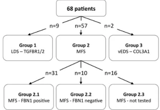
Genetic testing was performed in 76% of patients. Sixty per cent of
these patients were positive for FBN1 mutations associated with
MFS, and in 20%, noFBN1 mutation was found. Interestingly, 17% of patients (representing an absolute number of 9 patients) harboured TGFBR1/2 mutations associated with LDS, and in 3% of patients, a COL3A1 mutation could be detected. Patients were divided into three groups (Fig.1): Group 1 are genetically confirmed LDS patients, Group 2 are MFS patients and Group 3 are patients with vEDS. The MFS patient group is further divided into three subgroups: Group 2.1 areFBN1-positive patients, Group 2.2 are FBN1-negative patients fulfilling clinical criteria and Group 2.3 are patients fulfilling clinical criteria who never underwent mutation analysis.
Dissection at initial presentation or during
follow-up
Initial presentation was AAD in 37% of patients and aneurysmal disease in 63%. Interestingly, initial presentation with AAD differed neither between LDS and the overall MFS population (33 vs 37%, P = 0.48) nor between subgroups. Likewise, there was no differ-ence in the rate of overall dissection (initially or during follow-up)
between LDS and MFS patients (44 vs 42%,P = 1.0). Again, there
was no difference compared with FBN1-positive or -negative
patients or those who had never been tested. Mean age at initial surgery did not differ between LDS and MFS patients (34 ± 13 vs 35 ± 15 years,P = 0.85).
Primary aortic root and arch interventions
Aortic root size at initial surgery, respectively the initial presenta-tion, in adult LDS patients not presenting with type A dissection was 57 ± 9 mm. A 14-year old patient presented with a 42-mm
aortic root dilatation, which translated into a z-score of
8. Although there was a trend towards modified Bentall
proce-dures (78 vs 47%) compared with VSRR performed in the LDS population, there were no significant difference between LDS and MFS patients in the type of initial aortic root that was performed. Valve-sparing procedures were performed in 11% of the LDS patients and 23% of the MFS patients (P = 0.67) with a strong trend towards VSRR in the most recent period. In 11% of LDS patients and 14% of MFS patients, only the ascending aorta was replaced since the diagnosis of a connective tissue disorder was not estab-lished at the time of surgery. Simultaneous total arch replacement in patients presenting with acute type A dissection was carried out in none of the LDS and 12% of MFS patients. No LDS patient undergoing elective aortic root repair needed primary total arch replacement, whereas this was the case in 2.8% of MFS patients.
Reinterventions on aortic root and arch
Reinterventions on the aortic root became necessary in 3 of 7 MFS patients in whom only the ascending aorta had been replaced during initial surgery for type A dissection, since the root was not severely affected and MFS was not suspected at the time of surgery. One patient had initially received a modified Bentall procedure. Both the LDS patients presenting with type A
dissec-tion had received a modified Bentall procedure and no
reinter-ventions have been necessary so far. Reintervention rates after elective aortic root surgery did not differ between LDS and MFS patients and were mainly because of aortic regurgitation after VSRRs using the Yacoub technique (11 vs 14%,P = 1.0). There was no difference in the need for secondary total arch replacement
between LDS and MFS patients (11 vs 14%, P = 1.0) or between
MFS subgroups (FBN1 positive 16%, P = 1.0; FBN1 negative 10%, P = 1.0; not tested 13%, P = 1.0).
Reinterventions on the distal aorta
There were no significant differences between LDS and MFS
patients regarding the need for distal aortic reoperations during follow-up (33 vs 28%,P = 0.71). Furthermore, there was no signi fi-cant difference in the rate of total aortic replacement between LDS and MFS patients (22 vs 12%,P = 0.6), nor between subgroups (FBN1 positive 19%, P = 1.0; FBN1 negative 0%, P = 0.2; not tested 6%,P = 0.5).
Follow-up and mortality
The mean follow-up was 12.7 ± 7 years. All-cause 30-day, 6-month and 1-year mortality rates were 2.9, 4.4 and 7.3%, re-spectively. The mean follow-up was 12.7 ± 7 years. Late mortality did not differ between LDS and MFS patients (11 vs 16%,P = 1.0), or between subgroups (FBN1 positive 13%, P = 1.0; FBN1 negative 10%,P = 1.0; not tested 25%, P = 0.62).
Freedom from reoperation and survival
Freedom from reintervention in LDS patients was not different compared with that in MFS patients (Figs2and3). Freedom from reintervention at 5, 10 and 15 years was 61, 53 and 53% in LDS and 74, 61 and 40% in MFS patients, respectively. There were no
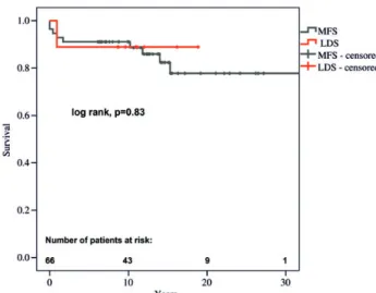
significant differences in survival between LDS and MFS patients
(Figs4and5). Survival at 5, 10 and 15 years was 89% in LDS and 91, 91 and 82% in MFS patients, respectively.
DISCUSSION
It has been repeatedly shown that AAD due to aortic aneurysm determines morbidity and mortality in patients with MFS, and there is substantial evidence that this is also true for patients with LDS [3,16]. Others and we have shown that patients initially pre-senting with AAD have an increased mortality because of a higher reintervention rate even if initial surgery is survived [12,17,18]. Therefore, the consistent results of elective aortic root surgery have fostered the concept of prophylactic aortic root surgery to prevent acute dissection. Guidelines have recognized patients with connect-ive tissue disease to be at a higher risk for AAD than the average patient and have continuously lowered the threshold to intervene.
The initial description of LDS as a subset of patients within the MFS spectrum with a rapid progression of aortic disease has high-lighted the need for identifying specific patient populations in patients with genetically mediated aortic disease.
In our population of patients diagnosed with MFS based on clinical criteria and mutation analysis of theFBN1 gene before the advent ofTGFBR1/2 testing, one-fifth of patients have been
retro-Figure 2: Kaplan–Meier curve showing no significant differences regarding
freedom from reoperation in LDS patients compared with MFS patients.
Figure 3: Kaplan–Meier curve showing no significant differences regarding
freedom from reoperation in LDS patients compared with patients with or without confirmed FBN1 mutations.
Figure 4:Kaplan–Meier curve showing no significant differences regarding
sur-vival in LDS patients compared with MFS patients.
Figure 5:Kaplan–Meier curve showing no significant differences regarding
survival in LDS patients compared with patients with or without confirmed FBN1 mutations. A O RTIC SURGE R Y
spectively diagnosed as having LDS. Patient selection for genetic testing in this study was certainly biased, as there was a tendency that patients with a strong phenotype who have been clinically diagnosed with MFS as children have never been tested. Furthermore, there is certain likelihood that those patients ful fill-ing Ghent criteria who have never been tested harbour mutations
other thanFBN1.
Recently, new mutations in patients sharing phenotypic features
with LDS such as SMAD3, TGFB2 and TGFB3 have been identified,
and it is likely that this will continue [19–21]. Our data stress the importance of genetic testing to allow a proper diagnosis, but also show that a negative mutation analysis certainly does not rule out the presence of a connective tissue disease. The diagnostic criteria for MFS have changed over time and will likely be challenged again in the future byfinding new mutations that correlate with a distinct phenotype within the MFS spectrum [22,23].
In our current series comprising 830 patient-years, LDS patients did not dissect earlier, did not dissect more frequently and did not have a higher need for reinterventions than MFS patients. Although LDS patients represented a significant percentage of the MFS patient population in this series, the absolute number of LDS patients involved is small and no definitive recommendations can be derived from this data. In our LDS patients, the mean aortic root diameter in patients not presenting with type A dissection was 57 ± 9 mm, with only 1 patient who underwent surgery as a child. In the initial report from the Baltimore group, the ratio of children vs adults who underwent surgery was 2 : 1, vascular events frequently occurred at diameters <45 mm and in patients younger than age 10, with the youngest being 6 months old.
Our results are in line with previously published data from the Hôpital Bichat group in Paris [24] and do not necessarily contradict the data that have already been published by the Baltimore group; it may rather be a proof for a wider phenotypic spectrum within the LDS population than previously thought. In a large
compara-tive study of 243 MFS and 70 patients withTGFBR2 mutation
pub-lished by Attiaset al. [24], there was no difference between MFS and LDS patients regarding average age at which aortic surgery was performed (35 ± 16 vs 39 ± 13 years) as well as incidence of aortic dissection (14 vs 10%). Mortality was higher inTGFBR2 families before diagnosis, but similar once patients had been diag-nosed correctly and underwent surgery.
Primary interventions on the root did not significantly differ between groups in our study and because there are no differences in the rate of AAD, numbers are comparable between LDS and MFS and in line with previously published data. There was a trend towards VSRR procedures performed in the MFS group, mainly because of the fact that in this historic cohort, LDS patients have
been identified very late and most patients already had an
enlarged annulus and/or multiple fenestrations in their valves, which resulted in a higher rate of Bentall procedures. Now that patients are prospectively identified, there seems to be no differences anymore.
Patel et al. [5] suggested that in LDS, the aortic arch is more prone to dilatation than in MFS patients, and that more aggressive arch repair should be considered. In the current series, using a liberal approach towards a short circulatory arrest to perform hemiarch replacement, the rate of secondary total arch replace-ments did not differ between LDS and MFS patients. The need for total aortic replacement in MFS is predominantly driven by the occurrence of type B dissection, and as the rate of type B dissec-tion did not differ between LDS and MFS patients, this seems to be true for the LDS patient population as well. It seems important
to point out that all LDS patients who underwent elective root surgery initially presented (or have been referred) with an aortic root diameter that would have prompted us to proceed to surgery regardless of the underlying disease. Since 2007, patients are spe-cifically evaluated for the presence of LDS and genetic testing is performed in all patients with a suspicious phenotype. The number of LDS patients has been constantly increasing most likely because of a growing awareness for this patient population, i.e. that many LDS patients are not missed anymore. Considering the well-documented series of patients seen at the Johns Hopkins
Hospital, we have adopted a modified approach published by this
group and consider surgery in adult LDS patients presenting with
an aortic root size of ≥4 and ≥3 cm in children if the aortic
annulus is adequately sized (≥2 cm) to allow for a durable solution that will last into adulthood.
CONCLUSION
In a population of patients diagnosed with MFS based on clinical criteria and mutation analysis of theFBN1 gene before the advent
of TGFBR1/2 testing, 17% of patients have been retrospectively
diagnosed as having LDS. Although early surgical intervention in LDS is warranted to avoid AAD, the current data suggest that once the diseased segment is repaired, there seems to be no additional burden in terms of mortality or reoperation rate compared with
MFS patients, with or without confirmed FBN1 mutation.
ACKNOWLEDGEMENT
We thank Brigitta Gahl, University Hospital Berne, for statistical assistance.
Conflict of interest: none declared.
REFERENCES
[1] Loeys BL, Schwarze U, Holm T, Callewaert BL, Thomas GH, Pannu Het al.
Aneurysm syndromes caused by mutations in the TGF-beta receptor. N Engl J Med 2006;355:788–98.
[2] Loeys BL, Chen J, Neptune ER, Judge DP, Podowski M, Holm Tet al. A
syn-drome of altered cardiovascular, craniofacial, neurocognitive and skeletal
development caused by mutations inTGFBR1 or TGFBR2. Nat Genet
2005;37:275–81.
[3] Williams JA, Loeys BL, Nwakanma LU, Dietz HC, Spevak PJ, Patel NDet al.
Early surgical experience with Loeys-Dietz: a new syndrome of aggressive thoracic aortic aneurysm disease. Ann Thorac Surg 2007;83:S757–63.
[4] Hiratzka LF, Bakris GL, Beckman JA, Bersin RM, Carr VF, Casey DE Jret al.
ACCF/AHA/AATS/ACR/ASA/SCA/SCAI/SIR/STS/SVM guidelines for the diagnosis and management of patients with Thoracic Aortic Disease. Circulation 2010;121:266–369.
[5] Patel ND, Arnaoutakis GJ, George TJ, Allen JG, Alejo DE, Dietz HCet al.
Valve-sparing aortic root replacement in Loeys-Dietz syndrome. Ann Thorac Surg 2011;92:556–60.
[6] Loeys BL, Dietz HC, Braverman AC, Callewaert BL, De Backer J, Devereux
RBet al. The revised Ghent nosology for the Marfan syndrome. J Med
Genet 2010;47:476–85.
[7] Beighton P, de Paepe A, Danks D, Finidori G, Gedde-Dahl T, Goodman R et al. International Nosology of Heritable Disorders of Connective Tissue, Berlin, 1986. Am J Med Genet 1988;29:581–94.
[8] De Paepe A, Devereux RB, Dietz HC, Hennekam RC, Pyeritz RE. Revised diagnostic criteria for the Marfan syndrome. Am J Med Genet 1996;62: 417–26.
[9] Matyas G, De Paepe A, Halliday D, Boileau C, Pals G, Steinmann B. Evaluation and application of denaturing HPLC for mutation detection in
Marfan syndrome: identification of 20 novel mutations and two novel
polymorphisms in theFBN1 gene. Hum Mutat 2002:19;443–56.
[10] Matyas G, Arnold E, Carrel T, Baumgartner D, Boileau C, Berger Wet al.
Identification and in silico analyses of novel TGFBR1 and TGFBR2
muta-tions in Marfan syndrome-related disorders. Hum Mutat 2006:27;760–9. [11] Schoenhoff F, Schmidli J, Czerny M, Carrel TP. Management of aortic
aneurysms in patients with connective tissue disease. J Cardiovasc Surg
2013;54:125–34.
[12] Schoenhoff FS, Jungi S, Czerny M, Roost E, Reineke D, Matyas Get al.
Acute aortic dissection determines the fate of initially untreated aortic segments in Marfan syndrome. Circulation 2013;16:1569–75.
[13] Schoenhoff F, Kadner A, Czerny M, Jungi S, Meszaros K, Schmidli Jet al.
Should aortic arch replacement be performed during initial surgery for aortic root aneurysm in patients with Marfan syndrome? Eur J Cardiothorac Surg 2013;44:346–51.
[14] Czerny M, Krähenbühl E, Reineke D, Sodeck D, Englberger L, Weber A et al. Mortality and neurologic injury after surgical repair with hypother-mic circulatory arrest in acute and chronic proximal thoracic aortic path-ology: effect of age on outcome. Circulation 2011;124:1407–13. [15] Krähenbühl ES, Clément M, Reineke D, Czerny M, Stalder M, Aymard T
et al. Antegrade cerebral protection in thoracic aortic surgery: lessons
from the past decade. Eur J Cardiothorac Surg 2010;38:46–51.
[16] Cameron DE, Alejo DE, Patel ND, Nwakanma LU, Weiss ES, Vricella LA et al. Aortic root replacement in 372 Marfan patients: evolution of opera-tive repair over 30 years. Ann Thorac Surg 2009;87:1344–9.
[17] Kari FA, Russe MF, Peter P, Blanke P, Rylski B, Euringer Wet al. Late
compli-cations and distal growth rates of Marfan aortas after proximal aortic
repair. Eur J Cardiothorac Surg 2013;44:163–71.
[18] Mimoun L, Detaint D, Hamroun D, Arnoult F, Delorme G, Gautier Met al.
Dissection in Marfan syndrome: the importance of the descending aorta. Eur Heart J 2011;32:443–9.
[19] Rienhoff HY Jr, Yeo CY, Morissette R, Khrebtukova I, Melnick J, Luo Set al. A
mutation in TGFB3 associated with a syndrome of low muscle mass, growth retardation, distal arthrogryposis and clinical features overlapping with Marfan and Loeys–dietz syndrome. Am J Med Genet A 2013;161:2040–6. [20] Wischmeijer A, Van Laer L, Tortora G, Bolar NA, Van Camp G, Fransen E
et al. Thoracic aortic aneurysm in infancy in aneurysms-osteoarthritis syn-drome due to a novel SMAD3 mutation: further delineation of the pheno-type. Am J Med Genet A 2013;161:1028–35.
[21] Lindsay ME, Schepers D, Bolar NA, Doyle JJ, Gallo E, Fert-Bober Jet al.
Loss-of-function mutations in TGFB2 cause a syndromic presentation of
thoracic aortic aneurysm. Nat Genet 2012;44:922–7.
[22] Aalberts JJ, Thio CH, Schuurman AG, van Langen IM, van der Pol BA, van
Tintelen JPet al. Diagnostic yield in adults screened at the Marfan
out-patient clinic using the 1996 and 2010 Ghent nosologies. Am J Med Genet
A 2012;158:982–8.
[23] Yang JH, Han H, Jang SY, Moon JR, Sung K, Chung TYet al. A comparison
of the Ghent and revised Ghent nosologies for the diagnosis of Marfan syndrome in an adult Korean population. Am J Med Genet A 2012;158:
989–95.
[24] Attias D, Stheneur C, Roy C, Collod-Béroud G, Detaint D, Faivre Let al.
Comparison of clinical presentations and outcomes between patients with
TGFBR2 andFBN1 mutations in Marfan syndrome and related disorders.
Circulation 2009;120:2541–9.
APPENDIX. CONFERENCE DISCUSSION
Dr T. Kuntze (Bad Berka, Germany): The authors present an interesting analysis of genetic testing for LDS in a series of consecutive patients with genetically mediated aortic disease. In a population of surgically treated patients with con-nective tissue disorder, primarily diagnosed as having MFS, 17% were
retro-spectively identified as having LDS by means of TGFBR testing.
In the series comprising a total of 830 patient-years, the authors were able to demonstrate that presence of LDS was not associated with an increased aortic dissection rate or higher reintervention rate during follow-up after primary suc-cessful aortic root surgery as compared to MFS patients. The rate of distal rein-terventions is known to be very high in these patients with a 30% prevalence in
this present study. Most notably, no significant difference was found in the rate
of proximal and distal reoperations among four analysed cohorts of patients with different genetic makeup. Moreover, the study clearly demonstrates that a negative mutation analysis does not rule out the presence of connective tissue disorder and is not associated with any major difference in the long-term outcome after initial surgery. Based on the data from this study and others, the
form of initial presentation of patients with connective tissue disorders and the type of surgical procedure on the diseased aortic segment seem to be major determinants of the long-term prognosis.
The authors observed, similarly to others, acute aortic dissection as an initial presentation in approximately one-third of their patients, which was independ-ent of the type of connective tissue disease involved. This fact highlights a crucial role of the echocardiographic screening and elective root surgery in families with genetically triggered aortic disease. My comments to the authors are following.
First, reoperation rate is known to be markedly elevated in this patient popu-lation. Based on the Kaplan-Meier curve presented in figure 3 in the
manu-script, reoperation rate seems to be higher during thefirst 5 to 7 years after the
primary surgery, at least in the LDS cohort andFBN1-positive MFS subgroup. Is
there any explanation for this increased reoperation rate in these patient
sub-groups during thefirst five years after primary surgery?
Second, the authors noticed in their manuscript that there was a trend toward fewer valve-sparing root procedures in LDS patients versus the Marfan patients. Are there any technical considerations to explain this difference in sur-gical strategy?
And third, do the results of the current study affect the authors’ current
man-agement of the aorta in patients who present with type A aortic dissection and
suspected LDS, e.g., in patients who have a bifid uvula?
Dr Schoenhoff: I will start with your second question which is the rate of David procedures in both groups. Indeed there was a slight difference in the rate of valve-sparing procedures performed, but this is mainly due to the fact
that the Loeys-Dietz patients in this historic cohort were identified very late or
have presented very late. The mean aortic root size in those patients not pre-senting with type A dissection was 57 mm, so it had presented late. Most patients already had an enlarged annulus and/or multiple fenestrations in their valves, which might explain the higher rate of Bentall procedures. Now that we actively look for these patients and see them earlier, I think just last month Dr Carrel performed two David procedures in Loeys-Dietz patients, so far there seems no technical difference in these patients.
Regarding your third question about the management of these patients, in a recently published paper, the Baltimore group raises the question whether the aortic arch should be more aggressively replaced in initial surgery in Loeys-Dietz patients. The data that we now have prompted us to treat them the same as Marfan patients, so very aggressively perform an open distal anasto-mosis even in the elective setting, but no primary total arch replacement if an anastomosis can be otherwise performed.
And so to thefirst question where you asked why there was a higher need
for reoperation in the subgroups ofFBN1 positive patients and Loeys-Dietz
patients. For the past decade there have been several groups who tried to show that there is a higher need for reoperation in patients that tested positive for FBN1, but they were never able to. And I think this is because in the FBN1
negative patients there are so many‘hidden’ patients, such as this group of
Loeys-Dietz patients, so this is why there was no difference. And if you look at the percentages, there was absolutely no difference in the need for reopera-tions. But it’s true, if you look at the Kaplan-Meier curve, there is a visible
differ-ence. This did not translate into a statistically significant difference, but I think
the obvious reason would be the fact that there are more dissections in these
groups. But this is not true in our patient population. So right now I can’t give
you a definite answer as to whether it’s just an artefact - I mean, it’s very few
patients in the Loeys-Dietz group - or if it’s real. But this is a very interesting
point and I will follow up on it.
Dr H. Schäfers (Homburg/Saar, Germany): In the last 5 to 10 years there has been some uncertainty about Loeys-Dietz syndrome, in that it dissects early and has such a bad prognosis. This is almost at the point that every patient who was diagnosed as LDS should be operated on the moment you see him. I’m exaggerating now a little.
Your data suggests that maybe these patients should be treated not more aggressively than Marfan patients but in a similar way. Would you be so open to propose that, or do you think the data is not there yet to be more conserva-tive in LDS?
Dr Schoenhoff: I think that we have too little data to make any definite state-ment. I think there are two important points. One is that if you look at the initial data from the Baltimore group, this clearly represents a different patient
popula-tion, it’s mostly children or young adults with a very strong phenotype, probably
referred from all over the U.S. to Baltimore. I think this is a very different patient population compared to the patients we regularly see. For example, the largest LDS family that we take care of is a family that was identified through the mother, who received a bi-iliac Y-prosthesis at the age of 56 and has only mod-erate aortic root enlargement. So I think this is a different phenotype as you said.
There was one paper from the Paris group who have similar results to ours so I think we have to look for more data.
A O RTIC SURGE R Y