doi:10.1017/S0967199410000092 First Published Online 13 July 2010
Review: Role of tubal environment in preimplantation
embryogenesis: application to co-culture assays
Pierre Guérin
1and Yves Ménézo
2VetAgro Sup, Marcy l’Etoile, France; UNILABs Genève, Switzerland; and Laboratoire d’Eylau, Paris, France Date submitted: 28.10.2009. Date accepted: 22.01.2010
Summary
The culture of early preimplantation stage embryo is still delicate and the metabolic pathways of embryos are not completely understood. Embryo needs are evolutionary during the preimplantation development, consequently it is difficult to meet embryo needs in vitro. Culture conditions have to respect several physical and chemical equilibria: such as redox potential, pH, osmotic pressure, metabolic flux of energetic compounds, endogenous pools of amino acids and transcripts, etc. Embryo culture media are generally supplemented with amino acids, glucose, other energetic metabolites and antioxidant compounds, vitamin, and growth factors etc. Furthermore autocrine and paracrine regulation of embryo development probably exist. In fact embryo culture conditions have to be as non-toxic as possible. Various types of co-culture systems have been devised to overcome these problems. Complex interrelations exist between embryos and co-cultured cells. The beneficial effects of co-cultured cells may be due to continuous modifications of the culture medium, i.e. the elimination of toxic compounds and/or the supply of embryotrophic factors.
Keywords: Co-culture, Culture medium, Embryo, Oviductal fluid
Introduction
Various culture media have been used to support early stages embryo development in human but also in farm animals. However embryo requirements are far from completely defined and length of culture time is still associated with loss of viability, especially at the individual level. In fact, during this preimplantation period, there are two phases. The first phase is one completely under maternal control, driven by the maternal material: mRNAs and proteins stored during oogenesis and the final stages of oocyte maturation. Then, secondly, after maternal to zygotic transition, the embryo will take the lead after the first real ‘embryonic’ transcriptions. If the culture time is shortened, embryo transfer is impossible in farm animals, due to their expulsion. In human, embryo transfer is usually performed in the uterus, at least 2 days before the normal entry time. Various types
1All correspondence to Pierre Guérin. VetAgro Sup, 69280 Marcy l’Etoile cedex, France. Tel: +33 4 78 87 26 08. Fax: +33 4 78 87 67 88. e-mail: p.guerin@vet-lyon.fr
2UNILABs, 12 Place Cornavin, Genève, Switzerland and
Laboratoire d’Eylau, 55 Rue St Didier, 75116 Paris, France.
of co-culture systems have been devised to overcome these problems: (1) co-culture with trophoblastic tissue (Heyman et al., 1987), the first system to overcome the early cell block in farm animals; (2) co-culture with cell monolayers of fibroblasts have really limited value for embryo culture; and (3) monolayers of oviduct cells or uterine cells are successful to allow culture from the fertilised embryo to the blastocyst stage (Gandolfi & Moor, 1987; Thibodeaux et al., 1992). In human, co-culture with Vero cells (Ménézo et al., 1990), and with autologous uterine cells obtained from biopsies has been widely used.
In this article, we will review the physiological and biochemical aspects of the tubal environment and their impact on in vitro co-culture.
Physiological and biochemical aspects
Oviduct fluid is generated by transudation into the lumen as well as by secretion of compounds synthesized by epithelial cells (Leese, 1988) (Fig. 1).
Collection of normal and physiologic tubal fluid has been performed in various ways (reviewed by Aguilar & Reyley, 2005): surgical installation of cannula in the lumen of the tube, aspiratory puncture of the
Figure 1 Mouse oviduct cells (A, C, D) and embryo into oviduct (B).
dilated oviduct 3 days after the organ was tied at its two extremities, and absorption with filter paper after oviduct dissection and opening.
We have observed that only two techniques permit the collection of physiologic tubal fluid: (1) cannulation; and (2) aspiratory puncture after double
ligature of the organ. Indeed the absorption technique with filter paper provides a fluid whose composition is close to that of the serum (personal observation). The absorption technique may select the transudate part of the oviduct fluid to the detriment of the secretory part. The flushing techniques do not allow the quantification of compounds.
The oviduct fluid amount is dependent on the stage of estrous cycle. The produced amount is increased by estradiol-17β and decreased by progesterone (Leese, 1988).
Oviduct fluid contains amino acids, ions, proteins, hormones, growth factors, carbohydrates and lipids. Oviduct fluid composition has been analysed in several species, including cow, ewe, sow, rabbit, mouse and human (Guérin et al., 1995).
Amino acids
Certain amino acids, particularly alanine, glycine, glutamate, hypotaurine and taurine, are present in strikingly high concentrations in the oviductal fluid of cow, ewe, sow, rabbit and mouse (Ménézo, 1976; Guérin et al. 1995) (Fig. 2). We have observed that oviduct cells support modifications of the amino acids environment. When methionine was incubated with oviduct epithelial cells we observed the production and release of taurine/hypotaurine (Guérin et al., 1995b), glutathione, cystathionine and homocysteine (Ouhibi et al., 1990). The particular pattern of amino acid concentrations may be an important factor to be
Table 1Concentration of ions in the oviduct fluid of various species collected from ampulla or the whole tube Ion concentration (mEq/l)
Cow (whole) Eww (ampulla) Rabbit (whole) Human (whole) Mare (whole)
Na 86.1 135 189.7 130 129.5 Cl 112.7 – 332.2 132 – K 65.7 8.1 16.8 21.1 7.9 Ca 3.2 7.6 2.7 1.1 2.3 Mg – 1.2 0.5 1.4 4.6 S – – – 12.3 – P 3.0 – 0.2 8.7 0.4 Zn – – 0.005 – – Bicarbonate – – 16.6 – –
Modified from Aguillar et al., 2005.
considered for the improvement of embryo culture media.
Ions
Concentrations of ions in tubal fluid are similar to those in serum for the majority of species. Nevertheless, high potassium levels in tubal fluid seem to be constant across species. Potassium concentrations are considerably higher in tubal fluid than in serum (Table 1). Consequently, potassium concentration in embryo culture media is an important factor to consider. In a mouse IVF system, more pregnancies were obtained by using a medium based on human oviduct fluid potassium composition (HOF) rather than Tyrode’s medium containing a low concentration of potassium (Quinn et al., 1985).
Glucose and its metabolites
Glucose levels in bovine oviduct are extremely low in comparison with serum concentrations suggesting that this is important for successful sperm capacitation and embryonic development. Preimplantation embryos gain their energy by oxidative phosphorylation, i.e. the oxidation of pyruvate and amino acids. Only at the compaction stage do embryos switch to glycolysis.
High levels of glucose in culture medium have been considered as major effectors of in vitro embryo developmental arrests. High glucose concentrations in the culture medium are implicated in the generation of reactive oxygen species (ROS) (Iwata et al., 1998). Gluc-ose inhibits hypoxanthine phosphoribosyl transferase (HPRT) activity. The result is the production of ROS via xanthine oxidase (Guérin et al., 2001). Metabolism of glucose may be impaired in rodent in relation to a metabolic block with a poor entry into Krebs’ cycle and a block at glucose-6-phosphate isomerase leading to useless accumulation of glycogen. High levels of glucose in culture media may have deleterious effects in diabetic mammals and probably in humans.
Pyruvate is an interesting compound in that it acts not only as an energy source, but can also detoxify ammonia in the embryo through transamination and export of alanine, formed as a result. It also plays a role in protection against oxidative stress (see Oviduct production against oxidative stress) by preventing peroxide-induced injury (Morales et al., 1999). Addition of lactate is questionable as it is a cul de sac product. To be re-introduced into the metabolism it has to be converted (oxidized) in pyruvate with the generation of NADH. Lactate can be used as an energy substrate by embryos and may serve to maintain the proper redox balance and pH. One of the interests of co-culture with oviduct cells may be the reduction of concentrations of glucose and the production of lactate (Ouhibi et al., 1990). Other glucose-derived metabolites (such as acetate, citrate, malate etc.) are sometimes added in media with no real established impact.
Vitamins
Ascorbic acid is present in follicular and tubal fluid (Hansen et al., 1991; Paszkowski & Clarke, 1999). Several animal experiments have claimed the interest of water-soluble vitamins in culture media, but still no unequivocal demonstration has been made. Vitamin C and vitamin E have protective effects on embryo development. Experiments concerning embryo culture medium supplementation with vitamin C in the presence or absence of vitamin E strongly suggest that vitamin C is active via the protection of vitamin E against oxidation reactions.
Folic acid is another parameter: reduced folic acid is a methyl donor (as methionine through S-adenosyl methionine). There is no evidence that supplementation in vitro is necessary. Lipid-soluble vit-amins (A and E) are natural potent anti-oxidants, but their introduction into culture media is problematic due to their low hydrosolubility.
Growth factors
Early embryos have receptors for a variety of growth factors, and co-cultured cells may secrete growth factors that stimulate embryo development. Several growth factors have been identified in oviduct and have beneficial effects on embryo development. Platelet-activating factor (PAF) is secreted by human and hamster embryo, and PAF receptor (PAF-R) has been identified in the oviduct. Bovine embryos synthesize growth hormone (GH) and its receptor in a stage-specific manner and bovine oviduct epithelial cells express the GH receptor. IGF binding proteins (IGFBPs) are present in early embryos (Pushpakumara et al., 1991). Locally produced IGFs, regulated by IGFBPs may act directly on the embryo or via mod-ulation of oviduct secretions and muscular activity to positively influence early embryonic development (Pushpakumara et al., 1991). Vascular endothelial growth factor (VEGF) and FGF are secreted by the bovine oviduct in different amounts depending on the stage of the estrous cycle. FGF-1 is mainly secreted at ovulation whereas FGF-2 is increased after ovulation. FGF-2 promotes development of bovine embryos beyond the 8-cell block (Larson et al., 1992). VEGF accelerates development of embryos (Luo et al., 2002), PDGF has beneficial effects on embryo development in vitro (Larson et al., 1992).
Nevertheless, no specific embryotrophic factors secreted by the genital tract has been identified.
Other compounds such as macromolecules (OVGP) and hyaluronic acid (HA) are present in tubal fluid and have beneficial effects on embryo development. Expanding cumulus cells produce large amounts of HA. HA has a positive effect on cell number and apoptosis in blastocysts (Ulbrich et al., 2004).
Serum is not necessary for IVF culture (Ménézo et al., 1984): serum addition affects mRNA content and is involved in the ‘large calf syndrome’. Serum albumin is not fully necessary for embryo culture, but has always been added into culture media as it helps with embryo handling. The beneficial role of albumin is not fully understood: it binds several compounds of various molecular weights, such as lipids, amino acids, peptides, catecholamines. It may be incorporated directly into the embryo and may be a carrier of nutrients.
Oviduct protection against oxidative stress
ROS are produced by gametes and embryo, particu-larly during capacitation, fertilization and the second cell cycle division in mouse (Noda et al., 1991). Superoxide anion O2−•) induces the production of
other ROS such as hydrogen peroxide (H2O2) and
the extremely reactive hydroxyl radicals (OH•) via the Haber–Weiss or de Fenton reactions (Guérin et al., 2001). Oxidative stress affects embryo viability via
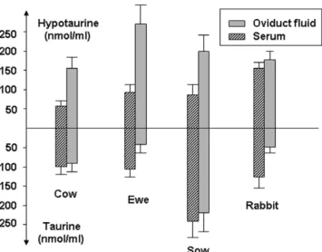
al-Figure 3Taurine and hypotaurine concentrations into serum and oviduct fluid.
teration of proteins, nucleic acids and membrane lipids (Guérin et al., 2001). Oxidative stress is particularly important during in vitro embryo production (Goto et al., 1993) and is implicated in the in vitro embryo block (Noda et al., 1991). Several factors may explain that: exposition to high levels of oxygen, traces of metallic ions and/or light (Nakayama et al., 1994), oxygen concentration etc. Furthermore, serum, used for in vitro maturation of animal oocytes contains amine oxidases (Parchment et al., 1990). Reduction of oxygen concentration from 20% to 5% limits ROS production by the mouse embryo (Goto et al., 1993).
Several enzymatic and non-enzymatic mechanisms present in oviduct are implicated in embryo protection against oxidative stress: hypotaurine, ascorbic acid, superoxide dismutase (SOD), catalase, gluthathione peroxidase (GPX) etc.
Hypotaurine and taurine have been detected in oviduct and follicular fluids of cow, ewe, sow, rabbit and mouse (Guérin et al., 1995b) (Fig. 3). Oviduct epithelial cells in vitro are able to synthesise taurine and hypotaurine from methionine (Guérin et al., 1995b). The enzyme cysteine sulphinate decarboxylase (EC 4.1.1.29.) is present and active in the oviduct of cow and goat (Guérin & Ménézo, 1995). Hypotaurine neutralise OH• radicals. The byproduct of this reaction is taurine, which is able to neutralise cytotoxic aldehydes produced by oxidative stress. Furthermore, hypotaurine addition to embryo culture medium has beneficial effects on bovine embryo develop-ment (Barnett & Bavister, 1992; Guyader-Joly et al., 1998).
Other antioxidant compounds are present in tubal fluid, such as vitamin C and pyruvate (see section on
Figure 4Schematic representation of some protective mechanisms of oocyte and embryo against reactive oxygen species. CSD, cysteine sulfinate decarboxylase; MnSOD, manganese superoxide dismutase; CuZnSOD, copper zinc superoxide dismutase; GPX, glutathione; GSH, reduced glutathione.
also have a pro-oxidant action via the reduction of ferric ions to the ferrous state (catalysing the Haber– Weiss reaction).
In addition the oviduct provides antioxidant en-zymes: the transcripts encoding for antioxidant enzymes SOD, catalase and GPX are present in oviduct epithelial cells (El Mouatassim et al., 1999). These redundant and complementary mechanisms are schematically presented in Fig. 4.
Furthermore, the low oxygen pressure in the oviduct (40–60 mmHg) (Fisher & Bavister, 1993), may limit auto-oxidation reactions. It must be emphasised that the benefit of co-culture may be partly related to the decrease of O2concentration in the medium by oviduct
epithelial cells (Nagao et al., 1994).
Impact onin vitro culture
Culture systems
Culture in microdrops under a layer of mineral oil is often recommended on the basis of mimicking the in vivo situation and following the ‘mouse model’ for embryo culture. Although the volume of liquid surrounding the embryo in vivo is minimal. or ‘virtual’, the embryo is in contact with an epithelial cell layer, with a film of liquid that is permanently renewed (Fig. 1). This is not the case for microdrop culture, and there are three features to be considered: (1)
Metabolic waste from the embryo may accumulate at a higher concentration, due to the sub-optimal volume of liquid. (2) The microdrop surface area allows a maximum exchange with oil, so that liposoluble compounds in the medium can be absorbed at a maximal level by the oil, thus depriving the embryo of these compounds. (3) Similarly, any micropollution of the oil with compounds that are water soluble will pass into the culture medium with a higher efficacy in a microdrop system.
Culture medium
Along with other teams, we began to design and define new, more complex culture media in the early 1970s that were based on the composition of genital tract secretions (Ménézo, 1976). In this way, culture media are supplemented with amino acids, especially glycine, glutamine, alanine, hypotaurine and taurine.
Early preimplantation development, i.e. until mater-nal to zygotic transition (MZT), is matermater-nally driven. Maternal mRNA and protein content of the oocytes, at the time of fertilization, is of major importance as there is an ineluctable turn over of these reserves. During MZT there is a minimal level of transcription only: the mature oocyte must contain a storage pool of proteins and/or mRNA transcripts in order to maintain its viability. Embryos’ needs before and after ZGA are not
alike and the environment, i.e. culture conditions, will have a direct impact on transcription and translation.
In vitro culture conditions and environmental factors are very different from the in vivo conditions (light, variations in pH and temperature, static medium etc.). pH is regulated by HCO3/CO2 buffer according
to the equation pH= pKa + log (HCO3)/(CO2). The
Henderson–Hasselbach equation allows the calcula-tion of pH under a 5% CO2 atmosphere, in relation
with the concentration of bicarbonate. The embryo has an alkaline (7.4) pH: it is not able to match with an acidic pH before ZGA. Gas phase can be 5% CO2 in
air or 5% CO2, 5% O2and 90% N2. Even if a reduced
O2atmosphere seems to be more physiological, based
on genital tract environment, and more susceptible to decrease the free radical formation, no clear cut data support the superiority of reduced oxygen tension in human especially in terms of ongoing pregnancy rates, especially if the medium is well protected by antireactive oxygen species (see Oviduct production against oxidative stress). In this way glutathione is sometimes added to culture media, however it has to be mentioned that it cannot enter the embryo.
Contaminants of the gas phase, such as NO and CO, are as deleterious as volatile organic compounds. An osmolarity between 280 and 300 mM allows early embryo development. No attention is generally paid to the red-ox potential, based on oviduct and uterine secretions it should be calibrated to−0.1 mV. The problem here is due to the absence of technical methods allowing the accurate measurement of the redox potential of a culture medium. A too strong reducing red-ox may have harmful effects on proteins. Serum addition destabilises the culture media through a contribution of enzymes and unknown metabolites and catabolites. It is useless and even deleterious as it may increase pathologies (especially linked to imprinting).
Addition of growth factors in embryo culture media is still a matter of controversy. Growth factors are present in the female genital tract and the corresponding receptors are present on the embryos sooner or later. Beneficial effects have been observed after supplementation of embryo culture medium with growth factors in various species: FGF-2, VEGF, LIF and PDGF in bovine (Larson et al., 1992; Luo et al., 2002). GH is synthesized by bovine embryo and bovine oviduct epithelial cells express the GH receptor (Hull & Harvey, 2001). In humans, GMCSF seems to have also a stimulatory and regulatory impact. Insulin is usually added for the second phase of culture: whether or not it has the same effect as IGF1 is questionable. Locally produced IGFs, regulated by IGFBPs, may act directly on the embryo or via modulation of oviduct secretions to positively influence the success of early embryonic development (Pushpakumara et al., 2002).
Growth factors may act through a balance between stimulatory and inhibitory effects. Mouse embryo development is significantly better when embryos are cultured in groups rather than alone. These results suggest an autocrine/paracrine effect but this effect has been denied in human systems.
Trophic effect of co-culture on embryo development
Even if it is now well established that tubal secre-tion is hormonally regulated, some pharmacological compounds may alter it weakly. There are, in vivo, relations between the oviduct and the passing embryo. The embryo probably regulates its transport and the microenvironment of the tubal cells and the cellular metabolism of the oviduct is modified during passage of the egg (Freese et al., 1973).
However, if we consider the trophic effect towards the embryo i.e. allowing a correct development without loss of viability, we can assess that hormones do not interfere in vitro with this process and that the exchanges between the embryo and the cells are not mandatory. Prepuberal oviducts can sustain early embryonic development in the mouse and in the bovine (Ménézo et al., 1989). Co-culture with genital tract cells collected at different stage of the estrous cycle effectively support embryonic development even if the cell morphology differs (Thibodeaux et al., 1992). The final demonstration was given with the use of Vero or BRL cells for co-culture in human and bovine (Ménézo et al., 1994).
Co-cultured cell metabolism leads to modifications of the culture medium, mimicking tubal secretions. In this way, co-cultured cells may control important physico-chemical parameters of the medium (ionic composition, redox potential, pH, osmotic pressure etc.). It is generally assumed that the feeder cells in the co-culture systems act, on the one hand, via removal of toxic compounds from the culture medium (heavy metal cations, metabolic inhibitors, oxygen, glucose etc.) and, on the other hand, via the production of compounds regulating embryo development (hypotaurine, glutathione, amino acids, lipids, growth factors etc.). Cell monolayers seem able to regulate metabolic pathways of the embryos.
The in vitro embryo development block appears to be related to embryonic genome activation. Thus the overcoming of the in vitro block by co-culture systems suggests that co-cultured cells are able to help in activation of the transcription mechanism.
Conclusion
It is important to understand some of the interactions that take place between oviduct and early embryo
in order to develop culture systems and culture media adapted to embryo needs. Prior to and during culture, it is important to be aware of, and to control, the interactions of different compounds with each other and with the gas phase. Sequential media are currently effective in human embryo culture, with blastocyst yields similar to those observed for co-culture systems. In order to evaluate new techniques and/or new culture media, in vitro grown embryos must be transferred to avoid misleading observations based upon morphology alone.
References
Aguilar, J. & Reyley, M. (2005). The uterine tubal fluid: secretion, composition and biological effects. Anim. Reprod.
2, 91–105.
Barnett, D.K. & Bavister, B.D. (1992). Hypotaurine require-ment for in vitro developrequire-ment of golden hamster one cell embryos into morulae and blastocysts, and production of term offspring from in vitro-fertilized ova. Biol. Reprod. 47, 297–304.
El Mouatassim, S., Guérin, P. & Ménézo, Y. (1999). Mammalian oviduct and protection against free oxygen radicals: expression of genes encoding antioxidant en-zymes in human and mouse. Eur. J. Obstet. Gynecol. Reprod.
Biol. 89, 1–6.
Fisher, B. & Bavister, B.D. (1993). Oxygen tension in the oviduct and uterus of rhesus monkeys, hamsters and rabbits. J. Reprod. Fertil. 99, 673–9.
Freese, V.E., Orman, S. & Paulos, P. (1973). An autoradio-graphic investigation of epithelium egg interaction in the mouse oviduct. Amer. J. Obstet. Gynecol. 117, 833–7. Gandolfi, F. & Moor, R.M. (1987). Stimulation of early
embryonic development in the sheep by coculture with oviduct epithelial cells. J. Reprod. Fertil. 81, 23.
Goto, K., Noda, Y., Mori, T. & Nakano, M. (1993). Increased generation of reactive oxygen species in embryos cultured
in vitro. Free Radic. Biol. Med. 15, 69–75.
Guérin, P. & Ménézo, Y. (1995). Hypotaurine and taurine in gamete and embryo environments: de novo synthesis via the cysteine sulfinic acid pathway in oviduct cells. Zygote
3, 33343.
Guérin, P., Gallois, E., Croteau, S., Revol, N., Maurin, F., Guillaud, J. & Ménézo, Y. (1995a). Techniques de récolte et aminogrammes des liquides tubaire et folliculaire chez les femelles domestiques. Revue Méd. Vét. 146, 805–14. Guérin, P., Guillaud, J. & Ménézo, Y. (1995b). Hypotaurine in
spermatozoa and genital secretions and its production by oviduct epithelial cells in vitro. Hum. Reprod. 10, 866–72. Guérin, P., El Mouatassim, S. & Ménézo, Y. (2001). Oxidative
stress and protection against reactive oxygen species in the preimplantation embryo and its surroundings. Hum.
Reprod. Update, 7, 175–89.
Guyader-Joly, C., Guérin, P., Renard, J.P., Guillaud, J., Ponchon, S. & Ménézo, Y. (1998). Precursors of taurine in female genital tract: effects on developmental capacity of bovine embryo produced in vitro. Amino Acids, 15, 27–42. Hansen, C., Srikandakumar, A. & Downey, B.R. (1991).
Presence of follicular fluid in the porcine oviduct and its
contribution to the acrosome reaction. Mol. Reprod. Dev. 30, 148–53.
Heyman,Y., Ménézo, Y., Chesne, P., Camous, S. & Garnier, V. (1987). In vitro cleavage of bovine and ovine early embryos: improved development using coculture with trophoblastic vesicles. Theriogenology 27, 59–68.
Hull, K.L. & Harvey, S. (2001). Growth hormone: role in female reproduction. J. Endocrinol. 168, 1–23.
Iwata, H., Akamatsu, S., Minami, N. & Yamada, M. (1998). Effects of antioxidants on the development of bovine IVM/IVF embryos in various concentrations of glucose.
Theriogenology 50, 365–75.
Larson, R.C., Ignotz, G.G. & Currie, W.B. (1992). Platelet derived growth factor (PDGF) stimulates development of bovine embryos during the fourth cell cycle. Development
115, 821–6.
Leese, H.J. (1988). The formation and function of oviduct fluid. J. Reprod. Fertil. 82, 843–56.
Luo, H., Kimura, K., Aoki, M. & Hirako, M. (2002). Effect of vascular endothelial growth factor on maturation, fertilization and developmental competence of bovine oocytes. J. Vet. Med. Sci. 64, 803–6.
Ménézo, Y. (1976). Milieu synthétique pour la survie et la maturation des gamètes et pour la culture de l’œuf fécondé. C.R. Acad. Sci., Paris, ser. D. 282, 1967–70.
Ménézo, Y., Testart, J. & Perdrone, D. (1984). Serum is not necessary for human in vitro fertilization, early embryo culture and transfer. Fertil. Steril. 42, 750–1.
Ménézo, Y., Hamidi, J., Khatchadourian, Ch. & Nardon, C. (1989). The murine prepuberal oviduct supports early embryo development in vitro. Dev. Growth Differ. 31, 551–5.
Ménézo, Y., Guérin, J.F. & Czyba, J.C. (1990). Improvement of human early embryo development in vitro by coculture on monolayers of Vero cells. Biol. Reprod. 42, 301–6.
Morales, H., Tilquin, P., Rees, J.F., Massip, A., Dessy, F. & Van Langendonckt, A. (1999). Pyruvate prevents peroxide-induced injury of in vitro preimplantation bovine embryos.
Mol. Reprod. Dev. 52, 149–57.
Nagao, Y., Saeki, M.H. & Kainuma, H. (1994). Effects of oxygen concentration and oviductal tissue on the development of in vitro matured and fertilized bovine oocytes cultured in protein-free medium. Theriogenology
41, 681–7.
Nakayama, T., Noda, Y. & Goto, Y, Mori, T. (1994). Effects of visible light and others environmental factors on the production of oxygen radicals by hamster embryos.
Theriogenology 41, 499–510.
Noda, Y., Matsumoto, H., Umaoka, Y., Tatsumi, K., Kishi, J. & Mori, T. (1991). Involvement of superoxide radicals in the mouse 2-cell block. Mol. Reprod. Dev. 28, 356–60.
Ouhibi, N., Hamidi, J., Guillaud, J. & Ménézo, Y. (1990). Co-culture of one cell mouse embryos on different cell supports. Hum. Reprod. 5, 737–43.
Parchment, R.E., Lewellyn, A., Swartzzendruber, D. & Pierce, B. (1990). Serum amine oxidase activity contributes to crisis in mouse embryo cell lines. Proc. Natl. Acad. Sci.
USA 87, 4340–4.
Paszkowski, T. & Clarke, R.N. (1999). The Graafian follicle is a site ofL-ascorbate accumulation. J. Assist. Reprod. Genet.
Pushpakumara, P.G., Robinson, R.S., Demmers, K.J., Mann, G.E., Sinclair, K.D., Webb, R. & Wathes, D.C. (2002). Expression of the insuline-like growth factor (IGF) system in the bovine oviduct at oestrus and during early pregnancy. Reproduction 123, 859–68.
Quinn, P., Warner, G.M., Kerin, J.R. & Kirby, C. (1985). Culture factors affecting the rate of in vitro fertilization and embryo transfer. Ann. NY Acad. Sci. 442, 195– 203.
Thibodeaux, J.K., Ménézo, Y., Roussel, D.J., Hansel, W., Goodeaux, L.L., Thompson, D.L., Jr & Godke, R.A. (1992). Co-culture of in vitro fertilized bovine embryos with oviductal epithelial cells originating from different stages of the estrous cycle. J. Dairy Sci. 75, 1448–55.
Ulbrich, S.E., Schoenfelder, M., Thoene, S. & Einspanier, R. (2004). Hyaluronan in the bovine oviduct: modulation of synthases and receptors during the estrous cycle. Mol. Cell.