International Immunology, Vol. 5, No. 12, pp. 1609- 1618 © 1993 Oxford University Press
Rearrangement and expression of x light
chain genes can occur without \L heavy
chain expression during differentiation of
pre-B cells
Ulf Grawunder, Dirk Haasner, Fritz Melchers, and Antonlus Rolink
Basel Institute for Immunology, Grenzacherstrasse 487, CH-4005 Basel, Switzerland
Key words: B cell differentiation, Ig gene rearrangement, xL chain protein expression, stromal cell/IL-7 reactive pre-B cells
Abstract
The kinetics of x light (xL) chain gene rearrangement and expression on mRNA and protein level has been studied with four stromal cell/IL-7 reactive, long-term In vitro proliferating pre-B cell lines and clones, two from fetal liver of normal mice and two from fetal liver of E/iH - 6c/-2 transgenlc (toc/-2-tg) mice. These pre-B cell lines and clones are DJH-rearranged on both H chain
alleles. Two of the clones harbor H chain rearrangements which do not allow the expression of VHDJH rearranged H chain genes as /iH chain proteins. Upon removal of IL-7 from the pre-B cell
cultures all four cell lines rearrange VH- D JH and VL- JL gene segments, loose the surface
expression of c-klt, CD43, and surrogate light chain, as well as the capacity to be clonable on stromal cells in the presence of IL-7. Pre-B cells from normal mice die by apoptosls during differentiation, while those from bc/-2-tg mice do not. All four lines and clones express comparable levels of mRNA for ^H and xL chains with the same time kinetics during 3 days of differentiation. However, only two of the four pre-B cell lines and clones express yH chain protein, whereas all four pre-B cell lines and clones express xL chain protein at comparable levels between 2 x 1 0 ' and 1.4 x 10s xL chain molecules per cell. These results suggest that y.H
chain expression Is not mandatory for rearrangement and normal expression of xL chain genes when pre-B cells differentiate to B cells.
Introduction
During B cell differentiation the genes encoding the variable regions of IgH and IgL chains are somatically assembled from VH, D, and JH, and VL and JL gene segments respectively (1).
The various rearrangement events apparently take place in an ordered fashion (2-4), first by rearranging D - JH segments,
folbwed by VH- D JH at the H chain locus (5). V ^ - J ,
rearrange-ments occur later (6,7), while Vx- Jx gene segments are
rearranged last (3,8). The ordered rearrangement of H and L chain genes has favored the hypothesis that the expression of y.H chain proteins is a prerequisite for xL chain gene rearrange-ments (2,9).
The jiH chains can associate with a surrogate light chain encoded by the precursor B cell specific genes \$ (10) and Vpr*e 01) to form an Ig-like complex (12,13). It has been
suggested that this Ig-like complex triggers L chain gene rearrangements during B cell differentiation (14-16).
However, the view that the ordered rearrangement of H and L chain genes is regulated by the expression of ^H protein was recently challenged by several studies, (i) Epstein - Barr virus-transformed human pre-B cell lines were found to express x, as well as X light chains, while H chain genes remained un-rearranged (17). (ii) Some Abelson murine leukemia virus (A-MuLV) transformed pre-B cells derived from SCID mice were found to attempt xL chain gene rearrangements in the apparent absence of jiH protein (18,19). (iii) Analysis of membrane /*H-deficient (/iMT) mice (20) revealed that surface deposition of a functional /iH chain into the cell membrane was not necessary to allow xL chain gene rearrangements in vivo (21). Furthermore, mice with a targeted deletion of the JH locus show normal
frequencies of xL chain gene rearrangements in sorted pre-B cell fractions of the bone marrow, although H chain genes cannot be rearranged in these mutant mice (22,23).
Correspondence to: U. Grawunder
Since all in vitro data have been obtained using transformed cell lines, we have tried to elucidate the question of a putative regulatory role of /«H chains for xL chain rearrangement by com-paring the in vitro differentiation of normal murine pre-B cells, which either can or cannot express /iH chains. This approach allows a detailed analysis of the kinetics of xL chain gene rear-rangements and of the expression of xL chain protein in the absence or presence of ^H chains.
Pro-B and pre-B I cells of the mouse can be cultured for long periods of time on stromal cells in the presence of IL-7 (24) (pro-B cells, all Ig loci in germline configuration; pre-B I cells, H chain loci DJH rearranged; pre-B II cells, at least one H chain locus
VHDJH rearranged; for nomenclature see also 25 and 26).
These cells retain the capacity to differentiate to slg+ B cells
upon removal of IL-7 in vitro (24). They are mostly DJH
re-arranged on both H chain loci and retain their L chain loci in germline configuration. They are generally B220+, express the
pre-B cell specific genes X5 and V ^ g , are c-kit+, CD43+, and
HSA+, while they may or may not express BP-1 (27-29).
Subcloning of pre-B cell lines sometimes results in pre-B cell clones which are DJH rearranged on both alleles in a way, that
a subsequent VH - DJH rearrangement cannot lead to a
pro-ductively rearranged H chain locus capable of expressing /JH chain protein. The in vitro differentiation of two such clones, CL18 derived from a normal BDF, mouse, and BCL-2-11, from a E^H -bd-2 transgenic (bd-2-lg) mouse (30,31), is compared with that of control pre-B cells, which are able to rearrange both their H chain and L chain loci non-productively and productively to generate slg~ and slg+ B cells. The kinetics of xL chain
rearrangement and the expression of xL chain protein is measured to answer the question whether or not expression of /iH chain protein is necessary for the rearrangement and expres-sion of xL chain genes.
Methods Mice
Female (C57 BL/6 xDBA/2)F, (BDF,) mice of different age were obtained from the Institut fur Medizinisch Forschung AG (Fullingsdorf, Switzerland), bcl-2-tg mice (30,31) were bred at our own animal facilities from breeding pairs originally obtained from Dr A. Strasser (The Walter and Eliza Hall Institute of Medical Research, Melbourne, Australia). BDF, embryos from pregnant C57 BL/6 females and bcl-2-\g embryos from time-pregnant heterozygous oc/-2-tg females were provided by the breeding facilities at the Basel Institute for Immunology. The appearance of vaginal plugs was counted as day 0 of gestation. Cell lines
The stromal cell line PA-6 (32) was kindly given to us by Dr H. Kodama (University of Ohu, Japan). Pre-B cell clones CL 18 (24) and PAL-1 (27) were established from day 14 fetal liver of normal BDF, embryos as previously described (24). Pre-B cell line BCL-2-5 and pre-B cell clone BCL-2-11 were obtained from fetal liver of 16 day old bcl-2-Xg embryos by limiting dilution as described (24). Cell-line 5-7 is a stromal cell independent, IL-7 dependent subline of a normal pre-B cell clone 5 (24), which had become slg+ (95% / i+, x+), and served as a positive control for
Western blots developed with nH and xL chain specific antibodies.
mAbs and flow cytometric analysis
The mAbs 187.1 (rat mouse xL chain) (33), ACK-4 (rat anti-mouse c-kit) (27), and S7 (rat anti-anti-mouse CD43) (29) were purified from hybridoma culture supernatants using Protein G -sepharose columns (Pharmacia, Uppsala, Sweden), and were biotinylated according to standard protocols. Biotinylated mAbs LM34 (rat anti-mouse X5) (34) and VP245 (rat anti-mouse V ^ a ) (34) were a kind gift of Dr H. Karasuyama, Basel Institute for Immunology. Biotinylated mAbs 6C3 (rat anti-mouse BP-1), M1/69 (rat anti-mouse HSA), and 7D4 (rat anti-mouse IL-2Rp55, CD25), and FITC-labeled mAbs C2 (rat anti-mouse CD71), RA3-6B2 (rat mouse B220, CD45R), and R6-60.2 (rat anti-mouse IgM) were obtained from Pharmingen (San Diego, CA). Polyclonal horseradish peroxidase-labeled rat anti-mouse xL chain and rat anti-mouse IgM antibodies for Western blotting were obtained from Southern Biological Associates (Birmingham, AL). Surface staining of cells and FACS analysis was carried out as previously described (24). Stainings using biotinylated anti-bodies were developed with streptavidin - FITC (Amersham, Buckinghamshire, UK), except for the Xs and V ^ g specific antibodies LM34 and VP245, which were developed with streptavidin-phycoerythrin (PE) (Southern Biotechnology Associates). Viable cells were either gated by exclusion of propidium iodide (in the case of FITC stainings) or by forward and side scatter (in the case of PE stainings). FACS analysis was carried out by means of a FACScan (Becton Dickinson, Mountain View, CA) equipped with an argon laser tuned to 488 nm. Data were acquired and analyzed using the Lysis software package (Becton Dickinson).
Cell cultures and limiting dilution analysis
Culture of pre-B cells and PA-6 stromal cells was carried out as described (35). Briefly, pre-B cells were cultured on a semiconfluent layer of 7-irradiated PA-6 stromal cells in medium containing 100-200 U/ml IL-7. To obtain differentiated pre-B cells, cells were washed three times in medium without IL-7 (to remove any IL-7 contamination) and were cultured on a semiconfluent layer of 7-irradiated PA-6 stromal cells in medium without IL-7. Differentiated cells were harvested after 1, 2, and 3 days of culture.
Limiting dilution analysis was carried out in 96-well flat-bottom plates containing an irradiated PA-6 stromal cell layer (104
cells/well). Pre-B cell suspensions were diluted by serial 2-fold dilutions in medium containing IL-7. Cultures were scored after 6 days of culture with an inverted microscope for growth of pre-B cell colonies.
Northern blot analysis
Total cellular RNA was extracted from differentiating pre-B cells with acid guanidinium thiocyanate - phenol - chloroform (36) and analyzed by Northern blotting exactly as described (35). Western blot analysis
Differentiated cells from normal pre-B cell clones CL18 and PAL 1 were enriched for viable cells by centrifugation using Rcoll-Paque (Pharmacia). Between 5 x i O6 and 2 x10⁷ living cells were
resuspended in 100 /J lysis buffer (2% NP40, 20 mM Tris - HCI, pH 8.0, 150 mM NaCI, 5 mM MgClj, 5 mM EDTA, 2 mM NaN3,
2 mM phenylmethyl sulfonyl fluoride) and were allowed to lyse for 20 min on ice. Nuclei were separated from cytoplasmic lysates
by centrifugation at 12 000 r.p.m , 4°C, 15 min. Supernatants were diluted in lysis buffer and appropriate dilutions were used to fractionate cytoplasmic proteins under reducing conditions on a 15% protein gel (37). Fractionated proteins were electrobtotted onto nitrocellulose filters (BioRad, Richmond, CA) in 192mM glycine, 25 mM Tris base, 20% methanol for 4 - 6 h at room temperature with constant stirring (38). Immunodetectjon of xL chains and /»H chains was carried out using horseradish peroxidase-labeled antibodies specific for either mouse xL chains or mouse IgM and an ECL Western blotting kit (Amersham) according to the manufacturers protocol. Chemiluminescence on the blots was detected using X-OMAT AR X-ray films (Eastman Kodak, Rochester, NY) with exposure times varying between 10 and 60 min.
Molecular analysis of preB cells
DJH rearrangements of the heavy chain locus can be amplified
and detected using a polymerase chain reaction (PCR) primed
xL chain protein expression without pH chain expression 1611 by oligonucleotides, which bind 5' of each D segment and 3' of the J ^ gene segment. In germline configuration the primers will be located too far apart to be amplified by a PCR, in case of a VHDJH rearrangement, the primer binding site 5' of D would
be deleted. The primers binding 5' of D and 3' of J ^ have been described by Gu et al. (39), and can detect all DJH joints except
those formed by DFL16.2 and DQ52 gene segments. To isolate genomic DNA, 5 x 10s cells were washed with
PBS, lysed by boiling (5 min) in 500 y.\ PBS, and treated with proteinase K (Boehringer, Mannheim, Germany, 0.2 mg/ml) at 55°C for 2 h. The preparations were boiled again to inactivate proteinase K, extracted once with phenol:chloroform:isoamyl-alcohol (25:24:1), and once with chloroform:isoamylphenol:chloroform:isoamyl-alcohol (24:1). DNA was precipitated with 1 volume of isopropanol and 0.1 volume of 3 M Na-acetate, pH 5.2, at -20°C for 1 h. After centrifugation at 12 000 r p.m. for 15 min at 4°C, the pellet was washed with 70% ethanol, air dried, and dissolved in 500 /xl 10 mM Tris-HCI, pH 8.3. Aliquots of 5 /xl of this preparation were subsequently used for PCR amplification.
PAL.1
CL18
B BCL-2-5
BCL-2-11
3 B220c-kit
IL2Ra2
K
B220 c-klt IL2RaLog fluorescence Intensity Log fluorescence Intensity
Urn* of dttfwwrtlrton (days) Um* of dlltwwUUUon (dm)
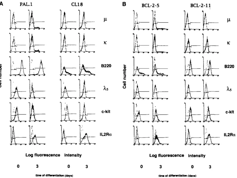
Fig. 1. Expression of a set of B cell differentiation markers (black histograms) on tong-term proliferating pre-B cell clones PAL.1 (left) and CL18
(right) from normal BDF, mice (a) and of pre-B cell Bne BCL-2-5 (left) and done BCL-2-11 (right) from bd-2-ig mice (b) before and after 3 days of differentiation in vitro. All stainings were carried out with either directly FfTC-labeled antibodies (p, B220) or btotinylated antibodies, wtiich were developed by means of streptavidin - FITC (x, c-kit, ll-2Ra) or streptavidin - PE (Xs). Histograms of control stainings (secondary reagent atone) are displayed in Dght grey color.
PCR reactions were performed according to the enzyme manufacturer's protocol (Perkin-Elmer Cetus, Norwalk, CT). The amplification protocol was 20, 94°C; 2 min at 72°C; 35 cycles on a Perkin-Elmer Cetus Thermal Cycler. The PCR products were finally separated and visualized on a 1.5% agarose gel containing 5 /tg/ml ethidium bromide.
Sequencing
The PCR primers used for amplification of the DJH rearranged
genes contain sites for restriction enzymes EcoRI and Xbal. After restriction enzyme digestion of the PCR products, the digestion mixture was separated on a 1.5% UltraPure Agarose gel (BRL, Bethesda, MD) and bands of interest were cut out. The DNA was isolated with the Nal glassbeads method (Geneclean II kit, Bio 101 Inc., La Jolla, CA), ligated into the M13mp19 vector (New England Biolabs, Beverly, MA) and sequenced (Sequenase, United States Biochemicals, Cleveland, OH).
Results
Phenotype of pre-B cells from normal and bcl-2-tg mice before and after 'in vitro' differentiation
Long-term in vitro proliferating pre-B cell clones CL18 and PAL.1 orginating from day 14 fetal liver of BDF, mice, as well as pre-B cell line bcl-2-5 and clone bcl-2-11 from day 16 fetal liver of £>c/-2-tg mice all divide every 2 0 - 2 4 h at 37°C on stromal cells in the presence of IL-7. All four cells lines and clones express various levels of B220, and are positive for c-kit, CD43, CD71 (transferrin receptor), and the surrogate light chain encoded by Xs and Vpjg.e (Fig. 1 and data not shown). Furthermore, undif-ferentiated pre-B cells do not express slg, evident from an in-ability to be stained for /iH and xL chain. They are only weakly positive or completely negative for the IL-2Ra chain p55 (Fig. 1). Upon differentiation in the absence of IL-7, pre-B cell clone PAL.1 and pre-B cell line BCL-2-5 gave rise to 7.0 and 5 6% respectively (/t,x)-slg+ B cells among living cells within 3 days
(Fig. 1). The pre-B cell clones CL18 and BCL-2-11, however, did not express detectable levels of Ig on the cell surface after differentiation (Fig. 1). Surface expression of c-kit, surrogate light chain, CD43, and CD71 was down-regulated on all four pre-B cell lines and clones, while B220 expression remained un-changed and IL-2Ra chain expression was upregulated (Fig. 1 and data not shown). Within 3 days of in vitro differentiation > 9 9 % of viable cells of all four pre-B cell lines and clones lost
the capacity to grow on stromal cells in the presence of IL-7 (Fig. 2). We conclude that the kinetics of differentiation, as measured by the changes in surface marker expression and by changes in the cloning frequencies of differentiated cells on stromal cells in the presence of IL-7, is comparable in all pre-B cell lines and clones independent of the expression of surface Ig. DJH rearrangements of pre-B cell clones CL18 and BCL-2-11 PCR amplification of DJH rearrangements (see Methods)
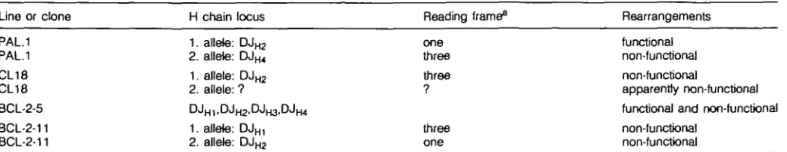
revealed that PAL.1 and BCL-2-11 are clonal, with two different DJH rearrangements on the two heavy chain alleles (Table 1).
The BCL-2-5 pre-B cell line had DJH rearrangements to all four
JH segments (Table 1), indicating that BCL-2-5 had continued
to rearrange D to JH in vitro (29). Only one DJH rearrangement
could be detected by a DJH PCR for pre-B cell clone CL18,
even when including D Q ^ and D R J ^ specific primers in the PCR (Table 1), while in Southern blots with CL18 two different DJH rearrangements were identified (24). Since transfer of CL18
pre-B cells into SCID mice did not result in the generation of measurable levels of serum IgM, it can be assumed that both chromosomes are non-functional for H chain expression (24).
• PAL.1 D CL18 • BCL-2-5 O BCL-2-11 1 2 3 d i j s of differentiation
Fig. 2. Cloning frequency of long-term proliferating pre-B cell lines and
clones on stroma) cells in the presence of IL-7 after various times of in vitro differentiation in the absence of IL-7. Frequencies of pre-B cells with the capacity to express iM chain protein (PAL. 1 and BCL-2-5) are represented by closed symbols, open symbols denote pre-B cell clones CL18 and BCL-2-11, which are not able to express nH chains upon differentiation. The data of one of three experiments was selected for this figure.
Table 1. DJH rearrangements of long-term in vitro differentiating pre-B cell lines and clones
Line oc clone H chain locus
1. allete: D J ^ 2. allele: DJH4 1. aJlele: DJm 2. allele: ? D JH 1, D J H 2 , D J H 3 , D JH 4 1. aJlele: DJH, 2. aJlele: DJH 2 Reading frame^ one three three ? three one Rearrangements functional non-functional non-functional apparently non-functional functional and non-functional non-functional non-functional PAL.1 PAL.1 CL18 CL18 BCL-2-5 BCL-2-11 BCL-2-11
xL chain protein expression without fiH chain expression 1613
PAL.1
1. alltki
•TT TAT TAC TAC GCT AGT AGC TACTAC T T T GAC TAC TGG •
PAL.1
Zallato
• • • T CTA CTA TGG T A ^ C T A ' T G J T TAC TAT GCT ATG GAC TAC T G G -STOP ' „ '
CL18
1. allots
JH3
•T TTA TTA CTA CGG TA^ TAX TTT GAC TAC TG« GGC • STOP BCL-2-11 Lallete BCL-2-11 2. «IMe - • ) - J H .
• T TTA TTA CTA CfiG T A ^ TAjC TGG TAC TTC GAT GTC TGG • • STOP ' • • • T C T A C TACTG 12bf>4p*car Iwpcimar TG dcATTG TG JH3 ACAACAATGATTAGACCCCTG •)H3 ACAATAAAT GATCCTTGGC TAGGGCTCCA GGATGATCTC AGATGGAGGC CAGTGAGGGA CAAAGAAAGC ATAGAAGAGA GGGACCTAGC GGCAATGCTG GCCAGGATCC CTATAAATCT CTGGCCATGA AGTATGGGAG CTGAGGATGT CTGTCTGCGT CAGCCAGGGC TCCCAATGAC CCTTTCTGAC TCCCAAGGTG TCCCTAGTCC TTCATGACCT GAAATTCAGA TACACACATT TCCCCCCCAA CAAATGCAGT L AAAATCTATT TAAGCTGAAT AGAAGAGAGA GGTTGTAAGG ACTCACC
. invrtwi J H 2 TCAGGAGACTGTGAGAGTGGTGCCTTGGCCCCAGTAGTCAAA Inywmj Dpi.ie.1 JH3 ^ GJTACTACCGTAGTAATAA G GCCTGGTTTGCTTACTGGGGCCAAGGG-1. rearranoement bv deletion JH 3 JH2"JH3 Int-Seq. JH2 "FL16.1
Rg. 3. (a) Nucleotide sequences of cloned DJH joints from pre-B cell clones PAL.1, CL18, and BCL-2-11. In the case of normal DJH joints, codons imposed by the fixed rfs of JH gene segments are emphasized by spaced typing. Stop codons are indicated by STOP. Nucleotide sequences originating from different D and JH gene segments as well as recombination signal sequences (RSS) are marked. Their transcriptional orientation is indicated by arrows. Although all pre-B cells are fetal Over-derived, PAL.1 contains two nucteotJdes of N-sequence, which correlates with the finding that PAL.1 is a rare clone expressing detectable levels of mRNA specific for terminal deoxynucleotidyl transferase (data not shown), (b) Schematic representation of the rearrangement events, which might have occured on the second H chain allele of pre-B cell done BCL-2-11. First a normal deletional DJH rearrangement between Do^g, and J ^ had taken place. This was apparentty followed by a VDJ recombinase-mediated, inversional rearrangement using the RSS 5' of J ^ (containing a 23 bp spacer) and the RSS 5' of Da t 6, (containing a 12 bp spacer).
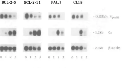
BCL-2-5 BCL-2-11 PAL.l
CL18
r • • • # # • * - O . 8 5 k b VpreBl
- 1.2kb
2.0kb |i-actin
0 1 2 3 0 1 2 3 0 1 2 3 0 1 2 3
time of differentiation (days)
Fig. 4. Northern blot analysis of all four pre-B cell lines and clones at different times after induction of differentiation in the absence of IL-7. All lanes were loaded with 15 HQ total RNA. The same blot was hybridized successively with the indicated probes. All autoradiographies were obtained by overnight exposure at - 70qC. IIH chain specific mRNA was also detectable with all four pre-B cell clones and lines upon differentiation (data not shown).
However, the primary structure of the DJH joint of the second
chromosome needs to be detemined.
Sequences for DJH rearrangements of all pre-B cell clones
are given in Fig. 3(a). They show that BCL-2-11 has both heavy chain alleles rearranged in a way which does not allow the expression of a functional heavy chain protein. On one allele a DJH1 rearrangement has occured in reading frame (rf) III (40).
In this rf the D segment contains a TAG stop codon. DFL161
used in this rearrangement is the most 5' located D segment, so that a secondary (and thus potentially functional) DJH
rearrangement can no longer be made on this allele. The second allele contains an inversion of a region spanning the DJH2 plus
the adjacent J ^ - J ^ intervening sequence, which should have occured after a normal DF L 1 6 1- JH 2 rearrangement in rfl
(Fig. 3b). This region apparently was inverted using the con-served RS sequences 5' of J ^ and 5' of DFL16,, evident in the
perfectly head to head fused signal joints 5' of the inverted sequence (Rg. 3a). Again, no additional D segment is left to allow a subsequent functional DJH joining. Thus we conclude, that
pre-B cell clone BCL-2-11 has two DJH-rearranged H chain
alleles, which do not allow expression of a /*H chain protein upon VHDJH rearrangement.
The DJHJ rearrangement of pre-B cell clone CL18 also uses the most 5'-located Da,6 1 in rflII, resulting in a stop codon
within the D segment (Fig. 3a). In this case the second allele has not yet been identified. It is expected that the second allele is in a configuration which does also not allow the expression of /iH chains after VHDJH rearrangement.
Kinetics of x chain mRNA expression
The extent of xL chain gene expression as a consequence of V, - Jx gene rearrangement in differentiating pre-B cells was first
assessed by measuring the amount of mRNA coding for xL chain protein relative to mRNA for 0-actin. In Northern blot analysis a mature V^J^C, mRNA is 1.2 kb in size and can thus be discriminated from a sterile transcript of C^ alone, which is detectable at a size of 0.8 kb (41). The kinetics of induction of
xL chain mRNA expression upon differentiation is nearly identical in pre-B cells with functional DJH rearrangements (BCL-2-5,
PAL.1) and those with non-functional DJH rearrangements
(BCL-2-11, CL18) (Fig. 4). The apparent decrease in the amount of xL chain mRNA in normal pre-B cells at day 3 of differentia-tion is probably due to extensive apoptosis which normal pre-B cells undergo upon differentiation and which affects the quality of the isolated RNA. This is also evident from the decrease of mRNA for j3-actin, although to a lesser extent. mRNA for the pre-B cell specific gene Vp,^ gradually decreased with time of differentiation in all four pre-B cell lines and clones. This indicates that the pre-B cells were indeed differentiating to more mature cells. These results suggest that the frequency of xL chain gene rearrangements and their subsequent expression as mature xL chain mRNA at different time points of in vitro differentiation in pre-B cells can occur in the absence or presence of ^H chain protein.
Expression of xL chain protein
The level of xL chain protein expression in pre-B cells differen-tiating for 3 days to B cells was determined by Western blotting. In order to enable a quantitative analysis of the amount of x protein produced by differentiated cells, a x protein standard (MOPC 41) was included and serial 2-fold dilutions of cytoplasmic proteins from cells of the same genetic background were carried out on the same blots. Furthermore, each blot included cytoplasmic protein preparations of a slg+ cell line 5-7 (as a
positive control) and of undifferentiated pre-B cells In Fig. 5 the results of one representative experiment out of three independ-ent experimindepend-ents are shown.
Undifferentiated pre-B cells do not produce detectable levels of either /tH or xL chain (Rg. 5a, lanes B and J; and Fig. 5b, lanes B and H). ^H chain protein is also not detectable in BCL-2-11 and CL18 cells differentiated for 3 days (Rg. 5a, lanes C - H ; and Rg. 5b, lanes C - G respectively), in contrast to BCL-2-5 and PaJ.1 cells differentiated for 3 days, in which /iH chain protein is detectable.
xL chain protein expression without /iH chain expression 1615
A B C D E F G H J K L M N O P Q
MW
kD
-97
-69
- 4 6
r
- 3 0
-21.5
-14.3
A B C D E F G H J K L M N O P Q
K
[ft
MW
kD
-97
-69
-46
-21.5
Fig. 5. (a) Western btot analysis of undifferentiated and differentiated cells of pre-B cell clone BCL-2-11 (lanes B - H) and pre-B cell line BCL-2-5
(lanes K-P) The btot was developed with antibodies specific for IgM (top) and xL chain protein (bottom) as indicated. Lane A contains proteins extracted trom the slg+ cell line 5-7 as a positive control (2 x 10s cells). Lane Q contains 30 ng of purified M0PC41 xL chain protein as standard. Lane B: 6 x 10⁶ undifferentiated BCL-2-11 pre-B cells; lanes C - H : serial 2-fold dilutions of proteins extracted from differentiated BCL-2-11 cells, beginning with 6 x 10⁶cells in lane C. Lane J: 6 x 10⁶ undifferentiated BCL-2-5 pre-B cells, lanes K - P : serial 2-fold dilutions of proteins extracted from differentiated BCL-2-5 cells, beginning with 6 x 10⁶ cells in lane K. (b) Western btot analysis of undifferentiated and differentiated cells of pre-B cell clones CL18 (lanes B-G)and PAL.1 (lanes H - N ) . Lane A: same as in Fig. 5(a). Lanes 0 - Q contain serial 2-fold dilutions of purified M0PC41 xL chain protein standard, beginning with 30 ng in lane O. Lane B: 2 x 10⁶ undifferentiated CL18 pre-B cells; lanes C - G : serial 2-fold dilutions of proteins extracted from differentiated CL18 cells, beginning with 2 x 10⁶ cells in lane C. Lane H: 2 x 10⁶ undifferentiated PAL.1 cells; lanes J - N: serial 2-fold dilutions of proteins extracted from differentiated PAL.1 cells, beginning with 2 x 10⁸ cells in lane J. Since pre-B cells derived from normal BDF, mice undergo extensive apoptosis upon differentiation, living cells were enriched by Rcoll centrifugation prior to protein extraction.
However, xL chain protein can be detected in differentiated cells from all four pre-B cell lines and clones. It appears that xL chain protein is always slightly more abundant in differentiated cells from normal mice tnan in those from bc/-2-tg mice. This could be due to a reduced rate of biosynthesis or an increased turn-over rate of xL chain proteins in bcl-2-tg cells. Normal, as well as bcl-2-tg cells, which are unable to express /iH chain protein, express - 3 to 4 fold lower levels of xL chain protein when compared with cells which also express /*H chains. This could either be a consequence of an increased stability of xL chains complexed by /iH chains or may just reflect an increased amount of xL chain protein which is retained by /iH chain protein in the
form of Ig complexes on the cell membrane and in the cytoplasm. We conclude that in normal differentiating pre-B cells x light chain protein can be expressed without /tH chain protein.
The average number of xL chain molecules in a differentiated cell can be calculated by a comparison of the intensity of the xL chain protein bands relative to the M0PC41 standard. In Fig. 5(a and b), - 7 x10° BCL-2-11 cells, 2 x i O8 BCL-2-5 cells,
3 xiO^CLIS cells, and 1 x i O6 PAL.1 cells contain ~3Ongof
xL chain protein. (These estimations are also in agreement with the additional Western blots performed with different prepara-tions of cytoplasmic proteins not shown in Figure 5.)
assum-ing that - 5 0 % of all differentiated cells may express xL chains from a productively rearranged xL chain locus, it follows that one differentiated CL18 and one PAL.1 cell contains - 4 . 8 x i O5
and 1.4 x i O6 xL chain molecules respectively. For BCL-2-11
and BCL-2-5 the numbers are 2 x 10⁵ and 7.1 x 10⁵ xL chain
molecules per cell respectively.
Discussion
Mammalian B cell differentiation is characterized by somatic DNA rearrangements of Ig genes located on three different chromosomes. There is a substantial body of evidence indicating that Ig genes are rearranged in an ordered fashion (see Introduc-tion). However, it is still controversial as to what controls the sequential rearrangement of first heavy, and later x and X light chain genes.
The protein products of rearranged Ig genes have been implied in the regulation of an ordered progression of H and L chain gene rearrangements, especially since it has been shown that xL chain gene rearrangement can be induced in a H chain negative (null) A-MuLV transformed pre-B cell line by expressing a membrane bound jtH protein (15). However, it has to be considered that the A-MuLV cell line used for this study was derived from a cell line previously shown to be prone to differentiation in vitro (7). No clear correlation between /»H chain protein expression and xL chain gene rearrangement could be seen, since many sublines retained the x locus in germline configuration, although nH chain proteins were expressed at high levels. Furthermore, analysis' of A-MuLV transformed pre-B cell lines derived from SCID mice revealed that some of these cells attempted xL chain gene rearrangements in the absence of /iH chain protein. More importantly, cells with silent xL chain loci could never be induced to attempt xL chain gene rearrangement after transfection with a membrane /iH chain expression vector (18,19).
The results presented in this study are not consistent with a model suggesting that H chain protein is necessary for xL chain gene rearrangement since differentiation and xL chain gene rearrangements appear to occur to the same extent in pre-B cells which can, and in pre-B cells which cannot, express /iH chain protein. Our findings support the conclusion reached earlier with human transformed cell lines, that xL chain rearrangements can occur in the absence of /iH chain expression. They are also in agreement with recently published data, showing that the frequency of xL chain gene rearrangement in sorted pro-B and pre-B cell fractions of bone marrow is not affected by the presence or absence of H chain protein or H chain gene rear-rangement (22,23).
We find that in vitro differentiation of pre-B cells measured by changes in surface marker expression and loss of clonability is also independent of H chain protein expression or surface deposi-tion. These changes upon in vitro differentiation are also observed with pre-B cell lines and clones from SCID mice, RAG-2 deficient mice (unpublished observation), and from X5T mice (42).
In vivo, the Ig-like complex of /iH chain with a surrogate L chain apparently exerts its role in selecting large B220+, c-kit + , CD43+ pre-B cells, with a productive H chain gene
rearrange-ment, into the pool of small B220+, c-kif-, CD43~ pre-B II cells in the bone marrow, since B cell differentiation is blocked at that
particular stage in XsT and /*MT mice (21,42,43). The /*H chain - surrogate L chain complex mediated positive selection and expansion of productively rearranged pre-B II cells in the bone marrow does apparently not occur in our pre-B cell culture system, probably because an important environmental factor for this selection is missing in vitro.
A mutation in the X5 gene impairs the formation of a pre-B II compartment in bone marrow, which is present in normal mice and comprises - 5 0 - 7 0 x 106 cells in a young ( 4 - 6 week old)
animal (26). However, XjT mice slowly fill up the pool of peripheral B cells from a pre-B I compartment [ - 2 x 10⁶ cells
in a normal 4 - 6 week old animal (26)]. This has led to the hypothesis that immature B cells in normal mice are not only generated from pre-B II cells, but also may be directly generated from the much smaller pre-B I compartment (22) If this is true, it can be expected that immature B cells exist, which harbor non-productively rearranged H chain loci and thus are not able to express ^H chains, but nevertheless express x or XL chain protein. Although these cells should be functionally inactive (since they cannot display Ig on their surface), they could be the target of a malignant transformation, which, in the end, may lead to Bence-Jones protein producing plasmacytomas. To test this hypothesis, an analysis of the configurations of H chain loci in such plasmacytomas is under way.
The independence of xL chain gene rearrangements from H chain protein expression in normal pre-B cells supports a model of an ordered Ig gene rearrangement that might mainly be regulated by the sequential accessibility of first H chain, and later x and X chain gene loci for the enzymes of the VDJ - recombinase complex (44 - 46). Thus, rearrangements of H and L chain gene segments are carried out in a stochastic way as proposed by Coleclough and colleagues (47), initially with a higher probability of H chain over L chain gene rearrangements. It remains further to be elucidated whether intrinsic preferences exist for VH - DJH over V, - Jx rearrangements, as they appear to exist for V , - Jx over Vx- J ^ rearrangements (48). The model
further implies that once a hematopoietic stem cell becomes commited to the B cell lineage, a molecular program of differentia-tion is initiated that can take place independent of external stimuli and can lead to the generation of immature slg+ B cells in the
absence of proliferation.
Acknowledgements
We like to thank Drs Klaus Karlajainen and Shunichi Takeda for critically reading the manuscript. The Base) Institute for Immunology was founded and is supported by F. Hoffmann-La Roche Ltd, Basel, Switzerland. Abbreviations A-MuLV PCR PE rf tg RSS MMT References
Abeteon murine leukemia virus polymerase chain reaction phycoerythrin
reading frame transgenic
recombinase signal sequence ^H-deftcient mice
1 Tonegawa, S. 1983. Somatic generation of antibody diversity. Nature 302:575.
2 Alt, F., Rosenberg, N., Lewis, S., Thomas, E., and Baltimore, D. 1981. Organization and reorganization of immunogbbulin genes in A-MuLV transformed cells: rearrangement of heavy but not light chain genes. Cell 27:381.
3 Korsmeyer, S., Hieter, P., Ravetch, J., Poplack, D., Waldmann, T., and Leder, P. 1981. Developmental hierarchy of immunoglobulin rearrangements in human leukemic preB cells. Proc. NatlAcad. Sci. USA 78:7096.
4 Siden, E., Alt, F., Shinefekj, L, Sato, V , and Baltimore, D. 1981. Synthesis of immunoglobulin n chain gene products precedes synthesis of light chains during B-tymphocyte development Proc. Natl Acad Sci. USA 78:1823.
5 Alt, F., Yancopoulos, G. D., Blackwell, T. K., Wood, C , Thomas, E., Boss, M , Coffman, R., Rosenberg, N , Tonegawa, S., and Baltimore, D. 1984. Ordered rearrangement of immunogbbufin heavy chain variable region segments. EMBO J. 3:1209
6 Lewis, S., Rosenberg, N., AJt, F., and Baltimore, D. 1982. Continuing kappa-gene rearrangement in a cell line transformed by Abelson murine leukemia virus. Cell 30:807
7 Reth, M., Ammirati, P., Jackson, S., and Alt, F. 1985. Regulated progression of a cultured preB cell line to the B-cell stage. Nature 317:353.
8 Persiani, D. M , Durdik, S., and Seising, E. 1987. ActiveX and x anti-body gene rearrangement in Abelson murine leukemia virus-transformed preB cell lines. J Exp. Med. 165:1655.
9 Levitt, D. and Cooper, M. D. 1980. Mouse preB cells synthesize and secrete n heavy chains but not light chains. Cell 19.617. 10 Sakaguchi, N. and Melchers, F. 1986. X5, a new light-chain-related
locus selectively expressed in preB lymphocytes. Nature 324:579. 11 Kudo, A. and Melchers, F. 1987. A second gene, V ^ ^ in the Xs locus of the mouse which appears to be selectively expressed in preB lymphocytes. EMBO J. 6:2267.
12 Karasuyama, H., Kudo, A., and Melchers, F. 1990. The proteins encoded by the Vp,aB and X5 preB cell specific genes can associate with each other and with n heavy chain. J. Exp. Med 172:969. 13 Tsubata.T. and Reth, M. 1990. The products of preB cell genes (Xs
and Vpfas) and the immunoglobulin /» chain form a complex that is transported onto the cell surface J. Exp. Med. 172:973
14 Iglesias, A., Kopf, M., Williams, G. S., Buhler, B., and Kohler, G. 1991. Molecular requirements for the /i-induced light chain gene rearrange-ment in preB cells. EMBO J. 10:2147.
15 Reth, M., Petrac, E., Wiese, P , Lobel, L , and Alt, F. 1987. Activa-tion of V, gene rearrangement in preB cells follows the expression of membrane-bound immunoglobulin heavy chains. EMBO J. 6:3299. 16 Tsubata.T , Tsubata, R.,and Reth, M. 1992. Crosslinking of the cell surface immunoglobulin (^-surrogate light chains complex) on preB cells induces activation of V gene rearrangements at the immuno-globulin x locus. Int. Immunol. 4:637.
17 Kubagawa, H., Cooper, M. D., Carrot), A. J., and Burrows, P D. 1989. Light-chain gene expression before heavy-chain gene rearrangement in preB cells transformed by Epstein - Barr virus. Proc. Natt Acad Set. USA 86.2356.
18 Blackwell, T. K , Malynn, B. A., Pollock, R. R., Ferrier, P., Covey, L. R., Fulop, G. M., Phillips, R. A., Yancopoulos, G. D., and Alt, F 1989. Isolation of scid preB cells that rearrange kappa light chain genes: formation of normal signal and abnormal coding joins. EMBO J. 8:735. 19 Blackwell, T. K.. Ferrier, P., Malynn, B. A., Pollock, R. R., Covey, L. R.,
Suh, H., Heinke, L. 8., Fulop, G. M., Philips, R. A., Yancopoulos, G. D., and Alt, F. W. 1989. The effect of the SCID mutation on mechanism and control of immunoglobufin heavy and Bght chain gene rearrange-ment. Curr. Top. Microbiol. Immunol. 152:85.
20 Kitamura, D., Roes, J., Kuhn, R., and Rajewsky, K. 1991. A B cell-deficient mouse by targeted disruption of the membrane exon of the immunoglobulin /i chain gene. Nature 350:423.
21 Kitamura, D. and Rajewsky, K. 1992. Targeted disruption of n chain membrane exon causes loss of heavy-chain allelic exclusion. Nature 356:154.
22 Ehlich, A., Schaal, S., Gu, H., Kitamura, D., MOIIer, W., and Rajewsky, K. 1993. Immunoglobulin heavy and light chain genes rearrange independently at early stages of B cell development. Cell 72:695.
xL chain protein expression without pH chain expression 1617 23 Chen, J., Trounstjne, M., AH, F. W., Young, F., Kurahara, C , Loring, J. F., and Huszar, D. 1993. Immunoglobulin gene rearrange-ment in B cell deficient mice generated by targeted deletion of the JH locus. Int. Immunol. 5:647.
24 Rolink, A., Kudo, A., Karasuyama, H., Kikuchi, Y., and Melchers, F. 1991. Long-term proliferating early preB cell lines and clones with the potential to develop to surface-lg positive mitogerweactive B cells 'in vitro' and 'in vivo'. EMBO J. 10:327.
25 Rolink, A. and Melchers, F. 1991. Molecular and cellular origins of B lymphocyte diversity. Cell 66:1081.
26 Rolink, A. and Melchers, F. 1993. Generation and regeneration of cells of the B-lymphocyte lineage. Curr. Opin. Immunol 5:207. 27 Rolink, A., Streb, M., Nishikawa, S. I., and Melchers, F. 1991. The
c-kit encoded tyrosine kinase regulates the proliferation of early preB cells. Eur. J. Immunol. 21:2609.
28 Hardy, R. R., Carmack, C. E., Shinton, S. A., Kemp, J. D., and Hayakawa, K. 1991. Resolution and characterization of proB and pre-pro B cell stages in normal mouse bone marrow. J. Exp. Med. 173:1213.
29 Rolink, A., Haasner, D., Nishikawa, S. I., and Melchers, F. 1993. Changes in frequencies of clonable preB cells during life in different lymphoid organs of mice. Blood 81:2290.
30 Strasser, A., WittJngham, S., Vaux, D. L., Bath, M. L, Adams, J. M., Cory, S., and Harris, A. W. 1991. Enforced BCL2 expression in B-lymphoid cells prolongs antibody responses and elicits autoimmune disease. Proc. Natl Acad. Sci. 88:8661.
31 Strasser, A., Harris, A. W., and Cory, S. 1991. oc/-2 transgene inhibits T cell death and perturbs thymic self-censorship. Cell 67:889. 32 Kodama, H., Amagai, Y., Koyama, H., and Kasai, S. 1982. Hormonal
responsiveness of a preadipose cell Bne derived from newborn mouse calvaria J. Cell Physbl. 11283.
33 Yelton, D , Desaymarol, C , and Scharff, M. 1981. Use of monoclonal anti-mouse immunogtobulins to detect mouse antibodies. Hybridoma 1:5.
34 Karasuyama, H., Melchers, F., and Rolink, A. 1993. A complex of glycoproteins is associated with Vpfeg/Xs surrogate light chain on the surface of n heavy chain-negative early precursor B cell lines. J. Exp. Med. 178.469.
35 Grawunder, U., Metchers, F., and Rolink, A. 1993. lnterferon-7 arrests proliferation and causes apoptosis in stromal cell/interleukin-7-dependent normal munne preB cell lines and clones in vitro, but does not induce differentiation to surface immunoglobulin-positive B cells. Eur J. Immunol. 23:544.
36 Chomczynski, P. and Sacchi, N. 1987. Single-step method of RNA isolation by acid guanidinium thiocyanate - phenol - chloroform ex-traction. Anal Bbchem. 162:156.
37 Laemmli, U. K. 1970. Cleavage of structural proteins during the assembly of the head of bacteriophage T4. Nature 227:680. 38 Towbin, H., Staehelin, T., and Gordon, J. 1979. Electrophoretic
transfer of proteins from potyacrylamide gels to nitrocellulose sheets: procedure and some applications. Proc. NatlAcad. Sci. USA 76:4350. 39 Gu, H., Kitamura, D., and Rajewsky, K. 1991. B cell development
regulated by gene rearrangement: arrest of maturation by membrane-bound D protein and selection of DH element reading frames. Cell 65:47.
40 Kaartinen, M. and Makela, 0.1985 Reading of D genes in variable frames as a source of antibody diversity. Immunol. Today 6:324. 41 Perry, R. P., Ketley, D. E., CcHeclough, C , Seidman, J. G., Leder, P.,
Tonegawa, S., Matthyssens, G., and Weigert, M. 1980. Transcrip-tion of mouse x chain genes: implicaTranscrip-tions for allelic exclusion. Proc. NatlAcad. Sci. USA 77:1937.
42 Rolink, A., Karasuyama, H., Grawunder, U., Haasner, D., Kudo, A., and Melchers, F. 1993. B cell development in mice with a defective X5 gene. Eur. J. Immunol. 23:1284.
43 Kitamura, D., Kudo, A., Schaal, S., MOIIer, W., Melchers, F., and Rajewsky, K. 1992. A critical role of Xs in B cell development. Cell 69:823.
44 Yancopoulos, G. D. and Alt, F. 1985. Devetopmentally controlled and tissue specific expression of unrearranged VH gene segments. Cell 40:271.
and Perry, R. P. 1981. Transcription of the unrearanged mouse C, 47 Coleclough, C, Perry, R P., Karlajainen, K., and Weigert, M 1981. locus: sequence of the initiation region and comparison of activity with Aberrant rearrangements contribute significantly to the allelic exclu-a reexclu-arrexclu-anged V ^ - C , gene CeH 27.593. sion of immunoglobulm gene expression. Nexclu-ature 290:372. 46 Schlissel, M. S. and Baltimore, D. 1989 Activation of immunoglobulin 48 Ramsden, D. A. and Wu, G. E. 1991. Mouse x light-chain
recombina-kappa gene rearrangement correlates with induction of germline tion signal sequences mediate recombination more frequently than kappa gene transcription. Cell 58:1001. do those of X light chain. Proc. Natl. Acad. Sci. USA 8810721.