Careinogenesis Vol.5 no.12 pp.1729-1732, 1984
Methylation of DNA in stomach and small intestine of rats after
oral administration of methylamine and nitrite
K.W. Huber and W.K. Lutz1
Institute of Toxicology, ETH and University of Zurich, CH-8603 Schwerzenbach, Switzerland
'To whom reprint requests should be sent
Young adult male Sprague-Dawley rats were given 30 /xmol/kg body weight [14C]methylamine hydrochloride and 700 /anol/
kg body weight sodium nitrite by oral gavage. DNA isolated from the stomach and from the first 15 cm of the small in-testine was methylated, containing 7-methylguanine (7mG) at a level of one 7mG molecule per 5xlO6 and lxlO7 nucleotides,
respectively. No 7mG was found in the liver at a limit of detection of one 7mG molecule per 2x10° nucleotides. In a se-cond experiment, the excised stomachs were incubated with deoxyribonudease before the isolation of the DNA in order to degrade DNA in the lumen and in the uppermost lining cells. This treatment resulted in a 30% decrease in the yield of DNA and a 90% reduction in the level of 7mG formation. The results show that nitrosation of a primary alkylamine yields a precursor of an alkylating agent which has a long enough lifetime to diffuse towards and react with intracellular DNA. A correlation of DNA methylation in the stomach with the corresponding tumor formation by the methylating carcinogen N-methyl-N' -nitro-N-nitroso-guanidine was used to estimate the role of DNA damage resulting from en-dogenous nitrosation of dietary methylamine in man. It was concluded that the risk resulting from this single amine must be negligible but that a similar evaluation of other primary amines is required before the over-all role of primary amine nitrosation in the etiology of human gastric cancer can be assessed.
Introduction
The generation of N-nitroso-compounds from secondary amines (1) or from amides (2) and nitrite (3,4) under acidic conditions could represent an important mechanism for the in
vivo formation of carcinogens in the stomach. The
nitrosa-tion of primary aliphatic amines (5) is known to the chemist as a deamination reaction because it leads to chemically unstable products which spontaneously hydrolyze to alcohols. Although the intermediates could also react with cellular nucleophiles such as protein, RNA and DNA, and although mutagenic effects to microorganisms have been reported (6—8), the interest of toxicologists in the nitrosation products of primary aliphatic amines has been negligible in comparison to secondary amines, probably because it was assumed that the reaction products were not stable enough to reach intracellular macromolecules in vivo.
Methylamine is by far the most widespread primary amine in the diet and is found above all in fish (9) and in vegetables (10). For this reason, and because the techniques for
deter-*Abbreviations: 7mG, 7-methylguanine. © IRL Press Ltd., Oxford, England.
mining DNA methylations have been previously established in this laboratory (11,12), methylamine was selected as a test compound. After oral administration of [14C]methylamine
and nitrite at high dose levels to rats, DNA was isolated from the stomach, the small intestine and the liver, and the level of formation of 7-methylguanine (7mG)* was determined. Bas-ed on the knowlBas-edge of the kinetics of DNA methylation by methylamine and nitrite in vitro (12), the experimental data were extrapolated to the concentrations expected in a human stomach. Upon comparison with another direct-acting alkylating carcinogen, N-methyl-N'-nitro-N-nitroso-guanidine, a tumorigenic risk resulting from the intake of methylamine and nitrite was estimated.
Materials and methods Chemicals and apparatus
All chemicals without specified distributor were purchased in the highest puri-ty available from E. Merck GmbH (Darmstadt, FRG). 7mG, sodium dodecylate and deoxyribonudease I from bovine pancrease (E.C.3.1.21.1.) were purchased from the Sigma Chemical Company (St. Louis, MO). ["CJMethylamine hydrochloride (mol. wt. 67.5) with a specific activity of 53.9 (experiment I) or 51.8 (experiment II) mCi/mmol, dissolved in ethanol (100 jiCi/ml), was obtained from New England Nuclear (Boston, MA). The radiochemical purity was 99%, as determined by t.l.c. on cellulose F254
(Merck, Darmstadt, FRG) using methanol:diethyl ethenl N hydrochloric acid:water (10+10+1 +3) as the solvent system and ninhydrin reagent for detection. The ethanol from the original [14C]methylamine hydrochloride
solution was evaporated under a slow stream of nitrogen and the salt was re-dissolved in distilled water to give a stock solution with a specific activity of 200fiCi/ml (experiment I) or 396/tCi/ml (experiment II). DNA from previous in vivo experiments with [14C]dimethylnitrosamine (carrying the label
mainly in form of a [14C]methyl group in the 7(N)-position of guanine) was used as radiolabelled DNA in control experiment I. This DNA had a specific activity of 2430 d.p.m./mg and was dissolved in 14 mM phosphate buffer pH 6.8 at a concentration of 0.8 mg/ml. Hydroxyapatite came from Bio-Rad Laboratories, Richmond, CA (DNA grade Bio-Gel HTP, batches selected for high yields; experiment I and control experiment I) and from Calbiochem-Behring, San Diego, CA (experiment II). L-15 (Leibovitz) medium (Ix) with L-glutamine was obtained from Gibco AG, Basel, Switzerland. Dialysis tub-ing (Visktub-ing type 20/32, mol. wt. exclusion of 12 000-14 000 Daltons; diameter 17 mm) was obtained from Union Carbide (Chicago, IL). Radioac-tivity measurements were carried out in 10 ml of Insta-Gel (Packard In-struments, Downers Grove, IL) in a liquid scintillation counter, model Packard Tri Carb 460 CD. H.p.l.c. analysis of the DNA purine bases was per-formed on a reverse-phase/iBondapak C18 column, 300x4 mm (experiment I) or 300x7.8 mm (experiment II; Waters Associates, Millford, MA) equipped with two h.p.l.c. pumps (model LC Pump 410 from Kontron, Zurich, Switzerland) controlled by a Kontron Programmer 200 to generate a linear gradient of two eluants.
Animals and treatments
General. Young adult male rats (Sprague-Dawley derived SIV-50,
200-300 g; Ivanovas, Kissleg, FRG) were housed in macrolone cages with free access to tap water and food (Laboratory chow no. 343, Klingental Miihle AG, Kaiseraugst, Switzerland) for an acclimatisation period of 1 week. Food was removed 16 h before the administration of the test compounds which were always given by gavage between 09.00 and 10.00 in a maximum volume of 2 ml per animal. Stock solutions of I mg sodium nitrite per ml distilled water were freshly prepared for each experiment. In experiments I and II, each animal was given 1 ml of [14C]methylamine hydrochloride stock
solution immediately followed by 1 ml of the sodium nitrite stock solution. In control experiment 1, the rats received 1.15 ml of radioactive DNA solution, while control experiment II was performed with untreated animals. Thirty
K.W. Huber and W.K. Lutz
Table 1. Formation of 7 raG in DNA isolated from stomach, small
intestine and liver of rats 30 min after successive oral administration of ['*C]methylamine and sodium nitrite
Experiment No. I II
Animal No./weight (g) Methylamine dose
Ounol/kg)
(109 d.p.m.["C]/kg)
Sodium nitrite dose (mmol/kg) DNase treatment of
stomach and small intestine
DNA Spec. Activity (d.p.m./mg) Stomach Small Intestine Liver Formation of 7mG (7mG-indexa) Stomach Small Intestine Liver Corrected for individual doses of reagents administered1"; mean ± S.D.: Stomach Small Intestine 1/214 19 2.3 0.68 -1013 5526 169 0.14 0.05 <0.02 12.8 ± 7.0 ± 2/196 16 1.9 0.71 -1092 4816 134 <0.65 0.09 <0.02 3/169 41 4.3 1.21 -2215 5600 302 0.58 0.25 < 0.005 4.4 (animals 1 and 3) 3.7 (animals 1 — 3) 4/234 30 3.9 0.70 + 24 108 no data 0.016 0.062 no data 5/219 28 3.7 0.73 + 15 69 no data 0.023 0.035 no data 1.3 ± 0.4 (4 and 5) 3.0 ± 1.:5 (4 and 5)
a7mG-index = /xmol 7mG/mol nucleotide
bCalculated according to 7mG-index divided by the methylamine dose (mmol/kg) and divided by the square of the sodium nitrite dose (mmolVkg2)
minutes (experiment I and II) or 5 min (control experiment I) after the ad-ministration of the appropriate solutions the animals were killed by an ether overdose. Organs were excised, cut lengthwise, washed twice in —50 ml 0.9% NaCl solution, and DNA was isolated either immediately or after additional treatments as indicated below.
Experiment I. Three rats were treated with [14C]methylamine hydrochloride
and nitrite at the dose levels given in Table I. After 30 min the stomachs, up-per small intestines (15 cm) and livers were excised and the methylation of DNA was determined as described below. Two untreated rats were used as controls for radioactivity background.
Control experiments. (I) Two rats were gavaged with radiolabelled DNA
(2230 d.p.m. 14C; 0.92 mg) and killed after 5 min. Stomachs were washed as
indicated above and DNA was isolated. (II) The freshly excised stomachs of six untreated rats were cut along the lesser curvature and turned inside out. Three stomachs were incubated for 26, 39 and 45 minutes at 37°C in L-15 cell culture medium. Three stomachs were incubated in 10 ml L-15 medium con-taining 25 units/ml DNase I at 37°C for the same periods of time. All stomachs were washed three times with 0.9% NaCl and minced. The yield of DNA was determined by spectroscopy before the hydroxyapatite step and was found to be 2.4, 4.1 and 4.0 mg/g stomach after incubation with DNase, whereas the respective controls yielded 4.9, 6.1 and 4.7 mg/g organ.
Experiment II. Two rats were treated with ["CJmethylamine hydrochloride
and nitrite at the dose levels given in Table I (animals 4 and 5). The excised stomachs and small intestines were incubated at 37°C for 30 min in 10 ml L-15 medium containing 25 units DNase/ml before the isolation of chromatin. The stomachs were prepared as described for the control experi-ment II. The small intestines were sliced along their whole length but they preserved their original shape during DNase incubation. The DNA of the stomachs and the small intestines of two rats used as controls for radioactivity background were isolated without addition of DNase to the L-15 medium, in order to assess the effectiveness of the DNase treatment.
Isolation of chromatin, DNA and purine bases
Organs were minced and homogenized in the cold in a Potter-Elvehjem-type homogenizer in 3 — 4 volumes 75 mM NaCl, 10 mM EDTA, 10 mM Tris/HCl pH 7.8, and DNA was isolated according to a method previously described in detail (13). In short, crude chromatin was prepared by
precipita-tion with a non-ionic detergent and the pellets were homogenized and depro-teinated by phenol/chloroform/isoamyl alcohol and extracted with ether. The crude DNA was purified by hydroxyapatite adsorption chromatography, dialysed against 0.2 M NaCl and precipitated with ethanol. The dried residue was redissolved in a 14 mM phosphate buffer, pH 6.8. The specific activity of the DNA was determined by counting the ["CJradioactivity and measuring the amount by u.v.-spectroscopy, assuming an absorption of 20 for a solution of 1 mg DNA/ml. DNA was hydrolyzed with 0.1 N HC1 for 1 h at 70°C to liberate thepurines, and unlabelled 7mG was added as standard. This mixture was loaded on an h.p.l.c. reverse-phase ^Bondapak C18 column and eluted for 20 min with 10 mM ammonium phosphate pH 4.0/1 % methanol, follow-ed by a linear gradient to 100% methanol over 40 min. The flow rate was 1.2 ml/min for the analytical column (experiment I, animals 1 and 2) and 3.5 ml/min for the semipreparative column (all other analyses).
Results Experiment I
Table I shows that the treatment of rats with [14
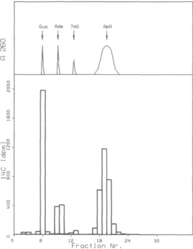
C]methyl-amine and nitrite resulted in radiolabelled DNA of the liver, stomach and upper small intestine. However, this radio-activity does not necessarily represent DNA methylations; it is also possible that biosynthetic incorporation of radioactivity occurred because radiolabelled methylamine is degradable to nucleic acid precursors of the carbon-1 pool. In order to dif-ferentiate between the various possible sources, DNA was depurinated with acid and the purine-bases were separated by h.p.l.c. Figure 1 shows, as an example, the DNA isolated from the small intestine of rat No. 3. Only a minute fraction of the total radioactivity eluted together with 7mG, the main part co-eluting with the natural purine bases and with apurinic acid. Upon conversion of the radioactivity of the 7mG fraction to the corresponding number of methyl groups, a 7-methylguanine index (7mG-index = /xmol 7-methyl-guanine/mol nucleotide) was calculated for each individual DNA sample and is given in Table I. For a comparison bet-ween different animals these values had to be normalized to the exact doses of methylamine and nitrite administered to each individual rat. For this purpose, the 7mG-index was divided by the dose of methylamine and by the second power of the dose of nitrite according to the equation derived for the reaction kinetics in vitro (12). On the basis of average doses of 0.03 mmol methylamine and 0.7 mmol nitrite per kg body weight, only one molecule 7mG was formed per 5xlO6
deox-yribonucleotides isolated from the stomach, one 7mG per 107
nucleotides in the small intestine. No 7mG was detectable at all in liver DNA at a limit of detection of one molecule 7-methylguanine per 2xl08 nucleotides (animal 3;
semipre-parative h.p.l.c. column).
Since 7mG is a natural constituent of tRNA it is important to exclude the possibility that tRNA contamination in the DNA samples could have given rise to the observed radioac-tivity coeluting with 7mG. The negative data shown for the liver provide an internal control that this potentially confoun-ding process is not operating in our experiments.
The DNA isolated from stomach and small intestine could have originated in part from dead lining cells in the gastro-intestinal lumen at the time of the formation of the alkylating agent. This DNA fraction would have been more readily methylated than intracellular DNA, leading to an over-estimate of DNA damage. Therefore, conditions for washing the organs and for enzymatic degradation of extracellular DNA were examined in two control experiments.
Control experiments
(I) Radiolabelled DNA which was gavaged 5 min before sacrifice was completely removed by washing the organs twice 1730
In vivo DNA methylation by methylamine and nitrite a "D Gua Rde 7mG flpfl
I I I
I
1
12 18 24 Fraction Nr. 30Fig. 1. H.p.l.c. elution profile of acid-hydrolyzed DNA isolated from the
small intestine of rat No. 3 in experiment I. Top chart: Optical density profile at 260nm, showing elution regions for guanine (Gua), adenine (Ade), 7mG added as unlabelled standard, and apurinic acid (ApA). Bottom chart: "C radioactivity profile.
in saline. No radioactivity was detectable in the DNA isolated from the stomach. Based on the limit of detection of 2.4 d.p.m./mg DNA this means that a maximum of 0.2% of the luminal DNA could have remained in the stomach during the washing procedure. (II) In order to also remove the DNA sticking to the mucus, stomachs were incubated with DNase. The yield of DNA was thereby reduced by - 3 0 % .
Experiment II
The methylation of DNA after oral administration of [14
C]-methylamine hydrochloride and sodium nitrite was determin-ed in stomach and small intestine. As opposdetermin-ed to the pro-cedure used in experiment I, the organs of the treated animals were incubated with DNase for 30 min whereas two controls were kept in the L-15 medium without DNase. The yield of purified DNA was reduced significantly for the stomachs
(p <0.05) from 1.02 ± 0.01 to 0.73 ± 0.09 mg/g organ
upon DNase digestion, whereas no difference was seen in the DNA yield from the small intestines (1.18 versus 1.23 mg/g organ). This lack of an effect could have been due to the lower exposure of the mucosa of the small intestines to DNase, because this organ could not be turned inside out as for the stomachs.
The results of the methylation of DNA are shown in the last two columns of Table I (animals No. 4 and 5). A com-parison with the results of experiment I shows that DNase treatment of the stomachs reduced the level of DNA
methyla-tion from 12.8 to 1.3, i.e., by a factor of 10, while the yield of DNA was reduced by - 3 0 % . In the small intestines, methylation was reduced from 7.0 to 3.0, or by 57% (p for a one-sided t-test = 0.13), while the yield of DNA after DNase treatment was not significantly reduced.
Discussion
The rate of formation of nitrosamines from secondary amines and nitrite in vitro at a given pH is proportional to the concentration of the amine and to the second power of the concentration of nitrite (14). It is a function of the pH and of the basicity of the amine and can be modulated by the presence of catalysts or inhibitors in the reaction mixture. Because primary aliphatic amines lead to chemically unstable products, it is more difficult to determine the rate constant k. For this reason we have recently determined a value k' by measuring the formation of 7mG in DNA present in the aqueous reaction mixture (12) to satisfy the equation /imol 7mG/mol nucleotide = k'x[amine]tota|X[nitrite]2tota| (1)
The values determined for k' for methylamine were 4xlO6
M ~3 at pH 4 and lxlO6 M ~3 at pH 2. Using k ' = 4xlO6
M ~3 and the molar concentrations of reactants calculated on
the basis of a stomach volume of 10 ml/kg rat, 7mG-indices of 35, 32 and 240 were calculated for animals No. 1 to 3 (ex-periment I) and of 59 and 60 for ex(ex-periment II. The 7mG in-dices actually measured in vivo were, however, only 0.14 and 0.58 in experiment I arid 0.016 and 0.023 in experiment II (which included incubating the stomachs with DNase). This means that the methylation of DNA in vivo (experiment I) was at least 330 times lower than after an in vitro incubation of DNA with the reactants, and DNase treatment reduced this level by a further factor of ten. Assuming that this cellular protection is primarily dependent on the physico-chemical nature of the alkylating species (e.g., rate of absorp-tion into the lining cells and into the nucleus; hydrolytic stability) and less influenced by species-specific differences of the gastric anatomy, the extent of DNA methylation can also be estimated for humans. Assuming a pH of 2, a maximal concentration of 1 mM methylamine (from 200 g squid)(9) and 2 mM nitrite (15), and using equation (1) with k' = 106
M~3, a 7mG-index of 4x10 ~3 can be calculated for the in
vitro reaction. Correcting this result with a minimum
in-tracellular protection factor of 330 as calculated from the above comparison of in vitro and in vivo data, a 7mG-index of 1.2x10 ~5 can be expected on average in the DNA isolated
from a human stomach. This means that only about every 7th cell would carry one 7-methylated guanine molecule from this source in its diploid set of DNA.
It is tempting here to relate this level of DNA damage to an expected induction of stomach tumors. Such a correlation might be possible for rats where both carcinogenicity data and levels of DNA methylation are available after administration of the wellknown carcinogen NmethylNnitrosoN' -nitroguanidine (MNNG). This agent liberates nitrosated methylamine under alkali or thiol catalysis (16), i.e., the same proximate carcinogen as the one generated in the present ex-periments. It can therefore be assumed that the relative abun-dance of the various DNA methylation products is the same, so that the most prevailing, 7mG, can be used as a marker of damage even if other lesions might be more critical. In fact, 7mG was the only methylated base detectable at all in our ex-1731
K.W. Huber and W.K. Lutz
periments because of the extremely low total methylation level.
It has been reported (17) that a single oral dose of 0.1 mmol MNNG/kg body weight in guinea pigs resulted in the forma-tion of 2.8 mmol 7mG/mol guanine in the stomach after 2 h. This result matches quite well with that of a study on rats (18), where an alkylation of 2 mmol 7mG/mol guanine was determined after a total dose of 0.37 mmol MNNG/kg body weight. This could indicate that guinea pigs and rats do not differ appreciably with respect to the initial DNA damage. Because the conditions used in reference 17 were closer to those of the present study, these data were further used to calculate that a dose of — 8 pmol MNNG/kg body weight would cause the same DNA damage as was estimated above for methylamine and nitrite in a human stomach. This dose is about 1 million times lower than the daily dose required to in-duce a tumor in half the animals treated life-long in a car-cinogenicity study (19). A daily human intake of the amounts of methylamine and nitrite assumed above (high, but not im-possible levels) could therefore be responsible for a life-time risk for one additional tumor per 106 people (extrapolated
along the 'one-particle curve'; reference 20). This incidence is many orders of magnitude below the epidemiologically observed incidence of stomach cancer. Such an estimation of a human risk remains tentative as long as we do not know more about the persistence and repair of the most critical alkylation products, the cumulative effects from chronic ex-posure, and the probabilities of subsequent stages in tumor formation. Nevertheless, the gap between the actual gastric tumor incidence and the consequences from endogenous nitrosation of methylamine is so large that modulatory in-fluences are unlikely to invalidate our comparison. Further-more, our assessment thus far has not taken into account that the DNA in the stem cells at the bottom of the gastric crypts, probably the most sensitive population for cancerous transformation, will be methylated to a lesser extent than the average DNA molecules isolated from the whole organ.
On the other hand, this calculation was based only on the intake of methylamine. Many more primary amines are in-gested with, our diet, all of which potentially give rise to alkylating nitrosation products. The most important biogenic amines should be investigated along these lines before we can conclude that the endogenous nitrosation of primary amines as a class does not represent an essential contribution to gastric tumor formation.
Acknowledgements
This work was supported by the Swiss League against Cancer. We thank Ms. Sarah Shephard for her help with the English translation.
References
1. Lijinsky.W. (1979), Current concepts in the toxicology of nitrates, nitrites, and nitrosamines, in Mehlman.M.A., Shapiro.R.E. and Blumenthal.H. (eds.), Advances in Modern Toxicology, Vol.1, Part 2, Hemisphere Publishing Corporation, Washington, New York and London, pp.149-164.
2. Mirvish.S.S. (1983), The etiology of gastric cancer, J. Nail. Cancer Insl., 71, 629-647.
3. Walters.C.L. (1980), The exposure of humans to nitrite, Oncology, 37, 289-296.
4. Walters.C.L., Carr.F.P.A., Dyke,C.S., Saxby.M.J., Smith,P.L.R. and Walker,R. (1979), Nitrite sources and nitrosamine formation in vitro and
in vivo, Fd. Cosmet. Toxicol., 17, 473-479.
5. Austin.A.T. (1960), The action of nitrous acid on aliphatic amines,
Nature, 188, 1086-1088.
6. Hussain.S. and Ehrenberg.L. (1974), Mutagenicity of primary amines combined with nitrite, Mutat. Res., 26, 419-422.
7. Boido.V., Bennicelli,C, Zanacchi.P. and De Flora.S. (1980), Formation of mutagenic derivatives from nitrite and two primary amines, Toxicol.
Lett., 6, 379-383.
8. Hecker.L.L, Saavedra.J.E., Farrelly.J.G. and Andrews.A.W. (1983), Mutagenicity of potassium alkanediazotates and their use as model com-pounds for activated nitrosamines, Cancer Res., 43, 4078-4082. 9. Lin,J.-K., Lee,Y.-J. and Chang, H.W. (1983), High concentrations of
dimethylamine and methylamine in squid and octopus and their implica-tions in tumour aetiology, Fd. Chem. Toxicol., 21, 143-149.
10. Neurath.G.B., Duenger.M., Pein.F.G., Ambrosius,D. and Schreiber.O. (1977), Primary and secondary amines in the human environment, Fd.
Cosmet. Toxicol., 15, 275-282.
11. Meier-Bratschi,A., Lutz.W.K. and Schlatter.Ch. (1983), Methylation of
liver DNA of rat and mouse by N-nitrosodimethylamine formed in vivo from dimethylamine and nitrite, Fd. Chem. Toxicol., 21, 285-289. 12. Huber.K.W. and Lutz.W.K. (1984), Methylation of DNA by incubation
with methylamine and nitrite, Carcmogenesis, 5, 403-406.
13. Sagelsdorff.P., Lutz,W.K. and Schlatter.C. (1983), The relevance of covalent binding to mouse liver DNA to the carcinogenic action of hexa-chlorocyclohexane isomers, Carcinogenesis, 4, 1267-1273.
14. Mirvish.S.S. (1975), Formation of N-nitroso compounds: Chemistry, kinetics and in vivo occurrence, Toxicol. Appl. Pharmacol., 31, 325-351. 15. Tannenbaum,S.R. (1980), Ins and outs of nitrites, The Sciences, 20, 7-10. 16. Lawley.P.D. (1976), Carcinogenesis by alkylating agents, in Searle.C.E. (ed.), Chemical Carcinogens, ACS Monograph 173, American Chemical Society, Washington, DC, pp.83-244.
17. Woolley.P.V., Pinsky.S.D. and Yerino.P. (1982), Distribution and bin-ding of the carcinogens 1-methyl-l-nitrosourea and l-methyl-3-nitro-l-nitrosoguanidine in the guinea pig after oral administration,
Car-cinogenesis, 3, 1443-1447.
18. Craddock,V.M. (1969/70), The reaction of N-methyl-N'-nitro-N-nitrosoguanidine with DNA in the intact animal, Chem.-Biol.
Interac-tions, 1, 234-237.
19. Parodi.S., Zunino.A., Ottaggio.L., De Ferrari.M. and Santi.L. (1983), Quantitative correlation between carcinogenicity and sister chromatid ex-change induction in vivo for a group of 11 N-nitroso derivatives, J.
Tox-icol. Environ. Health, 11, 337-346.
20. Food and Drug Administration Advisory Committee on Protocols for Safety Evaluation (1971), Panel on Carcinogenesis Report on Cancer Testing in the Safety Evaluation of Food Additives and Pesticides,
Tox-icol. Appl. Pharmacol., 20, 419-438.
(Received on 20 July 1984; accepted on 21 September 1984)
![Table 1. Formation of 7 raG in DNA isolated from stomach, small intestine and liver of rats 30 min after successive oral administration of ['*C]methylamine and sodium nitrite](https://thumb-eu.123doks.com/thumbv2/123doknet/14910097.658083/2.936.66.450.129.545/formation-isolated-stomach-intestine-successive-administration-methylamine-nitrite.webp)