Analysis of a new genetic cross between two East African
Trypanosoma brucei clones
J. SCHWEIZER
1, H. POSPICHAL
1*, G. HIDE
2, N. BUCHANAN
2, A. T A I T
2and L. JENNI
1 1Swiss Tropical Institute, Postfach, CH-4002 Basel, Switzerland
2
Wellcome Unit of Molecular Parasitology, University of Glasgow, Bearsden Road, Glasgow G61 1QH, Scotland (Received 15 July 1993; revised 8 November 1993; accepted 11 November 1993)
SUMMARY
Two clones of East African Trypanosoma brucei, with distinct homozygous isoenzyme patterns for one of three enzymes examined, were cotransmitted through the tsetse fly vector Glossina morsitans centralis. Flies with mature infections were individually fed on mice and the subsequent bloodstream form populations analysed for the presence of hybrid trypano-somes by isoenzyme analysis. Several combinations have previously been detected using this approach (Schweizer, Tait & Jenni, 1988; Sternberg et al. 1989). Four clones were isolated from one of the hybrid-containing populations. They showed a hybrid phenotype, as would be expected for the Fl progeny in a diploid Mendelian system. The analysis of the progeny clones, using two gene probes which detect restriction fragment length polymorphisms between the two parental stocks, showed that alleles had segregated at each locus and given rise to three different non-parental combinations of alleles in the hybrid progeny. Characterization of the hybrid progeny clones by PFGE (pulsed field gradient gel electrophoresis) revealed that all progeny clones were recombinant for the intermediate size chromosomes. From the analysis of the segregation of the larger chromosomes, marked by PGK (phosphoglycerate kinase) and CP (cysteine protease) gene probes, it was inferred that the progeny clones did not result from a direct fusion of diploid cells. Results with the PGK probe fit into a classical system with meiosis and subsequent fusion of the nuclei to form diploid progeny. On the other hand, blots with the CP probe as well as some of the ethidium bromide stained PFGE gels revealed the existence of non-parental size chromosomes in some of the hybrid progeny. This phenomenon was observed previously (Gibson, 1989) and further investigation is required to elucidate the mechanism.
Key words: Trypanosoma (T.) brucei, genetic exchange, isoenzyme analysis, restriction fragment length polymorphism, karyotype analysis.
INTRODUCTION
Until recently, Trypanosoma brucei, the causative agent of African sleeping sickness in man and nagana disease in cattle, was believed to multiply solely by binary fission. In the early 1980s the first evidence for the existence of a mating system was obtained indirectly from the electrophoretic analysis of en-zyme variation between stocks isolated from natural populations (Gibson, Marshall & Godfrey, 1980; Tait, 1980; Tait et al. 1985). Later, direct dem-onstration of genetic exchange after the cotrans-mission of two phenotypically different T. brucei clones through the tsetse fly vector was provided by Jenni et al. (1986). These results gave rise to a wide range of new questions and, as a consequence, investigations have concentrated mainly on three topics: (1) frequency of gene exchange (Schweizer et
al. 1988; Sternberg et al. 1989), (2) the localization
of the stage in the parasite's life-cycle when genetic exchange occurs (Schweizer & Jenni, 1991; Gibson & Whittington, 1993), and (3) the mechanisms
* Reprint requests to: Harry Pospichal, Swiss Tropical Institute, Postfach, CH-4002 Basel, Switzerland.
involved in hybrid formation. In order to understand trypanosome mating, the analysis of numerous hybrid progeny clones from different parental com-binations and independent crosses is indispensable. Up to now 8 different crosses have been described (Jenni et al. 1986; Paindavoine et al. 1986; Wells et
al. 1987; Sternberg et al. 1988, 1989; Gibson, 1989;
Gibson & Garside, 1990; Turner et al. 1990; Gibson & Whittington, 1993). I n this paper we provide additional data on gene exchange by the character-ization of four hybrid clones from a further cross between two East African Trypanosoma brucei par-ental clones.
MATERIALS AND METHODS
Trypanosomes
Two East African T. brucei clones — both showing distinct homozygous isoenzyme patterns for iso-citrate dehydrogenase (ICD; EC 1.1.1.42) - were chosen for simultaneous transmission through the tsetse fly vector.
STIB (Swiss Tropical Institute Basel) 247-LF is a
restabilate of clone STIB 247-L after passage
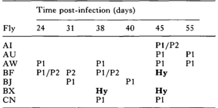
Table 1. Isolated and characterized (isoenzyme analysis for ICD) bloodstream-form populations from mixed infected Glossina morsitans centralis at different days after fly infection
(PI, P2, homozygous ICD (isocitrate dehydrogenase) patterns characteristic for bloodstream forms of parental clones PI (ST1B 247-LF) and P2 (STIB 777-A), re-spectively. Hy, heterozygous hybrid pattern. P1/P2, simultaneous homozygous patterns of both parental clones.) Fly AI AU AW BF B.T BX CN
Time post-infection (days)
24 PI P1/P2 31 P2 PI 38 40 PI P1/P2 PI H y PI 45 P1/P2 PI PI H y H y PI 55 PI PI
through Glossina; this stock originates from a population from a Coke's hartebeest in the Serengeti National Park, Tanzania (Geigy & Kaufmann, 1973) and was used for earlier mixed-infection experiments (Jenni et al. 1986; Sternberg et al. 1989). Clone
STIB 111-A is derived from the population KETRI
(Kenyan Trypanosome Research Institute) 7957 (Gibson & Gushumba, 1983), isolated in 1971 from a wild-caught Glossina fuscipes in eastern Uganda.
Tsetse flies and cyclical transmission
Teneral Glossina morsitans centralis were membrane fed on an equal mixture (3 x 10⁷ trypanosomes/ml) of bloodstream forms of both parental clones
STIB 247-LF (PI) and STIB 777-A (P2); fly
hand-ling and maintenance were performed as described by Schweizer et al. (1988). Infective flies - identified by probing on warmed slides — were repeatedly fed on Swiss ICR mice and the subsequent bloodstream-form trypsanosomes were cryopreserved. A total of 17 stabilates from 7 different flies were thus obtained and after expansion in mice and purification of the trypanosomes were subsequently screened for hy-brids by isoenzyme analysis for ICD.
Cloning
Cloning was performed on progeny population
STIB 787-1, isolated from fly BX on day 38 after
infection which was chosen at random from the three populations which had been shown to contain hybrid trypanosomes (Table 1). The bloodstream form trypsanosomes were therefore thawed and used to infect a lethally X-irradiated (650 rad; radiation absorbed dose) Swiss ICR mouse. After a sufficient
level of parasitaemia had been reached, single trypanosomes from tail blood were cloned intra-peritoneally into a total of 19 ICR mice using the hanging drop method (Van Meirvenne, Janssens & Magnus, 1975). Four clones were isolated (desig-nated as STIB 787-IA, 787-IB, 787-IC and 787-ID respectively), cryopreserved and subsequently characterized by isoenzyme and molecular karyotype analysis.
Isoenzyme analysis
The preparation of bloodstream-form lysates was performed as described by Schweizer et al. (1988). Starch gel electrophoresis, electrophoresis buffers and the stains used for ICD and AP (alkaline phosphatase; EC 3.1.3.1) have previously been described (Tait, Babiker & Le Ray, 1984). Isoelectric focusing for MEB (malic enzyme B; EC 1.1.1.40)
was carried out according to Richner et al. (1989) except for pre-focusing, which was carried out at 220 Vh.
Restriction fragment length polymorphism analysis
Isolation of trypanosome DNA (deoxyribonucleic acid) as well as DNA-electrophoresis, Southern hybridization and restriction fragment length poly-morphism (RFLP) analysis was carried out as previously described (Gibson et al. 1985; Jenni et al. 1986; Wells et al. 1987; Sternberg et al. 1989) using probes for phosphoglycerate kinase (Osinga et al. 1985) and RNA-polymerase I (Jess, Hammer & Cornelissen, 1989).
Pulsed field gradient gel electrophoresis
Molecular karyotype analysis by pulsed field gradi-ent gel electrophoresis, PFGE (Schwartz & Cantor, 1984) was performed on procyclic forms. Blood-stream-form trypanosomes of the different clones were transformed in vitro according to a slightly modified (Schweizer, Pospichal & Jenni, 1991) method of Brun & Schonenberger (1981).
The trypanosomes were prepared in blocks in low-gelling temperature agarose at 2-5 x 10⁸/ml accord-ing to the method of Van der Ploeg et al. (1984, 1989). The digestion of the blocks was carried out in 0-5 M EDTA (ethylenedinitrilotetraacetic acid, Ti-triplex III) pH 9-0, 1 % sodium N-lauryl sarcosinate supplemented with proteinase K (0-5 mg/ml) for 48 h at 50 °C. After rinsing the blocks 3 times in 50 mM EDTA, pH 8-0, the samples were stored in the same solution at 4 °C.
For electrophoresis, chromosome blocks were inserted into the sample slots of a 15x15 cm 1 % agarose gel in 89 mM Tris (tris[hydroxymethyl]-aminomethane), 89 mM boric acid, 0-2 mM EDTA. The gel was run in an LKB Pulsaphor system with
a hexagonal electrode array. T h e electrode buffer was the same as in the gel and was maintained at 14 °C. Pulses of different frequencies were applied in N - S and E-W directions. In order to resolve different size ranges of chromosomes, three different sets of running conditions (I, II and III) involving different pulse frequencies and durations of electro-phoresis were applied. Whereas program I (4 phases: 300 s (12 h), 200 s (18 h), 150 s (24 h), 100 s (18 h); 110 V) provides a good resolution of the intermediate (100—720 kilobases) size chromosomes, program II (5 phases: 900 s (24 h), 750 s (24 h), 700 s (24 h), 600 s (24 h), 500 s (24 h); 825 V) and program I I I (6 phases: 1200 s (24 h), 1000 s (24 h), 900 s (24 h), 800 s (24 h), 700 s (24 h), 600 s (24 h); 82-5 V) allow the separation of larger chromosomes ranging from 1 to 3 Mb. After separation, the gel was stained in an ethidium bromide solution (46 mM Tris, 46 mM boric acid, 0-1 mM EDTA, 2 /<g/ml ethidium bro-mide) for 2-8 h and subsequently destained in the same buffer without ethidium bromide.
PFGE gel separations were blotted as described by Hide et al. (1990) with the exception that the DNA was transferred to nylon filters (Hybond-N, Amersham) and cross-linked to the filter by exposure to long-wave ultra-violet light for 7 min. The filter was pre-hybridized for 30 min in 0-5 M phosphate buffer (pH7-2), 7 % S D S (dodecylsulfate sodium salt) at 65 °C and subsequently hybridized with 107 cpm (counts/min) of the appropriate labelled DNA probe overnight at the same temperature in the same buffer. Subsequently the filter was washed twice for 15 min in 0 - 1 % S D S , 2 x SSC (saline-sodium citrate buffer) at 65 °C before being exposed to the X-ray film at —70 °C. Filters were hybridized with D N A fragments coding for the phospho-glycerate kinase (PGK) gene (Osinga et al. 1985) and for the cysteine protease (CP) gene (Mottram et al. 1989). T h e probes were labelled using a random primer labelling kit (Boehringer) as directed by the manufacturer.
RESULTS
Frequency of hybrid formation
In order to obtain information on the frequency of hybrid formation with the new combination of parental clones, 7 infective tsetse flies were randomly chosen to feed on individual mice at different times after Glossina infection. The results of the character-ization of 17 of the subsequent bloodstream-form populations by isoenzyme analysis for I C D are summarized in Table 1; hybrids are identified by the presence of a heterozygous I C D pattern as described previously (Jenni et al. 1986). T h e results indicate that of the 7 Glossina examined, at least 2 extruded trypsanosome populations containing hybrid organ-isms on at least one occasion. This observation is in
Table 2. Phenotypes (isoenzymes) and genotypes (RFLPs) of parental and progeny clones
(Isoenzymes: the electrophoretic variants are denoted by numbers; a single number defining a single band of activity and a pair of hyphenated numbers indicating multiple bands of activity typical of a heterozygous pattern. ICD: isocitrate dehydrogenase; AP: alkaline phosphatase; MEB: malic enzyme B. RFLPs: markers were used for
which one of the parentals is heterozygous. Clones were screened with a cDNA-clone of phosphoglycerate kinase (PGK) or a polymerase I probe (Pol I) using DNA digested with the enzymes indicated.)
Clone Parental: STIB 247-LF STIB 777-A Progeny: STIB 787-IA STIB 787-IB STIB 787-IC STIB 787-ID Isoenzymes ICD 1 2 1-2 1-2 1-2 1-2 AP 1 1-2 1 1 1 1-2 MEB 3 2-3 2-3 2-3 2-3 2-3 RFLPs PGK Poll Pstl Hindlll + + H— H -1— + + H + 4- H + +
-accordance with earlier findings in mixed infection experiments in relation to the frequency of hybrids produced (Schweizer et al. 1988; Sternberg et al. 1989; Turner et al. 1990).
Enzyme phenotype of progeny clones
Parental and progeny clones were characterized by starch gel electrophoresis (ICD and AP) or iso-electric focusing (MEB) of extracts for 3 marker
enzymes. T h e results obtained are shown in T a b l e 2. T h e 4 progeny clones can be classified into 2 phenotypic classes (class 1 and class 2) which are nonparental and distinguishable solely by A P -pattern. Whereas all 4 progeny clones show a non-parental 3-banded hybrid ICD-pattern and a P2-type MEB-banding, 3 of them (class 1) express a
PI-type AP-phenotype (STIB 787-1 A, 78?'-IB and
787-IC) and 1 (STIB 787-ID) shows a P2-type
AP-pattern (class 2).
T h e enzyme phenotypes observed at each of these loci are compatible with a simple Mendelian pattern of inheritance. They represent 2 of 4 possible phenotypic F l progeny classes that would be predicted if meiosis had occurred either before or after the genetic exchange event which led t o hybrid formation.
Restriction fragment length polymorphism analysis
Bloodstream forms of all 4 progeny clones were analysed together with the parental stocks by Southern blotting of restriction digests of nuclear
A 86 PGK P2 P1 - I D - I C - I B - I A Poll P2 P1 - I D -IC - I B - I A 4-5 1-6 0-9 110 2-3 1-2 B H H* 110 1-8 B
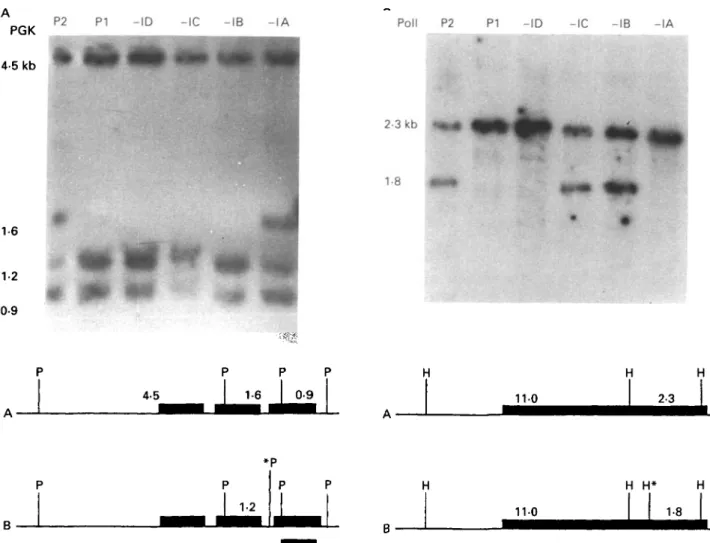
Fig. 1. (A) Autoradiograph of a Southern blot of total genomic DNA digested with Pst 1 and probed with a cDNA clone of phosphoglycerate kinase (PGK). The DNA was extracted from two parental stocks PI, P2: parental clones
STIB 247-LF and STIB 777-A and four progeny clones -IA, -IB, -IC, -ID: STIB 787-1 A, 787-IB, 787-IC and 787-ID. Below the panel is a map to show the alleles involved, with the 3 homologous PGK genes shown as black
boxes and an asterisk marking the polymorphic site; the position of the probe is indicated by a bar beneath the map. (B) Autoradiograph of Southern blot of total genomic DNA digested with HindWl and probed with a DNA probe of polymerase I(PolI), Parental stock: P2 : STIB 777-A; PI : STIB 247-LF; progeny: -IA, -IB, -IC, ID: STIB
787-IA, 787-IB, 787-IC and 787-ID.
DNA using a probe for the phosphoglycerate kinase gene (PGK-960 bp Pst 1 fragment from phospho-glycerate kinase cDNA clone pTcPGK8 (Osinga et
al. 1985)) and a probe for RNA-polymerase I (Pol I;
Jess et al. 1989). The restriction pattern of
STIB 247-LF (PI) can be interpreted as being
homozygous for the presence of a Pst 1 restriction site detected by PGK and is identical with the pattern of STIB 386 (Turner et al. 1990) whereas
STIB 777-A (P2) is heterozygous for the presence/
absence of this site. Of the 4 progeny clones,
STIB 787-1 A clearly shows the heterozygous
pat-tern of P2, while STIB 787-IB, STIB 787-IC and
STIB 787-ID show the homozygous pattern of PI
(Fig. 1A).
A further polymorphism is detected by the DNA probe Pol I: STIB 777-A (P2) shows a heterozygous pattern whereas STIB 247-LF (PI) has a homo-zygous 1-banded pattern (Fig. IB); the progeny clones STIB 787-IA and STIB 787-ID show the
same homozygous band as STIB 247-LF while the progeny clones STIB 787-IB and STIB 787-IC show a heterozygous pattern.
This finding underpins the postulate that the progeny are products of syngamy. Considering the segregation of the alleles at the loci for I CD, AP, MEB (isoenzymes) and PGK, Pol I (RFLPs) in the
cross, it can be seen that 3 classes of Fl progeny are generated (Table 2). The non-parental combinations of alleles of these 3 classes show that the alleles at each locus segregate. STIB IB and STIB
787-IC were identical according to all markers for which
they were analysed.
Molecular karyotype analysis
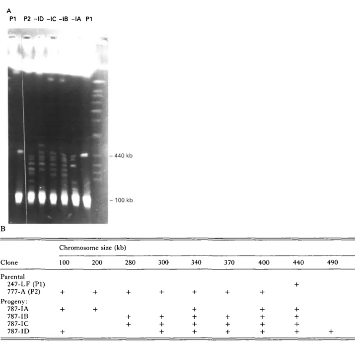
The analysis of intermediate size chromosomes (Fig. 2A) reveals that the parental clone STIB 247-LF (PI) shows a single band at 440 kb while clone
P1 P2 - I D -IC -IB - I A P1 H - 440 kb - 1 0 0 kb B Chromosome size (kb) Clone 100 200 280 300 340 370 400 440 490 Parental 247-LF (PI) 777-A (P2) Progeny: 787-IA 787-IB 787-IC 787-ID
Fig. 2. (A) Ethidium bromide-stained PFGE run using program I to give increased resolution in the size range 100-720 kb. Stocks are: PI, P2: parental clones STIB 247-LF and STIB 777-A respectively; -IA, -IB, -IC, -ID: progeny clones STIB 787-IA, 787-IB, 787-IC and 787-ID respectively. (B) Table t o (A) summarizing the data on chromosomes observed ranging in the size 100 and 490 kb.
from 100 to 400 kb. None of the 4 progeny clones shows a pattern identical to either parental clone or to the sum of both parental types; by this criterion the progeny are all recombinant. The intermediate size chromosomes are thought to contain primarily repetitive sequences and VSG genes; on the as-sumption that identical size indicates an identical chromosome, all progeny clones inherit the PI parental chromosomes of 440 kb and the P2 parental chromosomes of 340 kb and 400 kb together with a varying number - but never all - of the remaining P2 chromosomes. The progeny can be grouped into 3 classes. One class consists of STIB 787-IA with 5 chromosomes. A second includes both STIB 787-IB and STIB 787-IC which show an identical pattern with 6 bands. The third class consists of progeny
clone STIB 787-ID which has 7 bands, one repre-senting a non-parental chromosome of novel size, approximately equal to the sum of the sizes of the two chromosomes not present (200 and 280 kb). These results are summarized in Fig. 2B.
The large chromosomes were separated using two different running conditions which resolve chromo-somes in different size ranges. The ethidium bromide stained pattern of the parental stocks and resulting hybrid clones separated under program II are illustrated in Fig. 3A. It is clear that the hybrid progeny are different from either parental clone and that hybrids -IB and -IC are identical. Most of the chromosome bands in the hybrid clones can, on the basis of size, be accounted for as having originated from one or other parent; however, one band in
88
P1 - I A -IB -IC -ID P2 P1 - I A -IB/-IC -ID P2
3500
kb-*E3
•
5
C Dnu
4 E23 E52
•
CD• Ds
CD CD 4• 3
2500 1600 1125 kb-Y77X 2 d cz: cm 1 czi 2 • i int• • • • • mir
-PGKPI - I A -IB -IC -ID P2
4 3
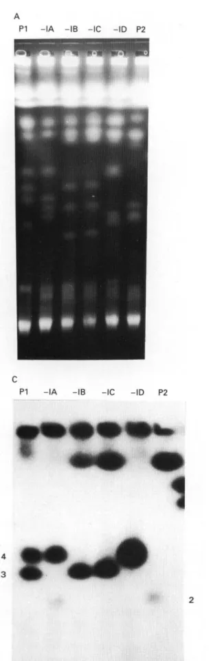
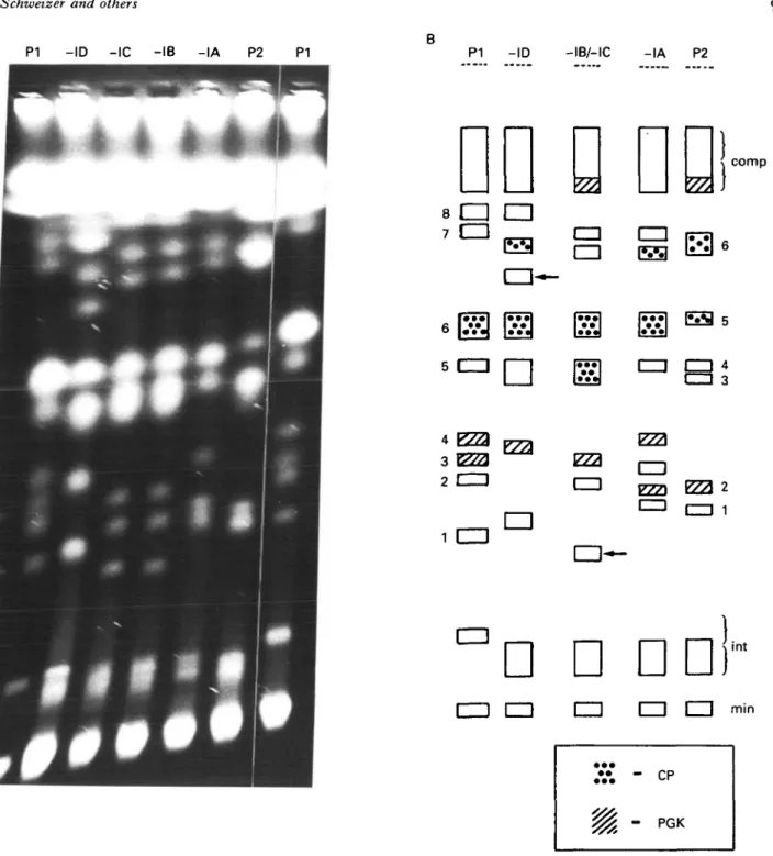
Fig. 3. PFGE analysis of P G K and CP gene inheritance. (A) Ethidium bromide-stained P F G E run using program II for separation of larger chromosomes (1-3 Mb); the designation of the tracks is as in Fig. 1. (B) Diagram of the bands in (A). For better identification the chromosomes of the parental clones within the range of interest are numbered. - I B / - I C shows the patterns of progeny, 787-IB and 787-IC respectively, and min and int indicate the position of the ' m i n i ' and the intermediate sized chromosomes. Arrows point to non-parental size chromosomes (novel size bands). Bands which hybridize with a probe for P G K are hatched. (C) Blot of the gel in (A) hybridized with the PGK-probe.
clones -IB and -IC is of novel size (arrows in Fig. 3B) not found in either parent. In order to identify homologous chromosomes, a blot of the gel was probed with a cDNA clone of PGK and the hybridization pattern obtained is illustrated schem-atically in Fig. 3B and C. Hybridization to bands 3 and 4 in PI and band 2 and the compression zone in P2 is observed. Hybrid -IA inherits band 2 (P2) and band 4 (PI) while hybrids -IB/-IC inherit the compression zone chromosome (P2) and band 3 (PI) as would be predicted if these chromosomes were homologues in the parental clones and the hybrid progeny inherited one homologue from each parent. Hybrid -ID shows only a single band of hybrid-ization of greater intensity than the other hybrids and of a similar mobility to band 4 (PI). Further data are presented and discussed below in relation to this pattern of hybridization.
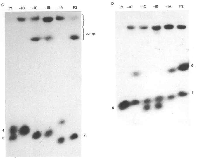
Greater resolution of the larger chromosomes was obtained using program III and the ethidium-stained gel using these conditions is shown in Fig. 4A. By comparison to the parental karyotype, it is clear that the hybrids are recombinant and in hybrids
-ID and -IB/-IC novel, non-parental size
chromo-somes are observed (arrowed, Fig. 4B). In order to confirm the results obtained with the PGK cDNA probe, the gel was blotted and hybridized with this probe. The results are presented in Fig. 4C and diagrammatically in Fig. 4B and are essentially similar to those in Fig. 3. Progeny clone STIB
787-ID shows an odd pattern with a single broad
hybridization band of the same size as band 4 of PI. A possible explanation could be that the single band consists of band 4 from PI and a novel size chromosome from P2; however, further data are required to establish this.
In order to establish whether other pairs of homologous chromosomes behave in the same way, the blot of the gel shown in Fig. 4 was probed with a cysteine proteinase gene probe (CP; Fig. 4D). The CP probe shows a single banding pattern for parental clone PI (band 6) and a double banding pattern (bands 5 and 6) for P2. From the intensity of the single band (see also Fig. 4A and B) one could assume that it contains 2 homologues of identical or nearly identical size. The progeny clones 787-IA and
-ID appear identical to P2, but could equally be due
to the inheritance of 1 homologue from PI and band 6 from P2. STIB787-IB and -IC respectively show a non-parental double banding pattern; one band is of the same size as band 6 (PI) or band 5 (P2) and the second represents a novel size band which could be derived from either parent.
These results indicate that there is evidence for normal Mendelian inheritance, i.e. of 1 homologue from each parent, but in addition we find chromo-somes of a novel size in some clones. This finding could be explained by recombination between
different-sized homologues at meiosis, and the generation of novel size chromosomes.
DISCUSSION
This publication reports an analysis of the products of a new genetic cross of two East African T. brucei clones. The inheritance of isoenzyme and RFLP markers from the parents to the progeny was examined. Clones were heterozygous for markers for which parents were homozygous and segregation of alleles was observed for phenotypic and genotypic markers. The four hybrid clones provided evidence that karyogamy and/or meiosis had occurred; this is in accordance with observations from other mixed infection experiments (Gibson & Garside, 1990; Tait & Turner, 1990; Turner et al. 1990).
Clones derived from hybrid-containing popula-tions showed heterozygous hybrid pattern for the isoenzyme I CD. The four hybrid clones were further analysed by both isoenzyme (AP and MEB) and gene
(PGK and pol I) markers for which the 777-A parent is heterozygous but the 247-LF parent is homozygous. Two of the clones (IB and
787-IC) were identical but recombinant while the
remaining two cones (787-IA and 787-ID) were distinct from each other and the first two clones but were also recombinant. Thus allelic segregation oc-curred at each locus and recombination between alleles at each locus. These results are those predicted of a normal diploid Mendelian system involving meiosis. The analysis of RFLPs showed three different non-parental combinations of alleles and showed that alleles segregate at each locus as reported for a different cross by Turner et al. 1990.
The inheritance of the intermediate chromosomes was examined and as has been reported with other crosses (Jenni et al. 1986; Wells et al. 1987; Gibson, 1989) these did not appear to be inherited in a Mendelian manner. The single (440 kb) intermediate chromosome of stock STIB 247-LF was inherited by all progeny clones but a variable number of the chromosomes from stock STIB 777-A leading to a range of intermediate karyotypes in the progeny clones. These results support the view that the intermediate chromosomes do not behave as pairs of homologues and so segregate at random into the progeny clones and are therefore inherited in a non-Mendelian manner. The additional detection of a non-parental size chromosome in one of the progeny clones is in accordance with results from Gibson (1989), who interpreted the novel chromosome as a product of the gene rearrangements associated with antigenic variation, or of a recombination associated with genetic exchange.
The analysis of the inheritance of large chromo-somes in four hybrid progeny clones revealed that
A
P1 -ID -IC -IB -IA P2 P1 - I D -IB/-IC - I A P2
77,
comp8CH •
7 C=l _ 4 3 2CZ3CD
PZ3 CD 1 = 1vzn
E22 2• *
-CD _
•
I inti i i i en im czi
Fig. 4. For legend see opposite.
three clones possessed novel-sized (non-parental) chromosomes. This finding could be explained by recombination between homologues differing in size at meiosis, although other explanations are possible. Evidence for such discrepancies in size between homologues can be shown by Southern analysis of chromosomes, a technique which represents a power-ful method for determining the fate of chromosomes. Thus the evidence presented shows that putative homologues harbouring the PGK genes were of different sizes in the two parentals; the Fl progeny clones -IA, -IB and -IC inherit one chromosome (hybridizing to PGK) from each parent (Fig. 4C)
which is consistent with the two bands identified in each of the parental stocks behaving as homologues and segregating at meiosis. Progeny clone -ID, however, shows a single broad band of hybridization; without additional markers for each putative parental homologue this result is difficult to interpret. Analysis of the chromosomes detected by the cys-teine proteinase gene probe for progeny clones -IA and -ID is also consistent with these hybrids in-heriting one homologue from each parent; how-ever, progeny clones -IB and -IC show a novel non-parental karyotype which, without further data, is difficult to interpret conclusively. Results which
C
PI - I D -IC - I B - I A P2 P1 - I D -IC -IB - I A P2
comp
if
Fig. 4. PFGE analysis of PGK and CP gene inheritance. (A) Ethidium bromide-stained PFGE gel using program III for the separation of the chromosomes in the range from 1 to 3 Mb. The lanes are designated as in Fig. 2. (B) Diagram of the bands in (A) including results from the PGK and CP blot. Numbering of the bands is as in Fig. 2; min, int and comp indicate the area of the mini-chromosomes, the intermediate size chromosomes and the compression zone respectively. The arrow indicates a non-parental band of novel size. (C) Blot of the gel in (A) hybridized with the PGK-probe. (D) Blot of the gel in (A) hybridized with the CP-probe. The hybridization observed at the slot region can be ignored on the assumption that it is due to trapping.
indicate that homologous chromosomes can differ in size have been provided by other authors (Gibson et
al. 1985; Gibson, 1989; Gottesdiener etal. 1990) but
the mechanisms by which these size differences arise remain to be elucidated. One interpretation of the data presented here is that such size differences are generated during or after genetic exchange but further markers for each homologue of a chromo-some pair are required in order to establish this.
The results imply that the progeny clones are not the products of a simple fusion but fit into a classical system with meiosis and subsequent fusion of the nuclei to form diploid progeny. Previous reports (Paindavoine et al. 1986; Wells et al. 1987; Gibson, Garside & Bailey, 1992) have identified hybrid progeny with raised DNA contents. Two of these clones (Gibson et al. 1992) have been characterized in terms of their molecular karyotype and shown to be trisomic at least for some of the chromosomes.
Our data, based on molecular karyotype analysis, suggest that the hybrids produced in the cross between STIB247-LF and STIB 777-A are dip-loid and provide further evidence that the production of hybrids with raised DNA content is a relatively rare event (Tait & Turner, 1990). However, it may be that such hybrids are generated at relatively high frequency but are selected against during the growth of trypanosomes from hybrid producing tsetse flies.
We are grateful to Dr D. Moloo from ILRAD, Nairobi, Kenya, for the supply of tsetse pupae and to Mrs J. M. Jenkins for critical reading of the manuscript. This study was supported by the Swiss National Science Foundation grant No. 31-25367.88 I/II (J. S., H. P., L. J) and by a grant from the Wellcome Trust (G. H., N. B., A. T.). This work forms part of the material for the Ph.D. theses of H. Pospichal and J. Schweizer at the University of Basel.
REFERENCES
BRUN, R. & SCHONENBERGER, M. (1981). Stimulating effect of citrate and cis-aconitate on the transformation of
Trypanosoma brucei bloodstream forms to procyclic
forms in vitro. Zeitschrift fur Parasitenkunde 66, 17-24.
GEIGY, R. & KAUFFMANN, M. (1973). Sleeping sickness survey in the Serengeti Area (Tanzania) 1971. Ada
Tropica 30, 12-48.
GIBSON, w. c. (1989). Analysis of a genetic cross between
Trypanosoma brucei rhodesiense and T. b. brucei. Parasitology 99, 3 9 1 ^ 0 2 .
GIBSON, w. c. & GARSIDE, L. (1990). Kinetoplast D N A minicircles are inherited from both parents in genetic hybrids of Trypanosoma brucei. Molecular and
Biochemical Parasitology 42, 45-54.
GIBSON, w. c , GARSIDE, L. & BAILEY, M. (1992). Trisomy
and chromosome size changes in hybrid trypanosomes from a genetic cross between Trypanosoma brucei
rhodesiense and T. b. brucei. Molecular and Biochemical Parasitology 52, 189-200.
GIBSON, w. c. GASHUMBA, j . K. (1983). Isoenzyme characterization of some Trypanozoon stocks from a recent trypanosomiasis epidemic in Uganda.
Transactions of the Royal Society of Tropical Medicine and Hygiene 77, 114-18.
GIBSON, W. C , MARSHALL, T. F. DE C. & GODFREY, D. G.
(1980). Numerical analysis of enzyme polymorphism: a new approach to the epidemiology and taxonomy of trypanosomes of the subgenus Trypanozoon. Advances
in Parasitology 18, 175-246.
GIBSON, W . C , OSINGA, K. A., MICHELS, P . A. M. & BORST,
p. (1985). Trypanosomes of subgenus Trypanozoon are diploid for housekeeping genes. Molecular and
Biochemical Parasitology 16, 231-42.
GIBSON, w. & WHITTINGTON, H. (1993). Genetic exchange in Trypanosoma brucei: selection of hybrid
trypanosomes by introduction of genes conferring drug resistance. Molecular and Biochemical
Parasitology 60, 19-26.
GOTTESDIENER, K., GARCIA-ANOVEROS, J., LEE, M. G . - S . &
VAN DER PLOEG, L. H. T. (1990). Chromosome organization of the protozoan Trypanosoma brucei.
Molecular and Cellular Biology 10, 6079-83.
HIDE, G., CATTAND, P., LERAY, D., BARRY, J. D. & TAIT, A.
(1990). The identification of Trypanosoma brucei subspecies using repetitive D N A sequences. Molecular
and Biochemical Parasitology 39, 213—26.
JENNI, L., MARTI, S., SCHWEIZER, J., BETSCHART, B . , LE PAGE, R. W. F . , WELLS, J. M., TAIT, A., PAINDAVOINE, P . ,
PAYS, E. & STEINERT, M. (1986). Hybrid formation between African trypanosomes during cyclical transmission. Nature, London 322, 173—5.
JESS, W . , HAMMER, A. & CORNELISSEN, A. W. C. A. ( 1 9 8 9 ) .
Complete sequence of the gene encoding the largest subunit of RNA polymerase I of Trypanosoma brucei.
FEBS Letters 249, 123-8.
MOTTRAM, J. C , NORTH, M. J., BARRY, J. D. & COOMBS,
G. H. (1989). A cysteine proteinase cDNA from
Trypanosoma brucei predicts an enzyme with an
unusual C-terminal extension. FEBS Letters 258, 211-15.
OSINGA, K. A., SWINKELS, B. W., GIBSON, W . C , BORST, P . , VEENEMAN, G. H . , VAN BOOM, J. H . , MICHELS, P . A. M. &
OPPERDOES, F. R. (1985). Topogenesis of microbody enzymes: a sequence comparison of the genes for the glycosomal (microbody) and cytosolic
phosphoglycerate kinases of Trypanosoma brucei.
EMBO Journal 4, 3811-17.
PAINDAVOINE, P . , ZAMPETTI-BOSSELER, F . , PAYS, E., SCHWEIZER, J., GUYAUX, M., JENNI, L. & STEINERT, M.
(1986). Trypanosome hybrids generated in tsetse flies by nuclear fusion. EMBO Journal 5, 3631-6.
RICHNER, D., SCHWEIZER, J . , BETSCHART, B. & JENNI, L.
(1989). Characterization of West African Trypanosoma
(Trypanozoon) brucei isolates from man and animals
using isoenzyme analysis and DNA hybridization.
Parasitology Research 76, 80-5.
SCHWARTZ, D. c. & CANTOR, c. R. (1984) Separation of yeast chromosome-sized DNAs by pulsed field gradient gel electrophoresis. Cell 37, 67-75. SCHWEIZER, j . & JENNI, L. (1991). Hybrid formation in
the life-cycle of Trypanosoma (T.) brucei: detection of hybrid trypanosomes in a midgut-derived isolate. Ada
Tropica 48, 319-21.
SCHWEIZER, j . , POSPICHAL, H. & JENNI, L. (1991). Hybrid
formation between African trypanosomes in vitro.
Ada Tropica 49, 237-40.
SCHWEIZER, j . , TAIT, A. & JENNI, L. (1988). T h e timing
and frequency of hybrid formation in African trypanosomes during cyclical transmission.
Parasitology Research 75, 98-101.
STERNBERG, J., TAIT, A., HALEY, S., WELLS, J. M., LE PAGE,
R. w. F., SCHWEIZER, j . & JENNI, L. (1988). Gene exchange in African trypanosomes: characterisation of a new hybrid genotype. Molecular and Biochemical
Parasitology 27, 191-200.
STERNBERG, J., TURNER, C. M. R., WELLS, J. M., RANFORD-CARTWRIGHT, L. C , LE PAGE, R. W. F. & TAIT, A. ( 1 9 8 9 ) .
Gene exchange in African trypanosomes: frequency and allelic segregation. Molecular and Biochemical
Parasitology 34, 269-80.
TAIT, A. (1980). Evidence for diploidy and mating in trypanosomes. Nature, London 287, 536-8.
TAIT, A., BABIKER, E. A. & LE RAY, D. (1984).' E n z y m e
variation in Trypanosoma brucei spp. 1. Evidence for sub-speciation of Trypanosoma brucei gambiense.
Parasitology 89, 311-26.
TAIT, A., BARRY, J. D., WINK, R., SANDERSON, A. & CROWE,
j . s. (1985). Enzyme variation in T. brucei spp. I I . Evidence for T. b. rhodesiense being a set of variants of
T. b. brucei. Parasitology 90, 89-100.
TAIT, A. & TURNER, c. M. R. (1990). Genetic exchange in
Trypanosoma brucei. Parasitology Today 6, 70-5. TURNER, C. M. R., STERNBERG, J . , BUCHANAN, N . , SMITH, E.,
HIDE, G. & TAIT, A. (1990). Evidence that the mechanism of gene exchange in Trypanosoma brucei involves meiosis and syngamy. Parasitology 101, 377-86.
VAN DER PLOEG, L. H. T . , SCHWARTZ, D. C , CANTOR, C. R. &
BORST, p. (1984). Antigenic variation in Trypanosoma
brucei analyzed by electrophoretic separation of
chromosome-sized D N A molecules. Cell 37, 77-84.
VAN DER PLOEG, L. H. T., SMITH, C. L., POLVERE, R. I. &
chromosome-sized D N A from Trypanosoma brucei WELLS, J. M., PROSPERO, T. D., JENNI, L. & LE PAGE, R. W. F. stock, 427-60. Nucleic Acids Research 17, 3217-27. (1987). D N A contents and molecular karyotypes of VAN MEIRVENNE, N., JANSSENS, P. G. & MAGNUS, E. (1975). hybrid Trypanosoma brucei. Molecular and Biochemical
Antigenic variation in syringe passaged populations of Parasitology 249, 103-16.
Trypanosoma (Trypanozoon) brucei: rationalization of
the experimental approach. Annales de la Socie'te beige