Regression of Left Ventricular Mass in
Hypertensive Patients Treated With Perindopril/
Indapamide as a First-Line Combination
The REASON Echocardiography Study
Nicola de Luca, Jean-Michel Mallion, Michael F. O’Rourke, Eoin O’Brien,
Karl-Heinz Rahn, Bruno Trimarco, Ramon Romero, Peter Wilhelmus De Leeuw,
Gerhart Hitzenberger, Edouard Battegay, Daniel Duprez, Peter Sever, and
Michel E. Safar, on behalf of the REASON Project*
Background: Increase in left ventricular mass (LVM)
may be linked to morbidity and mortality in hypertensive patients. Arterial stiffness, systolic blood pressure (BP), and pulse pressure (PP) seem to be the main determinants of LVM. The perindopril/indapamide combination nor-malizes systolic BP, PP, and arterial function to a greater extent than atenolol. The aim of this study was to compare the effects of perindopril (2 mg)/indapamide (0.625 mg) first-line combination with atenolol (50 mg) on LVM reduction in hypertensive patients.
Methods: Two hundred fourteen patients with essential
hypertension participating in the PREterax in Regression of Arterial Stiffness in a ContrOlled Double-BliNd (REASON), randomized, double-blind, parallel-group study, underwent M-mode two-dimensional-guided echocardiography.
Results: Perindopril/indapamide and atenolol were both
effective at brachial BP reduction during the 12-month pe-riod. The systolic BP reduction was significantly greater with perindopril/indapamide than with atenolol (⫺21.2 v ⫺15.3 mm Hg), whereas the reduction in diastolic BP was similar
between treatment groups (⫺12.1 v ⫺11.3 mm Hg). Reduc-tion in LVM was higher with perindopril/indapamide than with atenolol. The between-group difference was significant for LVM (⫺13.6 v ⫺4.3 g, P ⫽ .027), LVM/body surface area (LVMI1, P⫽ .032), and LVM/body height
2.7
(LVMI2,
P⫽ .013). The 124 patients with LV hypertrophy at baseline showed greatest LVM regression (LVM:⫺22.5 v ⫺8.9 g, P ⫽ .009; LVMI1, P⫽ .031; LVMI2, P⫽ .028). The reduction
in LVM adjusted for brachial systolic BP and heart rate was still significantly greater with perindopril/indapamide than with atenolol.
Conclusions: Treatment, based on a first-line
perindo-pril/indapamide combination in hypertensive patients, was more effective than atenolol on regression of echocardio-graphic indices of LVM and LV hypertrophy. Am J Hy-pertens 2004;17:660 – 667 © 2004 American Journal of Hypertension, Ltd.
Key Words: Hypertension, left ventricular
hypertro-phy, left ventricular mass, echocardiograhypertro-phy, perindopril/ indapamide combination.
I
t is well established that hypertension, defined as systolic blood pressure (BP)ⱖ140 mm Hg or dia-stolic BP ⱖ90 mm Hg,1–3represents a major riskfactor for morbidity and mortality. Much emphasis has previously been placed on risk reduction associated with diastolic BP control. However, recent epidemiologic
stud-Received November 12, 2003. First decision January 12, 2004. Accepted March 30, 2004.
From the Clinica Medica (NdL, BT), Napoli, Italy; Cardiologie et Hypertension Artérielle (J-MM), CHRU Grenoble, France; Saint Vin-cent’s Hospital (MFO’R), Darlinghurst, Australia; Department of Clini-cal Pharmacology, Royal College of Surgeons in Ireland, and Blood Pressure Unit, Beaumont Hospital (EO’B), Dublin, Ireland; Medizinische Poliklinik, University of Münster (K-HR), Münster, Germany; Hospital Germans Trias y Pujol (RR), Badalona, Spain; Academisch Ziekenhuis Maastricht (PWDL), Maastricht, The Netherlands; Institut für Hypertoni-ker (GH), Wien, Austria; Medizinische Universitäts Poliklinik (EB),
Basel, Switzerland; Universitair Ziekenhuis Gent (DD), Gent, Belgium; Saint Mary’s Hospital Medical School (PS), London, UK; and Hôpital Broussais (MES), Paris, France.
This study was supported by Institut de Recherches Internationales Servier.
*See Appendix for complete list of the REASON International Co-ordination Group.
Address correspondence and reprint requests to Prof. Nicola de Luca, Università degli Studi “Federico II” di Napoli, Facoltà di Medicina e Chiru-rgia, Via Sergio Pansini, 5, 80131 Napoli, Italy; e-mail: nideluca@unina.it
0895-7061/04/$30.00 © 2004 by the American Journal of Hypertension, Ltd.
ies as well as recommendations from hypertension guide-lines have shown that the brachial systolic BP is a better guide for evaluating cardiovascular risk than brachial di-astolic BP.2–5 Pulse pressure (PP) and arterial stiffness
may also be independent cardiovascular risk factors.6 –10
Hypertensive patients with concurrent cardiovascular structural changes such as left ventricular hypertrophy (LVH) have a greater mortality risk than those with hypertension alone.11–14There is evidence to suggest that a parallel cardiac
and vascular adaptation exists in arterial hypertension15with
a positive relationship between cardiac mass and arterial stiffness.16 A direct relationship between cardiac mass and
BP has been demonstrated, and the main pressure determi-nants of left ventricular mass (LVM) in essential hyperten-sion are systolic BP and PP.7,17–23 Reduced aortic
distensibility and compliance may participate in the genesis of cardiac hypertrophy in hypertension. Arterial compliance also determines the pulsatile amplitude of the pressure wave and its reduction induces a selective increase in systolic level and PP. Consequently a different response after antihyperten-sive treatment has been demonstrated in cardiac and arterial changes.24,25
A first-line combination of the angiotensin-converting enzyme (ACE) inhibitor perindopril (2 mg) and indapam-ide (0.625 mg), a sulphamoyl diuretic that belongs to the thiazide group, has been shown to have a favorable effi-cacy:safety ratio26,27 and superior antihypertensive
effi-cacy in comparison with enalapril, losartan, and irbesartan.28 –30 Recently, PREterax in Regression of
Ar-terial Stiffness in a ContrOlled Double-BliNd Study (REASON) has shown that the perindopril/indapamide antihypertensive treatment normalized systolic BP, PP, and arterial function to a greater extent than atenolol in hypertensive patients.31 Moreover, the
perindopril/indap-amide combination has shown a beneficial effect on car-diac hypertrophy and capillary density in experimental hypertension.32 Given such findings it is timely to
com-pare cardiac mass structural changes after perindopril/ indapamide combination or atenolol in the treatment of hypertensive patients.
In this article we present the results from the REASON echocardiography ancillary study. This study investigated the long-term effects of perindopril/indapamide first-line combination compared with atenolol on echocardio-graphic indices of LVM in hypertensive patients.
Methods
Study Design
The REASON study was a 12-month, randomized, dou-ble-blind, parallel-group study conducted in 13 countries, which has previously been published.31 REASON
com-pared the efficacy and safety of perindopril (2 mg)/ inda-pamide (0.625 mg) or atenolol (50 mg) in the treatment of 471 patients with essential hypertension.
Men and women aged 18 to 84 years, with a diagnosis of uncomplicated essential hypertension (systolic BP
ⱖ160 mm Hg and ⬍210 mm Hg; diastolic BP ⱖ95 mm Hg and⬍110 mm Hg), were eligible for inclusion.
After a 4-week placebo wash-out period, patients were randomized to receive one capsule of perindopril (2 mg)/ indapamide (0.625 mg) or atenolol (50 mg) once daily for 12 months. After 3 months of treatment and then at every 3-month period until the end of the study, the dose could be increased to two tablets per day if the systolic BP remained⬎160 mm Hg or the diastolic BP ⬎90 mm Hg (n⫽ 103 perindopril/ indapamide and 92 for atenolol; log rank test P⫽ not significant [NS]). Other antihypertensive drugs were not allowed during the study follow-up. At study end, dosage was progressively decreased during 8 to 15 days to avoid any complications that might result from abrupt atenolol withdrawal. This study was performed in accordance with the Declaration of Helsinki, the protocol was approved by the Ethic Committees according to na-tional regulations, and all patients provided written in-formed consent to participate.
Patients who participated in the echocardiography study (n⫽ 214) had an echocardiogram recorded at base-line (month 0) and after 12 months of effective treatment (end value).
Efficacy Assessments
Left ventricular dimensions were assessed using two-di-mensionally directed M-mode echocardiography. A qual-ity control procedure was established for the validation of the echocardiographic evaluations. Each examination was recorded on S-VHS tape and readings of the measure-ments were performed centrally, on five cycles by two experienced physician readers blind to treatment, patients, and visit, and the values of both readers were then aver-aged.
Echocardiographic measurements (left ventricular in-ternal dimension [LVID], posterior wall thickness [PWT], and interventricular septum thickness [IVST]) were as-sessed at the end of diastole, defined as the peak of the R wave of the QRS complex, and in accordance with the recommendations of the American Society of Echocardi-ography.33The intraobserver and interobserver
reproduc-ibility of the echocardiographic measurements was tested in a parallel series of 10 subjects with three replications, the methodology of which is described in detail else-where.34,35The coefficient of variation was below 3%, the
intraobserver correlation was r⫽ 0.99 mm, and the inter-observer correlation was r⫽ 0.96 mm (mean difference, 0.05⫾ 0.03 mm).
The LVM was calculated from the Penn convention according to the standard Devereux formula36: LVM (g)⫽
1.04 [(IVSTd⫹ PWTd ⫹ LVIDd)3⫺ LVIDd3]⫺ 13.6,
and converted into LVM index (LVMI1) by dividing by
body surface area. Due to a possible overcalculation in overweight patients, LVM divided by body height2.7was
also calculated (LVMI2).37
Brachial systolic BP, diastolic BP, and heart rate were determined after a 10-min rest in the supine position using
a mercury sphygmomanometer and stethoscope. Brachial PP was calculated from individuals’ values of systolic BP and diastolic BP.
Tolerability Assessments
Any adverse events were recorded at clinic visits. Varia-tions in laboratory parameters were measured between baseline and end values. Tolerability data are presented for the entire REASON study population.
Statistical Analyses
All analyses were performed using SAS software version 8.2 (SAS Institute, Cary, NC). Quantitative variables are presented as mean changes (with standard deviation) from baseline to end value, unless otherwise stated. Patients who received at least one dose of treatment were included in an intent-to-treat analysis. A last observation carried forward approach was used if data were missing. Patients with LVH at baseline (LVMI1⬎100 g/m2for women and
⬎120 g/m2 for men) were included in a per-protocol
subgroup analysis prespecified in the protocol.
Within-group effect was assessed using a two-tailed Student t test for paired samples. Between-group compar-isons were assessed on the change at last evaluation using a covariance analysis, which included the treatment group as a factor, and age, sex, and the baseline value of the variable as covariates. A 5% threshold was considered significant. The relationship between LVM parameters and influence parameters was investigated using a stepwise linear regression procedure.
Results
Patient Characteristics
A total of 214 randomized patients, including 124 patients with LVH, were recruited to the echocardiography study. The baseline characteristics (Table 1) were similar in the two treatment groups and did not differ from those of the
entire population.31Seventy-eight percent of patients com-pleted 12 months of treatment. Forty-eight patients with-drew, 21 in the perindopril/indapamide group and 27 in the atenolol group because of adverse events (10 v 11, respec-tively), lack of efficacy (4 v 11), major protocol violations (1 v 2), and nonmedical reasons (6 v 3). Overall, the mean exposure to study drug was 312.3 days.
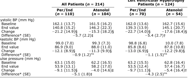
Brachial BP Assessment
Perindopril/indapamide and atenolol were both effective at BP reduction during the 12-month period. However, re-duction in systolic BP was significantly greater with per-indopril/indapamide than with atenolol (⫺21.2 v ⫺15.3 mm Hg, respectively; P⬍ .01). Significant reductions in diastolic BP were achieved in both treatment groups, but with no significant between-group differences (⫺12.1 v ⫺11.3 mm Hg). The PP reduction was significantly greater for the perindopril/indapamide group than for the atenolol group (⫺9.1 v ⫺4.0 mm Hg; P ⬍ .01). As expected, heart rate was lowered to a significantly greater extent with atenolol than perindopril/indapamide (⫺6.7 v ⫺2.5 beats/min; P ⬍ .001;Table 2).
Echocardiographic Assessments
Left ventricular diameter, thickness, and mass did not differ between the two treatment groups at baseline (Table 3). At the end of the treatment period LVID, PWT, and IVST were all significantly reduced in the perindopril/ indapamide group. There was no significant difference with atenolol (LVID:⫺0.62 v ⫺0.02 mm; PWT: ⫺0.29 v ⫺0.10 mm; IVST: ⫺0.22 v ⫺0.12 mm, respectively;
Table 3).
Consequently, LVM was considerably more reduced with the perindopril/indapamide than with atenolol. The between-group difference was significant, irrespective of whether this indice was expressed as LVM (⫺13.6 v ⫺4.3 g; P ⬍ .05), LVMI1 (⫺6.6 v ⫺2.1 g/m2; P ⬍ .05), or
LVMI2(⫺3.3 v ⫺0.9 g/m2.7; P ⬍ .05, respectively). Table 1. Baseline characteristics for patients included in the study
All Patients (n ⴝ 214) Left Ventricular HypertrophyPatients (n ⴝ 124) Per/Ind
(n ⴝ 110) (n ⴝ 104)Atenolol (n ⴝ 70)Per/Ind Atenolol(n ⴝ 54)
Sex (M:W) (%) 61:39 68:32 57:43 63:37 Age (y) 53.2 (11.8) 53.7 (11.4) 54.3 (11.5) 51.8 (12.1) BMI (kg/m2) 26.7 (2.8) 26.8 (2.9) 26.8 (3.0) 26.9 (2.7) Systolic BP (mm Hg) 162.1 (13.7) 161.5 (16.2) 162.0 (13.6) 162.7 (15.6) Diastolic BP (mm Hg) 99.0 (7.0) 99.3 (7.3) 98.8 (7.0) 100.0 (7.8) Mean BP (mm Hg) 120.0 (6.7) 120.0 (7.9) 119.9 (6.3) 120.9 (7.9) Pulse pressure (mm Hg) 63.1 (15.0) 62.2 (16.5) 63.2 (15.5) 62.8 (16.4)
Heart rate (beats/min) 72.7 (10.4) 71.8 (7.9) 72.8 (11.0) 71.1 (7.1)
BMI ⫽ body mass index; BP ⫽ blood pressure; Per/Ind ⫽ perindopril/indapamide combination. Mean (SD) values presented unless otherwise stated.
Patients with LVH at baseline demonstrated an even greater regression in LVM (LVM: ⫺22.5 v ⫺8.9 g; LVMI1⫺11.3 v ⫺5.3 g/m
2
, respectively;Table 4,Fig. 1). The relationship between LV mass reduction and influ-ence parameters was investigated using a stepwise linear regression procedure. Last step with systolic BP and heart rate for covariates showed that between treatment groups difference in adjusted mean LVM reduction was still sig-nificant (LVM ⫺8.7, P ⫽ .034). Systolic BP variation estimate was 0.23 and the statistical significance was
bor-derline (P⫽ .060). Heart rate variation was not significant (estimate 0.02, P⫽ .92).
Tolerability
Tolerability was assessed for the entire REASON popula-tion (n⫽ 471). Both treatments were well tolerated and no differences in the incidence of side effects were reported. The most frequently reported adverse events (all occurring atⱕ5%) were headache, dizziness, asthenia, and cough.31
Table 2. Blood pressure (BP) changes, comparison between the two treatments
All Patients (n ⴝ 214) Left Ventricular HypertrophyPatients (n ⴝ 124) Per/Ind
(n ⴝ 110) (n ⴝ 104)Atenolol (n ⴝ 70)Per/Ind Atenolol(n ⴝ 54) Systolic BP (mm Hg) Baseline 162.1 (13.7) 161.5 (16.2) 162.0 (13.6) 162.7 (15.6) End value 140.8 (15.2) 146.2 (22.3) 139.3 (13.9) 145.1 (20.7) Change ⫺21.2 (14.9)§ ⫺15.3 (18.2)§ ⫺22.7 (14.0)§ ⫺17.6 (18.4)§ Difference* (SE) ⫺5.7 (2.2)‡ ⫺5.4 (2.7)† Diastolic BP (mm Hg) Baseline 99.0 (7.0) 99.3 (7.3) 98.8 (6.8) 10.0 (7.8) End value 86.9 (9.0) 88.0 (11.0) 85.8 (8.6) 87.8 (10.8) Change ⫺12.1 (8.9)§ ⫺11.3 (9.9)§ ⫺13.0 (6.9)§ ⫺12.2 (9.8)§ Difference* (SE) ⫺0.9 (1.2)ns ⫺1.1 (1.5)ns Pulse pressure (mm Hg) Baseline 63.1 (15.0) 62.2 (16.5) 63.2 (15.5) 62.8 (16.4) End value 53.9 (13.1) 58.2 (17.0) 53.5 (12.4) 57.4 (16.7) Change ⫺9.1 (11.5)§ ⫺4.0 (14.6)‡ ⫺9.7 (11.1)§ ⫺5.4 (16.4)† Difference* (SE) ⫺5.1 (1.8)‡ ⫺4.3 (2.5)ns Abbreviations as inTable 1.
* Adjusted for baseline; † P ⬍ .05; ‡ P ⬍ .01; § P ⬍ .001; ns ⫽ not significant [versus baseline for change values and between groups for difference values].
Mean (SD) values presented unless otherwise stated.
Table 3. Changes in left ventricular morphology from baseline to end value
All Patients (n ⴝ 214) Left Ventricular HypertrophyPatients (n ⴝ 124) Per/Ind
(n ⴝ 110) (n ⴝ 104)Atenolol (n ⴝ 70)Per/Ind (n ⴝ 54)Atenolol
LVID (mm) Baseline 49.98 (3.79) 50.72 (4.54) 50.56 (3.29) 51.72 (3.84) End 49.36 (3.86) 50.70 (4.11) 49.68 (3.29) 51.56 (3.98) Change ⫺0.62 (1.97)† ⫺0.02 (2.71)ns ⫺0.88 (2.06)‡ ⫺0.17 (1.87)ns PWT (mm) Baseline 10.04 (1.12) 9.83 (1.04) 10.46 (1.01) 10.24 (0.95) End 9.75 (1.03) 9.73 (0.96) 9.91 (1.02) 10.00 (0.86) Change ⫺0.29 (0.79)‡ ⫺0.10 (0.62)ns ⫺0.55 (0.76)‡ ⫺0.24 (0.70)* IVST (mm) Baseline 11.57 (1.70) 11.18 (1.35) 12.04 (1.70) 11.57 (1.31) End 11.35 (1.60) 11.06 (1.37) 11.68 (1.69) 11.36 (1.29) Change ⫺0.22 (0.76)† ⫺0.12 (0.84)ns ⫺0.36 (0.79)‡ ⫺0.21 (0.79)ns
IVST ⫽ interventricular septal thickness; LVID ⫽ left ventricular internal dimension; PWT ⫽ posterior wall thickness; other abbreviations as inTable 1.
* P ⬍ .05; † P ⬍ .01; ‡ P ⬍ .001; ns ⫽ not significant [versus baseline for change values]. Mean (SD) values presented.
In the echocardiography study, withdrawal rate due to adverse events was similar between atenolol and perindo-pril/indapamide (11 v 10, respectively).
Discussion
In this REASON echocardiography study, the first-line combination perindopril/indapamide was superior to atenolol in regression of echocardiographic indices of LVM, as shown by the significant reductions in LVM, LVM/body surface area, and LVM/body height2.7. These
benefits were more pronounced in patients who had LVH at entry. Reduction in systolic BP was significantly greater with perindopril/indapamide than with atenolol, whereas the reductions in diastolic BP were similar between treat-ment groups. The results are robust as the study design complies with criteria for informative trials on LVH re-gression,38 namely, sufficient number of subject (214),
representative population, long study duration (12 months), double-blind randomization, and blinded echo-cardiographic readings. On average, atenolol decreased heart rate significantly more than the combination of per-indopril/indapamide. Nevertheless, the distribution of the individual heart rates was great and the intersection be-tween the values under atenolol and the values without atenolol was large. The blindness to treatment, patient, and visits could be considered as sufficient to avoid a strong probability that the reader could guess from which treat-ment group a tracing came. Antihypertensive drugs other than the randomized treatments were not allowed during the study and the comparison between the treatment
groups reflects only the difference between treatment with the perindopril/indapamide combination and treatment with atenolol.
These results are consistent with previous studies sug-gesting that some antihypertensive drugs may reverse LVM more effectively than others. A meta-analysis39 shows that after correction for BP, LVM regression is more evident with ACE inhibitors than with other antihy-pertensive agents. The Losartan LVH Regression (REGAAL) study40 compared treatment with either an angiotensin II antagonist (losartan) or atenolol in 225 hypertensive patients with LVH during a 36-week period. The reduction in LVMI was⫺4.5 g/m2with atenolol and
⫺7.4 g/m2
with losartan. In our study the reduction in LVMI with atenolol was similar (⫺5.3 g/m2) taking into account the longer study duration (48 weeks).
The larger reduction after treatment with perindopril/ indapamide (⫺11.3 g/m2) could be due to the specific effect of this combination. A direct beneficial effect of treatment on myocyte hypertrophy, myocardial fibrosis periarteriolar, total interstitial collagen, structural and functional coronary microcirculation, or vascular thick-ness are possible explanations.32,41– 43
The greater effect of perindopril/indapamide on bra-chial systolic BP could explain its superior effect on LVM reduction, as the main pressure determinants of LVM are systolic BP and PP.21–23Moreover, the lowering heart rate
effect of atenolol might also modify LVM.44Nevertheless,
to assess the effect on LVM beyond the peripheral hemo-dynamic changes, the tests were adjusted for brachial
Table 4. Left ventricular mass indices changes, comparison between the two treatments
All Patients (n ⴝ 214) Left Ventricular HypertrophyPatients (n ⴝ 124) Per/Ind
(n ⴝ 110) (n ⴝ 104)Atenolol (n ⴝ 70)Per/Ind Atenolol(n ⴝ 54)
LVM (g) Baseline 241.68 (58.07) 238.01 (55.84) 259.77 (51.33) 258.20 (49.64) End value 228.09 (51.67) 233.66 (51.90) 237.26 (45.16) 249.28 (47.06) Change ⫺13.58 (28.88)§ ⫺4.34 (28.05)ns ⫺22.51 (28.06)§ ⫺8.92 (26.83)† Difference* (SE) ⫺7.88 (3.55)† ⫺11.95 (4.49)‡ LVMI1(g/m2) Baseline 128.22 (26.58) 126.52 (25.20) 138.39 (21.71) 137.45 (21.60) End value 121.61 (23.82) 124.48 (22.95) 127.10 (19.91) 132.13 (21.34) Change ⫺6.61 (15.64)§ ⫺2.05 (14.47)ns ⫺11.29 (15.43)§ ⫺5.32 (15.06)* Difference* (SE) ⫺4.03 (1.86)† ⫺5.58 (2.55)† LVMI2(g/m2.7) Baseline 58.10 (12.76) 57.31 (12.27) 63.10 (11.31) 62.54 (11.17) End value 54.83 (11.31) 56.40 (11.75) 57.67 (10.25) 60.45 (11.09) Change ⫺3.27 (6.98)§ ⫺0.92 (7.21)ns ⫺5.43 (6.76)§ ⫺2.09 (6.79)† Difference* (SE) ⫺2.22 (0.89)† ⫺3.14 (1.15)‡
LVM ⫽ left ventricular mass; LVMI1⫽ left ventricular mass/body surface area; LVMI2⫽ left ventricular mass/height2.7; other abbreviations
as inTable 1.
* Adjusted for age, sex, and baseline.
Within group comparisons for change value and between group comparisons for difference value: † P ⬍ .05; ‡ P ⬍ .01; § P ⬍ .001; ns ⫽ not significant.
systolic BP and heart rate variation without altering the difference (⫺8.7 mm; P ⬍ .05), which can, therefore, be considered to reflect differences in the two drugs that are independent of their ability to lower brachial systolic BP or heart rate.
The main REASON results31,45demonstrated that per-indopril/indapamide reduced central (carotid) and periph-eral (brachial) systolic BP and PP to a significantly greater extent than atenolol, and that these benefits were more pronounced for central systolic BP and PP. Perindopril/ indapamide attenuated carotid wave reflections to a greater extent than atenolol, which accounted for the beneficial effects on central systolic BP and PP. Given that LVM is affected more by central rather than peripheral hemody-namic changes, the more pronounced effects of perindo-pril/indapamide on central systolic BP and PP lowering could explain its greater effect on LVM reduction. It has already been demonstrated that aortic impedance15,16and PP amplification7,17–19,21–23are involved in cardiovascular factors influencing LVM, and that the effects of antihy-pertensive agents on these parameters may differ accord-ing to the drug involved.46
It will be interesting to determine whether the specific effect of the perindopril/ indapamide combination on LVM regression will result in morbidity and mortality reduction over that observed with adequate BP control. The effect of
the perindopril/indapamide combination on morbidity and mortality is the subject of an ongoing double-blind con-trolled study in more than 10,000 subjects,49 the main results of which are expected in 2006. The study of LVM in the Hypertensive Subject (MAVI)47 showed that
ele-vated LVM is a cardiovascular disease risk independently of brachial systolic BP. In the Losartan Intervention for Endpoint Reduction (LIFE) study, after adjustment for treatment and other factors, regression of LVH was found to be associated with an improved prognosis indepen-dently of brachial BP.48
In conclusion, the findings have shown that the first-line combination perindopril (2 mg)/indapamide (0.625 mg) was more effective than atenolol in systolic BP and PP reduction and improvement of LVM parameters, during a 12-month treatment period. Perindopril/indapamide may be a valuable treatment choice, with a view to reducing the long-term morbidity and mortality risks associated with hypertension or LVH.
Appendix
The REASON International Coordination Group:
Main coordinator: M.E. Safar (Paris, France). Echocardi-ography coordinator: N. de Luca (Napoli, Italy).
Steering Committee: R. Asmar (Paris, France); E. Bat-tegay (Basel, Switzerland); A. Benetos (Vandoeuvre, France); N. De Luca (Napoli, Italy); P.W. De Leeuw (Maastricht, the Netherlands); D. Duprez (Gent, Belgium); D. Fitzgerald (Dublin, Ireland); T. Hedner (Göteborg, Sweden); G. Hitzenberger (Wien, Austria); G. London (Sainte-Geneviève-des-Bois, France); J.P. Ollivier (Paris, France); M.F. O’Rourke (Darlinghurst, Australia); J. Po-lonia (Porto, Portugal); K.H. Rahn (Münster, Germany); R. Romero (Badalona, Spain); P. Sever (London, UK); B. Trimarco (Napoli, Italy).
Contributors to echocardiography: Australia: B. Mac Grath, Clayton; T. Morgan, West Heidelberg; M.F. O’Rourke, Darlinghurst; R. Zacest, North Adelaide. Aus-tria: G. Hitzenberger, Wien. Belgium: D. Duprez, Gent; P.G. Silance, Bruxelles. France: P. Gosse, Bordeaux; B. Levy, Paris; R. Luccioni, Marseille; J.M. Mallion, Grenoble; H. Michalski, Perpignan; A. Mimran, Montpel-lier; D. Stephan, Strasbourg; Y. Weiss, Meaux. Germany: R.M. Lederle, Dortmund; N. Prokynitopoulos, Nienburg-Langendamm; K.H. Rahn, Münster; A. Schreckenberg, Weyhe-Mitte. Ireland: E. O’Brien, Dublin. Italy: G. Cera-sola, Palermo; R. De Cesaris, Foggia; M. Giardinieri, Roma; E. Uslenghi, Cuneo. Spain: C. Calvo, Santiago de Compostela; C. Fernandez Andrade, Sevilla; J. Herrera Pérez De Villar, Oviedo. Switzerland: E. Battegay, Basel. The Netherlands: P.W. De Leeuw, Maastricht. United Kingdom: J.L. Reid, Glasgow.
FIG. 1. Change from baseline to end value for left ventricular
in-ternal dimension (LVID), posterior wall thickness (PWT), interven-tricular septum thickness (IVST) and left veninterven-tricular mass (LVM) for patients with left ventricular hypertrophy (n ⫽ 124). Mean ⫾ SE. Shaded bars ⫽ perindopril/indapamide combination; open bars ⫽ atenolol. *P ⬍ .05; ***P ⬍ .001; ns ⫽ not significant.
References
1. Guidelines Committee: 2003 European Society of Hypertension-European Society of Cardiology guidelines for the management of arterial hypertension. J Hypertens 2003;21:1011–1053.
2. Chobanian AV, Bakris GL, Black HR, Cushman WC, Green LA, Izzo JL Jr, Jones DW, Materson BJ, Oparil S, Wright JT Jr, Roccella EJ: The Seventh Report of the Joint National Committee on Pre-vention, Detection, Evaluation, and Treatment of High Blood Pres-sure: the JNC 7 report. JAMA 2003;289:2560 –2572.
3. Chalmers J, MacMahon S, Mancia G, Whitworth J, Beilin L, Hans-son L, Neal B, Rodgers A, Ni Mhorchu C, Clark T: 1999 World Health Organization–International Society of Hypertension Guide-lines for the management of hypertension. GuideGuide-lines sub-commit-tee of the World Health Organization. Clin Exp Hypertens 1999; 21:1009 –1060.
4. National Institutes of Health: National Heart, Lung, and Blood Institute. Morbidity & Mortality: 2002 Chart Book on Cardiovascular, Lung, and Blood Disease. Bethesda, MD, NIH Publication, 2002.
5. Staessen JA, Fagard R, Thijs L, Celis H, Arabidze GG, Birkenhager WH, Bulpitt CJ, de Leeuw PW, Dollery CT, Fletcher AE, Forette F, Leonetti G, Nachev C, O’Brien ET, Rosenfeld J, Rodicio JL: Ran-domised double-blind comparison of placebo and active treatment for older patients with isolated systolic hypertension. The Systolic Hypertension in Europe (Syst-Eur) Trial Investigators. Lancet 1997; 350:757–764.
6. Safar M, Levy B, Struijker-Boudier H: Current perspectives on arterial stiffness and pulse pressure in hypertension and cardiovas-cular disease. Circulation 2003;107:2864 –2869.
7. Bonner G: Risk evaluation and therapeutic implications of pulse pressure in primary arterial hypertension. Dtsch Med Wochenschr 2002;127:2396 –2399.
8. Baguet JP, Mallion JM, Moreau-Gaudry A, Noirclerc M, Peoc’h M, Siche JP: Relationships between cardiovascular remodelling and the pulse pressure in never treated hypertension. J Hum Hypertens 2000;14:23–30.
9. Celentano A, Palmieri V, Di Palma Esposito N, Pietropaolo I, Arezzi E, Mureddu GF, de Simone G: Relations of pulse pressure and other components of blood pressure to preclinical echocardio-graphic abnormalities. J Hypertens 2002;20:531–537.
10. Haider AW, Larson MG, Franklin SS, Levy D: Systolic blood pressure, diastolic blood pressure, and pulse pressure as predictors of risk for congestive heart failure in the Framingham Heart Study. Ann Intern Med 2003;138:10 –16.
11. Koren MJ, Devereux RB, Casale PN, Savage DD, Laragh JH: Relation of left ventricular mass and geometry to morbidity and mortality in uncomplicated essential hypertension. Ann Intern Med 1991;114:345–352.
12. Liao Y, Cooper RS, McGee DL, Mensah GA, Ghali JK: The relative effects of left ventricular hypertrophy, coronary artery disease, and ventricular dysfunction on survival among black adults. JAMA 1995;273:1592–1597.
13. Levy D, Garrison RJ, Savage DD, Kannel WB, Castelli WP: Prog-nostic implications of echocardiographically determined left ven-tricular mass in the Framingham Heart Study. N Engl J Med 1990;322:1561–1566.
14. Ghanem WM, Murin J, Sleiman O, Bulas J, Jaber J, Mikes P, Baqi L, Radman A, Kozlikova K, Reptova A: Is left ventricular hyper-trophy a risk factor in hypertensive patients? Bratisl Lek Listy 2002;103:215–222.
15. Roman MJ, Saba PG, Pini R, Spitzer M, Pickering TG, Rosen S, Alderman MH, Devereux RB. Parallel cardiac and vascular adap-tation in hypertension. Circulation 1992;86:1909 –1918.
16. Bouthier JD, De Luca N, Safar ME, Simon AC: Cardiac hypertro-phy and arterial distensibility in essential hypertension. Am Heart J 1985;109:1345–1352.
17. Franklin SS, Khan SA, Wong ND, Larson MG, Levy D: Is pulse pressure useful in predicting risk for coronary heart disease? The
Framingham Heart Study. Circulation 1999;100:354 –360. 18. Gasowski J, Fagard RH, Staessen JA, Grodzicki T, Pocock S,
Boutitie F, Gueyffier F, Boissel; INDANA Project Collaborators: Pulsatile blood pressure component as predictor of mortality in hypertension: a meta-analysis of clinical trial control groups. J Hy-pertens 2002;20:145–151.
19. Jokiniitty JM, Majahalme SK, Kahonen MA, Tuomisto MT, Tur-janmaa VM: Pulse pressure is the best predictor of future left ventricular mass and change in left ventricular mass: 10 years of follow-up. J Hypertens 2001;19:2047–2054.
20. Pini R, Cavallini MC, Bencini F, Silvestrini G, Tonon E, De Alfieri W, et al: Cardiovascular remodelling is greater in isolated systolic hypertension than in diastolic hypertension in older adults: the Insufficienza cardiaca negli Anziani residenti (ICARE) a Dicomano Study. J Am Coll Cardiol 2002;40:1283–1289.
21. Armario P, del R, Martin-Baranera M, Andreu-Valls N, Ceresuela LM, Pardell H: The effect of age on the relationship of pulse pressure and left ventricular mass in untreated patients with mild to moderate hypertension. Blood Press 2002;11:13–17.
22. Viazzi F, Leoncini G, Parodi D, Ravera M, Ratto E, Vettoretti S, et al: Pulse pressure and subclinical cardiovascular damage in primary hypertension. Nephrol Dial Transplant 2002;17:1779 –1785. 23. Verdecchia P, Schillaci G, Borgioni C, Gattobigio R, Ambrosio G:
Prevalent influence of systolic over pulse pressure on left ventricular mass in essential hypertension. Eur Heart J 2002;23:658 – 665. 24. Olsen MH, Wachtell K, Hermann KL, Frandsen E, Dige-Petersen H,
Rokkedal J, et al. Is cardiovascular remodelling in patients with essential hypertension related to more than high blood pressure? A LIFE substudy. Losartan Intervention for Endpoint-Reduction in Hypertension. Am J Heart 2002;144:530 –537.
25. Asmar RG, Pannier B, Santoni JP, Laurent S, London GM, Levy BI, et al: Reversion of cardiac hypertrophy and reduced arterial com-pliance after converting enzyme inhibition in essential hypertension. Circulation 1988;78:941–950.
26. Laurent S. Clinical benefit of very-low-dose perindopril–indapamide combination in hypertension. J Hypertens 2001;19(Suppl 4):S9 –S14. 27. Safar M, Zanchetti A, Sever PS, Analauf M, Calvo Gomez C, De
Leeuw PW, et al. Perindopril and indapamide as a combination in the treatment of mild to moderate hypertension: a double-blind, randomized placebo controlled European multicenter study. Am J Hypertens 1994;7:42A– 43A.
28. Chanudet X, de Champvallins M: Antihypertensive efficacy and tolerability of low-dose perindopril/indapamide combination com-pared with losartan in the treatment of essential hypertension. Int J Clin Pract 2001;55:233–239.
29. Morgan T, Anderson A: Low dose combination therapy with per-indopril and indapamide compared with irbesartan. Clin Drug Invest 2002;22:553–560.
30. Mogensen CE, Viberti G, Halimi S, Ritz E, Ruilope L, Jermendy G, et al: Effect of low-dose perindopril/indapamide on albuminuria in diabetes: preterax in albuminuria regression: PREMIER. Hyperten-sion 2003;41:1063–1071.
31. Asmar RG, London GM, O’Rourke ME, Safar ME, for the REA-SON project coordinators and investigators: Improvement in blood pressure, arterial stiffness and wave reflections with a very-low-dose perindopril/indapamide combination in hypertensive patient. A comparison with atenolol. Hypertension 2001;38:922–926. 32. Tobli JE, DeRosa G, Rivas C, Cao G, Piorno P, Pagano P, et al:
Cardiovascular protective role of a low dose antihypertensive com-bination in obese Zucker rats. J Hypertens 2003;21:611– 620. 33. Cheitlin MD, Alpert JS, Armstrong WF, Aurigemma GP, Beller
GA, Bierman FZ, et al: ACC/AHA guidelines for the clinical application of echocardiography. A report of the American College of Cardiology/American Heart Association Task Force on practice guidelines (Committee on Clinical Application of Echocardiogra-phy). Developed in collaboration with the American Society of Echocardiography. Circulation 1997;95:1686 –1744.
34. Trimarco B, Ricciardelli B, De Luca N, De Simone A, Cuocolo A, Galva M, Picotti GB, Condorelli M. Participation of endogenous catecholamines in the regulation of left ventricular mass in progeny of hypertensive patients. Circulation 1985;72:38 – 46.
35. Trimarco B, De Luca N, Rosiello G, Ricciarelli B, Marchigiano R, Condorelli G, Raponi M, Condorelli M: Effects of long term anti-hypertensive treatment with tertatolol on diastolic function in hy-pertensive patients with and without left ventricular hypertrophy. Am J Hypertens 1989;2:278 –283.
36. Devereux BJ, Reichek N: Echocardiographic determination of left ventricular mass in man. Anatomic validation of the methods. Cir-culation 1977;55:613– 618.
37. De Simone G, Daniels S, Devereux R, Meyer R, Roman M, De Divitis O: Left ventricular mass and body size in normotensive children and adults: assessment of allometric relations and impact of overweight. JACC 1992;20:1251–1260.
38. Devereux RB, Dahlöf B: Criteria for an informative trial of left ventricular hypertrophy regression. J Hum Hypertens 1994;8:735– 739.
39. Jennings G, Wong J: Regression of left ventricular hypertrophy in hypertension: changing patterns with successive meta-analyses. J Hypertens 1998;16:S29 –S34.
40. Dahlof B, Zanchetti A, Diez J, Nicholls MG, Yu C, Barrios V, Aurup P, Smith RD, Johansson M, for the REGAAL Study Inves-tigators: Effects of losartan and atenolol on left ventricular mass and neurohormonal profile in patients with essential hypertension and left ventricular hypertrophy. J Hypertens 2002;20:1855–1864. 41. Schwartzkopff B, Brehm M, Mundhenke M, Strauer BE: Repair of
coronary arterioles after treatment with perindopril in hypertensive heart disease. Hypertension 2000;36:220 –225.
42. Mourad JJ, Hanon O, Deverre JR, Camici PG, Sellier P, Duboc D, Safar ME: Improvement of impaired coronary vasodilator reserve in
hypertensive patients by low dose ACE inhibitor/diuretic therapy: a pilot PET study. JRAAS 2003;4:94 –95.
43. Levy BI, Duriez M, Samuel JL: Coronary microvasculature alter-ation in hypertensive rats. Effect of treatment with a diuretic and an ACE inhibitor. Am J Hypertens 2001;14:7–13.
44. De Simone G, Devereux R, Kimball T, Roman M, Palmieri V: Relation of heart rate to left ventricular dimensions in normotensive, normal weight children, adolescents and adults. Ital Heart J 2001; 2:599 – 604.
45. London GM, Asmar RG, O’Rourke MF, Safar ME, on behalf of the REASON Project Investigators. Mechanism(s) of selective systolic blood pressure reduction following a low-dose combination of per-indopril/indapamide in hypertensive subjects: comparison with atenolol. J Am Coll Cardiol 2004;43:92–99.
46. Mancia G, Grassi G: Systolic and diastolic blood pressure control in antihypertensive drug trials. J Hypertens 2002;20:1461–1464. 47. ADVANCE Management Committee: Study rationale and design of
ADVANCE: action in diabetes and vascular disease—Preterax and diamicron MR controlled evaluation. Diabetologia 2001;44:1118 – 1120.
48. De Simone G, Verdecchia P, Pede S, Gorini M, Maggioni AP: Prognosis of inappropriate left ventricular mass in hypertension. The MAVI study. Hypertension 2002;40:470 – 476.
49. Kjeldsen SE, Dahlof B, Devereux RB, Julius S, Aurup P, Edelman J, Beevers G, de Faire U, Fyhrquist F, Ibsen H, Kristianson K, Lederballe-Pedersen O, Lindholm LH, Nieminen MS, Omvik P, Oparil S, Snapinn S, Wedel H, for the LIFE Study Group. Effects of losartan on cardiovascular morbidity and mortality in patients with isolated systolic hypertension and left ventricular hypertrophy. A Losartan Intervention For Endpoint Reduction (LIFE) substudy. JAMA 2002;288:1491–1498.