Dominant partial epilepsies : A clinical, electrophysiological and genetic study of 19 European families
16
0
0
Texte intégral
(2) 1248. F. Picard et al.. 1995b, 1998). Three different mutations of the α4 neuronal nicotinic acetylcholine receptor (nAChR) subunit have been identified in four families with ADNFLE (Steinlein et al., 1995, 1997a; Saenz et al., 1999; Hirose et al., 1999). However, linkage to the α4 nAChR gene on chromosome 20q13 (Phillips et al., 1995) has been excluded in most families with ADNFLE (Steinlein et al., 1995; Phillips et al., 1998). The involvement of this subunit in a subset of ADNFLE families makes the other neuronal nAChR subunits major candidates, especially the β2 subunit, which combines with α4 to form the major brain nAChR in humans. Recently, linkage to 15q24, close to a cluster containing the α3, α5 and β4 nicotinic subunit genes, has been demonstrated in a family with ADNFLE (Phillips et al., 1998). There are only two families with FTLE in which linkage, to chromosome 10q, has been revealed. These are the family described by Ottman and colleagues (Ottman et al., 1995) and more recently another family with lateral temporal epilepsy (Poza et al., 1999). Lastly, possible linkage to chromosome 2 has been reported in the only family reported so far with familial partial epilepsy with variable foci (Scheffer et al., 1998). In this paper we present a detailed clinical and genetic study of 19 families with non-lesional autosomal dominant partial epilepsy. The clinical and EEG analysis showed that there is no clear boundary between the different syndromes that have been described, and that at present the familial partial epilepsies cannot always be classified reliably based on the lobe-related location of the epilepsy. This genetic study included analysis of the α4 subunit gene (CHRNA4) and the β2 subunit gene (CHRNB2). Linkage analysis of the α7 subunit gene (CHRNA7) (chromosome 15q14) and chromosome 10q markers (Ottman et al., 1995) was also performed.. Patients, material and methods Families and patients Nineteen unrelated families (18 from Western Europe and one from Algeria) with a non-lesional autosomal dominant partial epilepsy were identified in France (14 families), Italy (four families) and Germany (one family). These families have not been reported previously except for family B (Thomas et al., 1998), and a single member of family C (C/ III-2) has been described previously in the series of children with ‘idiopathic frontal lobe epilepsy’ by Vigevano and Fusco (Vigevano and Fusco, 1993). The families were identified on the basis of (i) definite partial epilepsy that was non-lesional (implying a normal clinical neurological examination and a normal cerebral MRI) in the index cases, and (ii) the presence of at least two affected first-degree relatives, with an inheritance pattern suggestive of autosomal dominant transmission. A history of febrile convulsion constituted an exclusion criterion. The partial epilepsy of the index cases was validated by EEG–video recording of seizures or by the existence of focal interictal epileptiform EEG activity whose. location was in agreement with the focus deduced from the ictal semiology. Focal epileptiform EEG abnormalities were defined as focal spikes, polyspikes, spike-and-waves, polyspike-and-waves or sharp waves that were consistently located over the same area in the same individual. Interictal and ictal EEGs were interpreted by two independent clinical neurophysiologists, who were blinded to the status of individuals. In addition, an epileptologist who specialized in the presurgical evaluation of epilepsies (P.K.) analysed the topography of the epileptic foci on the basis of EEG–video ictal recordings or, if these were not available, anamnestic features and interictal EEG recordings. In each family all the affected and as many non-affected members as possible were investigated clinically by means of a questionnaire and clinical examination, performed mostly at their home. The 19 families included 86 epileptic subjects (for pedigrees, see Fig. 1). Fifteen patients were excluded for the following reasons: presence of only generalized tonic– clonic seizures (n ⫽ 5); presence of an anatomical cerebral lesion (n ⫽ 2); the patient was deceased (n ⫽ 8). Detailed clinical and electrophysiological data were obtained for 71 patients. A waking EEG recording with 17 derivations according to the 10–20 system was carried out in most affected individuals (62 individuals), as well as a sleep EEG recording of a daytime nap or a complete night (40 individuals), and neuroimaging (50 individuals). For each family, the subtype of dominant partial epilepsy (ADNFLE, FTLE or autosomal dominant partial epilepsy with variable foci) was specified according to classical criteria (Berkovic et al., 1994, 1996; Scheffer et al., 1995a, b, 1998). ADNFLE was diagnosed if all affected members presented shortduration hyperkinetic or tonic seizures occurring mainly during sleep, frequently in clusters, with onset in childhood. FTLE was diagnosed if the seizure semiology was consistent with the diagnosis of temporal lobe epilepsy in all affected family members (e.g. simple partial seizures with autonomic or psychic symptomatology with or without complex partial seizures) and if there were no clinical or investigatory features suggestive of extratemporal partial epilepsy. Focal temporal epileptiform EEG abnormalities were not a prerequisite for diagnosis (Berkovic et al., 1996). Lastly, diagnosis of autosomal dominant partial epilepsy with variable foci was attributed to a family when epilepsy seemed to involve different lobes in the different affected individuals, while ictal symptoms were stereotyped for a given individual, as in the other syndromes. In families J, N, P and R, 33 non-epileptic at-risk individuals underwent a waking EEG recording; in eight of them a sleep EEG was also obtained. A single patient (A/III-2) underwent presurgical evaluation of his epilepsy using stereotaxic intracerebral EEG recordings. Detailed neuropsycho-logical testing (Wechsler Adult Intelligence Scale—Revised, Wechsler Memory—Revised, Wisconsin Card Sorting Test, verbal fluency, reaction time to elementary visual stimuli, Rorschach projective tests) was carried out in eight patients. Statistical tests were performed using Pearson’s.
(3) Dominant partial epilepsies χ2 test. Informed consent was obtained from all participants. This study was approved by the ethical committee at Pitie´ Salpeˆ trie`re Hospital (1992).. Estimation of penetrance A method derived from Johnson and colleagues was used (Johnson et al., 1996). In each family, we took into account all the siblings with one affected parent, and those with at least one affected child when parents were both unaffected. Each pedigree was divided into one or more sibships of children at risk. Because of the inclusion of families with at least two affected first-degree relatives, two probands per family were excluded for the calculation of penetrance. This evaluation was performed for each syndrome.. Genotyping Genotyping and linkage analyses Blood samples were taken from 137 consenting individuals (60 epileptic individuals and 77 asymptomatic family members), and high molecular weight genomic DNA was extracted. Family members were genotyped using standard procedures (Gyapay et al., 1994; Stevanin et al., 1994) with the following markers: 10q markers: D10S583 – 1.3 cM – D10S185 – 3.5 cM – D10S1709 – 4.4 cM – D10S1265 – 0.7 cM – D10S1692 – 2.8 cM – D10S566 – 1.6 cM – D10S1663; 15q14 markers: D15S165 – 1 cM – D15S1031 – 0.2 cM – D15S976 – 2 cM – D15S1010 – 1.9 cM – D15S144 – 6.3 cM – D15S971 – 0.6 cM – D15S118 (cM ⫽ centimorgan). These latter have been used previously for analysis of linkage between CHRNA7 and schizophrenia (Freedman et al., 1997) and juvenile myoclonic epilepsy (Elmslie et al., 1997). Pairwise and multipoint lod scores were calculated using the MLINK and LINKMAP programs (Lathrop et al., 1985) of the FASTLINK package version 3.0 (Scha¨ ffer et al., 1994). The status of non-epileptic at-risk individuals with focal EEG abnormalities was considered as unknown. Lod scores were calculated under the assumption of equal allele frequencies. Recombination fractions were assumed to be equal for males and females. Disease was considered to be autosomal dominant, with a gene frequency of 10–4, and with the penetrance calculated for each syndrome.. Search for the S248F mutation, non-isotopic single-strand conformation polymorphism (SSCP) analysis and genomic sequencing of the CHRNA4 gene The S248F mutation in exon 5 was sought in all families except K and S, by means of a polymerase chain reaction (PCR)–restriction fragment analysis using HpaII (Steinlein et al., 1995). The entire coding region of the CHRNA4 gene was analysed in one epileptic individual (most often corresponding to the index case) in each family except F, K. 1249. and S. PCR reactions were performed as previously described (Steinlein et al., 1996). The 1.5 kb-long exon 5 was analysed using seven overlapping PCR fragments (5a, 5b, 5c, 5d, 5e, 5f, 5g). PCR products were analysed by non-radioactive SSCP as described previously (Rouger et al., 1997). For the index cases presenting SSCP profiles different from controls, DNA was amplified by PCR, sequenced using the Big Dye Deoxy Terminator Cycle Sequencing Kit (Perkin Elmer, France) with a 377 Applied Bio-Instruments automatic sequencer. Both strands were analysed using Sequencing Analysis (version 2.1.2) and AutoAssembler (version 1.4.0) software. For each base change, restriction sites distinguishing the normal from the putative mutated sequence were found, which permitted rapid characterization of the genotypes of relatives by restriction endonuclease digestion of the appropriate PCR fragment. Base changes which did not affect a restriction site were analysed in relatives by SSCP.. Sequencing of CHRNB2 The entire coding sequence of the CHRNB2 gene was amplified for the same 16 index cases and sequenced directly using the primers described previously (Rempel et al., 1998). Sequence analysis was performed using the same procedure as for the CHRNA4 gene.. Results Clinical study Diagnosis Ictal EEG–video recordings confirmed the clinical diagnosis of partial epilepsy in the index cases in all families except H, I and M. In families H and M, diagnosis was validated by meaningful interictal focal EEG abnormalities. It was not validated in family I because of normal interictal EEGs, but the diagnosis was highly probable as the affected members had both episodes, suggestive of partial seizures and generalized tonic–clonic seizures.. Syndromic classification and clinical characteristics According to the syndromic nosology proposed by Scheffer and colleagues (Scheffer et al., 1995a, b, 1998) and Berkovic and colleagues (Berkovic et al., 1996), families were classified as ADNFLE (n ⫽ 8, families A to H), FTLE (n ⫽ 7, families I to O) and partial epilepsy with variable foci (n ⫽ 4, families P to S). In the case of family R, containing six individuals with partial epilepsy in four generations, ictal symptoms (n ⫽ 6) and interictal (n ⫽ 6) and ictal (n ⫽ 3) EEG abnormalities suggested a temporal focus in individual III-1, a parietal focus in individual IV-1 and a central focus in individuals V-1 and V-2. In family S, containing six affected individuals in three generations, electroclinical seizure phenotypes were parietal (individual III-14), temporal.
(4) 1250. F. Picard et al.. (III-15), central (IV-6) and frontal (IV-9), with seizures lasting several minutes for the first two individuals and 20–50 s for the other two; the focus was not determined for individuals II-1 and IV-2. Tables 1 and 2 present some clinical characteristics for the different syndromes. Mean seizure duration was ~30 s in ADNFLE and 80 s in FTLE. Rare secondarily generalized seizures, which were inaugural or occurred in the course of the disease, were reported by many. patients. They were more frequent in FTLE (71% of the patients) and partial epilepsy with variable foci (86%) than in ADNFLE (48%). The difference between partial epilepsy with variable foci and ADNFLE was significant (P ⬍ 0.05). Classification was difficult for at least five families. The distinction between FTLE and autosomal dominant partial epilepsy with variable foci was unclear for four families (J, L, N, P), and we hesitated between a diagnosis of ADNFLE,.
(5) Dominant partial epilepsies. 1251. Fig. 1 Pedigrees of the 19 families. Families A to H may correspond to autosomal dominant nocturnal frontal lobe epilepsy (ADNFLE), families I to O to familial temporal lobe epilepsy, and families P to S to autosomal dominant partial epilepsy with variable foci. Index cases are indicated by arrows.. FTLE and ‘temporofrontal’ epilepsy for family O. A brief description of families O, J and N, which were particularly difficult to classify, is reported in the Appendix. Generally, the ictal symptoms were variable within a family; however, certain families (C, D, F, L, R) showed homogeneity between two individuals. For instance, in individuals L/II-1 and III-1,. a ‘shock-like feeling in the head’ followed by a vision of coloured phosphenes were described. In individuals R/V-1 and V-2, deviation of the head to the left and then a focal motor seizure limited to the left hemibody with preservation of consciousness were reported. Family A was exceptional in that all four affected individuals displayed identical.
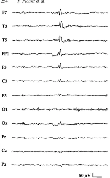
(6) 1252. F. Picard et al.. Table 1 Main clinical and electrical features observed in the different syndromes Sex ratio (M/F). Mean age at onset (years). Mean maximal seizure frequency. Patients with only SPS (no CPS) (%). Patients with mainly nocturnal seizures (%). PharmacoBehavioural resistance (%) disorders (%). ADNFLE (n ⫽ 29). 1.23. 8.2 ⫾ 11.3 Range 2 months to 56 years Median 5 years. 11/day. 42. 90. 29. 43. FTLE (n ⫽ 26). 1.17. 20.1 ⫾ 13.2 Range 1–54 years Median 15 years. 2/day. 42. 62. 9. 13. AD partial epilepsy with variable foci (n ⫽ 16). 1.28. 13.2 ⫾ 9.4 Range 2 months to 31 years Median 12 years. 3/day. 29. 62. 15. 23. All patients. 1.22. 13.6 ⫾ 12.6 Median 10 years. 5/day. 39. 75. 18. 27. M ⫽ male; F ⫽ female; SPS ⫽ simple partial seizures; CPS ⫽ complex partial seizures; AD ⫽ autosomal dominant.. symptoms. The side of the epileptic focus was homogeneous in two families (left temporal focus in the four affected individuals of FTLE family M, and right focus, rolandic, temporal or parietal, in the four affected individuals of family R). However, although the lateralization of the focus could not be determined in all patients in the other families, the distribution between left and right-sided foci within a given family seemed to be similar. Carbamazepine was the most effective antiepileptic medication. It completely suppressed seizures in 71% of treated ADNFLE patients (17 out of 24), 91% of FTLE patients (21 out of 23) and 85% of patients with familial partial epilepsy with variable foci (11 out of 13). The efficacy of valproate was low as seizures disappeared in 0 out of 3 ADNFLE patients, 2 out of 7 (29%) FTLE patients and 0 out of 3 patients with partial epilepsy with variable foci who were treated with this drug. In five patients in whom valproate was totally ineffective (two with ADNFLE and three with FTLE), the substitution of carbamazepine for valproate led to disappearance of the seizures. Pharmacoresistance to carbamazepine and other antiepileptic drugs was observed in 9–29% of the patients treated, depending on the syndrome (Table 1). Most of the families with ADNFLE contained one pharmacoresistant patient; however, in the other affected members a good response to the antiepileptic medication was observed. Whatever the syndrome, seizures tended to disappear with age (around the fourth or fifth decade), even in those patients who had been refractory to antiepileptic medication for several years.. Electrophysiology As shown in Table 3, interictal EEG abnormalities were frequently observed during the active phase of the epilepsy (when seizures occur at their maximal frequency). They. consisted of focal theta or delta slow waves and/or sharp waves or sparse spikes, at the same location when both types of abnormalities were present. The location of the interictal EEG abnormalities was concordant with the clinically presumed epileptic focus, except for three ADNFLE patients with clinically apparent frontal lobe seizures and EEG abnormalities in the temporal lobe (individuals G/II-1, H/III5, H/III-8). Four of the 40 patients who underwent a sleep EEG recording in addition to waking EEG presented EEG abnormalities only during sleep (3 out of 16 ADNFLE patients, 1 out of 15 FTLE patients, 0 out of 9 patients with partial epilepsy with variable foci). Interictal EEG abnormalities could still be detected ⬎2 years after the disappearance of the clinical seizures in 2 out of 6 (33%) FTLE patients and in 2 out of 9 (22%) patients with familial partial epilepsy with variable foci. To test the possibility that EEG abnormalities are present in asymptomatic gene carriers, EEGs were performed in 33 non-epileptic at-risk members from two families with FTLE (families J, N) and two families with partial epilepsy with variable foci (families P, R). They revealed significant abnormalities in 3 out of 16 (19%) individuals from FTLE families (J/III-5, III-6, N/II-2) and in 3 out of 17 (18%) individuals from families with partial epilepsy with variable foci (P/II-2, III-8, III-12). They consisted of focal epileptiform abnormalities, as shown in Fig. 2, sometimes accentuated by hyperpnoea (n ⫽ 3). These asymptomatic individuals with focal EEG abnormalities may be considered carriers, because (i) these kinds of EEG abnormalities are exceptional in the general population (estimated at ⬍0.2%) (Trojaborg, 1992), and (ii) the six individuals included two of the three asymptomatic obligate carriers in whom an EEG was carried out. Abnormalities were observed during a waking EEG recording for one obligate carrier but only during a sleep recording for the other (P/II-2). This suggests that sleep EEG,.
(7) Dominant partial epilepsies. 1253. Table 2 Seizure patterns in the different syndromes of autosomal partial epilepsy Symptoms. ADNFLE (n ⫽ 28). Autonomic Breathless feeling Flushing or pallor. FTLE (n ⫽ 26). 5 1. Psychic Dreamy perceptions Fear De´ ja` vu Forced thinking. 5 2. AD partial epilepsy with variable foci (n ⫽ 15). 2. 2 4 8 1. 1. Visual illusions, hallucinations. 4. 4. Auditory symptoms (buzzing, humming). 5. 2. Vertiginous. 1. 2 1. Somatosensory Epigastric Paraesthesia, pain. 2 4. Speech arrest. 1. Motor aphasia, verbal automatisms. 2. 3 4. 2. 1. 3. 2. Jargonphasia. 1. 2. Oroalimentary automatisms. 1. 4. 3. Gestural, ambulatory automatisms. 1. 7. 8. 20. 5. 4. 8. 1. 1. 3. 3. 1. 13. 14. 10. Motor Tonic, dystonic or clonic components Thrashing hyperkinetic attacks Vocalization Altered awareness AD ⫽ autosomal dominant.. Table 3 Type of interictal EEG abnormalities observed in the different syndromes Syndrome. Presence of interictal abnormalities. Focal slow waves. Focal spikes or sharp waves (unilateral/bilateral). ADNFLE (n ⫽ 25) FTLE (n ⫽ 23) AD partial epilepsy with variable foci (n ⫽ 14) All patients (n ⫽ 62). 19 (76%) 15 (65%) 12 (86%). 5 8 8. 19 (15/4) 13 (10/3) 8 (7/1). 46 (74%). 21. 40 (32/8). Sixty-two out of 71 affected individuals underwent an interictal EEG.. which was not performed in the third obligate carrier, is necessary to detect epileptiform abnormalities in asymptomatic gene carriers when waking EEG is normal. Three of the individuals with abnormal EEGs were. adolescents (J/III-5, III-6, P/III-12). The follow-up will reveal whether the electrical abnormalities precede the appearance of clinical seizures. Photosensitivity type 1 (posterior sharp waves after intermittent photic stimulation) was detected in.
(8) 1254. F. Picard et al. Intracerebral stereotaxic EEG recordings were performed in patient A/III-2, who suffered from a pharmacoresistant form of ADNFLE. His seizures were mainly nocturnal (he had up to 30 seizures per night). At the onset of the illness, his seizures were similar to those described by Scheffer and colleagues in ADNFLE patients (Scheffer et al., 1995a), consisting of a breathless sensation with fear followed by polypnoea and sudden movements of the body and upper limbs with dystonic postures. Their duration was ⬍1 min. Later, the ictal symptomatology changed and seizures consisted mainly of sustained right predominant bilateral hypertonia of the upper limbs with tonic anteflexion of the trunk, mandibular constriction, apnoea, and sometimes sialorrhoea. During more prolonged episodes, a throaty moaning occurred, and motor tonic manifestations were followed by violent and uncoordinated movements of the limbs with fear mimicry and gasping. Sometimes, an initial tingling sensation in the back was reported by the patient. There was no postictal deficit. Ictal surface EEG showed a diffuse arrest reaction or left frontoprecentral fast activity at the onset of seizures. Intra-cerebral EEG recordings, conducted with stereotactically implanted intracerebral electrodes (stereo EEG), demonstrated that seizures originated from the left operculo-insular cortex (Fig. 3). In addition the inaugural tingling could be reproduced by electrical stimulation of the insular cortex alone. The fact that the primary epileptogenic focus was localized in the operculo-insular area of the dominant hemisphere for language indicated that this patient could not be surgically treated.. Fig. 2 Isolated spike-and-waves predominant in the temporal region of the left hemisphere, recorded in a non-epileptic at-risk member of family P (P/III8). Monopolar derivations (recalculated against the average reference) covering the left hemisphere. No simultaneous right hemisphere recordings were obtained during this recording on paper. However, no discharge was noted in the midline electrodes.. a single individual (N/II-5). Given the high frequency of this electrical trait in the general population, we considered that it was not related to partial epilepsy. Ictal EEGs were recorded in 12 ADNFLE patients, five FTLE patients and five patients with familial partial epilepsy with variable foci, one of whom presented with frontal lobe epilepsy. Thirteen patients with frontal lobe seizures showed only artefacts (n ⫽ 1), diffuse flattening (n ⫽ 1), bifrontal abnormalities (n ⫽ 6) or unilateral frontal abnormalities (n ⫽ 5, with spreading towards the temporal lobe in two cases). These abnormalities consisted of either sharp waves/repetitive 8–11 Hz spikes (n ⫽ 9, with a recruiting pattern in one case) or rhythmic theta activity (n ⫽ 2). The nine extrafrontal lobe seizures were characterized by a classical focal recruiting pattern with increasing amplitude and decreasing frequency.. Neuropsychology and psychiatry Clinical neurological examination was normal in all patients. However, a detailed neuropsychological study performed in eight right-handed epileptic individuals from one ADNFLE family, two FTLE families and three families with partial epilepsy with variable foci showed some moderate specific disturbances in seven of them: (i) attention deficit and verbal perseverations in a frontal lobe epilepsy in the dominant hemisphere (individual A/III-2); (ii) difficulty in the Wisconsin Card Sorting Test and deficits in verbal fluency (figural fluency not tested) in a frontal lobe epilepsy of the non-dominant hemisphere (individual R/V-1); (iii) verbal memory and verbal fluency impairment in frontotemporal epilepsies in the dominant hemisphere (individuals J/III-4, N/III-3, P/II-3); (iv) verbal memory deficit in a temporal lobe epilepsy in the dominant hemisphere (individual Q/I-2); and (v) visuospatial memory impairment in a temporal lobe epilepsy in the non-dominant hemisphere (individual R/III-1). The study was performed during the active phase of the epilepsy only for individual A/III-2. Moreover, a history of psychiatric disturbance was found in 13–43% of the patients (Table 1). They were mostly observed during the active phase of their epilepsy. Strikingly, they were more frequent in ADNFLE. They.
(9) Dominant partial epilepsies consisted of personality and behavioural disorders, including irritability, aggressiveness, impulsive behaviour, with fugues during adolescence. Four individuals required transitory neuroleptic treatment. One patient with right temporal lobe epilepsy (patient R/III-1) presented such disorders associated with hypersexuality. This manifestation is a rare behavioural manifestation in temporal lobe epilepsy, and is considerably rarer than hyposexuality (Shukla et al., 1979). One patient (M/III-10) suffered from panic attacks and three patients (F/II-2, I/II-2, S/IV-9) from depression.. Neuroimaging Neuroimaging was available in 50 patients: 16 ADNFLE patients (14 MRI brain scans and two CT scans), 21 FTLE patients (15 MRI and six CT scans) and 13 patients with partial epilepsy with variable foci (nine MRI and four CT scans). Two patients belonging to the same FTLE family (J/I-1, II-3) had a discrete cortical atrophy with predominance in the left insular and opercular area, one ADNFLE patient (G/II-2) had a left frontal atrophy. These anatomical changes seemed to correspond to the suspected location of their epileptic focus. Other, minor changes were small high-signal areas in the right basal ganglia on T2-weighted images (patient L/II-1) and pineal cyst (patients O/III-8, III-9). No hippocampal sclerosis was observed. An ictal SPECT (single photon emission computed tomography) using 99mTc-ethyl cysteinate dimer (ECD) was performed in one of the ADNFLE patients (A/III-2). It showed a left frontal hyperperfusion that correlated with the right predominance of his ictal motor symptoms.. Clinical genetics Families with a non-lesional partial epilepsy and a probable dominant mode of inheritance were investigated in our study. No family had consanguinity. Male-to-male transmission occurred in seven families, which excluded X-linked inheritance. The male : female ratio ranged from 1.17 to 1.28 depending on the syndrome of familial partial epilepsy (Table 1). Clinical penetrance reached 0.72 in the eight families with ADNFLE. Asymptomatic obligate carriers were observed in three FTLE families (M, N, O) and in three families with autosomal dominant partial epilepsy with variable foci (P, R, S). Penetrance was 0.54 in the seven families with FTLE and 0.44 in the four families with epilepsy with variable foci. The last value is low because of family P, which contains many non-epileptic at-risk individuals. Penetrance increased with age. The distribution of age at onset was variable among the three types of familial partial epilepsy: mean age at onset was 8.2 years (ranging from 2 months to 56 years) in ADNFLE, 20 years (1–54 years) in FTLE and 13.2 years (from 2 months to 31 years) in familial partial epilepsy with variable foci (Fig. 4). There were significant differences between. 1255. ADNFLE and FTLE (P ⬍ 0.0001) and between ADNFLE and partial epilepsy with variable foci (P ⬍ 0.05), but not between FTLE and partial epilepsy with variable foci (P ⫽ 0.11). Expressivity of the different syndromes was variable, with different ages at onset, ictal symptoms and courses of the disease, even within families.. Molecular genetics Linkage analysis Two-point and multipoint linkage analyses using seven microsatellite markers from the 10q22–q24 region were performed in the 10 most informative families (families A, C, D, H, J, L, M, N, P, R). None of these families was informative enough to establish linkage but they could be used for exclusion of a given locus. For five families (C, H, J, M, N), two-point lod scores were negative at θ ⫽ 0.00 for all markers of the interval, and multipoint lod scores were below the threshold value of –2 in the entire candidate interval, excluding linkage with this locus (data not shown). For family R, the lod scores were negative but did not reach the threshold of –2 in the interval D10S185–D10S1265 to exclude the locus. For families A, D, L and P, haplotype reconstruction showed cosegregation of the disease with markers in 10q, and positive but not statistically significant lod scores were obtained in the entire candidate interval D10S583–D10S1663 (data not shown). For these four families, linkage to the tested locus can be neither established nor excluded. The CHRNA7 region was studied by linkage analysis with markers located in 15q14 in the same 10 families mentioned above. For six families (A, H, M, N, P, R), lod scores were negative and below the threshold value of –2 at θ ⫽ 0.00 by two-point analysis for all markers tested. Exclusion of the candidate region was confirmed using multipoint analysis. For families J and L, parts of the candidate region, the 11.4 cM D15S165–D15S971 interval and the 6.9 cM D15S144–D15S118 interval, respectively, were not excluded, probably because of the low level of informativeness of the markers. For ADNFLE families C and D, lod scores were slightly positive for the 5.1 cM interval D15S165–D15S144 by two-point and multipoint analyses, but did not reach the significance threshold of ⫹3.. Search for mutation in the CHRNA4 and CHRNB2 genes In the index cases of 17 families (all families except families K and S), the search for the S248F mutation in the CHRNA4 gene was negative. The entire coding sequence of CHRNA4 was explored by non-radioactive SSCP in 16 families. Band-shifts were observed for exon 1 (in families I, M, O), intron 2 (in families E, L, P), intron 3 (in families A, E, Q, R) and exons 5b (in families A, E, P).
(10) 1256. F. Picard et al.. and 5c (in family L), but did not cosegregate with the disease and were present in relatives who were not at risk. However, PCR products that migrated abnormally. were sequenced directly. Five different base changes, which were all silent, were identified in the CHRNA4 gene (Table 4). Polymorphisms in introns 2 and 3 and in exons 5b.
(11) Dominant partial epilepsies. 1257. Fig. 3 Stereo EEG recorded a partial seizure of left operculo-insular origin in a 12-year-old righthanded boy suffering from an ADNFLE (individual A/III-2). The stereo EEG trace shows that a fast activity occurs within the central operculum (first arrow) at the clinical onset of the fit, involving the insular cortex 3 s later (second arrow). These changes are visible partly in the central cingulate gyrus and in the supplementary motor area (SMA). When the tonic motor signs occur, the fast activity becomes more evident, increasing in amplitude within the insular cortex, and is followed by a fast spike discharge within the central operculum. Only the most relevant recording sites are shown. Letters in brackets refer to the recording electrodes. The intracerebral placement of the electrodes is represented on the stereotaxic scheme (left lateral and frontal view of the skull). Bipolar stereo EEG recordings were performed between contiguous contacts of each electrode, at different levels along the electrode axis. Thus, various mesial and lateral cortical areas were evaluated, including the sulcal cortex. The insular cortex was explored by the deepest contacts of the electrode Y⬘.. and 5c modified restriction sites, allowing rapid analysis by PCR–restriction enzyme digestion of the genotypes in each family.. In the index cases of 16 families (all except F, K and S), we searched for mutations in the six coding exons and the flanking exon–intron boundaries of the CHRNB2 gene..
(12) 1258. F. Picard et al. Table 4 Polymorphisms observed in the CHRNA4 gene in the families tested in the present study Position. Base change. Amino acid. Exon 1 Intron 2 Intron 3 Exon 5b Exon 5c. G51→A IVS2⫹20C→G IVS3–26G→A C555→T T594→C. Leu17Leu Asp185Asp Cys198Cys. Restriction site. Heterozygosity (%). NlaIII Sau96.I HgaI CfoI. 16 18 11 28 18. IVS2 corresponds to intron 2 and IVS3 to intron 3. The percentage of heterozygosity was estimated from the DNA of 50 control subjects.. Fig. 4 Cumulative curves for ages at onset for the three syndromes of familial partial epilepsy. VF ⫽ autosomal dominant partial epilepsy with variable foci.. Direct sequencing of the corresponding PCR fragments failed to detect any base change.. Discussion Dominant partial epilepsies can be subdivided into three groups: the frontal form (ADNFLE), the temporal form (FTLE) and the form with variable foci (Scheffer et al., 1995a, b; Berkovic et al., 1996; Scheffer et al., 1998). Forty ADNFLE families, 20 temporal lobe epilepsy families without history of familial febrile convulsions, and one family with autosomal dominant partial epilepsy with variable foci have been reported to date (Ottman et al., 1995; Scheffer et al., 1995a, b; Berkovic et al., 1996; Oldani et al., 1996, 1998; Magnusson et al., 1996; Cendes et al., 1998; Phillips et al., 1998; Scheffer et al., 1998; Hirose et al., 1999; Poza et al., 1999; Saenz et al., 1999). The present paper raises the number of reported families with non-lesional dominant partial epilepsy to 80, and confirms the existence of other families with autosomal dominant partial epilepsy with variable foci. In contrast to the previous studies cited above, which selected homogeneous families and led to the identification of the different syndromes, the present study analysed a heterogeneous group of familial partial epilepsies, similar to the population encountered in clinical practice. This allinclusive approach resulted in difficulty in classification for. five families within the current nosology. The classification of a family depends on the definition of the seizure location in each affected member, which is usually based only on the ictal clinical symptoms and on surface EEG data, and may therefore be approximate. From a clinical point of view, commonly reported auras (for instance fear, de´ ja` vu, auditory symptoms and epigastric discomfort) have been described in both the ADNFLE and the FTLE syndrome (Scheffer et al., 1995a; Berkovic et al., 1996). However, although there is still debate about the localizing value of auras, most authors agree that de´ ja` vu and auditory symptoms indicate temporal lobe dysfunction (Penfield and Jasper, 1954; Gloor et al., 1982; Bancaud et al., 1994). Their description as auras in ADNFLE is therefore surprising. In addition, some classical features of frontal lobe seizures may be observed in temporal lobe seizures: (i) nocturnal occurrence; (ii) occurrence in clusters; and (iii) brief duration. In this respect we have noticed unusually frequent nocturnal occurrence of temporal lobe seizures in our FTLE families. From an electrophysiological point of view, it is known that scalp EEG, even if ictal, may be misleading (Rektor et al., 1997). In some patients who present non-localizing ictal symptoms and unilateral EEG abnormalities, in both the frontal and the temporal areas, the term ‘frontotemporal’ epilepsy should be considered, particularly because an epileptogenic focus is not necessarily limited by the anatomical borders that define the cerebral lobes (Munari et al., 1995). The difficulty of classification of the familial partial epilepsies is supported by the description of some members with probable temporal lobe seizures (Magnusson et al., 1996; Nakken et al., 1999) in a Norwegian ADNFLE family carrying a mutation in the nAChR α4 subunit (Steinlein et al., 1997a). A further demonstration of this problem was our observation in one ADNFLE patient (A/III-2) of a discrepancy between the results of the non-invasive examinations and the intracranial depth recordings. In this patient, the ictal semiology, ictal SPECT and EEG suggested a frontal lobe epilepsy of mesial midfrontal or frontopolar origin, as described by Hayman and colleagues in ADNFLE patients (Hayman et al., 1997). However, the intracerebral exploration found a primary focus in the central opercular area and insular cortex. It is possible that the current.
(13) Dominant partial epilepsies classification of familial partial epilepsies fails in some cases because variable patterns of seizure propagation from junction areas, such as the insular cortex, may lead to more ‘temporal’ or more ‘frontal’ symptoms. In conclusion, the clinical spectra of the different syndromes of autosomal dominant partial epilepsy are not clear-cut, but present overlaps. The diagnosis of familial forms of partial epilepsy is of clinical importance. First, carbamazepine is much more effective than valproate in these types of epilepsy, as reported by Scheffer and colleagues in ADNFLE (Scheffer et al., 1995a). In relation to the high sensitivity of familial partial epilepsies to carbamazepine, it is interesting to note that α4β2 nicotinic receptors with an ADNFLE mutation, reconstituted in Xenopus oocytes, are more sensitive to this drug than normal receptors, and are readily inhibited at pharmacological concentrations (Picard et al., 1999). Secondly, we observed less pharmacoresistance in dominant partial epilepsies (18%) than has been described in classical cryptogenic partial epilepsies (30% according to Schmidt, 1991; Bourgeois, 1994; 55% according to Semah et al., 1998, who used the criterion of 1 year without seizure for ‘seizure control’). However, it reached 30% in our ADNFLE subgroup, indicating that dominant partial epilepsies do not have a consistently favourable outcome and that surgical treatment can sometimes be considered. In other respects, many patients presented transitory behavioural disturbances in their history, and the detailed neuropsychological evaluation that was performed in some patients showed disturbances that usually appeared congruent with the presumed location of their epileptogenic focus. This emphasizes that non-lesional autosomal dominant partial epilepsies have wider consequences than were initially presumed. The 19 families we analysed showed wide interfamilial and intrafamilial clinical variability. The ictal symptoms varied greatly from one individual to another within a family, which is concordant with the ‘absence of obvious intrafamily clustering of simple partial seizure symptomatology’ reported by Berkovic and colleagues in FTLE families (Berkovic et al., 1996). Recently, Hayman and colleagues analysed ictal video–EEG recordings and functional neuroimaging in two pairs of patients from unrelated ADNFLE families and showed that the frontal lobe foci were in variable locations (Hayman et al., 1997). Because of the diversity of ictal symptoms and the types and locations of EEG abnormalities, the dominant partial epilepsies are very similar to the socalled cryptogenic focal epilepsies, except perhaps for the high frequency of nocturnal attacks. The tendency of seizures to disappear with age that we observed in our families is also observed in non-familial partial epilepsies, since approximately half of the patients with non-familial intractable epilepsy become seizure-free over the years (Juul-Jensen, 1986). Consequently, the role of genetic factors has to be examined in apparently isolated cases of cryptogenic focal epilepsy. The reduced penetrance of the dominant partial epilepsies reinforces the hypothesis that gene mutations. 1259. can account for some sporadic cases. As we have demonstrated the presence of epileptiform focal EEG abnormalities in asymptomatic family members, including obligate carriers, we suspect that such mutations can be expressed in a subclinical form. Confirmation of this must await the identification of the genes responsible in the corresponding families. We screened all the families for all the candidate loci tested in the present study, namely the α4, β2 and α7 subunit genes and the locus on chromosome 10q [implicated in a family with autosomal dominant partial epilepsy with auditory symptoms (Ottman et al., 1995)]. So far, the only gene implicated in an autosomal dominant partial epilepsy syndrome is the CHRNA4 gene, coding for the neuronal nAChR α4 subunit. This gene was studied by the direct detection of mutation in the coding region by SSCP. We detected the previously described polymorphisms CfoI, Sau96.I and HgaI/FokI (Steinlein et al., 1995, 1997b; Guipponi et al., 1997) as well as new polymorphisms in exon 1 and in intron 2, which do not correspond to an amino acid change. Their identification may be useful for future linkage studies. The failure to detect mutation in CHRNA4 in our families confirms the low frequency of mutations in this gene in autosomal dominant partial epilepsies, as previously reported in ADNFLE (Steinlein et al., 1995; Phillips et al., 1998). In total, only four families, all presenting with ADNFLE, were linked to the α4 subunit gene (Steinlein et al., 1995, 1997a; Hirose et al., 1999; Saenz et al., 1999). The genes encoding other subunits of the neuronal nAChR are promising candidates in all other families with ADNFLE or in families with other forms of autosomal dominant partial epilepsy, especially the β2 subunit, which usually assembles with α4. In this study, its entire coding sequence was analysed by direct sequencing. No mutations were identified in 16 index cases. This confirms that the CHRNB2 gene does not represent a major locus for ADNFLE, in agreement with Rempel and colleagues (Rempel et al., 1998), and further shows that it is not a major locus for the other syndromes of familial partial epilepsy. It has been proposed that the CHRNA7 gene, coding for the α7 subunit, another widely expressed nAChR subunit (Gotti et al., 1997), represents a major susceptibility locus for juvenile myoclonic epilepsy (Elmslie et al., 1997), and benign epilepsy of childhood with centrotemporal spikes has been linked to markers on chromosome 15q14 in the vicinity of the CHRNA7 gene (Neubauer et al., 1998). We performed linkage analyses in 10 families informative enough for exclusion testing of loci. This locus was excluded in six pedigrees. In the four remaining families in which linkage to the CHRNA7 gene was not excluded, a search for mutations in this gene will be important when its genomic structure has been fully determined. Lastly, the linkage study of the 10q22–q24 candidate region revealed positive but not significant lod scores for four of the 10 tested families, two with ADNFLE (A, D), one with FTLE (L) and one with partial epilepsy with variable.
(14) 1260. F. Picard et al.. foci (P). In the future, the identification of candidate genes in the 10q region will permit their analysis in these families, especially in families L and P, in each of which there is one affected individual having seizures with auditory hallucinations. This genetic study demonstrates that previously implicated genes and loci are rarely involved in families with familial partial epilepsy, reflecting the underlying genetic heterogeneity. Combination of genetic mapping in large informative families and candidate gene approaches (involving genes encoding ion channels or their regulatory subunits) will contribute to the identification of additional causative genes. As for other inherited complex neurological disorders, a more appropriate classification of the familial partial epilepsies will be possible only when their molecular basis has been elucidated.. Acknowledgements We thank Drs H. De Grissac (Clinique de Chateaulin, France), M. Gavaret (Centre Saint Paul, Marseille, France) and M. Wolff (Universita¨ t Kinderklinik, Abt. Neuropa¨ diatrie, Tu¨ bingen, Germany), who each referred one of the families, C. Penet, Y. Pothin, J. Bou and A. Camuzat for technical support, Drs M. Morris and M. Seeck for helpful suggestions and Dr L. Curtis for critical reading of the manuscript. We are grateful to the PHRC Strasbourg 1996 UF 9617, the ARGE (Association pour la Recherche sur la Ge´ ne´ tique des Epilepsies; Professor O. Dulac) and the Association Franc¸ aise contre les Myopathies for financial support.. References Bancaud J, Brunet-Bourgin F, Chauvel P, Halgren E. Anatomical origin of de´ ja` vu and vivid ‘memories’ in human temporal lobe epilepsy. Brain 1994; 117: 71–90. Berkovic SF, Howell RA, Hopper JL. Familial temporal lobe epilepsy: a new syndrome with adolescent/adult onset and a benign course. In: Wolf P, editor. Epileptic seizures and syndromes. London: John Libbey; 1994. p. 257–63. Berkovic SF, McIntosh A, Howell RA, Mitchell A, Sheffield LJ, Hopper JL. Familial temporal lobe epilepsy: a common disorder identified in twins. Ann Neurol 1996; 40: 227–35. Bourgeois BFD. Establishment of pharmacoresistance. In: Wolf P, editor. Epileptic seizures and syndromes. London: John Libbey; 1994. p. 591–7. Cendes F, Lopes-Cendes I, Andermann E, Andermann F. Familial temporal lobe epilepsy: a clinically heterogeneous syndrome. Neurology 1998; 50: 554–7. Elmslie FV, Rees M, Williamson MP, Kerr M, Kjeldsen MJ, Pang KA, et al. Genetic mapping of a major susceptibility locus for juvenile myoclonic epilepsy on chromosome 15q. Hum Mol Genet 1997; 6: 1329–34. Freedman R, Coon H, Myles-Worsley M, Orr-Urtreger A, Olincy A, Davis A, et al. Linkage of a neurophysiological deficit in. schizophrenia to a chromosome 15 locus. Proc Natl Acad Sci USA 1997; 94: 587–92. Gloor P, Olivier A, Quesney LF, Andermann F, Horowitz S. The role of the limbic system in experiential phenomena of temporal lobe epilepsy. Ann Neurol 1982; 12: 129–44. Gotti C, Fornasari D, Clementi F. Human neuronal nicotinic receptors. [Review]. Prog Neurobiol 1997; 53: 199–237. Guipponi M, Baldy-Moulinier M, Malafosse A. A fok1 polymorphism in the human neuronal nicotinic acetylcholine receptor alpha 4 subunit gene. Clin Genet 1997; 51: 78–9. Gyapay G, Morissette J, Vignal A, Dib C, Fizames C, Millasseau P, et al. The 1993–94 Genethon human genetic linkage map. Nat Genet 1994; 7: 246–339. Hayman M, Scheffer IE, Chinvarun Y, Berlangieri SU, Berkovic SF. Autosomal dominant nocturnal frontal lobe epilepsy: demonstration of focal frontal onset and intrafamilial variation. Neurology 1997; 49: 969–75. Hirose S, Iwata H, Akiyoshi H, Kobayashi K, Ito M, Wada K, et al. A novel mutation of CHRNA4 responsible for autosomal dominant nocturnal frontal lobe epilepsy. Neurology 1999; 53: 1749–53. Johnson WG, Kugler SL, Stenroos ES, Meulener MC, Rangwalla I, Johnson TW, et al. Pedigree analysis in families with febrile seizures. Am J Med Genet 1996; 61: 345–52. Juul-Jensen P. Epidemiology of intractable epilepsy. In: Schmidt D, Morselli PL, editors. Intractable epilepsy. New York: Raven Press; 1986. p. 5–11. Lathrop GM, Lalouel JM, Julier C, Ott J. Multilocus linkage analysis in humans: detection of linkage and estimation of recombination. Am J Hum Genet 1985; 37: 482–98. Magnusson A, Nakken KO, Brubakk E. Autosomal dominant frontal lobe epilepsy [letter]. Lancet 1996; 347: 1191–2. Munari C, Francione S, Kahane P, Hoffmann D, Tassi L, Lo Russo G, et al. Multilobar resections for the control of epilepsy. In: Schmidek HH, Sweet WJ, editors. Operative neurosurgical techniques, Vol. 2. 3rd ed. Philadelphia: W.B. Saunders; 1995. p. 1323–39. Nakken KO, Magnusson A, Steinlein OK. Autosomal dominant nocturnal frontal lobe epilepsy: an electroclinical study of a Norwegian family with ten affected members. Epilepsia 1999; 40: 88–92. Neubauer BA, Fiedler B, Himmelein B, Ka¨ mpfer F, La¨ ssker U, Schwabe G, et al. Centrotemporal spikes in families with rolandic epilepsy: linkage to chromosome 15q14. Neurology 1998; 51: 1608–12. Oldani A, Zucconi M, Ferini-Strambi L, Bizzozero D, Smirne S. Autosomal dominant nocturnal frontal lobe epilepsy: electroclinical picture. Epilepsia 1996; 37: 964–76. Oldani A, Zucconi M, Asselta R, Modugno M, Bonati MT, Dalpra` L, et al. Autosomal dominant nocturnal frontal lobe epilepsy. A video-polysomnographic and genetic appraisal of 40 patients and delineation of the epileptic syndrome. Brain 1998; 121: 205–23..
(15) Dominant partial epilepsies Ottman R, Risch N, Hauser WA, Pedley TA, Lee JH, BarkerCummings C, et al. Localization of a gene for partial epilepsy to chromosome 10q. Nature Genet 1995; 10: 56–60. Penfield W, Jasper H. Epilepsy and the functional anatomy of the human brain. Boston: Little, Brown; 1954. Phillips HA, Scheffer IE, Berkovic SF, Hollway GE, Sutherland GR, Mulley JC. Localization of a gene for autosomal dominant nocturnal frontal lobe epilepsy to chromosome 20q13.2 [letter]. Nat Genet 1995; 10: 117–18. Phillips HA, Scheffer IE, Crossland KM, Bhatia KP, Fish DR, Marsden CD, et al. Autosomal dominant nocturnal frontal lobe epilepsy: genetic heterogeneity and evidence for a second locus at 15q24. Am J Hum Genet 1998; 63: 1108–16. Picard F, Bertrand S, Steinlein OK, Bertrand D. Mutated nicotinic receptors responsible for autosomal dominant nocturnal frontal lobe epilepsy are more sensitive to carbamazepine. Epilepsia 1999; 40: 1198–209. Poza JJ, Saenz A, Martinez-Gil A, Cheron N, Cobo AM, Urtasun M, et al. Autosomal dominant lateral temporal epilepsy: clinical and genetic study of a large Basque pedigree linked to chromosome 10q. Ann Neurol 1999; 45: 182–8. Rektor I, Svejdova M, Kanovsky P, Landre´ E, Bancaud J, Lamarche M. Can epileptologists without access to intracranial EEG use reliably the International League Against Epilepsy classification of the localization-related epileptic syndromes? J Clin Neurophysiol 1997; 14: 250–4. Rempel N, Heyers S, Engels H, Sleegers E, Steinlein OK. The structures of the human neuronal nicotinic acetylcholine receptor β2- and α3-subunit genes (CHRNB2 and CHRNA3). Hum Genet 1998; 103: 645–53. Rouger H, LeGuern E, Birouk N, Gouider R, Tardieu S, Plassart E, et al. Charcot–Marie–Tooth disease with intermediate motor nerve conduction velocities: characterization of 14 Cx32 mutations in 35 families. Hum Mutat 1997; 10: 443–52. Saenz A, Galan J, Caloustian C, Lorenzo F, Marquez C, Rodriguez N, et al. Autosomal dominant nocturnal frontal lobe epilepsy in a Spanish family with a Ser252Phe mutation in the CHRNA4 gene. Arch Neurol 1999; 56: 1004–9. Scha¨ ffer AA, Gupta SK, Shriram K, Cottingham RW Jr. Avoiding recomputation in linkage analysis. Hum Hered 1994; 44: 225–37. Scheffer IE, Bhatia KP, Lopes-Cendes I, Fish DR, Marsden CD, Andermann E, et al. Autosomal dominant nocturnal frontal lobe epilepsy. A distinctive clinical disorder. Brain 1995a; 118: 61–73. Scheffer IE, Phillips H, Mulley J, Sutherland G, Harvey AS, Hopkins IJ, et al. Autosomal dominant partial epilepsy with variable. 1261. foci is not allelic with autosomal dominant nocturnal frontal lobe epilepsy [abstract]. Epilepsia 1995b; 36 Suppl 3: S28. Scheffer IE, Phillips HA, O’Brien CE, Saling MM, Wrennall JA, Wallace RH, et al. Familial partial epilepsy with variable foci: a new partial epilepsy syndrome with suggestion of linkage to chromosome 2. Ann Neurol 1998; 44: 890–9. Schmidt D. Medical intractability in partial epilepsies. In: Lu¨ ders HO, editor. Epilepsy surgery. New York: Raven Press; 1991. p. 83–91. Semah F, Picot MC, Adam C, Broglin D, Arzimanoglou A, Bazin B, et al. Is the underlying cause of epilepsy a major prognostic factor for recurrence? Neurology 1998; 51: 1256–62. Shukla GD, Srivastava ON, Katiyar BC. Sexual disturbances in temporal lobe epilepsy: a controlled study. Br J Psychiatry 1979; 134: 288–92. Steinlein OK, Mulley JC, Propping P, Wallace RH, Phillips HA, Sutherland GR, et al. A missense mutation in the neuronal nicotinic acetylcholine receptor α4 subunit is associated with autosomal dominant nocturnal frontal lobe epilepsy. Nat Genet 1995; 11: 201–3. Steinlein O, Weiland S, Stoodt J, Propping P. Exon-intron structure of the human neuronal nicotinic acetylcholine receptor α4 subunit (CHRNA4). Genomics 1996; 32: 289–94. Steinlein OK, Magnusson A, Stoodt J, Bertrand S, Weiland S, Berkovic SF, et al. An insertion mutation of the CHRNA4 gene in a family with autosomal dominant nocturnal frontal lobe epilepsy. Hum Mol Genet 1997a; 6: 943–7. Steinlein OK, Sander T, Stoodt J, Kretz R, Janz D, Propping P. Possible association of a silent polymorphism in the neuronal nicotinic acetylcholine receptor subunit alpha4 with common idiopathic generalized epilepsies. Am J Med Genet 1997b; 74: 445–9. Stevanin G, Le Guern E, Ravise N, Chneiweiss H, Du¨ rr A, Cancel G, et al. A third locus for autosomal dominant cerebellar ataxia type I maps to chromosome 14q 24.3–qter: evidence for the existence of a fourth locus. Am J Hum Genet 1994; 54: 11–20. Thomas P, Picard F, Hirsch E, Chatel M, Marescaux C. Epilepsie frontale nocturne autosomique dominante. Rev Neurol (Paris) 1998; 154: 228–35. Trojaborg W. EEG abnormalities in 5,893 jet pilot applicants registered in a 20-year period. Clin Electroencephalogr 1992; 23: 72–8. Vigevano F, Fusco L. Hypnic tonic postural seizures in healthy children provide evidence for a partial epileptic syndrome of frontal lobe origin. Epilepsia 1993; 34: 110–19. Received April 15, 1999. Revised August 25, 1999. Accepted January 24, 2000.
(16) 1262. F. Picard et al.. Appendix Family J The location of the seizures was difficult to define in individual III4, whose seizures consisted of a thoracic sensation of fear followed a few seconds later by loss of consciousness and tonic posturing of the limbs, lasting ⬍1 min in total. Interictal EEGs revealed spikes in the left anterior region and ictal EEGs showed spikes in the left and right anterior areas with left predominance. The two other affected relatives suffered from typical temporal lobe seizures and had the same aura as individual III-4. We assumed individual III-4 may have temporal lobe seizures with rapid frontal propagation.. Family N Three members (III-3, III-4 and III-6) had typical temporal lobe seizures while two others (II-4 and III-7) had essentially hyperkinetic motor symptoms. However, individual III-7 had an aura consisting of bilateral paraesthesia of a pleasant nature in the genital area, which may have had a mesiotemporal origin, although the second. sensory area and the orbitofrontal area were not excluded. Interictal EEG abnormalities in the two individuals with motor symptoms consisted of right temporal and central abnormalities (patient III7), and left posterior temporal abnormalities during waking and sleep recordings (patient II-4). We classified the family as presenting FTLE, although a diagnosis of autosomal dominant partial epilepsy with variable foci is also possible.. Family O The index case (III-8) had seizures lasting ~90 s, consisting of a brief strange feeling and fear, followed by deviation of the head and eyes associated with tonic posturing of the upper limbs. While ictal EEGs showed right frontotemporal spikes, interictal EEG displayed slow waves and sharp waves in the right temporal area. Her brother (III-9) also presented seizures with head deviation, and obnubilation, which were preceded by a feeling of de´ ja` vu. They lasted several minutes. His interictal EEG showed left temporal slow waves. We assumed temporal lobe seizures in individual III9, but a diagnosis of ‘temporofrontal’ epilepsy would seem more appropriate in individual III-8..
(17)
Figure


+3


Documents relatifs
In this paper, we extend the uniqueness theorem for meromorphic mappings to the case where the family of hyperplanes depends on the meromorphic mapping and where the
a) - The whole set of experimental measurements on both the thin unsupported layers and the tiles is coherent with the modeling of a glaze containing two families of
D r. Sulaimon Giwa, Assistant Professor in the School of Social Work at Memorial University, broke down a common miscon- ception that not all families on the move are
(“I think that the Guidance Counselors in the schools need to have a solid education on diabetes for sure, because it doesn’t happen. I mean, like this year’s difficult because
It is found that the MLE and the classical Rao's score test can be misleading in the presence of model misspecification which in the context of logistic regression means either
In this study, a prototype of Etest® KPC (bioMérieux, La Balme-les-Grottes, France), containing meropenem and boronic acid, and two commercially available Etest® MBL strips
— We can find a finite morphism S" —>• S of an integral scheme 6" to S', and a finite alteration X' —> X with purely inseparable function field extension such that X'
The main result of [6] deals with Thue equations twisted by a set of units which is not supposed to be a group of rank 1, but it involves an assumption (namely that at least two of
