Respiratory dysfunction by AFG3L2 deficiency
causes decreased mitochondrial calcium uptake
via organellar network fragmentation
Francesca Maltecca
1, Diego De Stefani
2, Laura Cassina
1,4, Francesco Consolato
1,5,
Michal Wasilewski
3, Luca Scorrano
3, Rosario Rizzuto
2and Giorgio Casari
1,∗1
San Raffaele Scientific Institute, Vita-Salute San Raffaele University and Center for Translational Genomics and Bioinformatics, Milan-I, Italy,2Department of Biomedical Sciences, University of Padova, Padova-I,
Italy, 3Department of Cell Physiology and Metabolism, University of Geneva Medical School, Gene`ve-CH, Switzerland and Dulbecco-Telethon Institute, Padova-I, Italy,4Department of Genetics, Biology and Biochemistry, University of Turin, Turin-I, Italy,5PhD school of Neurobiology, University of Insubria, Varese-I, Italy
Received April 27, 2012; Revised and Accepted May 29, 2012
The mitochondrial protein AFG3L2 forms homo-oligomeric and hetero-oligomeric complexes with paraplegin in the inner mitochondrial membrane, named m-AAA proteases. These complexes are in charge of quality control of misfolded proteins and participate in the regulation of OPA1 proteolytic cleavage, required for mitochondrial fusion. Mutations in AFG3L2 cause spinocerebellar ataxia type 28 and a complex neurodegen-erative syndrome of childhood. In this study, we demonstrated that the loss of AFG3L2 in mouse embryonic fibroblasts (MEFs) reduces mitochondrial Ca21uptake capacity. This defect is neither a consequence of global alteration in cellular Ca21homeostasis nor of the reduced driving force for Ca21 internalization within mitochondria, since cytosolic Ca21transients and mitochondrial membrane potential remain unaffect-ed. Moreover, experiments in permeabilized cells revealed unaltered mitochondrial Ca21uptake speed in Afg3l22/2 cells, indicating the presence of functional Ca21uptake machinery. Our results show that the de-fective Ca21handling in Afg3l22/2cells is caused by fragmentation of the mitochondrial network, secondary to respiratory dysfunction and the consequent processing of OPA1. This leaves a number of mitochondria devoid of connections to the ER and thus without Ca21elevations, hampering the proper Ca21 diffusion along the mitochondrial network. The recovery of mitochondrial fragmentation in Afg3l22/2MEFs by overex-pression of OPA1 rescues the impaired mitochondrial Ca21buffering, but fails to restore respiration. By link-ing mitochondrial morphology and Ca21 homeostasis, these findings shed new light in the molecular mechanisms underlining neurodegeneration caused by AFG3L2 mutations.
INTRODUCTION
Mitochondria are interconnected dynamic organelles central to energy production, fatty acid metabolism and Ca2+buffering. Dysfunction of mitochondria causes oxidative stress, per-turbed cellular Ca2+ homeostasis and cell death and is linked to aging and neurodegeneration.
Several surveillance mechanisms have evolved to prevent the accumulation of dysfunctional mitochondria, such as the continuous remodelling of the mitochondrial network and mitophagy (1). At the organellar level, highly conserved pro-teases conduct protein quality control (2). Among them, m-AAA proteases belong to the AAA-protein superfamily (ATPases associated with a variety of cellular activities) and
∗To whom correspondence should be addressed at: Vita-Salute San Raffaele University & Center for Translational Genomics and Bioinformatics,
San Raffaele Scientific Institute, Via Olgettina 58, Milan, Italy. Tel:+39 0226433502; Fax: +39 0226436352; Email: [email protected]
#The Author 2012. Published by Oxford University Press.
This is an Open Access article distributed under the terms of the Creative Commons Attribution Non-Commercial License (http://creativecommons.org/ licenses/by-nc/2.5), which permits unrestricted non-commercial use, distribution, and reproduction in any medium, provided the original work is prop-erly cited.
Human Molecular Genetics, 2012, Vol. 21, No. 17 3858–3870 doi:10.1093/hmg/dds214
are present as AFG3L2 homo-oligomers or paraplegin-AFG3L2 hetero-oligomers in the inner mitochon-drial membrane. The m-AAA complexes mediate the selective degradation of non-assembled and damaged proteins (2) and exert chaperone-like activity on respiratory chain complexes (3–5). In addition, they perform proteolytic cleavage of sub-strates, which are the nuclear-encoded subunit of mitochon-drial ribosomes MrpL32 and the regulator of mitochonmitochon-drial fusion OPA1 (6,7). Both paraplegin and AFG3L2 have been linked to neurodegenerative disorders: mutations in the paraplegin-coding gene cause a recessive form of hereditary spastic paraplegia (8), while heterozygous and homozygous mutations in AFG3L2 have been associated to spinocerebellar ataxia type 28 (SCA28) (9–11) and to a novel progressive myoclonic epilepsy-ataxia-polyneuropathy syndrome of child-hood, respectively (12). Whether these diverse diseases reflect tissue-specific expression level of paraplegin and AFG3L2 (13) or different substrate specificity of the two proteases remain to be clarified.
We provided evidence that Afg3l22/2 mice resemble patients carrying homozygous mutations, showing a severe neurological syndrome that leads to lethality at P16 (5). Ac-cordingly, Afg3l2 haploinsufficient mice recapitulate most fea-tures of SCA28 patients, displaying defects in motor coordination and balance due to dark degeneration of Purkinje cells (PC-DCD) (14). The latter is a phenomenon documented in other SCAs as secondary to excitotoxicity and high levels of intracellular Ca2+, which cause calpain-mediated cytoskeletal breakdown (15,16). Peculiarly, in the SCA28 model, PC-DCD originates from mitochondrial dysfunction, as demonstrated by the fact that alterations in mitochondrial morphology and me-tabolism precede PC degeneration (14).
We hypothesize that Afg3l2-depleted mitochondria have de-fective Ca2+ buffering capacity, leading to dysregulation of cytosolic Ca2+ homeostasis and finally to PC-DCD. Indeed, compelling evidence in the literature points at the central role of mitochondrial Ca2+buffering in shaping the amplitude and the spatio-temporal dimension of intracellular Ca2+waves (17,18). This is particularly crucial for neurons, and especially for PCs, which uniquely receive glutamatergic afferents and experience large and sudden Ca2+influx (19).
Mitochondria take up Ca2+ rapidly from the cytoplasm during agonist-induced Ca2+ signals, thanks to the strong driving force ensured by the mitochondrial membrane poten-tial (Dcm) via a low-affinity Ca
2+channel of the inner
mem-brane (20). Impairment of mitochondrial Ca2+buffering can result from dysfunction of mitochondrial metabolism or from functional defects of the mitochondrial Ca2+uptake machin-ery (20,21). Also, fragmentation of the mitochondrial network has been shown to interrupt intramitochondrial Ca2+wave and to reduce mitochondrial Ca2+uptake (22).
In this study, we investigated the mitochondrial Ca2+ hand-ling capacity in Afg3l2- depleted fibroblasts. Our results dem-onstrate that the loss of AFG3L2 reduces the average mitochondrial Ca2+ uptake leaving Dcm and the Ca
2+
import machinery unaffected. The defective Ca2+ load is caused by fragmentation of the mitochondrial network, due to increased processing of OPA1, which reduces the volume of individual organellar particles and disrupt the matrix con-tinuity of the mitochondrial network.
RESULTS
Loss of Afg3l2 causes reduced mitochondrial Ca21uptake To examine the effect of the loss or haploinsufficiency of Afg3l2 on mitochondrial Ca2+buffering, we performed Ca2+ measurements on mouse embryonic fibroblasts (MEFs) by employing aequorin chimeras targeted to mitochondria and cytosol compartments (mtAEQmut and cytAEQ) (23). Cells were challenged with bradykinin, which leads to an inositol (1,4,5) triphosphate-triggered release of Ca2+ from the ER stores and its accumulation in the mitochondrial matrix. In Afg3l22/2 cells, the mitochondrial Ca2+ peak response ([Ca2+]m) elicited by bradykinin stimulation (50.71 +
7.54 mM) was strongly reduced compared with Afg3l2+/2
(74.47 + 7.95 mM, P , 0.001) and wild-type cells (82.87 +
10.79 mM, P , 0.001), as measured by the low-affinity
mtAEQmut probe (Fig. 1A). The decreased mitochondrial Ca2+response in Afg3l22/2cells was specific of mitochondria and not secondary to global cellular Ca2+signalling. In fact, cytosolic Ca2+ response ([Ca2+]c) measured by the cytAEQ
probe after bradykinin stimulation revealed no alterations in Afg3l22/2 cells (1.52 + 0.13 mM) compared with wild-type
cells (1.52 + 0.2 mM) (Fig. 1B). Afg3l2+/2 cells showed a
decreased [Ca2+]c trend (1.35 + 0.2 mM) that may explain
the reduction in [Ca2+]m(Fig.1A).
We then verified whether the reduction in [Ca2+]m in
Afg3l22/2 cells was dependent on ER as Ca2+ source. We therefore induced capacitative Ca2+ entry to raise intracellular-free Ca2+(24). We treated cells with thapsigar-gin, an irreversible inhibitor of the sarcoplasmic reticulum Ca2+ATPase (SERCA pump), in the absence of extracellular Ca2+ to deplete ER stores and to activate store-operated Ca2+ channels at the plasma membrane. The re-addition of 1 mM Ca2+ resulted in comparable [Ca2+]m responses in the
three genotypes (1.65 + 0.39 mM in the wild-type; 1.95 +
0.43 in heterozygous and 1.74 + 0.45 in Afg3l22/2 cells) (Fig. 1C). These experiments demonstrate that Afg3l2 null mitochondria show decreased average [Ca2+]m elevations in
the presence of a maximal Ca2+stimulation, as at the mouth of IP3 receptor channels (Fig.1C).
Latent mitochondrial dysfunction of Afg3l22/2MEFs in the presence of unaltered Ca21uptake speed and basal Dcm
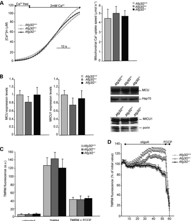
We next examined whether mitochondria of Afg3l22/2 cells have an intrinsic defect in Ca2+accumulation by measuring mitochondrial Ca2+ uptake in digitonin-permeabilized cells and by imposing an ethylene glycol tetraacetic acid (EGTA)-buffered Ca2+ concentration of 2 mM (see Materials and
Methods). In these conditions, all mitochondria are allowed to take up Ca2+ independently of their proximity to the Ca2+source. The Ca2+uptake speed, estimated by linear re-gression from the slope of the ascending phase of [Ca2+]m
rise (see Materials and Methods), was unchanged in Afg3l22/2 cells compared with syngenic controls (Fig. 2A). These data indicate that individual mitochondria lacking AFG3L2 have intact ability to internalize Ca2+, meaning that the uptake machinery is likely not altered. We then direct-ly verified the mRNA and protein levels of the mitochondrial
Ca2+ uniporter (MCU) and of its regulator mitochondrial calcium uptake 1 (MICU1). Real-time qPCR revealed compar-able levels of both transcripts in the three genotypes. Accord-ingly, western blot showed unchanged protein levels of MCU and MICU1 in Afg3l22/2 cells compared with controls (Fig.2B).
Since mitochondrial Ca2+uptake is dependent on Dcmand
we previously reported respiratory defects in neuronal tissues from Afg3l2 knockout and haploinsufficient mouse models (5,14), we tested whether the defective mitochondrial Ca2+ loading of Afg3l22/2 cells could be secondary to altered mem-brane polarization. The measurement of Dcmby the evaluation Figure 1. Loss of Afg3l2 reduces mitochondrial Ca2+uptake. (A and B) [Ca2+]mand [Ca2+]cin Afg3l2 MEFs of the indicated genotypes measured by aequorin probes (mtAEQmut and cytAEQ). On the left: representative graphs; on the right: means + SD of [Ca2+]mand [Ca2+]cpeak responses after bradykinin stimu-lation, respectively (16 traces obtained from four independent experiments). Student’s t-test:∗∗P , 0.001,∗P , 0.05. (C) Measurement of [Ca2+]m(mtAEQ) after capacitative Ca2+influx. On the left: representative graph; on the right: means + SD of mitochondrial peak Ca2+responses (10 – 12 traces obtained from four independent experiments).
of the steady-state loading of the potentiometric dye tetramethyl rhodamine methyl ester (TMRM) into mitochondria of Afg3l22/2, Afg3l2+/2and wild-type MEFs revealed no differ-ences in the three genotypes (Fig.2C). From these data, we con-clude that the reduced Ca2+uptake ability of Afg3l22/2cells is
due neither to defective Ca2+uptake machinery nor to a reduced driving force for Ca2+influx.
Nevertheless, a latent mitochondrial dysfunction of Afg3l22/2MEFs could be masked by the ATP synthase oper-ating in the reverse mode and maintaining Dcm during the Figure 2. Mitochondrial Ca2+uptake speed and electrochemical gradient are unaffected in Afg3l22/2cells. (A) [Ca2+]
min permeabilized MEFs measured by mtAEQmut. On the left: representative graph; on the right: means + SD of the slope of [Ca2+]mrise-ascending phase (10 – 12 traces obtained from four inde-pendent experiments). (B) On the right: quantification (real-time qPCR) of MCU and MICU1 mRNA relative to GAPDH mRNA (wild-type was set to 1). On the left: MCU and MICU1 immunoblot analysis of total cell extracts from Afg3l2 MEFs of the indicated genotypes. Bars represent means + SD of three independent experiments. (C) Analysis of Dcmby uptake of the potentiometric dye TMRM. TMRM fluorescence intensity in Afg3l22/2 MEFs and controls relative to unloaded cells was measured by cytofluorimetry. Bars represent means + SD of four independent experiments. a.u.: arbitrary units. (D) Real-time imaging of TMRM fluorescence intensity of Afg3l2+/+, Afg3l2+/2and Afg3l22/2MEFs loaded with 20 n
MTMRM. Cells were imaged every 60 s and mitochondrial
TMRM fluorescence intensity was calculated as described. Where indicated, 4 mg/ml oligomycin A (OligoA) and 4 mMFCCP were added. Data represent
experiment, a mechanism that has already been described in the literature (25). To test this hypothesis, we measured Dcm
by real-time imaging of TMRM fluorescence intensity during incubation of cells with oligomycin A (OligoA), an inhibitor of mitochondrial F1FO-ATP synthase (25,26).
As expected, addition of OligoA to healthy respiring cells like wild-type MEFs resulted in hyperpolarization of the inner membrane. In fact, protons pumped by the respiratory chain in the intermembrane space cannot cross the F1FO-ATP synthase and remain in the intermembrane space.
In contrast, OligoA addition resulted in a mild mitochondrial hyperpolarization of Afg3l2+/2 MEFs and in a slow but pro-gressive mitochondrial depolarization in Afg3l22/2cells, indi-cating that the maintenance of Dcmin Afg3l22/2cells relies
on the reverse activity of the ATP synthase (Fig.2D). To exclude that the drop of Dcmwas secondary to the
in-duction of the mitochondrial permeability transition (MPT), which depends on the opening of the permeability transi-tion pore (PTP) (27), we performed real-time imaging of TMRM fluorescence pre-incubating the cells with a known inhibitor of MPT, cyclosporine A (CsA), that desensitizes the PTP via its mitochondrial receptor cyclo-philin D (27). The treatment with CsA did not prevent the Dcm drop that is induced by OligoA in Afg3l22/ 2 MEFs,
demonstrating that MPT is not occurring in these cells (data not shown).
Altogether, these data indicate that Dcmin Afg3l2 2/2
cells is not maintained by respiration but rather by a reverse activity of the F1FO-ATP synthase, which pumps protons from the
matrix to the intermembrane space, thereby consuming ATP. This mechanism masks the mitochondrial dysfunction in Afg3l22/2cells, in line with an impairment of the respiratory chain activity.
Afg3l22/2cells show increased mitochondrial fragmentation
Alterations of mitochondrial shape may account for the ineffi-cient mitochondrial Ca2+handling of Afg3l22/2cells. Indeed, it has been demonstrated that fragmentation of the mitochon-drial network impairs mitochonmitochon-drial Ca2+uptake by modifying the spatial pattern of the intra-mitochondrial Ca2+ diffusion (22,28).
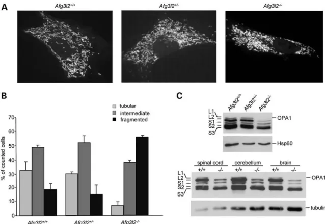
We thus evaluated the effect of the loss of Afg3l2 on the regulation of mitochondrial network morphology by perform-ing live imagperform-ing confocal microscopy in cells overexpressperform-ing a mitochondria-targeted yellow fluorescent protein (mt-YFP). Similarly to what reported by Ehses et al. (29) upon concomi-tant downregulation of both Afg3l1 and Afg3l2 in MEFs, we observed a marked alteration in mitochondrial network morphology in Afg3l22/2 cells. Morphometric analysis revealed that wild-type and heterozygous MEFs have
Figure 3. Increased mitochondrial fragmentation in Afg3l22/2cells. (A) Representative pictures of mitochondrial morphology in Afg3l2 MEFs transfected with mt-YFP and visualized by confocal microscopy. (B) Morphometric analysis of mitochondrial morphology in Afg3l2 MEFs. One hundred and fifty randomly selected cells were analysed in each experiment. Bars represent means + SD of three independent experiments. The Chi-squared test (2 degrees of freedom): Afg3l22/2versus either heterozygous or wild-type MEFs ¼ P , 0.001. (C) Immunoblot analysis of total cell extracts from Afg3l2 MEFs (upper panel) or neuronal tissues of the indicated genotypes (lower panel). The five OPA1 bands are indicated as long (L1 and L2) and short isoforms (S1, S2 and S3) (30). Hsp60 and tubulin were used to verify equal loading, respectively.
elongated and interconnected mitochondria. In contrast, Afg3l22/2 cells showed more than 50% of cells with small, round-shaped organelles and only 5% of cells with a tubular mitochondrial network (Fig.3A and B).
The proper balance of long and short OPA1 isoforms is crit-ical for maintenance of tubular mitochondrial morphology (30). MEFs express four alternative splicing mRNAs coding for OPA1 isoforms, resulting from the inclusion or the exclu-sion of exons 4b and 5b (31). These isoforms undergo different proteolytic cleavages (7,30,32) resulting in five bands on SDS – PAGE, corresponding to long (L1 and L2) and short (S1 to S3) processed OPA1 isoforms. The analysis of OPA1 band pattern in wild-type, Afg3l2+/2 and Afg3l22/2 cells by western blotting revealed that the loss of AFG3L2 results in alteration of OPA1 processing: we detected an increased amount of short OPA1 forms and the almost-complete dis-appearance of the long bands in Afg3l22/2 MEFs (Fig. 3C). No apparent differences were observed between Afg3l2 wild-type and heterozygous cells, indicating that AFG3L2 haploin-sufficiency does not affect OPA1 processing and mitochon-drial morphology in this cellular system (Fig. 3A – C). Mitochondrial fragmentation in Afg3l22/2 MEFs is rescued by the overexpression of wild-type AFG3L2, but this effect is not achieved with the overexpression of AFG3L2M666R, one of the most severe SCA28-causing mutations (10) (Sup-plementary Material, Fig. S1A).
We also evaluated OPA1 processing in the neuronal tissues of Afg3l22/2mice, in particular in the brain, cerebellum and spinal cord, which are the most affected by Afg3l2 loss and also by AFG3L2 mutations (5,10,12). As shown in the western blot in Figure 3C, OPA1 processing is increased in Afg3l22/2 mouse neuronal tissues, thus confirming the pathogenetic relevance of mitochondrial fragmentation in AFG3L2-associated diseases.
Fission of the mitochondrial network reduces organellar matrix continuity and leaves a fraction of mitochondria devoid of connection to the ER
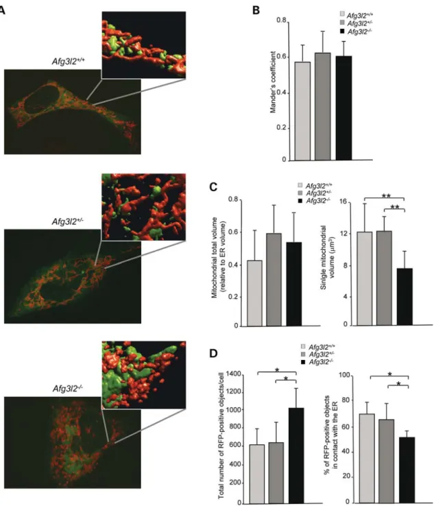
We evaluated whether the mitochondrial fragmentation observed in Afg3l22/2 cells could impact on the number of mitochondria/ER contacts, specialized sites where IP3R/RyR-mediated Ca2+oscillations are propagated locally to the mito-chondria. The close vicinity of mitochondria to Ca2+-release sites on the ER is crucial for the activation of the MCU and thus for the rapid and efficient accumulation of Ca2+ inside the organelles (33,34). To measure the total number of con-tacts in the whole cell volume, we analysed the colocalization between mitochondria and ER with three-dimensional (3D) re-construction and volume rendering of confocal stacks of ER-targeted green fluorescent protein (ER-GFP) and mitochondrial-targeted red fluorescent protein (mt-RFP) in the three genotypes, as previously reported (22,35) (Fig. 4A). In this analysis, we considered the overlapping mitochondrial and ER spots as the sites where most-efficient Ca2+transfer between the two organelles occurs, thus satisfy-ing the high Ca2+concentration demand for the opening of the MCU. As measured by Manders’ colocalization coefficient (36), we found that the evident fragmentation of the
mitochondrial network does not affect the average number of mitochondria-ER contact sites in Afg3l22/2cells (Fig.4B). Similarly, the overall volume of the organellar network of both mitochondria and ER is unaffected by the loss of AFG3L2, as demonstrated by quantitative morphological imaging of the mitochondrial and ER networks by transfecting cells with mt-RFP or ER-GFP, respectively. After 3D recon-struction of confocal stacks, the images were analysed evalu-ating the total volume of the organellar network. ER architecture was unaffected by the loss of Afg3l2, appearing as an interconnected network of cistarnae spanning the whole cellular volume. We also observed that fragmentation of the mitochondrial network does not alter either the mito-chondrial or the ER total volume, the ratio of the two organel-lar volumes being comparable in the three genotypes (Fig. 4C). According to the observed fragmentation, we found a significant reduction in the average size of individual mitochondrial particle in Afg3l22/2cells compared with con-trols (6.94 + 2.3 versus 12.68 + 3.35 mm3 in wild-type and 12.3 + 1.91 mm3 in heterozygous cells, P , 0.001) (Fig.4C). Having the three cell lines the same mitochondrial total volume, these data indicate that Afg3l22/2 cells have an increased number of individual mitochondria compared to wild-type and heterozygous cells. This hypothesis was verified by counting the total number of RFP-positive objects per cell, which is almost doubled in Afg3l22/2 cells compared with control cells, while the percentage of objects that are in contact with the ER is reduced (Fig.4D). These data demon-strate that a significant number of Afg3l2 null mitochondria lacks connections to the ER, and thus to Ca2+ sources, and therefore remains without significant [Ca2+]mrises.
Rescue of mitochondrial fragmentation by OPA1 and MFN1 overexpression restores normal mitochondrial Ca21 uptake in Afg3l22/2 cells
To validate our hypothesis, we decided to investigate whether rescue of mitochondrial fragmentation could ameliorate mito-chondrial Ca2+uptake in Afg3l22/2cells. For this purpose, we generated wild-type, heterozygous and knockout MEF lines stably expressing wild-type OPA1 isoform1 (OPA1v1) or a mutant version of OPA isoform1 carrying a mutation within the GTP-binding domain (OPA1v1Q297V). By mimicking the GTP-bound form of OPA1, this mutant has been described as constitutively active and disassembly resistant form (37). In order to avoid strong overexpression of OPA1, being known that this can cause clustering of small mitochondria (38), we transfected cells with OPA1v1 or pMSCV-OPA1v1Q297V constructs, where OPA1 cDNA expression is under the control of the weak murine stem cell virus promoter.
As already reported by other groups in transient transfection experiments (37), the expression levels of exogenous OPA1v1 and OPA1v1Q297V revealed by western blot analysis were modest. We indeed detected a slight increase in the expected second (L2) and fifth bands (S3) in cells expressing OPA1v1 and OPA1v1Q297V compared with those transfected with the empty vector (EV) (Supplementary Material, Fig. S2B). Nevertheless, we observed that the moderate overexpression of OPA1v1 and OPA1v1Q297V clearly affects mitochondrial morphology in stable cell lines. In fact, morphometric analysis
revealed that .50% of the Afg3l22/2::EV analysed cells dis-played fragmented mitochondria, exactly like immortalized Afg3l22/2 MEFS. This percentage is strikingly reduced in
Afg3l22/2::OPA1v1 cells, where most mitochondria appear in the intermediate state. The effect is even more evident in Afg3l22/2::OPA1v1Q297V MEFs, in which tubulated mito-chondria are the most represented (Fig. 5A). Accordingly, imaging of Afg3l2+/2::OPA1v1 cells revealed an increased
percentage of tubular mitochondria compared with Afg3l2+/2::EV cells. In Afg3l2+/2::OPA1v1Q297V, most of the organelles are highly elongated and interconnected in a branched network (Supplementary Material, Fig. S3A and B). These findings indicate that the modest restoration of OPA1 L2 band is able to rescue mitochondrial fragmentation in Afg3l22/2cells and to increase the fusion rate in Afg3l2+/2 cells.
Figure 4. Mitochondrial fragmentation leaves a fraction of organelles free of connections to the ER. (A) Representative images of Afg3l2 knockout, heterozygous and wild-type MEFs cotransfected with ER-GFP and mt-RFP. Insets show 3D reconstructed confocal stacks. (B) Quantitative analysis of ER-mitochondria colo-calization estimated by Manders’ coefficient. (C) Effect of Afg3l2 depletion on total mitochondrial volume and on individual mitochondrial particles, respect-ively. (D) Quantification of the total number of RFP-positive objects and quantification of the percentage of RFP-positive objects in contact with the ER in the three genotypes. Data were obtained from an average of 60 randomly selected cells per genotype. Bars represent means + SD of three independent experiments. Student’s t-test:∗P , 0.05,∗∗P , 0.001.
Figure 5. The defective mitochondrial Ca2+uptake is rescued by OPA1 or MFN1 overexpression. (A) Morphometric analysis of mitochondrial morphology of Afg3l22/2cells stably expressing wild-type OPA1v1 or OPA1v1-Q297V. One hundred and fifty randomly selected cells were analysed in each experiment. Bars represent means + SD of three independent experiments. EV, empty vector. The Chi-squared test (2 degrees of freedom): Afg3l22/2::EV MEFs versus either ::OPA1v1 or ::OPA1v1Q297V ¼ P , 0.001, ::OPA1v1 versus ::OPA1v1Q297V ¼ P , 0.001. (B) [Ca2+]mmeasured by aequorin probes (mitAEQmut) of Afg3l22/2cells stably expressing wt OPA1v1 or OPA1v1-Q297V. On the left: representative graphs; on the right: means + SD of [Ca2+]
mpeak responses after bradykinin stimulation (12 traces obtained from four independent experiments). Student’s t-test: ∗∗P , 0.001, ∗P , 0.05. (C) Real-time imaging of TMRM fluorescence intensity of Afg3l2+/2and Afg3l22/2 cells stably expressing wt OPA1v1 or OPA1v1-Q297V loaded with 20 n
M TMRM. Cells were
imaged every 60 s and mitochondrial TMRM fluorescence intensity was calculated as described. Where indicated, 4 mg/ml oligomycin A (OligoA) and 4 mM
FCCP were added. Data represent mean + SE of five independent experiments. (D) Morphometric analysis of mitochondrial morphology of Afg3l22/2 overexpres-sing MFN1. One hundred and fifty randomly selected cells were analysed in each experiment. Bars represent means + SD of three independent experiments. The Chi-squared test (2 degrees of freedom): Afg3l22/2::EV MEFs versus either ::MFN1 or Afg3l2+/2::EV ¼ P , 0.001, Afg3l2+/2::EV versus Afg3l22/2::MFN1 ¼ P , 0.001. (E) [Ca2+]mmeasured by aequorin probes (mitAEQmut) of Afg3l22/2cells transfected with MFN1. On the left: representative graphs; on the right: means + SD of [Ca2+]mpeak responses after bradykinin stimulation (10 traces obtained from two independent experiments). Student’s t-test:∗∗P , 0.001.
We next examined whether the rescue of mitochondrial morphology could ameliorate the defective Ca2+ handling capacity of Afg3l22/2cells. We thus measured mitochondrial Ca2+ uptake in stable cell lines using the mitochondria-targeted aequorin mtAEQmut. Bradykinin stimulation of Afg3l22/2::EV MEFs resulted in [Ca2+]m rises similar to
those measured in Afg3l22/2 MEFs (51.46 + 10.48 mM). On
the contrary, OPA1v1 overexpressing cells showed a signifi-cant increase in the bradykinin-evoked [Ca2+]m rise (66.7 +
4.4 mM, P , 0.05), that was greater in OPA1v1Q297V
overex-pressing cells (76.1 + 9.1 mM, P , 0.001) (Fig.5B).
To verify that the rescue of the defective Ca2+ uptake in Afg3l22/2::OPA1v1 and Afg3l22/2::OPA1v1Q297V was spe-cifically caused by the recovery of mitochondrial morphology and not by a different effect of OPA1 overexpression in the inner membrane, we tested whether it can be achieved by stimulating mitochondrial fusion through a different molecular system. Namely, we assessed mitochondrial morphology in Afg3l22/2 cells after overexpression of MFN1, essential for outer membrane fusion (39), and we observed a significant re-duction in the amount of cells harbouring fragmented orga-nelles and an increased quote of cells harbouring fused organelles (Fig. 5D). Of note, we found that MFN1 overex-pression is able to increase bradykinin-evoked [Ca2+]m rise
in Afg3l22/2 MEFs (68.61 + 4.3 mM, P , 0.001) compared
with mock-transfected cells (55.49 + 9.18 mM) (Fig. 5E).
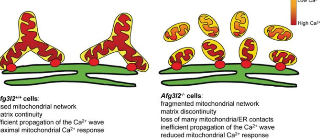
These data prove that Afg3l22/2 cells are still fusion compe-tent and clearly demonstrate that the defective ability of Afg3l22/2cells to internalize Ca2+is dependent on fragmen-tation of the mitochondrial network (Fig.6).
Since stable expression of OPA1v1 or OPA1v1-Q297V rescued the mitochondrial network fragmentation in Afg3l22/2 MEFs, we then examined whether OPA1 can rescue also the progressive mitochondrial depolarization of OligoA-treated Afg3l22/2 MEFs. The real-time imaging of TMRM in these stable cell lines exposed to OligoA showed no recovery of the Dcmin Afg3l22/2cells stably expressing
wild-type OPA1v1 or OPA1v1-Q297V to levels comparable to the control cells (Fig. 5C). Therefore, we conclude that the rescue of mitochondrial network morphology by OPA1
is not sufficient to recover the latent mitochondrial dysfunction of Afg3l22/2cells.
DISCUSSION
In this study, we evaluated the mitochondrial Ca2+uptake cap-acity of Afg3l2-depleted cells. Our results show that the loss of Afg3l2 causes a marked reduction in mitochondrial Ca2+ buf-fering. This defect depends neither on impaired organellar Ca2+ uptake machinery nor on alteration of Dcm, since
MCU mRNA and protein levels and TMRM loading resulted comparable between mutant and controls. These observations are in line with the fact that Afg3l22/2 individual mitochon-dria present intact ability to accumulate Ca2+, as demonstrated by unchanged Ca2+ uptake speed in digitonin-permeabilized cells experiments. We showed that the defective Ca2+ uptake of Afg3l22/2 cells is instead caused by fragmentation of the mitochondrial network, secondary to enhanced process-ing of OPA1. Evidence for this mechanism is provided by rescue experiments, where we stably expressed wild-type OPA1v1 or a constitutively active form of OPA1 (OPA1v1-Q297V) in Afg3l22/2MEFs. By slightly increasing the amount of the long OPA1 isoforms (L2 band) that promote organellar fusion, the overexpression of OPA1v1 is able to recover fragmentation of the mitochondrial network in Afg3l22/2 cells. This effect is even more pronounced with OPA1v1-Q297V overexpression, which increases the number of cells with a tubular network, besides reducing mitochon-drial fragmentation. This can be explained by the fact that Q297V mutation, as previously proposed (37,40), should make OPA1 constitutively active by mimicking the GTP-bound state. Notably, we showed that fusion restoration in Afg3l22/2::OPA1v1 MEFs significantly ameliorates mito-chondrial Ca2+buffering respect to Afg3l22/2::EV cells and that the defect is completely recovered in Afg3l22/2:: OPA1v1Q297V MEFs. We also demonstrated that overexpres-sion of MFN1, an outer membrane regulator of mitochondrial fusion, exerts the same effect of OPA1v1 on both mitochon-drial morphology and Ca2+ buffering in Afg3l22/2 MEFs.
Figure 6. Model explaining the defective Ca2+buffering in Afg3l22/2cells. The red dots represent ER-mitochondria Ca2+transmission sites. In Afg3l22/2cells, many mitochondria remain without contacts with ER, and thus without Ca2+elevations. The Ca2+ions reaching mitochondria propagate less efficiently in a fragmented organellar network compared with a tubular one.
These findings prove that mitochondrial fusion can occur in the absence of AFG3L2 and can be enhanced by overexpres-sing mitochondrial fusion regulators. In addition, since MFN1 can efficiently tubulate mitochondria in the presence of OPA1 (38), they indicate that the overall amount of long and short OPA1 isoforms present in Afg3l2-depleted cells is sufficient to respond to fusion induction triggered by MFN1. Moreover, these data demonstrate that impaired Ca2+ buffer-ing in the absence of AFG3L2 is indeed caused by mitochon-drial fragmentation secondary to enhanced OPA1 processing. The mechanism through which the loss of AFG3L2 induces increased processing of OPA1 is not fully understood. OMA1, a metallopeptidase of the inner mitochondrial membrane, mediates the cleavage of OPA1 in the absence of AFG3L2 (29). Moreover, the loss of Dcmor decreased ATP levels is
known to augment OPA1 processing (41,42). Whether OMA1 is directly regulated by AFG3L2 itself or indirectly by the respiratory deficiencies mediated by the loss of AFG3L2 (5) remains to be determined. Here, we provide evi-dence that respiratory dysfunction is the primary defect fol-lowing AFG3L2 depletion and associates to mitochondrial fragmentation.
In fact, our studies on the regulation of Dcmin the absence
of AFG3L2 in MEFs show that Dcmin basal conditions is not
altered. On the other hand, treatment with the ATP-synthase inhibitor Oligo A revealed depolarization in Afg3l22/2 cells, indicating that Dcmis maintained through the reverse activity
of the F1FO-ATP synthase and not by respiration. This
pheno-type is in line with a dysfunction in the activity of the respira-tory chain complexes, which affects proton pumping across the inner mitochondrial membrane and requires reversion of the activity of ATP synthase (i.e. consuming ATP) for proton pumping from the matrix to the intermembrane space to achieve the maintenance of Dcm. As a direct consequence,
the inhibition of the ATP synthase activity by Oligo A pro-duces a decline of Dcm, as observed in Afg3l2 knockout
MEFs. Interestingly, stable expression of OPA1v1 or OPA1v1-Q297V is not able to rescue the progressive mito-chondrial depolarization of OligoA-treated Afg3l22/2 MEFs to levels comparable with the wild-type demonstrating that the rescue of mitochondrial network morphology is not suffi-cient to recover the latent mitochondrial dysfunction. These data indicate that the impaired mitochondrial respiration is the primary defect following depletion of Afg3l2 that we hy-pothesize is the cause of the increase in OPA1 processing and of the following mitochondrial fragmentation.
This is in line with the fact that in Afg3l2+/2MEFs, OligoA addition resulted in a milder hyperpolarization compared with wild-type cells. This can indicate a reduced efficiency of mito-chondrial respiration in Afg3l2+/2 cells, although mitochon-drial morphology and OPA1 processing are not affected in this cellular system.
All this considered, we demonstrate the consequential ar-rangement of events, starting from loss of AFG3L2, to respira-tory dysfunction, OPA1 cleavage and finally to organellar fragmentation, although some mechanistic steps of this process are indeed still lacking and maybe include some new effectors still to be identified.
We observed that mitochondrial fragmentation in Afg3l22/2 cells reduces the volume of individual
mitochondrial particles without changing the total volume of the organellar network. As a direct consequence, a fraction of mitochondria in Afg3l22/2cells lose their link to the ER and result positioned at a higher distance from the Ca2+ source, failing to match the MCU-activation needs (33) and therefore remaining without substantial [Ca2+]m rises.
More-over, studies have shown that Ca2+ entering mitochondria can spread and equilibrate rapidly within the matrix along large and tubular, but not fragmented organelles. In fact, the small size of mitochondrial particles in a fragmented network and the consequent lack of matrix connectivity limit the proper diffusion of the Ca2+wave and increases the het-erogeneity of mitochondrial Ca2+responses (22,28). Together, these phenomena account for the overall reduction in the mito-chondrial Ca2+load in Afg3l22/2cells.
Heterozygous and homozygous mutations in AFG3L2 cause neurodegeneration that involves PCs in the cerebellum and results in ataxia (9,12). The fact that cerebellum is more sen-sitive to AFG3L2 dysfunction compared with other neuronal tissues can be explained by the high expression of this gene in PCs (9), but also with the special requirement of mitochon-drial functionality by PCs. These are indeed unique neurons with very large and highly branched dendritic trees that receive only excitatory inputs. They are therefore exposed to high Ca2+ influxes due to glutamatergic stimulation of both mGluR1 and AMPA receptors (19). For these reasons, mito-chondria play a pivotal role in maintaining Ca2+homeostasis in PCs by direct uptake of Ca2+and, also, by providing ATP to pump Ca2+across membranes.
We therefore hypothesize that the inefficient buffering and shaping of the Ca2+waves operated by mutant mitochondria provokes a local increase in [Ca2+]cand that this, in chronic
conditions like as in AFG3L2-associated diseases, triggers PCs degeneration.
Considering what mentioned above, it is conceivable that Afg3l2 haploinsufficiency causes inefficient Ca2+ buffering in PCs in a progressive degenerative situation. Nonetheless, we failed to demonstrate mitochondrial fragmentation and de-fective Ca2+uptake in Afg3l2+/2fibroblasts likely because in culture they rely marginally on mitochondria for ATP produc-tion and Ca2+ buffering compared with neurons and, also, because in these cells the Ca2+wave could only be elicited by ER stimulation. Moreover, we performed acute experi-ments that do not properly mimic the physio-pathological con-dition of chronic degeneration.
We can hypothesize that fragmentation of the mitochondrial network in Afg3l2-depleted PCs causes defective mitochon-drial Ca2+ buffering, but also inefficient trafficking of the organelles to dendritic stalks and distal branches. Indeed, it has been demonstrated that in PCs lacking mitochondrial fusion, both increased mitochondrial diameter due to swelling and aggregations of mitochondria seem to block efficient entry into neurites, resulting in a dearth of mitochondria in axons and dendrites (43). In the worst scenario, the two events may negatively synergize causing not only the inefficient op-position to Ca2+wave close to the ER and at in dendrites, but the increased mitochondria retention in the cell soma, too.
In conclusion, our results functionally link for the first time AFG3L2 to mitochondrial Ca2+ buffering. The molecular pathogenesis of SCA28 can now be drawn in the light of the
inefficient mitochondrial Ca2+uptake as one of the triggering events of PC-DCD.
MATERIALS AND METHODS MEF lines
Primary MEFs were established from embryonic day E16.5 from Afg3l2 knockout, heterozygous and wild-type embryos (5) and immortalized by SV40 (44), using 300 mg/ml geneticin for selection.
For the generation of stable cell lines, MEFS were
transfected with EV, OPA1v1 and pMSCV-OPA1v1Q297V constructs and selected with 1 mg/ml puro-mycin for 2 weeks. Transfection was performed using Metafectene (Biontex Laboratories, San Diego, CA, USA) according to the manufacturer’s instruction.
Aequorin measurements
MEFs grown on 13 mMround glass cover slips at 50%
conflu-ence were transfected with the cytosolic (cytAEQ) or mito-chondrial aequorin probes (mtAEQmut or mtAEQ). Cells were reconstituted with 5 mM coelenterazine for 90 min in
DMEM supplemented with 1% fetal bovine serum, incubated in KRB (Krebs – Ring modified buffer: 125 mM NaCl, 5 mM
KCl, 1 mM Na3PO4, 1 mM MgSO4, 5.5 mM glucose, 20 mM
4-(2-hydroxyethyl)-1-piperazineethanesulfonic acid (HEPES), pH 7.4, 378C) supplemented with 1 mMCaCl2and then
trans-ferred to the perfusion chamber. When indicated, 100 nM
bradykinin was added. The experiments were concluded by lysing MEFs with 100 mM digitonin in a hypotonic
Ca2+-rich solution (10 mM CaCl2 in H2O), thus discharging
the remaining aequorin pool. The light signal was collected in a purpose-built luminometer as previously described (23).
[Ca2+]mtfollowing capacitative Ca2+influx was measured
by perfusing MEFs with the SERCA inhibitor thapsigargin (1 mM) in a KRB solution containing no Ca2+ and 100 mM
EGTA. Mitochondrial Ca2+ uptake was evoked by adding 1 mMCaCl2to the medium and was measured by mtAEQ.
In the experiments with permeabilized cells, a buffer mim-icking the cytosolic Ca2+ ionic composition (intracellular buffer) was used: 130 mM KCl, 10 mM NaCl, 2 mMK2HPO4,
5 mM succinic acid, 5 mM malic acid, 1 mM MgCl2, 20 mM
HEPES, 1 mM pyruvate, 0.5 mM ATP and 0.1 mM ADP
(378C, pH 7). Intracellular buffer was supplemented with either 100 mM EGTA (intracellular buffer/EGTA) or a 2 mM
EGTA and 2 mM hydroxy-2-ethylenediaminetriacetic
acid-buffered [Ca2+] of 2 mM(intracellular buffer/Ca2+) calculated
with the Chelator software (45).
RNA extraction and gene expression analysis
Total RNA from cells was purified using Trizol (Invitrogen, Carlsbad, CA, USA) following the manufacturer’s instructions. cDNA was generated using a Thermo script RT – PCR system kit (Invitrogen). For the expression of MCU and MICU1, cDNA was analysed by real-time PCR using the SYBR green chemistry (Light cycler 480, SYBR green I master, Roche, Basel, Switzerland). MCU and MICU1-PCR-primer sequences were
previously described by De Stefani et al. (20). glyceraldehyde 3-phosphate dehydrogenase was used for normalization. Western blot analysis
OPA1 band pattern was analysed by standard western blot pro-cedure. Total cell lysate was prepared by protein extraction using a Triton-containing buffer (10 mM Tris – HCl, pH 7.4,
150 mM NaCl, 1% Triton, 1 mM ethylenediaminetetraacetic
acid (EDTA) pH 8, 1X Protease Inhibitor Cocktail, Roche, Basel, Switzerland). Twenty micrograms of protein extracts were dissolved in sample buffer (60 mM Tris – HCl, pH 6.8,
5% glycerol, 1.7% SDS, 0.1M DTT, 0.002% bromophenol
blue), were separated on 7% SDS – PAGE and analysed by standard immunoblotting procedures. Anti-OPA1 antibody was from BD Transduction Laboratories (Franklin Lakes, NJ, USA), anti-Hsp60 and Hsp70 antibodies were from Stressgen (San Diego, CA, USA). For the detection of MCU and MICU1, cells were lysed with radio immunoprecipitation assay buffer (NaCl 150 mM, Tris–HCl 50 mM, EDTA 1 mM, pH 8, NP40
1%, SDS 0.1%, Sodium Deoxycholate 1%). Fifty micrograms of protein extracts were dissolved in sample buffer and loaded on a 7% SDS–PAGE followed by immunoblotting. Anti-MCU (CCDC109A) and anti-MICU1 antibodies were from SIGMA (St Louis, Missouri, USA), anti-tubulin was from Invitrogen. Measurements of mitochondrial membrane potential DCm was measured using the mitochondrial potentiometric
dye TMRM (Invitrogen) and analysing cells uptake of the compound cytofluorimetrically. Cells were seeded in six-well multi-dishes, 300 000 cells/well. Twenty-four hours after plating, cells were collected, washed in PBS 1× and then suspended in phenol-red free Hank’s buffered salt solu-tion (HBSS) 1× supplemented with 10 mM HEPES, 2 mM
Ciclosporin H (Alexis Biochemicals, San Diego, CA, USA) and 20 nMTMRM and incubated for 30 min at 378C. In
paral-lel, after incubation with TMRM containing medium, cells were treated for 5 min with 4 mM trifluorocarbonylcyanide
phenylhydrazone (FCCP), as control of specific mitochondrial staining. TMRM fluorescence was analysed using Cytomics FC500 Flow Cytometry System (Beckman Coulter). Output files were analysed using flow cytometry data analysis program WinMDI 2.8.
Real-time analysis of Dcm was performed as follows.
Afg3l2+/+, Afg3l2+/2 and Afg3l22/2 MEFs were plated in 12-well multi-dishes, 50 000 cells/well. After 40 h, cells were incubated with 20 nM TMRM, 2 mM CsH and 2 mg/ml
Hoechst 33 342 dissolved in phenol-red free HBSS 1× supple-mented with 10 mMHEPES for 30 min at 378C. For CsA
pre-treatment, cells were incubated for 30 min at 378C in the abovementioned medium supplemented with 2 mM CsA.
Images were acquired with IN Cell Analyzer 1000 (GE Healthcare, Little Chalfont, UK). Sequential images of TMRM fluorescence from at least three regions of interest (ROIs) were acquired every 60 s. Four micrograms per milli-litre oligomycin A and 4 mM FCCP were added in each well,
after minute 5 and minute 55 of the acquisition, respectively, by In Cell Analyzer motorized injector. Images were stored for subsequent analysis using IN Cell Investigator software. Five 3868 Human Molecular Genetics, 2012, Vol. 21, No. 17
independent experiments were performed. The average TMRM fluorescence intensity of at least 15 ROIs minus back-ground was calculated for each frame and normalized for com-parative purposes.
Imaging analyses
Analysis of mitochondrial network morphology was per-formed as described (38) by expressing a mt-YFP (Clontech, Mountain View, CA, USA) and live imaging confocal micros-copy. An average of 150 cells was analysed for each experi-mental condition. Cells were divided into three classes accordingly to mitochondria shape: tubular mitochondrial network, intermediate or fragmented mitochondrial structures. Experiments were repeated at least three times, and three operators analysed the images, independently. The Chi-squared test on row data was applied for significance cal-culation (degrees of freedom ¼ 2).
For the analysis of mitochondrial and ER volumes, cells were transfected with mitochondrial-targeted red fluorescent protein mt-RFP (Clontech) and ER-targeted green fluorescent protein (ER-GFP, kindly provided by R. Sitia) and stacks of consecutive confocal images taken at 0.1 mm intervals were acquired. 3D reconstruction and volume rendering were per-formed using Volocity 3D Image Analysis Software version 5.5.1, Perkin Elmer, Norwalk, MA, USA.
Manders’ coefficient was applied for the quantitative ana-lysis of mitochondria-ER colocalization. In all the experi-ments, confocal microscopy was performed using Perkin Elmer UltraVIEW Spinning Disk Confocal Microscope and EMCCD Hamamatsu C9100 imaging camera. The system is equipped with a stage incubator from OkoLab (www.okolab. com), allowing to work with live cells maintained under stable conditions of temperature, CO2and humidity.
For each cell, the total number of red objects (mitochondria) was identified using the IN Cell Developer Toolbox 1.8 (GE Healthcare). After the optimization of segmentation para-meters, a mask was created to cover the mitochondrial network. This mask was projected on the ER. At this point, a threshold was set in order to classify mitochondria using the green (ER) intensity. Mitochondria with a green signal in-tensity greater than or equal to the threshold were classified as in contact with the ER, whereas mitochondria with a green signal intensity less than threshold were classified as free of contacts.
SUPPLEMENTARY MATERIAL
Supplementary Material is available at HMG online.
ACKNOWLEDGEMENTS
We thank our internal imaging facility ALEMBIC and, in par-ticular, Miriam Ascagni for technical assistance on real-time analysis of Dcm and for organelle quantification. We thank
Riccardo Vago for FACS analysis technical assistance. Conflict of Interest statement. None declared.
FUNDING
This work has been supported by the Italian Telethon Foundation (GGP 08138), Compagnia di San Paolo (Turin, Italy) and National Ataxia Foundation (Minneapolis, MN-USA). Funding to pay the Open Access publication charges for this article was provided by the Italian Telethon Foundation.
REFERENCES
1. Tatsuta, T. and Langer, T. (2008) Quality control of mitochondria: protection against neurodegeneration and ageing. EMBO J., 27, 306 – 314. 2. Koppen, M. and Langer, T. (2007) Protein degradation within
mitochondria: versatile activities of AAA proteases and other peptidases. Crit. Rev. Biochem. Mol. Biol., 42, 221 – 242.
3. Arlt, H., Steglich, G., Perryman, R., Guiard, B., Neupert, W. and Langer, T. (1998) The formation of respiratory chain complexes in mitochondria is under the proteolytic control of the m-AAA protease. EMBO J., 17, 4837 – 4847.
4. Atorino, L., Silvestri, L., Koppen, M., Cassina, L., Ballabio, A., Marconi, R., Langer, T. and Casari, G. (2003) Loss of m-AAA protease in mitochondria causes complex I deficiency and increased sensitivity to oxidative stress in hereditary spastic paraplegia. J. Cell Biol., 163, 777 – 787.
5. Maltecca, F., Aghaie, A., Schroeder, D.G., Cassina, L., Taylor, B.A., Phillips, S.J., Malaguti, M., Previtali, S., Guenet, J.L., Quattrini, A. et al. (2008) The mitochondrial protease AFG3L2 is essential for axonal development. J. Neurosci., 28, 2827 – 2836.
6. Nolden, M., Ehses, S., Koppen, M., Bernacchia, A., Rugarli, E.I. and Langer, T. (2005) The m-AAA protease defective in hereditary spastic paraplegia controls ribosome assembly in mitochondria. Cell, 123, 277 – 289.
7. Ishihara, N., Fujita, Y., Oka, T. and Mihara, K. (2006) Regulation of mitochondrial morphology through proteolytic cleavage of OPA1. EMBO J., 25, 2966– 2977.
8. Casari, G., De Fusco, M., Ciarmatori, S., Zeviani, M., Mora, M., Fernandez, P., De Michele, G., Filla, A., Cocozza, S., Marconi, R. et al. (1998) Spastic paraplegia and OXPHOS impairment caused by mutations in paraplegin, a nuclear-encoded mitochondrial metalloprotease. Cell, 93, 973 – 983.
9. Di Bella, D., Lazzaro, F., Brusco, A., Plumari, M., Battaglia, G., Pastore, A., Finardi, A., Cagnoli, C., Tempia, F., Frontali, M. et al. (2010) Mutations in the mitochondrial protease gene AFG3L2 cause dominant hereditary ataxia SCA28. Nat. Genet., 42, 313 – 321.
10. Cagnoli, C., Stevanin, G., Brussino, A., Barberis, M., Mancini, C., Margolis, R.L., Holmes, S.E., Nobili, M., Forlani, S., Padovan, S. et al. (2010) Missense mutations in the AFG3L2 proteolytic domain account for approximately 1.5% of European autosomal dominant cerebellar ataxias. Hum. Mutat., 31, 1117 – 1124.
11. Edener, U., Wollner, J., Hehr, U., Kohl, Z., Schilling, S., Kreuz, F., Bauer, P., Bernard, V., Gillessen-Kaesbach, G. and Zuhlke, C. (2010) Early onset and slow progression of SCA28, a rare dominant ataxia in a large four-generation family with a novel AFG3L2 mutation. Eur. J. Hum. Genet., 18, 965 – 968.
12. Pierson, T.M., Adams, D., Bonn, F., Martinelli, P., Cherukuri, P.F., Teer, J.K., Hansen, N.F. and Cruz, P., Mullikin For The Nisc Comparative Sequencing Program, J.C., Blakesley, R.W. et al. (2011) Whole-exome sequencing identifies homozygous AFG3L2 mutations in a spastic ataxia-neuropathy syndrome linked to mitochondrial m-AAA proteases. PLoS Genet., 7, e1002325.
13. Koppen, M., Metodiev, M.D., Casari, G., Rugarli, E.I. and Langer, T. (2007) Variable and tissue-specific subunit composition of mitochondrial m-AAA protease complexes linked to hereditary spastic paraplegia. Mol. Cell Biol., 27, 758 – 767.
14. Maltecca, F., Magnoni, R., Cerri, F., Cox, G.A., Quattrini, A. and Casari, G. (2009) Haploinsufficiency of AFG3L2, the gene responsible for spinocerebellar ataxia type 28, causes mitochondria-mediated Purkinje cell dark degeneration. J. Neurosci., 29, 9244– 9254.
15. Custer, S.K., Garden, G.A., Gill, N., Rueb, U., Libby, R.T., Schultz, C., Guyenet, S.J., Deller, T., Westrum, L.E., Sopher, B.L. et al. (2006)
Bergmann glia expression of polyglutamine-expanded ataxin-7 produces neurodegeneration by impairing glutamate transport. Nat. Neurosci., 9, 1302– 1311.
16. Mansouri, B., Henne, W.M., Oomman, S.K., Bliss, R., Attridge, J., Finckbone, V., Zeitouni, T., Hoffman, T., Bahr, B.A., Strahlendorf, H.K. et al. (2007) Involvement of calpain in AMPA-induced toxicity to rat cerebellar Purkinje neurons. Eur. J. Pharmacol., 557, 106 – 114. 17. Duchen, M.R. (2000) Mitochondria and calcium: from cell signalling to
cell death. J. Physiol., 529, 57 – 68.
18. Berridge, M.J., Bootman, M.D. and Roderick, H.L. (2003) Calcium signalling: dynamics, homeostasis and remodelling. Nat. Rev. Mol. Cell Biol., 4, 517 – 529.
19. Ito, M. (2002) The molecular organization of cerebellar long-term depression. Nat. Rev. Neurosci., 3, 896 – 902.
20. De Stefani, D., Raffaello, A., Teardo, E., Szabo, I. and Rizzuto, R. (2011) A forty-kilodalton protein of the inner membrane is the mitochondrial calcium uniporter. Nature, 476, 336 – 340.
21. Perocchi, F., Gohil, V.M., Girgis, H.S., Bao, X.R., McCombs, J.E., Palmer, A.E. and Mootha, V.K. (2010) MICU1 encodes a mitochondrial EF hand protein required for Ca(2+) uptake. Nature, 467, 291–296. 22. Szabadkai, G., Simoni, A.M., Chami, M., Wieckowski, M.R., Youle, R.J.
and Rizzuto, R. (2004) Drp-1-dependent division of the mitochondrial network blocks intraorganellar Ca2+ waves and protects against Ca2+-mediated apoptosis. Mol. Cell, 16, 59–68.
23. Chiesa, A., Rapizzi, E., Tosello, V., Pinton, P., de Virgilio, M., Fogarty, K.E. and Rizzuto, R. (2001) Recombinant aequorin and green fluorescent protein as valuable tools in the study of cell signalling. Biochem. J., 355, 1 – 12.
24. Glitsch, M.D., Bakowski, D. and Parekh, A.B. (2002) Store-operated Ca2+ entry depends on mitochondrial Ca2+ uptake. EMBO J., 21, 6744–6754. 25. Nicholls, D.G. (1974) The influence of respiration and ATP hydrolysis on the proton-electrochemical gradient across the inner membrane of rat-liver mitochondria as determined by ion distribution. Eur. J. Biochem./FEBS, 50, 305 – 315.
26. Irwin, W.A., Bergamin, N., Sabatelli, P., Reggiani, C., Megighian, A., Merlini, L., Braghetta, P., Columbaro, M., Volpin, D., Bressan, G.M. et al. (2003) Mitochondrial dysfunction and apoptosis in myopathic mice with collagen VI deficiency. Nat. Genet., 35, 367 – 371.
27. Bernardi, P. (1999) Mitochondrial transport of cations: channels, exchangers, and permeability transition. Physiol. Rev., 79, 1127 – 1155. 28. Frieden, M., James, D., Castelbou, C., Danckaert, A., Martinou, J.C. and
Demaurex, N. (2004) Ca(2+) homeostasis during mitochondrial fragmentation and perinuclear clustering induced by hFis1. J. Biol. Chem., 279, 22704 – 22714.
29. Ehses, S., Raschke, I., Mancuso, G., Bernacchia, A., Geimer, S., Tondera, D., Martinou, J.C., Westermann, B., Rugarli, E.I. and Langer, T. (2009) Regulation of OPA1 processing and mitochondrial fusion by m-AAA protease isoenzymes and OMA1. J. Cell Biol., 187, 1023– 1036. 30. Song, Z., Chen, H., Fiket, M., Alexander, C. and Chan, D.C. (2007) OPA1
processing controls mitochondrial fusion and is regulated by mRNA splicing, membrane potential, and Yme1L. J. Cell Biol., 178, 749 – 755.
31. Akepati, V.R., Muller, E.C., Otto, A., Strauss, H.M., Portwich, M. and Alexander, C. (2008) Characterization of OPA1 isoforms isolated from mouse tissues. J. Neurochem, 106, 372 – 383.
32. Duvezin-Caubet, S., Jagasia, R., Wagener, J., Hofmann, S., Trifunovic, A., Hansson, A., Chomyn, A., Bauer, M.F., Attardi, G., Larsson, N.G. et al. (2006) Proteolytic processing of OPA1 links mitochondrial dysfunction to alterations in mitochondrial morphology. J. Biol. Chem., 281, 37972 – 37979.
33. Rizzuto, R., Pinton, P., Carrington, W., Fay, F.S., Fogarty, K.E., Lifshitz, L.M., Tuft, R.A. and Pozzan, T. (1998) Close contacts with the endoplasmic reticulum as determinants of mitochondrial Ca2+ responses. Science, 280, 1763 – 1766.
34. Csordas, G., Varnai, P., Golenar, T., Roy, S., Purkins, G., Schneider, T.G., Balla, T. and Hajnoczky, G. (2010) Imaging interorganelle contacts and local calcium dynamics at the ER-mitochondrial interface. Mol. Cell, 39, 121 – 132.
35. de Brito, O.M. and Scorrano, L. (2008) Mitofusin 2 tethers endoplasmic reticulum to mitochondria. Nature, 456, 605 – 610.
36. Manders, E.M., Verbeek, F.J. and Aten, A. (1993) Measurement of colocalization of objects in dual color confocal images. J. Microsc., 169, 375 – 382.
37. Yamaguchi, R., Lartigue, L., Perkins, G., Scott, R.T., Dixit, A., Kushnareva, Y., Kuwana, T., Ellisman, M.H. and Newmeyer, D.D. (2008) Opa1-mediated cristae opening is Bax/Bak and BH3 dependent, required for apoptosis, and independent of Bak oligomerization. Mol. Cell, 31, 557 – 569.
38. Cipolat, S., Martins de Brito, O., Dal Zilio, B. and Scorrano, L. (2004) OPA1 requires mitofusin 1 to promote mitochondrial fusion. Proc. Natl Acad. Sci. USA, 101, 15927 – 15932.
39. Legros, F., Lombes, A., Frachon, P. and Rojo, M. (2002) Mitochondrial fusion in human cells is efficient, requires the inner membrane potential, and is mediated by mitofusins. Mol. Biol. Cell, 13, 4343 – 4354. 40. Misaka, T., Miyashita, T. and Kubo, Y. (2002) Primary structure of a
dynamin-related mouse mitochondrial GTPase and its distribution in brain, subcellular localization, and effect on mitochondrial morphology. J. Biol. Chem., 277, 15834 – 15842.
41. Baricault, L., Segui, B., Guegand, L., Olichon, A., Valette, A., Larminat, F. and Lenaers, G. (2007) OPA1 cleavage depends on decreased mitochondrial ATP level and bivalent metals. Exp. Cell Res., 313, 3800– 3808.
42. Griparic, L., Kanazawa, T. and van der Bliek, A.M. (2007) Regulation of the mitochondrial dynamin-like protein Opa1 by proteolytic cleavage. J. Cell Biol., 178, 757 – 764.
43. Chen, H., McCaffery, J.M. and Chan, D.C. (2007) Mitochondrial fusion protects against neurodegeneration in the cerebellum. Cell, 130, 548 – 562. 44. Schuermann, M. (1990) An expression vector system for stable expression
of oncogenes. Nucleic Acids Res., 18, 4945– 4946.
45. Schoenmakers, T.J., Visser, G.J., Flik, G. and Theuvenet, A.P. (1992) CHELATOR: an improved method for computing metal ion
concentrations in physiological solutions. Biotechniques, 12, 870 – 874, 876 – 879.
![Figure 1. Loss of Afg3l2 reduces mitochondrial Ca 2+ uptake. (A and B) [Ca 2+ ] m and [Ca 2+ ] c in Afg3l2 MEFs of the indicated genotypes measured by aequorin probes (mtAEQmut and cytAEQ)](https://thumb-eu.123doks.com/thumbv2/123doknet/14911208.658674/3.918.212.699.115.884/figure-reduces-mitochondrial-indicated-genotypes-measured-aequorin-mtaeqmut.webp)