Nephrol Dial Transplant (1995) 10: 287-289
case Report
Nephrology
Dialysis
Transplantation
Recurrence of Fabry's disease in a renal allograft 14 years after
transplantation
H. Gantenbein
1, E. Bruder
2, H. R. Burger
2, J. Briner
2and U. Binswanger
1'Department of Internal Medicine, Section of Nephrology,2 Department of Pathology, University Hospital Zurich, Switzerland
Key words: Fabry's disease; renal transplantation; recurrence of disease
Introduction
Fabry's disease (a-galactosidase-deficiency), an inborn error of glycosphingolipid metabolism, results from the defective activity of the lysosomal enzyme a-galac-tosidase. The enzymatic defect, transmitted by an X-linked recessive gene, leads to the progressive accu-mulation of neutral glycosphingolipids with terminal a-galactosyl moieties in most visceral tissues and body fluids. Characteristic clinical manifestations of the dis-ease are onset of pain and paraesthesias in the extrem-ities, vessel ectasia (angiokeratomata) in skin and mucous membranes, and hypohidrosis during child-hood or adolescence. Corneal and lenticular opacities are early findings. With increasing age renal failure and a dilated cardiomyopathy can develop. Kidney transplantation successfully corrects renal insufficiency [1] and was regarded by some authors as a possible way for correcting not only renal insufficiency, but also the enzymatic deficiency by replacement of a-galactosidase through the transplanted kidney [2,3]. We report a case of Fabry's disease in which the disease recurred in the transplanted kidney 14 years after successful renal transplantation.
Case report
Our male patient was born in 1947, and did well until 1974 when for the first time impaired renal function was described. In 1978 the diagnosis of Fabry's disease was made. There were typical opacities in the corneas as well as skin changes (angiokeratomatas). Ceramid-trihexosid concentration was elevated in the urine. In cultured fibroblasts of the skin a diminished activity
Correspondence and offprint requests to: Prof. U. Binswanger,
Departement fur Innere Medizin, Abteilung Nephrologie, Ramistrasse 100, CH-8091 Zurich, Switzerland.
of a-galactosidase was found. In the same year the kidney function deteriorated and haemodialysis was required.
One year later, the patient was successfully trans-planted. A few days after transplantation a rejection was postulated and the patient received 3 x 1000 mg solumedrol i.v. The following course was unevent-ful and renal function was stable until 1993. Immunosuppressive maintenance therapy included 10 mg prednisone and 100 mg azathioprine. After transplantation the patient became HBs-Ag and HBe-Ag positive; it was suspected that he became infected through six blood transfusions during the transplanta-tion period. During follow-up chronic aggressive hep-atitis B was diagnosed and the patient developed cirrhosis of the liver. In 1981 he exhibited femur head necrosis on both sides, probably related to steroid medication after transplantation. Hip replacement was performed in 1981 on the left side, and on the right side in 1982. The patient was hospitalized in August 1985 because of deep-vein thrombosis of the left leg. In 1987 a cholecystectomy was performed because of acute cholecystitis; in the same year osteonecrosis of the lateral femur condylus was diagnosed, eventually due to Fabry's disease or steroid therapy.
During 1992 infection of the right hip joint with
Salmonella enteritidis was observed. Treatment with
ciprofloxacin was begun and a new artificial joint was implanted.
In 1993 deterioration of the clinical status of the patient began. Liver function became worse, the patient gained weight and the kidney function deteriorated. The patient was hospitalized. The diagnosis of activa-tion of hepatitis B and liver cirrhosis was made and a hepatorenal syndrome was suspected. Renal function became worse despite treatment with vasoactive sub-stances and volume substitution. In the end stage of the disease, a continuous venovenous haemofiltration was begun. The patient died 2 days later from car-diac failure.
Autopsy findings
Autopsy was performed 27.5 h after death. Morphological findings were: Excentric hypertrophy
1
288
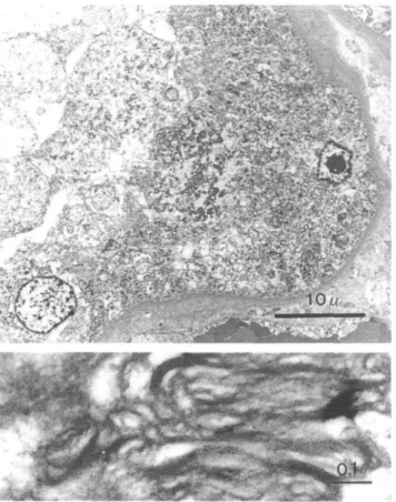
of both cardiac ventricles with signs of relative coron-ary insufficiency, micronodular cirrhosis and cholest-asis of the liver, adenoma of Papilla vateri, focuses of infarction of the spleen, and atrophy of the adrenal cortex. Deposits of ceramid-trihexosid were demon-strable in walls of blood vessels, myocardial cells, Kupffer cells and also in liver cells. The patient's own kidney showed massive cortical atrophy and complete loss of glomeruli. Residual tubules were dilated and tubular epithelium appeared vacuolated in paraffin sections (Figure 1). Electron-microscopy revealed mas-sive deposits of ceramid-trihexosid in tubular epithe-lium, tubular lumen, interstitial macrophages, and endothelial cells. Although the kidney transplant was of normal light-microscopic appearance, ultrastruc-turally deposits of ceramid-trihexosid in endothelial cells (Figure 2) as well as in tubular epithelium (Figure 3) were observed.
Discussion
In this 46-year-old male patient, recurrent Fabry's disease was noted in the transplanted kidney 14 years after successful allotransplantation. There was an accu-mulation of ceramid-trihexosid in several organs as well as in tubular epithelial cells and endothelial cells of the transplanted kidney.
The description of recurrence of Fabry's disease in the transplanted kidney is rare. We found only four other cases in the literature [4-7]. In 1981 we reported a case from our centre in which, 14 years after success-ful transplantation, no signs of de novo Fabry's disease in the transplanted kidney could be found [8].
Four possibilities for recurrence can be thought of: 1. Inadequate enzyme replacement for complete
meta-bolization of the ceramid-trihexosid produced. 2. Increased production of ceramid-trihexosid, which
cannot be totally metabolized in presence of normal enzyme activity. This seems to us the most probable hypothesis. It explains why only small amounts of
H. Gantenbein et al
*
Hi
Fig. I. Patient's kidney (H&Ex460). Vacuolization of tubular
epithelium.
Fig. 2. Kidney transplant (EM x 3700, inset x 83 700). Formalin
fixed and refixed in glutaraldehyde. Endothelial cell showing extens-ive ceramid-trihexosid deposition. Inset demonstrates lamellar struc-tures with periodicity.
ceramid-trihexosid are found (that are seen only by electron-microscopy) and why the deposits are always in the vascular endothelium. Friedlander
et al. [4] performed a renal biopsy in a patient 8
years after transplantation with normal renal func-tion. Light-microscopy showed no abnormalities, but electron-microscopy revealed occasional small myelin figures in the vascular endothelium compat-ible with early accumulation of ceramid-trihexosid. Mosnier et al. [5] also described a case in which autopsy was performed II years after successful transplantation. Sphingolipid inclusions were found in endothelial cells of capillaries by electron-microscopy.
Recolonization of the graft vascular system through host endothelium. This theory was formulated by McMahon et al. [6]. They described a male patient with blood group B, who was transplanted with a kidney from a donor with blood group O. They showed, that the donor allograft endothelium was slowly replaced by cells displaying both cytoplasmic metabolic inclusions and surface immunological markers specific to the host phenotype with com-plete recolonization of the graft vascular system by host endothelium.
Presence of a circulating inhibitor of a-galactosid-ase. Faraggiana et al. [7] described a patient who died of heart failure 6 months after kidney
trans-Recurrence of Fabry's disease in a renal allograft
Fig. 3. Kidney transplant (EM x 900, inset x 83 700). Formalin fixed and refixed in glutaraldehyde. Tubular epithelium snowing ceramid-trihexosid deposition. Inset demonstrates lamellar structure with periodicity.
plantation. They found a small number of typical deposits in the transplanted kidney. In this case a circulating a-galactosidase inhibitor was demon-strated, but this was not investigated in our patient. Survival of the allograft in our patient for 14 years with well-maintained structure of the kidney at autopsy suggests that the indication for kidney allotransplant-ation is not limited by the possibility of graft failure from recurrence of Fabry's disease.
For other organ damage it is expected that progres-sion of pre-existing leprogres-sions should be slowed down. Cardiac pathology in the patient reported included mainly dilatation, which is compatible with normal blood pressure without medical treatment observed clinically. The possibility that the amount of
ceramid-289
trihexosid deposited in myocardial cells contributed to terminal cardiac insufficiency cannot be totally excluded.
Concerning the course of hepatitis B, high antigen titres up to 280 000 E/ml in the absence of anti-Hbs antigen and positive Hbe antigenaemia in 1991 were observed. GOT and GPT levels were elevated slightly and intermittently during the whole course. This con-stellation is very likely to be related to immunosuppres-sive therapy. An influence of Fabry's disease of the liver on the course of the hepatitis B infection seems rather remote.
In summary, this case illustrates that kidney trans-plantation cannot heal Fabry's disease by enzyme transplantation in all patients. Several explanations for recurrent disease are discussed which may explain the variable occurrence of Fabry's deposits in transplanted kidneys. Nevertheless a patient can be successfully treated for kidney failure during an extended time period. The extent of heart damage might decide the long-term survival of patients (8, this report). Ethical concern to include patients with Fabry disease in transplantation programmes seems not to be justified even in times of transplant organ shortage.
References
1. Desnick RJ, Bishop DF. Fabry disease: a-galactosidase defi-ciency. In: Scriver R, Beaudet AL, Sly WS, Valle D. The
metabolic basis of inherited disease. McGraw-Hill, New York,
1989: 6th edn: 1751-1780
2. Clarke JTR, Guttman RD, Wolfe LS, Beaudoin JG, Morehouse DD. Enzyme replacement therapy by renal allotransplantation in Fabry's disease. N Engl J Med 1972; 287: 1215
3. Krivit W, Desnick RJ, Bernloko RW, Wold F. Enzyme trans-plantation in Fabry's disease. N Engl J Med 1972; 287: 1248 4. Friedlaender MM, Kopolovic J, Rubinger D et al Renal biopsy
in Fabry's disease eight years after successful renal transplanta-tion. Clin Nephrol 1987; 27: 206-211
5. Mosnier JF, Degott C, Bedrossian J et al. Recurrence of Fabry's disease in a renal allograft eleven years after successful renal transplantation. Transplantation 1991; 51: 759-762
6. MacMahon J, Tubbs R, Gephardt G, Steinmuller D. Pseudo recurrence of Fabry's disease in renal allograft (Abstract). Lab
Invest 1986; 54: 42A
7. Farragiana T, Churg J, Grishman E et al Light- and electron-microscopic histochemistry of Fabry's disease. Am J Pathol 1981; 103: 247-262
8. Bannwart F. Morbus Fabry. Licht- und elektronen mikroskop-ischer Herzbefund 12 Jahre nach erfolgreicher Nierentransplantation. Schweiz Med Wocheuschr 1982; 112: 1742-1747
Received for publication: 9.8.94 Accepted: 20.10.94