In the adult nervous system, glutamatergic neurotransmission is
tightly controlled by neuron–glia interactions through glial glutamate
reuptake by the specific transporters GLT-1 and GLAST. Here, we
have explored the role of these transporters in the structural and
functional maturation of the somatosensory cortex of the mouse. We
provide evidence that GLT-1 and GLAST are early and selectively
expressed in barrels from P5 to P10. Confocal and electron
microscopy confirm that the expression is restricted to the astroglial
membrane. By P12, and despite an increased global expression as
observed by immunoblotting, the barrel pattern of GLAST and GLT-1
staining is no longer evident. In P10 GLT-1 –/– and GLAST –/– mice,
the cytoarchitectural segregation of the barrels is preserved.
However, at P9–10, the functional response to whisker stimulation,
measured by deoxyglucose uptake, is markedly decreased in GLT-1
–/– and GLAST –/– mice. The role of GLAST is transient since the
metabolic response is already restored at P11–12 in GLAST –/– mice
and remains unchanged in adulthood. However, deletion of GLT-1
seems to impair the functional metabolic response until adulthood.
Our data suggest that astrocyte–neuron interactions via the glial
glutamate transporters are involved in the functional maturation of
the whisker representation in the somatosensory cortex.
Introduction
Glutamate plays a major role during brain development (McDonald and Johnston, 1990). Glutamate modulates neuronal migration (Komuro and Rakic, 1993), outgrowth of neuronal processes (Rajan and Cline, 1998) and synapse elimination (Rabacchi et al., 1992). This widespread role is mediated through glutamatergic receptors whose properties and expres-sion change during development (Bahn et al., 1994; Catania et al., 1994; Ozawa et al., 1998). Pre- and post-synaptic markers of glutamatergic pathways are commonly over-expressed during development, and periods of over-expression often coincide with times of great synaptic plasticity when appropriate neur-onal connections are consolidated (Rema and Ebner, 1996; Romano et al., 1996; Blue et al., 1997; Brennan et al., 1997). Besides neurons, glial cells are active components of gluta-matergic transmission (Araque et al., 2001; Bezzi and Volterra, 2001). The glial glutamate transporter proteins GLAST and GLT-1 represent the main mechanism of glutamate removal from the extracellular f luid, thereby preventing excessive stimulation of glutamate receptors [for review see (Danbolt, 2001)]. Knockout of GLT-1, either by chronic antisense oligonucleotide adminis-tration (Rothstein et al., 1996) or by inactivation of the gene, results in excitotoxic neurodegeneration and epilepsy. The preferential localization of glial glutamate transporters around synapses (Chaudhry et al., 1995) suggests that in addition to carrying out a protective role, these transporters play a role in neurotransmission. Electrophysiological recordings have indeed provided evidence that glial glutamate transporters are im-portant in determining the time-course of synaptic responses at excitatory synapses (Bergles and Jahr, 1997; Diamond and Jahr,
1997). Recently, a reduction in glutamate clearance associated with a decrease of glial coverage in the vicinity of synapses was shown to affect transmitter release through modulation of pre-synaptic metabotropic glutamate receptors (Oliet et al., 2001). Astrocytic wrapping of neurons may therefore contribute to the regulation of synaptic efficacy in the central nervous system.
The developmental changes of glial glutamate transporters suggest that these proteins play an active role in the develop-ment of the CNS. GLAST and GLT-1 mRNAs and proteins are present in the spinal cord, in telencephalic and mesencephalic CNS proliferative zones at early developmental stages [embryonic days (E) 11–15] (Shibata et al., 1996, 1997; Sutherland et al., 1996; Yamada et al., 1998). GLAST expression is low at birth, except in the cerebellum, and increases in all regions during maturation. Near birth, GLT-1 protein levels are very low but the expression dramatically increases in many brain areas at the beginning of the second postnatal week (Furuta et al., 1997; Ullensvang et al., 1997). The fact that the levels of GLAST and GLT-1 markedly increase during the most active period of synaptogenesis (Furuta et al., 1997; Ullensvang et al., 1997) corroborates previous experiments indicating that the vesicular uptake of glutamate also increases in parallel with synapto-genesis (Christensen and Fonnum, 1992). In adult animals, GLAST and GLT-1 are expressed in almost all brain regions but GLT-1 is the quantitatively dominating glutamate transporter with the notable exception of the cerebellum (Lehre and Danbolt, 1998). Using double-labeling post-embedding immuno-cytochemistry, it has been shown that the two excitatory amino acid transporters GLT-1 and GLAST can occur side by side in the same astroglial plasma membrane (Haugeto et al., 1996). While evidence suggests a close connection between patterns of glu-tamate transporters expression, synapse formation and astrocyte development, no direct link has been yet demonstrated.
The rodent somatosensor y system, with its one-to-one correspondence between each vibrissa follicle and its cortical, barrel-like projection area within the somatosensor y cortex, provides a useful model to study the role of the glial glutamate transporters during development. Numerous experiments have been performed to identif y the molecular mechanisms underlying the establishment of these patterned topographic connections [for review see (Killackey et al., 1995)]. These patterns are consolidated during the first postnatal week, and are subject to alterations if the sensor y periphery is perturbed by nerve or whisker lesions during a critical period. Early neural activity might, therefore, be essential for the development and plasticity of central patterns. There is strong evidence for a major role of excitatory amino acid transmission in both development and plasticity of the barrel cortex. The thalamocortical afferents are glutamatergic (Agmon and O'Dowd, 1992) and excitatory glutamatergic synapses are functional at birth (Kim et al., 1995). More recent studies indicate that cortical N-methyl-D-aspartate
Glial Glutamate Transporters and
Maturation of the Mouse Somatosensory
Cortex
Brigitte Voutsinos-Porche1, Graham Knott2, Kohichi Tanaka3,4, Charles Quairiaux2, Egbert Welker2and Gilles Bonvento1,5 1CNRS FRE 2363, Paris, France,2Institut de Biologie Cellulaire et Morphologie, University of Lausanne, Lausanne, Switzerland, 3Medical Research Institute, Tokyo Medical and Dental
University, Tokyo, Japan,4PRESTO, Japan Science and Technology Corporation, Saitama, Japan and5UR A CEA CNRS 2210, Service Hospitalier Frederic Joliot, CEA, Orsay, France
(NMDA) glutamate receptors are essential for the aggregation of layer I V cells into barrels and development of the thalamo-cortical patterns (Fox et al., 1996; Iwasato et al., 1997, 2000). Metabotropic glutamate receptors (mGluRs) have also been identified as components of a signaling pathway that couples thalamic activity to the cytoarchitectural differentiation of the barrel cortex (Hannan et al., 2001).
Finally, our recent experimental data support the notion that the glial glutamate transporters are key components of a metabolic signaling pathway that lead to the transfer of energy substrates to activated glutamatergic neurons in the mouse developing cortex (Voutsinos-Porche et al., 2003).
All these observations prompted the suggestion that the glial glutamate transporters may play an important role during formation of the barrels. However, the precise contribution of GLAST and GLT-1 to this process is unknown. Here, we sought to determine in the developing and adult barrel cortex (i) the spatial and temporal regulation of both glutamate transporters, and (ii) the anatomical and functional consequences of in-activating GLAST or GLT-1. Using knockout mice for each glutamate transporter, the data suggest that astrocyte–neuron interactions are involved in the functional maturation of the somatosensory cortex.
Materials and Methods
The experimental protocols were approved by the Bureau de la Protection A nimale of the Ministère de l’Agriculture (authorization no. 75–01 to G.B.). Postnatal pups were obtained from pregnant C57 BL/6 mice (JA N VIER, France). The day of birth was designated as postnatal day 0 (P0). Homozygote wild-type +/+, GLAST –/– and GLT-1 –/– mice of different postnatal days were obtained from GLAST and GLT-1 hetero-zygote +/– breader pairs (Tanaka et al., 1997; Watase et al., 1998). They were maintained under standard conditions of feeding and lighting (12 h/12 h light/dark cycle, 22°C). The genotyping of all mice was a priori (adult mice) or a posteriori (infant mice) determined by polymerase chain reaction.
Immunohistochemistry/Cytochrome Oxidase Histochemistry Brains from infant mice were removed and immersed in 4% para-formaldehyde in 0.1 M phosphate buffer (PB) at pH 7.4 for 24 h, while adult mice were perfused transcardially with 4% paraformaldehyde in 0.1 M PB, pH 7.4. Brain tissues were cr yoprotected in 20% sucrose in 0.1 M PB for 24 h. The forebrain was separated from the brainstem at the level of the superior colliculus. The forebrain was bisected mid-sagittally, and each hemisphere was fitted to a small plastic wedge allowing tangential cutting of the cortical barrel field. The tissue with wedge were together immersed and frozen in isopentane at –40°C and mounted onto a cryostat object holder with cooled embedding matrix (M-1, Lipshaw). Each hemisphere was processed for cytochrome oxidase histochemistr y or GLAST, GLT-1 and glutamine synthetase (GS) immunohistochemistry. The tangential sections for cytochrome oxidase histochemistry were incubated in the dark at 40°C for 6 h in 0.1 M PB containing 4% sucrose, 0.6 mg/ml 3,3′-diaminobenzidine tetrahydro-chloride (D-5905 Sigma) and 0.4 mg/ml cytochrome C (C-7752 Sigma).
For GLAST or GLT-1 immunohistochemistry, postnatal (P5, P7, P10, P12) and adult tangential sections were pretreated with 0.3% H2O2in phosphate-buffered saline (PBS, 0.02 M, pH 7.4) for 15 min, rinsed in PBS and incubated in 5% normal goat serum in PBS (1 h), primary antibody GLAST or GLT-1 (0.1µg/ml) (Shibata et al., 1997; Yamada et al., 1998) with 2% normal goat serum and 0.1% Tween-20 in PBS (12 h), secondary antibody (biotinylated goat anti-rabbit IgG) diluted at 1:200 with 0.1% Tween-20 in PBS for 1 h, and then avidin–biotin complex diluted at 1:200 for 1 h (Vector Laboratories). Diaminobenzidine tetrahydrochloride with H2O2was used as the peroxidase substrate to visualize sites of antibody binding. For GS immunohistochemistr y, P10 tangential sections were pretreated with 0.3% H2O2in PBS for 15 min, rinsed in PBS and incubated sequentially in 5% normal horse serum in PBS (1 h), and similarly processed with 0.25µg/ml mouse polyclonal anti GS (Interchim) and
secondary antibody (biotinylated goat anti-mouse IgG) diluted at 1:200. For double labeling of GS/GLAST and GS/GLT-1, coronal free-f loating sections (10 µm) from P10 mice were incubated sequentially in 5% normal goat serum in PBS for blocking (1 h), primary antibody GS 0.25µg/ml with GLAST or GLT-1 (0.1 µg/ml) with 2% normal goat serum and 0.1% Tween-20 in PBS (12 h), secondary antibody (A lexa Fluor 488-conjugated goat anti-rabbit IgG and A lexa Fluor 568-conjugated goat anti-mouse IgG; Interchim) diluted at 1:200 with 0.1% Tween-20 in PBS for 1 h. Sections were examined under a confocal laser scanning microscope (BioRad MRC 600, Hertfordshire, UK) attached to a Nikon Optophot microscope using epif luorescence. Images were acquired using double excitation at 488 and 568 nm and were recorded through separate channels. Serial plane images were collected at 1µm intervals at a given depth or throughout the whole thickness of the f luorescent preparation.
Electron Microscopy
To localize the GLT-1 and GLAST transporters at the ultrastructural level, two separate pre-embedding immunocytochemistr y protocols were carried out which used two different secondar y antibodies: the first (protocol 1) used a biotinylated secondar y antibody, subsequently enhanced with DA B, and the second (protocol 2), a gold-labeled antibody enhanced with silver.
Anesthesia and Fixation
Infant (P9 and P12) and adult (P60) mice, C57 BL/6, were anesthetized with sodium pentobarbitone (6 mg/100 g body wt, i.p.) and perfused immediately with 100 ml of 0.2% glutaraldehyde, 2% paraformaldehyde and 0.2% picric acid in 0.1 M PB. One hour after perfusion, the brains were removed and 50µm vibratome (Leica VT100) sections cut tan-gentially from the barrel cortex. Sections containing the barrels at the level of layer I V were identified under a dissecting microscope, and prepared for immunocytochemistry.
Protocol 1
Sections were cryoprotected in 2% glycerol and 20% DMSO in 0.1 M PBS, for 15 min, and freeze–thawed twice in liquid nitrogen to improve the antibody penetration. A fter washing three times in PBS (0.1 M, pH 7.4) sections were then pretreated in 0.3% hydrogen peroxide in PBS for 10 min. Prior to first antibody exposure, sections were blocked in PBS containing 5% NGS and 0.1%BSA-c (Aurion, The Netherlands), three times, 10 min each rinse, and then incubated overnight in the primary antibody (GLT-1, 1/5000; GLAST, 1/10 000) in PBS containing (0.1 M, pH 7.4; 0.1% BSA-c) overnight at 4°C. A fter washing, the sections were added to the biotinylated secondar y antibody [1/500 goat antirabbit (F)ab fragment, Jackson Laboratories, USA], in the same buffer and left for 4 h before being washed in TBS (0.1 M, pH 8.0), incubated in avidin–biotin peroxidase complex (A BC Elite, Vector Laboratories, USA) for 2 h, and finally incubated in 3,3′-diaminobenzidine tetrachloride (Fluka, Switzer-land) and 0.015% H2O2. Following enhancement, the sections were again washed in the same TBS buffer and then in cacodylate buffer (0.1 M, pH 7.4) prior to secondary fixation and embedding.
Protocol 2
Sections were cryoprotected in 25% sucrose and 10% glycerol, in 0.1 M PB, for 15 min, and freeze–thawed twice in liquid nitrogen. A fter washing three times in PBS (0.1 M, pH 7.4) sections were then pretreated in 0.05 M glycine in PBS for 10 min. Prior to first antibody exposure, sections were blocked in PBS containing 5% NGS and 0.1%BSA-c (Aurion), three times, 10 min each rinse, and then incubated overnight in the primary antibody (GLT-1, 1/5000; GLAST, 1/10 000) in PBS containing (0.1 M, pH 7.4; 0.1% BSA-c) overnight at 4°C. A fter washing, the sections were added to the gold-labeled secondary antibody [1/100 goat antirabbit (F)ab fragment (Ultra Small ImmunoGold, Aurion)], all in the same buffer. These were left for 4 h at room temperature and then thoroughly washed in HEPES buffer (0.15 M, pH 8.0). Ultra small gold particles were silver enhanced using pre-prepared solutions (Aurion R-Gent SE-EM, Aurion) and left for a total of 50 min in the dark at room temperature. Following enhancement, the sections were again washed in the same HEPES buffer
and then in cacodylate buffer (0.1 M, pH 7.4) prior to secondary fixation and embedding.
Secondary Fixation and Embedding
Sections were postfixed in 1% osmium tetroxide in cacodylate buffer (0.1 M), dehydrated in alcohol and then embedded between silicon-coated glass slides in Durcapan ACM resin (Fluka). Once the resin had cured, after 48 h at 60°C, the cortical barrels could be identified and a trapezoid prepared of a layer IV barrel hollow only. Silver/grey (60 nm thickness) thin sections were cut using an ultramicotome (Reichert, Ultracut E) and placed on formvar-coated, single-slot, gold grids. Images were taken using a Phillips CM100 electron microscope at a filament voltage of 80 kV and collected using a charge-coupled device (Gatan, USA).
Immunoblotting
Fresh frozen cortical samples containing∼10 barrel columns were taken in the middle of the barrel field of P6, P9 and P40 mouse. They were homogenized in 10 mM HEPES/KOH, 100µm DTT, 0.32 M sucrose and 1% Triton X-100 with protease inhibitors in a total volume of 300µl. The homogenate was centrifuged at 14 000 g (10 min at 4°C) and the supernatant was collected. Protein concentrations were determined by the method of Lowry (Lowry et al., 1951) using bovine serum albumin as standard. Samples were mixed (v/v) with buffer (63 mM Tris–HCl, pH 6.8, 2% sodium dodecyl sulfate, 1% glycerol, 0.05% bromophenol blue and 5% 2-mercaptoethanol). Electrophoresis of total proteins (30µg) was performed on a SDS–polyacrylamide gel (8% acrylamide/bis-acrylamide, Sigma). The proteins were transferred to PVDF membrane (Biorad) with buffer (25 mM Tris–HCl, pH 8.3, 20% methanol, 0.1% SDS,192 mM glycine) by electroblotting. Blots were blocked with 5% nonfat dry milk in TBST (20 mM Tris–HCl, pH 7.5, 500 mM NaCl, 0.1% Tween-20) at room temperature and then incubated with primary antibody anti-actin (42 kDa) (1/2000) (A2066 Sigma) plus GLAST (65 kDa) or GLT-1 (72 kDa) (0.05µg/ml) or GS (45 kDa) (0.25µg/ml) (Interchim), overnight at 4°C. A fter primar y antibody incubation, membranes were washed and incubated with peroxidase-conjugated secondary antibodies (1/2000) and developed with ECL (PPN 2108-Amersham) using Kodak X-OMAT film. The resulting bands were digitized and immunoreactive proteins semi-quantitatively evaluated by densitometry using the NIH image analysis system. For semi-quantitative evaluation of the relative level of expression of GLAST, GLT-1 and GS, the optical densities of the bands were normalized to the optical density of actin in each animal.
W hisker Stimulation and Analysis of 2-Deoxyglucose (2-DG) Uptake
Homozygote wild-type +/+, GLAST –/– and GLT-1 –/– infant (P9–10, P11–12) and adult mice were gently restrained and all whiskers except the two most caudal whiskers of row C (C1 and C2) of both whiskerpads were clipped. Mu metal pieces (1.5 mm long, 0.2 mm diameter) were fastened onto the C1C2 whiskers of both sides with cyanoacrylic glue. The mice were immobilized on a support and received an i.p injection of 2-[1–14C]deoxy-D-glucose (16.5 µCi/100g body wt) (New England Nuclear) in 0.9% NaCl. They were immediately placed in the Lausanne whisker stimulator as previously described (Melzer et al., 1985). The stimulation consisted of magnetic field bursts that were delivered at 50 Hz, the burst duration was 46 ms and the interval between bursts 90 ms. Forty-five minuntes after 2-DG injection, the forebrain was pro-cessed as previously described (see above). The tangential sections were cut at 20µm, mounted on slides and dried on a 60°C hot plate. The slides were then processed for autoradiography together with calibrated 14C-standards (A RC) on BIOM A X film (Kodak) for 10 days at room temperature. Thereafter, the slides were Nissl stained, dehydrated and cover-slipped. The outlines of the sections and the barrels were drawn using a microscope fitted with a drawing tube. The autoradiograms were digitized and analyzed using a computer-based image analysis system (Biocom 2000, France). The gray levels determined on the autoradio-grams were calibrated using the co-exposed densitometric microscales and converted to nCi/g tissue. The [14C] concentration was determined within the C1–C2 barrels and was repeated on all serial sections through layer I V that contained these barrels (4–6). For each animal, a reference value (background) was calculated as the mean 2-DG uptake in
unstimulated barrels measured in the same consecutive series of section. This reference value was used to calculate the relative 2-DG uptake using the formula: relative 2-DG uptake = (2-DG uptake – reference value)/ reference value. This value, expressed as a percentage, was determined in each animal. Pseudo-colored images were generated by replacing the range of 256 gray level values with 16 colors. The correspondence between the color code and the 2-DG uptake is presented (in nCi/g tissue) in each case on a scale.
Results
Expression of GLAST and GLT-1 in the Somatosensory Barrel Cortex of Mice During Development
Immunolabeling of GLAST and GLT-1, performed on tangential sections through layer IV of the primary somatosensory cortex of P5, P7 and P10 mice, revealed the entire vibrissae-related pattern (Fig. 1). Therefore, at these ages, barrels of the five major rows are enriched in both glial glutamate transporters. The immuno- reactivity was confined to the barrel hollows, leaving septa relatively free of staining. By P12, and despite an increased expression (see below), the barrel pattern of GLAST and GLT-1 staining was no longer evident since the individual barrels were not visible within these rows.
To investigate further the cellular localization of GLAST and GLT-1 within the barrels, we performed double immunostaining of GLAST and GLT-1 with GS, a specific cytoplasmic astrocytic protein. Within the barrel cortex of P10 mice, we obser ved a strong GS immunolabeling of astrocytes, which also revealed the entire vibrissae-related pattern (Fig. 2A). As for GLAST and GLT-1, the immunoreactivity was confined to the hollows (Fig. 2B). Using immunof luorescent confocal laser-scanning microscopy, GS reaction product was localized within the cytoplasm around the nucleus and in the processes of the astrocytes. GLAST and GLT-1 immunostaining was restricted to membrane profiles including the outer limiting membrane, suggesting that GLAST and GLT-1 were strictly localized in astroglial cells (Fig. 3).
Immunoelectron microscopy at P9, P12 and adulthood revealed both GLAST (Fig. 4b,d,f), and GLT-1 (Fig. 4a,c,e) labeling within only the astrocytic processes, which was most easily seen with DA B. Gold labeling revealed the presence of GLT1 and GLAST at the level of the astrocytic membrane. No labeling was seen in neurons at any age. The highest density of labeling was seen in regions of neuropil that were not abutting cell bodies: neuronal, glial or endothelial. GLT-1 labeling appeared in the close vicinity of asymmetric synapses-presumed excitatory (Baude et al., 1995), particularly those on dendritic spines. The comparison of labeling between the two antibodies showed that GLAST was more prolific, throughout the neuropil, with dense labeling seen along astrocytic processes for larger distances in comparison with GLT-1, which appeared more sporadic.
Immunoblot Analysis
Immunoblotting was used to measure changes in the relative level of expression of the glutamate transporters GLAST, GLT-1 and of GS in the barrel cortex during development. Samples of barrel cortex from P6, P9 and P40 mice (n = 3 for each age) were studied. GLAST and GLT-1 antibodies detected distinct proteins with molecular weights of∼65 and ∼72 kDa respectively (Fig. 5). GS antiserum recognized a major protein of ∼45 kDa. During postnatal development, GLAST, GLT-1 and GS expression increased progressively with age to reach adult levels (Fig. 5). Compared to GLAST, the expression of both GLT-1 and GS increased very rapidly and with a similar time course after P6. At
P9, the level of expression of all proteins studied is already∼75% of the adult level.
Anatomical Analyses of the Somatosensory Cortex in GLAST and GLT-1 KO Mice
To explore the possible implication of glial glutamate trans-porters in the mechanisms underlying the formation of the barrels, we examined the somatosensory cortex of mutant mice that lack the expression for either GLAST or GLT-1. As previously
reported (Tanaka et al., 1997) (also K. Tanaka et al., unpublished data), we obser ved that GLT-1 –/– and GLAST –/– mice gained weight more slowly than wildtype mice. At P9–10, body weight (g) of GLAST and GLT-1 null mice were significantly smaller compared to their respective wildtype mice: 4.4 ± 0.7 (n = 3) vs. 6.2 ± 0.7 (n = 5) (P < 0.05) (mean ± SD, Student’s paired t-test) and 4.8 ± 1.1 (n = 12) vs. 5.9 ± 1.1 (n = 9) (P < 0.05). Such a difference was still observed at P11–12: 5.4 ± 0.6 (n = 5) vs. 6.9 ± 1.0 (n = 8) (P < 0.05) and 5.5 ± 0.7 (n = 7) vs. 6.2 ± 0.3 (n = 9) (P < 0.05) for GLAST and GLT-1, respectively. The body weight (g) and general appearance of the adult GLAST wildtype and –/– mice were normal: 17.3 ± 5.6 vs. 17.6 ± 5.7, respectively (n = 5 for each group; Ns). Homozygote GLT-1 mice were 30% smaller than wildtype mice: 11.4 ± 2.5 (n = 5) vs. 16.4 ± 1.7 (n = 6) (P < 0.05) and tended to die prematurely.
In tangential sections from P10 and adult GLAST or GLT-1 null mice, Nissl staining revealed the cytoarchitectural contours of individual barrels typical of wild-type mice (Fig. 6A,C,E). To determine whether barrels from null mice receive functional thalamocortical afferents, we performed cytochrome oxidase (CO) staining. In P10 and adult GLAST or GLT-1 (–/–) mice, CO staining, which labels highly active synaptic regions at the terminals of thalamic fibers, revealed dense patches which were organized into five rows in a similar orientation and with a similar density as in wild-type mice (Fig. 6B,D,F).
Functional Analyses of the Somatosensory Cortex in GLAST and GLT-1 KO Mice
Accumulation of 2-DG is commonly used to determine the level of neuronal activation in specific brain areas. This method has been used successfully in several paradigms including activation of a somatosensory pathway such as the whisker-to-barrel system (Cholet et al., 1997; Welker et al., 1996). Therefore, we inves-tigated whether the absence of either GLAST or GLT-1 might have a functional consequence within the barrel field during development. For all experiments, P9–10, P11–12 and adults
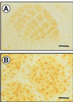
Figure 1. GLAST and GLT-1 immunohistochemical staining in the mouse barrel cortex
during development. From P5 to P10, GLAST and GLT-1 immunohistochemical staining performed on tangential sections shows individual cell clusters corresponding to mystacial vibrissae. From P12 until adulthood, immunolabeling of GLAST and GLT-1 shows immunoreactive punctae but does not delineate the adult barrel field giving rise to a mottled image at this magnification. Scale bar = 500µm.
Figure 2. Glutamine synthetase (GS) immunohistochemical staining in the mouse
barrel cortex at postnatal day 10. Individual barrels fields are parceled by immuno-positive GS reactivity (A) which is associated with astrocytes and their processes. Few GS-positive astrocytes arborize the septa (B). Scale bar: A, 400µm; B, 100 µm.
+/+ and –/– from the same littermates were comparatively examined. In wildtype mice, stimulation of the two most caudal follicles of row C (C1 and C2) on the left side, evoked an increase in 2-DG uptake in the corresponding part of the somatosensory cortex (Figs 7 and 8). The activated zone was centered in C1 and C2 barrels of primary somatosensory cortex and was observed in 5–8 consecutive tangential sections. In wildtype mice, the relative level of stimulus evoked 2-DG uptake increased sig-nificantly during development, from∼15% at P9–10 to >35% in the adult (Figs 7 and 8).
In P9–10 GLAST –/– mice, the 2-DG uptake was lower than the uptake in the wildtype (5.9 ± 4.9% vs. 16.8 ± 4.9%, Student’s t-test). However, at P11–12 and in adult GLAST –/– mice, the metabolic response to whisker stimulation was not significantly different from that found in the wildtype: 29.9 ± 9.6% vs. 32.8 ± 7.8% and 39.3 ± 2.2% vs. 38.3 ± 4.0%, respectively (Fig. 7). In P9–10 GLT-1 –/– animals, the 2-DG uptake was also lower than in the wildtype (6.3 ± 5.6% vs. 15.8 ± 5.0%). However, in these mice, the metabolic response remained lower than that in wildtype animals until adulthood: 20.3 ± 4.6% vs. 24.8 ± 5.7% and
22.8 ± 7.7% vs. 33.2 ± 5.8% for the P11-P12 and the adult mice, respectively (Fig. 8).
Discussion
The primary finding reported here is that both glial glutamate transporters GLAST and GLT-1 proteins are expressed selectively and very early in the barrels of the mouse somatosensory cortex. Confocal and electron microscopy confirm that this expression is restricted to the astroglial membrane and localized in the close vicinity of excitatory synapses. While the lack of expression of either glial glutamate transporter does not appear to modif y significantly the cellular organization of the barrel cortex nor alter the expression of cytochrome oxidase, functional response to whisker stimulation, measured by 2-deoxyglucose uptake, is markedly decreased in P9–10 GLT-1 and GLAST –/– mice. The role of GLAST would appear to be transient since the metabolic response is already restored in P11-P12 GLAST –/– mice and unchanged in adult GLAST –/– mice. However, deletion of GLT-1 seems to impair the functional response continuously until adult-hood. These data suggest that astrocyte–neuron interactions via
Figure 3. Confocal microscopic images of GLAST, GLT-1 and glutamine synthetase (GS) in the mouse barrel cortex at postnatal day 10. Confocal microscopic images were acquired
sequentially through the green channel (GLT-1, GLAST) and the red channel (GS), and subsequently merged for the visualization of double labeling. GLAST and GLT-1 immunostaining was restricted to membrane profiles including the outer limiting membrane. GS reaction product was localized within the cytoplasm around the nucleus and in the processes of astrocytes. Merged images strongly shows that GLAST and GLT-1 were strictly localized in astroglial cells. Scale bar = 10µm.
Figure 4. Ultrastructural localization of GLT1 and GLAST in neuropil of layer IV barrel hollows of somatosensory cortex from P9, P12 and adult mice. Electron micrographs on the left
show the localization of the GLT1 transporter (a, c, e) and those on the right show that for GLAST (b, d, f). (a) GLT1 labeling, using DAB seen at P9, in astrocytic processes (arrow) close to a dendritic spine and its asymmetric synapse (arrowheads) as well adjacent to a neuronal cell body (star). (b) GLAST labeling, using DAB, seen at P9 in astrocytic processes (arrowed) surrounding an asymmetric synapse (arrowheads). (c) GLT-1 labeling, localized with gold conjugated antibody, at P12 in astrocytic elements surrounding asymmetric synapses (arrowheads) seen on a dendritic shaft and a dendritic spine (inset). (d) GLAST labeling, using gold-conjugated secondary antibody, at P12 in astrocytic processes surrounding a dendrite. An axodendritic synapse (arrowheads) is bordered by astrocytic elements, which show immunoreactivity for the glutamate transporter. (e) GLT-1 labeling shown with DAB in neuropil at P60 (adult) mice. Two asymmetric synapses are indicated (arrowheads), both axospinous, however the DAB reaction product localized in astrocytic elements (arrowed) is only seen in close proximity to one. (f) GLAST labeling shown with DAB in neuropil at P60 (adult) mice. Three asymmetric synapses are indicated, two axospinous and one axodendritic (lower left). Each synapse can be seen bordered by astrocytic elements, each containing dense reaction product. Micrographs a–d are the same magnification and the scale bar in a is 0.5µm. Micrographs e and f are at the same magnification, scale bar in f is 0.5 µm.
the glial glutamate transporters may be involved in the functional maturation of the whisker-to-barrel pathway. Developmental Expression of GLAST and GLT-1 in the Barrel Somatosensory Cortex
In the rodent trigeminal system, the topographic organization of the central whisker representations develops in a peripheral-to-central progression, from brainstem through thalamus to cerebral cortex. A fter thalamic barreloid maturation at P2, the cortical barrel field first appears at P3-P5 (Rice and Van der Loos, 1977; Senft and Woolsey, 1991). At this date, we have provided evidence showing that both GLAST and GLT-1 proteins are already expressed in the barrels of the somatosensory cortex. Therefore, GLT-1 expression can be obser ved in the cortex during the first postnatal week, earlier than previously demon-strated (Furuta et al., 1997; Ullensvang et al., 1997). Until P10, the staining of both glutamate transporters was higher in barrel hollows and much lower in the septa and in the surrounding cortex. The staining of GLAST and GLT-1 processes is often seen to ensheathe axospinous synapses with asymmetric (presumably glutamatergic) contact. This apposition to the synaptic cleft represents a favorable position for capturing synaptically released glutamate and for keeping the tonic synaptic glutamate
concentration low. This astroglial localization of GLT-1 and GLAST is in agreement with the notion that these glial cells have the largest glutamate uptake activity (Danbolt, 2001). Since GLAST and GLT-1 are almost exclusively located on astrocytes, results reported here strongly suggest a significant role of these glial cells during formation of the barrels. Previous experiments were mainly focused on the role of radial glial cells as guides for migrating neurons (Cooper and Steindler, 1986a; Crandall et al., 1990). A number of adhesion molecules synthesized by glial cells, such as cytotactin (Crossin et al., 1989), tenascin (Steindler et al., 1989) or lectins (Cooper and Steindler, 1986b), has been shown to be expressed transiently within the barrel boundaries. Our present obser vations indicate that molecules expressed locally by mature and not by radial astrocytes may also be involved in the functional maturation of the somatosensor y system. A fter P10, both GLT-1 and GLAST expression drama-tically increase in the whole cortex including the barrel cortex. Due to an increased expression at the level of the septa, between the barrels, the visualization of the barrel boundaries gets lost. This ‘septal expression’ from P10 onwards could well be the consequence of a functional maturation of intracortical con-nectivity within this compartment of the barrel cortex that, in time, follows the maturation of the neuropil in the barrel hollow. The expression of a somatototopic pattern by glutamate transporters is closely correlated with the establishment of a whisker-specific patterning of serotoninergic fibers (Fujimiya et al., 1986; D’A mato et al., 1987). This serotonergic pattern actually ref lects thalamocortical terminals and not raphe projec-tions (Lebrand et al., 1996) and is only transiently expressed during the first 10 postnatal days (D'Amato et al., 1987). In vitro recording from thalamocortical slices indicates that 5-HT exerts, via presynaptic 5-HT1B receptors, an inhibitor y effect on glutamatergic transmission (Rhoades et al., 1994; Laurent et al., 2002). Therefore, 5-HT and glial glutamate transporters could act as coordinated regulators of glutamatergic transmission during patterning of thalamocortical axon terminals.
Neuronal Activity-induced Patterning of the Somatosensory System: Role of Glial Glutamate Transporters?
Electrophysiological recordings from glial cells and neurons have shown that glial glutamate transporters may determine the time-course and spread of synaptic responses at excitator y synapses (Bergles and Jahr, 1997; Diamond and Jahr, 1997). Any manipulation of the glial glutamate transporters may therefore alter the activation of NMDA receptors which play a critical role in the patterning of the somatosensory cortex [for review see (Erzurumlu and Kind, 2001)]. Indeed, whole-cell recordings on thalamocortical synapses at developing (P3–8) layer IV neurons in the barrel cortex reveal that pharmacological inhibition of glutamate transport induces a massive activation of NMDA receptors and a reversible blockade of A MPA and kainate receptor-mediated dual component excitator y postsynaptic currents (A MPA/K A EPSCs) (Kidd and Isaac, 2000). The ob-ser vation that GLAST and GLT-1 are expressed early during development in the somatosensory cortex highlights the view that they could play an active role in the mechanisms leading to the structural and functional maturation of connections in the somatosensor y cortex. The consequences upon synaptic activity-induced by whisker stimulation in the somatosensory cortex of these mutant immature mice is not known. Electrophysiological results obtained so far in adult mice suggest that the alteration may be limited. Indeed, the decay of EPSCs measured in the hippocampus or in the cerebellum is not
Figure 5. Immunoblots for glutamate transporters subtypes and glutamine synthetase
(GS) in developing mouse barrel cortex. While the expression of GLAST at P6 is already 50% of the adult level and increases gradually with age, the expression of both GLT-1 and GS is relatively low at P6 (20–30% of the adult level) and increases very rapidly during the critical period of functional maturation to reach 75% of adult level at P9. Each point corresponds to the mean optical density measured in three different animals at each age.
significantly modified in adult GLT-1 and GLAST –/– mice (Tanaka et al., 1997; Watase et al., 1998), suggesting that glutamatergic neurotransmission is preser ved in these mice despite an alteration of the clearance of glutamate from the synaptic cleft. Our present observation indicates that the lack of expression of one or other glial glutamate transporter does not appear to modif y significantly the cellular organization of the barrel field nor alter the expression of cytochrome oxidase. Therefore, an apparently normal development of the whisker-to-barrel pathway occurred in the GLAST –/– and GLT-1 –/– mice. It should be mentioned that in these –/– mice, part of the glutamate transport is likely to be preser ved and carried out by the other non-targeted transporter so that any structural alteration, if any, may be limited. Indeed, we have shown that the level of expression of glutamine synthetase, the glial enzyme that converts glutamate to glutamine once transported via GLAST or GLT-1, is unchanged in P10 GLAST and GLT-1 –/– mice (data not shown).
Impairment of the Functional Metabolic Response to Whisker Stimulation in GLAST and GLT-1 –/– Mice During Development
Our functional analysis of the somatosensor y barrel system, using the uptake of 2-DG as a marker of neuronal activity, revealed that an alteration of the glutamate transport induces a
marked metabolic functional deficit. We provide evidence that the functional response to whiskers stimulation, measured by 2-DG uptake, is markedly decreased in P9–10 GLT-1 and GLAST –/– mice. The role of GLAST is transient since the metabolic response is already restored in P11–12 GLAST –/– mice and unchanged in adult GLAST –/– mice, while deletion of GLT-1 continuously impairs the functional response until adulthood. These results are in line with the observation that the develop-ment of the total glutamate uptake activity in the forebrain parallels that of GLT-1, so that GLT-1 accounts for∼93% of the total transport activity in adult rat forebrain (Ullensvang et al., 1997). Therefore, a good correlation exists between the capacity of glutamate uptake and the uptake of glucose during whisker activation for both glutamate transporters during development. Interestingly, the functional impairment as observed in both of the null mice at the earliest postnatal age studied, does not seem to prevent barrel formation nor does it seem to delay the structural development of the somatosensory cortex.
A classical role attributed to astrocytes has been to ensure an adequate metabolic supply to neurons. Based on observations that (i) astrocytes display a particular localization between blood vessels, (ii) neurons possess specialized processes called end-feet that come in close contact with capillaries (Kacem et al., 1998), and (iii) neurons express glucose transporters (Morgello et al., 1995), it has been suggested that astrocytes may constitute
Figure 6. Visualization of barrels in the primary somatosensory cortex in GLAST and GLT-1 P10 KO mice. (A, C, E) Nissl staining; (B, D, F) cytochrome oxidase (CO) histochemistry.
GLAST (C, D) and GLT-1 (E, F) P10 –/– mice display control barrel segregation as clustered granular cells surrounding a cell-sparse hollow and control CO staining, which label highly active synaptic regions at the terminals of thalamic fibers, as revealed by dense patches organized into five rows in a similar orientation and with a similar density as in wild-type mice. Scale bar: A, C, E, 100µm; B, D, F, 300 µm.
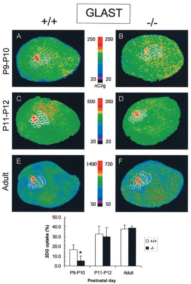
Figure 7. Effect of C1C2 whisker stimulation on 2-DG uptake in the somatosensory cortex of P9–10, P11–12 and adult GLAST +/+ and –/– mice. Representative pseudo-colored
digitized autoradiograms obtained from tangential sections through layer IV of the primary somatosensory cortex. The level of 2-DG uptake is color-coded according to the respective colored scales. Stimulation of C1C2 in +/+ mice (P9 to adult) produced a significant increase in 2-DG uptake in the somatosensory cortex that was restricted to the C1C2 barrels (A, C, E). In P9–10 GLAST –/– mice (B), the increase in 2-DG uptake was significantly lower (–64%) than in +/+ mice, while this metabolic response was not different from control matched +/+ mice in the C1C2 barrels of P11–12 (D) and adult GLAST –/– mice (F). Data are presented as mean ± SD; n = 3–8 animals per group (P < 0.05 ; Student’s t-test).
Figure 8. Effect of C1C2 whisker stimulation on 2-DG uptake in somatosensory cortex of P9–10, P11–12 and adult GLT-1 +/+ and –/– mice. Representative pseudo-colored digitized
autoradiograms obtained from tangential sections through layer IV of the primary somatosensory cortex. The level of 2-DG uptake is color-coded according to the respective colored scales. Stimulation of C1C2 in +/+ mice (P9 to adult) produced a significant increase in 2-DG uptake in the somatosensory cortex that was restricted to the C1C2 barrels (A, C, E). In P9–10 GLT-1 –/– mice (B), the increase in 2-DG uptake was significantly lower (–60%) than in +/+ mice. In P11–12 GLT-1 –/– mice (D), this metabolic response was slightly lower than that in wildtype P11–12 animals (–20%) and was significantly different from matched adult +/+ mice (–31%) in the C1C2 barrels of adult GLT-1 –/– mice (F). Data are presented as mean ± SD; n = 3–6 animals per group (P < 0.05; Student’s t-test).
a privileged site of glucose uptake as it penetrates the brain parenchyma. A series of results obtained in vitro have led to the hypothesis that synaptic activity at glutamatergic synapses could be detected by astrocytes via glutamate transport activity and would be translated into a metabolic signal (Pellerin and Magistretti, 1994; Poitry-Yamate et al., 1995; Takahashi et al., 1995). The mechanism proposed would provide a way of delivering energy substrates on demand to face energy needs resulting from an increased neuronal activity (Magistretti et al., 1999). Our recent in vitro investigation indeed demonstrates that glial glutamate transport represents a critical step for triggering enhanced glucose utilization, but also lactate release from astrocytes through a mechanism involving changes in intracellular Na+concentration (Voutsinos-Porche et al., 2003).
In conclusion, we show that the glial glutamate transporters GLAST and GLT-1 are expressed early in the mouse barrel somatosensory cortex. This observation suggests that molecules expressed locally by astrocytes may also be involved in the functional maturation of the somatosensory system. In addition to their essential glutamate removal role, these transporters are active components of a metabolic cross-talk between neurons and astrocytes. Since this metabolic dialog is regulated by synaptic glutamatergic activity and follows a developmental regulation, it further emphasizes the role of glutamatergic transmission in the development and plasticity of central patterns such as the whisker-to-barrel pathway.
Notes
The authors acknowledge the expert technical assistance provided by Pascal Steiner and Niels Daubolt. This work was supported by a Human Frontier Science Program grant no. RG118/1998-B (to G.B.) and by a Swiss National Science Foundation grant no. 3100-062112.00 (to E.W.).
Address correspondence to Gilles Bonvento, Service Hospitalier Frederic Joliot, UR A CEA CNRS 2210, 4 place du Général Leclerc, 91401 Orsay, France. Email: bonvento@shfj.cea.fr.
References
Agmon A, O’Dowd DK (1992) NMDA receptor-mediated currents are prominent in the thalamocortical synaptic response before maturation of inhibition. J Neurophysiol 68:345–349.
Araque A, Carmignoto G, Haydon P (2001) Dynamic signaling between astrocytes and neurons. Annu Rev Physiol 63:795–813.
Bahn S, Volk B, Wisden W (1994) Kainate receptor gene expression in the developing rat brain. J Neurosci 14:5525–5547.
Baude A, Nusser Z, Molnar E, McIlhinney R A, Somogyi P (1995) High-resolution immunogold localization of A MPA type glutamate receptor subunits at synaptic and non-synaptic sites in rat hippocampus. Neuroscience 69:1031–1055.
Bergles DE, Jahr CE (1997) Synaptic activation of glutamate transporters in hippocampal astrocytes. Neuron 19:1297–1308.
Bezzi P, Volterra A (2001) A neuron–glia signalling network in the active brain. Curr Opin Neurobiol 11:387–394.
Blue ME, Martin LJ, Brennan EM, Johnston MV (1997) Ontogeny of non-NMDA glutamate receptors in rat barrel field cortex: I. Metabotropic receptors. J Comp Neurol 386:16–28.
Brennan EM, Martin LJ, Johnston MV, Blue ME (1997) Ontogeny of non-NMDA glutamate receptors in rat barrel field cortex: II. A lpha-A MPA and kainate receptors. J Comp Neurol 386:29–45. Catania MV, Landwehrmeyer GB, Testa CM, Standaert DG, Penney JB Jr,
Young A B (1994) Metabotropic glutamate receptors are differentially regulated during development. Neuroscience 61:481–495.
Chaudhr y FA, Lehre KP, van Lookeren Campagne M, Ottersen OP, Danbolt NC, Storm-Mathisen J (1995) Glutamate transporters in glial plasma membranes: highly differentiated localizations revealed by quantitative ultrastructural immunocytochemistr y. Neuron 15:711–720.
Cholet N, Seylaz J, Lacombe P, Bonvento G (1997) Local uncoupling of the cerebrovascular and metabolic responses to somatosensor y
stimulation after neuronal nitric oxide synthase inhibition. J Cereb Blood Flow Metab 17:1191–1201.
Christensen H, Fonnum F (1992) The ontogeny of the uptake systems for glutamate, GABA, and glycine in synaptic vesicles isolated from rat brain. Neurochem Res 17:457–462.
Cooper NG, Steindler DA (1986a) Monoclonal antibody to glial fibrillary acidic protein reveals a parcellation of individual barrels in the early postnatal mouse somatosensory cortex. Brain Res 380:341–348. Cooper NG, Steindler DA (1986b) Lectins demarcate the barrel subfield in
the somatosensor y cortex of the early postnatal mouse. J Comp Neurol 249:157–169.
Crandall JE, Misson JP, Butler D (1990) The development of radial glia and radial dendrites during barrel formation in mouse somatosensory cortex. Brain Res Dev Brain Res 55:87–94.
Crossin KL, Hoffman S, Tan SS, Edelman GM (1989) Cytotactin and its proteoglycan ligand mark structural and functional boundaries in somatosensor y cortex of the early postnatal mouse. Dev Biol 136:381–392.
D’Amato RJ, Blue ME, Largent BL, Lynch DR, Ledbetter DJ, Molliver ME, Snyder SH (1987) Ontogeny of the serotonergic projection to rat neocortex: transient expression of a dense innervation to primary sensory areas. Proc Natl Acad Sci USA 84:4322–4326.
Danbolt NC (2001) Glutamate uptake. Prog Neurobiol 65:1–105. Diamond JS, Jahr CE (1997) Transporters buffer synaptically released
glutamate on a submillisecond time scale. J Neurosci 17:4672–4687. Erzurumlu RS, Kind PC (2001) Neural activity: sculptor of ‘barrels’ in the
neocortex. Trends Neurosci 24:589–595.
Fox K, Schlaggar BL, Glazewski S, O’Lear y DD (1996) Glutamate receptor blockade at cortical synapses disrupts development of thalamocortical and columnar organization in somatosensory cortex. Proc Natl Acad Sci USA 93:5584–5589.
Fujimiya M, Kimura H, Maeda T (1986) Postnatal development of sero-tonin nerve fibers in the somatosensory cortex of mice studied by immunohistochemistry. J Comp Neurol 246:191–201.
Furuta A, Rothstein JD, Martin LJ (1997) Glutamate transporter protein subtypes are expressed differentially during rat CNS development. J Neurosci 17:8363–8375.
Hannan AJ, Blakemore C, Katsnelson A, Vitalis T, Huber KM, Bear M, Roder J, Kim D, Shin HS, Kind PC (2001) PLC-beta1, activated via mGluRs, mediates activity-dependent differentiation in cerebral cortex. Nat Neurosci 4:282–288.
Iwasato T, Erzurumlu RS, Huerta PT, Chen DF, Sasaoka T, Ulupinar E, Tonegawa S (1997) NMDA receptor-dependent refinement of somatotopic maps. Neuron 19:1201–1210.
Iwasato T, Datwani A, Wolf A M, Nishiyama H, Taguchi Y, Tonegawa S, Knopfel T, Erzurumlu RS, Itohara S (2000) Cortex-restricted disruption of NMDA R1 impairs neuronal patterns in the barrel cortex. Nature 406:726–731.
Kacem K, Lacombe P, Seylaz J, Bonvento G (1998) Structural organization of the perivascular astrocyte endfeet and their relationship with the endothelial glucose transporter: a confocal microscopy study. Glia 23:1–10.
Kidd FL, Isaac JT (2000) Glutamate transport blockade has a differential effect on A MPA and NMDA receptor-mediated synaptic transmission in the developing barrel cortex. Neuropharmacology 39:725–732. Killackey HP, Rhoades RW, Bennett-Clarke CA (1995) The formation of a
cortical somatotopic map. Trends Neurosci 18:402–407.
Kim HG, Fox K, Connors BW (1995) Properties of excitatory synaptic events in neurons of primary somatosensory cortex of neonatal rats. Cereb Cortex 5:148–157.
Komuro H, Rakic P (1993) Modulation of neuronal migration by NMDA receptors. Science 260:95–97.
Laurent A, Goaillard JM, Cases O, Lebrand C, Gaspar P, Ropert N (2002) Activity-dependent presynaptic effect of serotonin 1B receptors on the somatosensor y thalamocortical transmission in neonatal mice. J Neurosci 22:886–900.
Lebrand C, Cases O, Adelbrecht C, Doye A, Alvarez C, El Mestikawy S, Seif I, Gaspar P (1996) Transient uptake and storage of serotonin in developing thalamic neurons. Neuron 17:823–835.
Lehre KP, Danbolt NC (1998) The number of glutamate transporter subtype molecules at glutamatergic synapses: chemical and stereological quantification in young adult rat brain. J Neurosci 18:8751–8757.
Lowry OH, Rosebrough N, Farr AL, Randall R (1951) Protein measure-ment with the Folin phenol reagent. J Biol Chem 193:265–275.
Magistretti PJ, Pellerin L, Rothman DL, Shulman RG (1999) Energy on demand. Science 283:496–497.
McDonald JW, Johnston MV (1990) Physiological and pathophysiological roles of excitator y amino acids during central nervous system development. Brain Res Brain Res Rev 15:41–70.
Melzer P, Van der Loos H, Dorf l J, Welker E, Robert P, Emery D, Berrini JC (1985) A magnetic device to stimulate selected whiskers of freely moving or restrained small rodents: its application in a deoxyglucose study. Brain Res 348:229–240.
Morgello S, Uson RR, Schwartz EJ, Haber RS (1995) The human blood– brain barrier glucose transporter (GLUT1) is a glucose transporter of gray matter astrocytes. Glia 14:43–54.
Oliet SH, Piet R, Poulain DA (2001) Control of glutamate clearance and synaptic efficacy by glial coverage of neurons. Science 292:923–926. Ozawa S, Kamiya H, Tsuzuki K (1998) Glutamate receptors in the
mammalian central nervous system. Prog Neurobiol 54:581–618. Pellerin L, Magistretti PJ (1994) Glutamate uptake into astrocytes
stimu-lates aerobic glycolysis: a mechanism coupling neuronal activity to glucose utilization. Proc Natl Acad Sci USA 91:10625–10629. Poitry-Yamate CL, Poitry S, Tsacopoulos M (1995) Lactate released by
Muller glial cells is metabolized by photoreceptors from mammalian retina. J Neurosci 15:5179–5191.
Rabacchi S, Bailly Y, Delhaye-Bouchaud N, Mariani J (1992) Involvement of the N-methylD-aspartate (NMDA) receptor in synapse elimination during cerebellar development. Science 256:1823–1825.
Rajan I, Cline HT (1998) Glutamate receptor activity is required for normal development of tectal cell dendrites in vivo. J Neurosci 18:7836–7846.
Rema V, Ebner FF (1996) Postnatal changes in NMDA R1 subunit expression in the rat trigeminal pathway to barrel field cortex. J Comp Neurol 368:165–184.
Rhoades RW, Bennett-Clarke CA, Shi MY, Mooney RD (1994) Effects of 5-HT on thalamocortical synaptic transmission in the developing rat. J Neurophysiol 72:2438–2450.
Rice FL, Van der Loos H (1977) Development of the barrels and barrel field in the somatosensor y cortex of the mouse. J Comp Neurol 171:545–560.
Romano C, van den Pol A N, O’Malley KL (1996) Enhanced early developmental expression of the metabotropic glutamate receptor mGluR5 in rat brain: protein, mRNA splice variants, and regional distribution. J Comp Neurol 367:403–412.
Rothstein JD, Dykes-Hoberg M, Pardo CA, Bristol LA, Jin L, Kuncl RW, Kanai Y, Hediger MA, Wang Y, Schielke JP, Welty DF (1996) Knockout of glutamate transporters reveals a major role for astroglial transport in excitotoxicity and clearance of glutamate. Neuron 16:675–686.
Senft SL, Woolsey TA (1991) Growth of thalamic afferents into mouse barrel cortex. Cereb Cortex 1:308–335.
Shibata T, Watanabe M, Tanaka K, Wada K, Inoue Y (1996) Dynamic changes in expression of glutamate transporter mRNAs in developing brain. Neuroreport 7:705–709.
Shibata T, Yamada K, Watanabe M, Ikenaka K, Wada K, Tanaka K, Inoue Y (1997) Glutamate transporter GLAST is expressed in the radial glia–astrocyte lineage of developing mouse spinal cord. J Neurosci 17:9212–9219.
Steindler DA, Cooper NG, Faissner A, Schachner M (1989) Boundaries defined by adhesion molecules during development of the cerebral cortex: the J1/tenascin glycoprotein in the mouse somatosensory cortical barrel field. Dev Biol 131:243–260.
Sutherland ML, Delaney TA, Noebels JL (1996) Glutamate transporter mRNA expression in proliferative zones of the developing and adult murine CNS. J Neurosci 16:2191–2207.
Takahashi S, Driscoll BF, Law MJ, Sokoloff L (1995) Role of sodium and potassium ions in regulation of glucose metabolism in cultured astroglia. Proc Natl Acad Sci USA 92:4616–4620.
Tanaka K, Watase K, Manabe T, Yamada K, Watanabe M, Takahashi K, Iwama H, Nishikawa T, Ichihara N, Hori S, Takimoto M, Wada K (1997) Epilepsy and exacerbation of brain injury in mice lacking the glutamate transporter GLT-1. Science 276:1699–1702.
Ullensvang K, Lehre KP, Storm-mathisen J, Danbolt NC (1997) Differential developmental expression of the two rat brain glutamate transporter proteins GLAST and GLT. Eur J Neurosci 9:1646–1655. Voutsinos-Porche B, Bonvento G, Tanaka K, Steiner P, Welker E,
Chatton JY, Magistretti PJ, Pellerin L (2003) Glial glutamate trans-porters mediate a functional metabolic crosstalk between neurons and astrocytes in the mouse developing cortex. Neuron 37:275–286. Watase K, Hashimoto K, Kano M, Yamada K, Watanabe M, Inoue Y,
Okuyama S, Sakagawa T, Ogawa S, Kawashima N, Hori S, Takimoto M, Wada K, Tanaka K (1998) Motor discoordination and increased susceptibility to cerebellar injur y in GLAST mutant mice. Eur J Neurosci 10:976–988.
Welker E, Armstrong-james M, Bronchti G, Ourednik W, Gheorghita-baechler F, Dubois R, Guernsey DL, Van DLH, Neumann PE (1996) Altered sensory processing in the somatosensory cortex of the mouse mutant barrelless. Science 271:1864–1867.
Yamada K, Watanabe M, Shibata T, Nagashima M, Tanaka K, Inoue Y (1998) Glutamate transporter GLT-1 is transiently localized on growing axons of the mouse spinal cord before establishing astrocytic expression. J Neurosci 18:5706–5713.