Neurologisches Labor/
Redaktion: F. Deisenhammer
Neurology Laboratory
Recent biomarker approaches in the diagnosis of
frontotemporal lobar degeneration
Neurochemische Ans ä tze in der Diagnose der Frontotemporalen
Lob ä rdegeneration
Emily Feneberg 1 , Petra Steinacker 1 , Stefan Lehnert 1 ,
Manuela Neumann 2 and Markus Otto 1, *
1 Department of Neurology , University of Ulm, Ulm ,
Germany
2 Institute of Neuropathology , University of Z ü rich, Z ü rich , Switzerland
Abstract
Frontotemporal lobar degeneration (FTLD) is a heterogene-ous group of syndromes with different symptoms. Frontotem-poral lobar degeneration is mostly used as a clinical umbrella term for different diseases. In some clinical subtypes of the FTLD spectrum, a close correlation with underlying pathol-ogy can be found. Neuroimaging techniques, such as mag-netic resonance imaging and position emission tomography help to detect neuroanatomical lesions and therefore obtain relevance for in vivo prediction of neurodegeneration. How-ever, there is still a lack of neurochemical biomarkers help-ing to differentiate between underlyhelp-ing histopathologies. The following review gives an overview about present neu-rochemical biomarker studies and perspective approaches in the diagnosis of FTLD.
Keywords: cerebrospinal fl uid (CSF); C9ORF72; frontotem-poral lobar degeneration; neurochemistry; primary progres-sive aphasia; progranulin; TDP-43 .
Zusammenfassung
Die frontotemporale Lob ä rdegeneration umfasst eine
heterogene Gruppe von Syndromen, deren klinische
Symptomatik sehr vielf ä ltig ist. Die frontotemporale
Lobärdegeneration ein Ü berbegriff f ü r unterschiedliche
*Correspondence: Prof. Dr. Markus Otto, Department of Neurology, University of Ulm, Oberer Eselsberg 45, 89081 Ulm, Germany Tel.: + 49-731-500-63010
Fax: + 49-731-500-63012 E-Mail : markus.otto@uni-ulm.de
Erkrankungen. F ü r einige dieser klinischen Untergruppen
im FTLD-Spektrum kann ein enger Zusammenhang mit der zugrundeliegenden Neuropathologie gefunden werden. Mit Hilfe von Bildgebungstechniken, wie der Kernspintomo-graphie (MRT) und der Positronen-Emissions-TomoKernspintomo-graphie (PET), k ö nnen neuroanatomische Ver ä nderungen festgestellt werden und sind damit von hoher Relevanz neurodegenerative Ver ä nderungen zu Lebzeiten vorherzusagen. Jedoch gibt es immer noch keine neurochemischen Biomarker, mit deren Hilfe man einzelne histopathologische Subtypen unterscheiden kann. Die folgende Ü bersicht stellt die derzeitigen
Biomarker-Studien und k ü nftige Ans ä tze zur Diagnosestellung der
frontotemporalen Lob ä rdegeneration dar.
Schl ü sselw ö rter: C9ORF72; frontotemporale
Lob ä rdegeneration; Liquor; Marker; Neurochemie; prim ä r progrediente Aphasie; Progranulin; TDP-43.
Introduction
Frontotemporal dementia (FTD) comprises the second most common type of early onset dementia accounting for 5 – 10 % of cases of dementia. FTD, however, as a diagnostic spec-trum refers to subgroups of clinical syndromes with differ-ent symptoms. There is some consensus that FTD is used as a clinical umbrella term for clinical subtypes and fronto-temporal lobar degeneration (FTLD) is used for the neuro-pathological description. Recently, the consensus criteria for a behavioral variant frontotemporal dementia (bvFTD) and for a language variant known as primary progressive aphasia (PPA) were revised [1, 2] . The spectrum of FTD also includes atypical parkinsonian syndromes: corticobasal syndrome (CBS) and progressive supranuclear palsy (PSP). Addi-tionally, amyotrophic lateral sclerosis (ALS) patients who develop a bvFTD in the course of the disease are included in this spectrum (ALS-FTD). Approximately 10 % of patients with FTD develop ALS and patients with ALS show behavio-ral and language changes (FTLD-ALS) [3 – 6] .
On the basis of neuropathological fi ndings, FTD is associ-ated with FTLD and abnormal protein aggregates in almost all cases. Historically, FTLD patients were subclassifi ed as
tau-positive cases (FTLD-tau) and those with tau-negative, ubiquitin-positive inclusions (FTLD-U). In most FTLD-U cases, the ubiquitinated protein is TAR DNA-binding pro-tein-43 (TDP-43) [7] and the term FTLD-TDP was recently introduced for this subgroup [8] , whereas in approximately
10 % of FTLD-U cases FUS (fused in sarcoma) protein
pathology could be found [9, 10] , and the term FTLD-FUS was introduced for this FTLD subtype [11] . TDP-43 and FUS pathology are also found in patients with ALS. Recently, as a specifi c feature of FTLD-FUS, but not of ALS-FUS, the co-accumulation of all members of the FET protein family that also includes Ewing ' s sarcoma protein (EWS), TATA-binding protein-associated factor 15 (TAF15) and the
Droso-phila ortholog Cabeza, was recently described by Neumann
et al. and proposed to be designated as FTLD-FET cases
[12] . In a small subset of cases, inclusions are positive
for proteins of the ubiquitin proteasome system, but nega-tive for tau protein, TDP-43 and FUS (FTLD-UPS), sug-gesting that additional protein abnormalities will be found in FTLD [11] . The link between the protein tau and FTLD was further strengthened by the discovery that mutations in the microtubule-associated protein tau gene ( MAPT ) cause familial FTLD-tau [13] . Later, other mutations, e.g., in the progranulin gene ( GRN ) were discovered in the majority of familial FTLD-TDP cases [14] . Rare forms of FTLD-TDP are associated with mutations in the TAR-DNA- binding pro-tein [15 – 17] or valosin-containing propro-tein gene ( VCP ). Most recently a hexanucleotide repeat expansion in C9ORF72 was described to be one cause of chromosome 9p21-linked ALS-FTD [18, 19] . In addition, mutations in the charged multive-sicular body protein 2B gene ( CHMP2B ) are associated with FTLD-UPS [20, 21] .
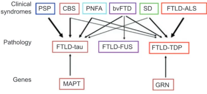
As shown in Figure 1 for some clinical subtypes of the FTLD spectrum, a close correlation with underlying pathol-ogy can be found. PSP with predominantly parkinsonian symptoms is positive for tau protein inclusions [22, 23] , other FTLD variants, such as CBS and progressive nonfl uent apha-sia (PNFA) show tau protein and TDP-43 pathology [24] . The most common clinical type bvFTD can be associated with all three molecular subtypes [9, 25] , whereas the motor neuron disease variant is mainly associated with TDP-43 positive inclusions. As TDP-43 is also found in familiar and sporadic ALS a pathophysiological continuum of both diseases is sug-gested [5] .
Neurochemical markers are therefore needed for early and differential diagnosis and for clinical defi nition of molecular subtypes of FTLD to allow the perspective of subtype specifi c treatment.
Two research approaches can be considered for the labo-ratory diagnosis of FTLD: (i) the direct detection of patho-logical hallmark proteins and (ii) the detection of surrogate markers in biological materials that show an altered pattern of expression in early stages of the disease or are used in the differential diagnosis of other dementias and thus enable an exclusion diagnosis. Thus far, information on specifi c mark-ers, such as tau protein, TDP-43 or FUS aggregates is lim-ited or does not exist, which might also be due to the fact that these neuropathological hallmarks were only detected
PSP Clinical syndromes Pathology Genes CBS PNFA bvFTD FTLD-ALS FTLD-tau FTLD-FUS FTLD-TDP GRN MAPT SD
Figure 1 Clinical syndromes (top row), pathological subtypes (middle row), and common gene mutations (bottom row) in FTLD are shown.
Arrows represent links between clinical syndromes, genes, and underlying histopathology, with thicker arrows corresponding to stronger relationships. bvFTD, behavioral variant frontotemporal dementia; CBS, corticobasal syndrome; FTLD-ALS, FTLD with amyotrophic lateral sclerosis; FTLD-TAU, FTLD with tau-positive inclusions; FTLD-FUS, FTLD with fused in sarcoma (FUS)-positive inclusions; FTLD-TDP, FTLD with TAR DNA-binding protein 43 (TDP-43)-positive inclusions; MAPT, microtubule-associated pro-tein tau; PGRN, progranulin; PNFA, progressive nonfl uent aphasia; PSP, progressive supranuclear palsy; SD, semantic dementia [3] . Reproduced by kind permission from Rabinovici GD and Miller BL, CNS Drugs 2010. Clinical, pathological and genetic spectrum of frontotemporal lobar degeneration (FTLD).
recently. Thus, most of the studies have predominantly con-centrated on evaluating biomarkers which are used for the differential diagnosis of FTLD vs. Alzheimer ' s disease (AD).
However, the combination of symptoms in FTLD can be highly variable and multifaceted, and for a biomarker study either a neuropathological verifi cation or at least highly stand-ardized protocols for the clinical examination are necessary. However, ascertainment of a pathological diagnosis is seldom done and in the absence of reliable and valid measuring meth-ods usually non-standardized “ tests ” are used. This also holds true for magnetic resonance imaging and additional neuroim-aging techniques. These are major drawbacks in determining the quality of a biomarker study for FTLD.
Among the studies investigating cerebrospinal fl uid (CSF) as a diagnostic tool, only a few comprehensively deal with FTLD. In several studies, FTLD patients were analyzed in “ control ” groups, but because the clinical information was so limited these studies were not included here.
tau and amyloid- β in FTLD
For tau protein and amyloid- β mainly mild changes have
been described (Table 1 ). Bian et al. [29] show that tau
protein is a potentially valuable biomarker for differenti-ating FTLD from AD. Four histopathological tau-negative patients of the FTLD spectrum had signifi cantly reduced CSF tau levels compared with AD, whereas three of the FTLD-tau cases showed elevated CSF tau levels. None of
the FTLD cases presented elevated tau/amyloid- β 1 – 42 levels.
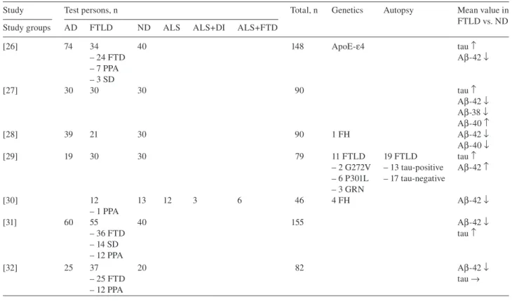
Table 1 Studies for standard CSF biomarkers: protein tau and amyloid- β in 2002 – 2011.
Study Test persons, n Total, n Genetics Autopsy Mean value in FTLD vs. ND Study groups AD FTLD ND ALS ALS + DI ALS + FTD
[26] 74 34 40 148 ApoE- ε 4 tau ↑ – 24 FTD A β -42 ↓ – 7 PPA – 3 SD [27] 30 30 30 90 tau ↑ A β -42 ↓ A β -38 ↓ A β -40 ↑ [28] 39 21 30 90 1 FH A β -42 ↓ A β -40 ↓ [29] 19 30 30 79 11 FTLD 19 FTLD tau ↑ – 2 G272V – 13 tau-positive A β -42 ↑ – 6 P301L – 17 tau-negative – 3 GRN [30] 12 13 12 3 6 46 4 FH A β -42 ↓ – 1 PPA [31] 60 55 40 155 A β -42 ↓ – 36 FTD tau ↑ – 14 SD – 12 PPA [32] 25 37 20 82 A β -42 ↓ – 25 FTD tau → – 12 PPA
AD, Alzheimer ' s disease; FTLD, frontotemporal lobar degeneration; ND, non-demented controls; ALS, amyotrophic lateral sclerosis; ALS + DI, amyotrophic lateral sclerosis with frontal disinhibition; ALS + FTD, amyotrophic lateral sclerosis with frontotemporal dementia; FTD, fronto-temporal dementia; PPA, primary progressive aphasia; SD, semantic dementia; FH, family history.
lower in FTLD than in AD and discriminated FTLD from AD with a sensitivity of 79 % and specifi city of 97 % . How-ever, there was no discrimination between FTLD and non-demented control groups.
In summary, the mean concentration for total tau protein
and amyloid- β 1 – 42 is between the mean values of the AD
group on the one hand and the control group (non-demented)
on the other hand. The average value of amyloid- β 1 – 42 is
higher in FTLD than in the respective AD group and lower than the non-demented group. However, in another study with autopsy and genetically proven FTLD the mean value of
amyloid- β 1 – 42 was with 531 pg/mL higher compared with the
lower mean value of 429 pg/mL in non-demented controls. The average value of tau protein in the respective groups is lower in FTLD compared with AD and higher compared
with the control group. Within the amyloid- β peptides, the
mean value of amyloid- β 1 – 38 in the FTLD group is 160 pg/mL,
which is lower than in the respective AD group (217 pg/mL) and the control group (226 pg/mL) [27] . Whereas one study
shows that amyloid- β 1 – 40 is increased in the FTLD group
and the ratio of amyloid- β 1 – 38 and amyloid- β 1 – 40 seems to
achieve the best discrimination with a sensitivity of 87 % and
a specifi city of 90 % [27] , another study shows decreased
amyloid- β 1 – 40 levels with an increased ratio of amyloid- β 1 – 42
to amyloid- β 1 – 40 in comparison to the AD and non-demented
control groups [28] .
Other neurochemical markers for the diagnosis of FTLD
Apart from tau protein and amyloid- β 1 – 42 several other
mark-ers were investigated (Table 2 ). Galimberti et al. [41] attracts
attention with study groups with more than 100 patients inves-tigated. Unfortunately, the biomarker result for MCP-1 in the CSF could only be evaluated in a subset of 23 FTLD patients. Others concentrated on the genetic contribution to the etiol-ogy of FTLD. As mutations in the GRN gene were identifi ed
as a causal mechanism underlying FTLD [14] , some
stud-ies refer to FTLD cases with a positive proven GRN mutation (FTLD + GRN). It was shown that progranulin levels in plasma and CSF are lower in FTLD + GRN than in FTLD without GRN mutation. This was also evident when FTLD + GRN patients were compared with controls. Unfortunately, progranulin levels were only tested on eight FTLD ( + ) cases. Ghidoni et al. [38] established that in CSF a cut-off level of 518 pg/mL reaches a specifi city and sensitivity of 100 % and therefore seems to be a promising method of screening such cases. For plasma, the pro-granulin protein cut-off level was 74.4 ng/mL with a specifi city and sensitivity of 100 % for mutation carriers among unaffected subjects. In FTLD values ≤ 110.9 ng/mL give a specifi city of 92.8 % and a sensitivity of 100 % for PGRN mutations. Finch et al. distinguished GRN mutation carriers from non- GRN carriers at a progranulin plasma cut-off level of 112 ng/mL [42] .
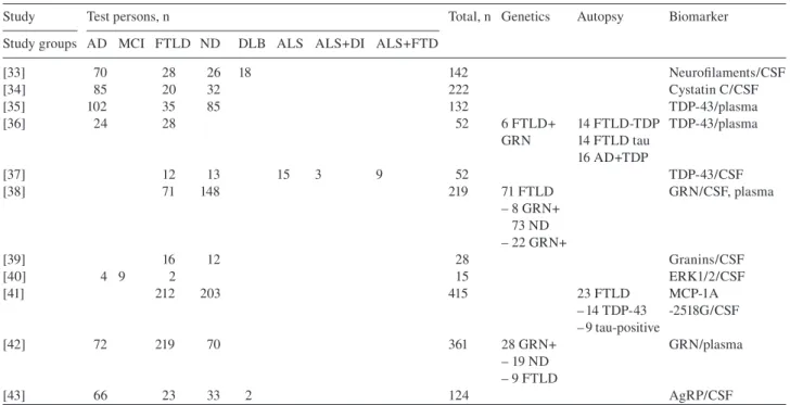
Table 2 Studies for different biomarkers in CSF and plasma 2007 – 2010.
Study Test persons, n Total, n Genetics Autopsy Biomarker Study groups AD MCI FTLD ND DLB ALS ALS + DI ALS + FTD
[33] 70 28 26 18 142 Neurofi laments/CSF [34] 85 20 32 222 Cystatin C/CSF [35] 102 35 85 132 TDP-43/plasma [36] 24 28 52 6 FTLD + 14 FTLD-TDP TDP-43/plasma GRN 14 FTLD tau 16 AD + TDP [37] 12 13 15 3 9 52 TDP-43/CSF [38] 71 148 219 71 FTLD GRN/CSF, plasma – 8 GRN + 73 ND – 22 GRN + [39] 16 12 28 Granins/CSF [40] 4 9 2 15 ERK1/2/CSF [41] 212 203 415 23 FTLD MCP-1A – 14 TDP-43 -2518G/CSF – 9 tau-positive [42] 72 219 70 361 28 GRN + GRN/plasma – 19 ND – 9 FTLD [43] 66 23 33 2 124 AgRP/CSF
AD, Alzheimer ' s disease; MCI, mild cognitive impairment; FTLD, frontotemporal lobar degeneration; ND, non-demented controls; DLB, dementia with Lewy bodies; ALS, amyotrophic lateral sclerosis; ALS + DI, amyotrophic lateral sclerosis with frontal disinhibition; ALS + FTD, amyotrophic lateral sclerosis with frontotemporal dementia; GRN + , mutation in the progranulin gene.
Another study focuses on further classifi cation of patients with neurodegenerative diseases with a negative biomarker profi le for current AD markers using multi-analyte profi ling (MAP). MAP biomarkers combined with current AD bio-markers achieved a sensitivity and specifi city of 92 % and 97 % for diagnosing AD. In a subanalysis of this study, Hu et al. [43] analyzed 23 autopsy proven FTLD subtypes, including 14 TDP-43 positive and 9 tau protein positive FTLD cases. In
the study, amyloid- β 1 – 42 , p-tau 181 and agouti-related peptide
(AgRP) were seen as useful classifying analytes: with AgRP levels elevated in a higher proportion of FTLD-TDP cases, compared with a small proportion of FTLD-tau cases. This study can be seen as a fi rst approach for the differentiation between the subgroups of FTLD as autopsy proven diagno-ses are applied. As TDP-43 inclusions are found in a major subgroup of FTLD, it is therefore becoming an important biomarker to clarify pathophysiologically regulated pathways and to create an opportunity to develop disease-modifying therapies. Increased amounts of TDP-43 and phosphoryl-ated TDP-43 plasma levels have been found in patients with FTLD and AD compared with controls and thereby index
TDP-43 pathology within the brain [35, 36] . Our studies
focus on TDP-43 as a biological marker in CSF. As TDP-43 is mainly found in sporadic ALS cases and signifi cantly higher levels of TDP-43 are found in their CSF when compared with age-matched controls [44] , we decided to investigate these patients in a proof-of-principle type of manner, as here the neuropathology can be easier predicted compared with the other diagnosis of the FTLD spectrum, especially the bvFTD cases [37] . This included FTLD, FTLD + ALS and ALS cases.
Here, no evidence of pathologically altered TDP-43 proteins in CSF could be seen. Methodological diffi culties, such as the heterogeneity of commercially available antibodies, anti-body cross-reaction with IgG, and the possible dysfunction of the CSF-blood barrier with high TDP-43 immunoblot bands in plasma may account for variation in results. However, TDP-43 levels might aid in characterizing subgroups of patients across the ALS and FTLD disease spectrum.
In summary, sustainable success of biomarker projects will mainly depend on comparable protocols in either the preanalytical or clinical phases. This makes it necessary to specify and agree upon diagnostic criteria for the inclusion of test individuals into studies. Current contradicting results are most likely due to these methodical drawbacks.
In conclusion, the main explorative studies show that a relation between biomarkers and diagnostic groups cannot be excluded. As a minimum requirement, future studies with biomarkers should be based on an appropriate number of test individuals and clear predefi ned diagnostic criteria. Therefore, genetically or neuropathologically defi ned cohorts are mandatory. Newly set-up consortia between institutions
specializing in the diagnostic spectrum of FTLD ( www.
ftld.de ) are very promising to build-up a crucial number of patients allowing in-depth research in this area.
Acknowledgments
We thank Gil Rabinovici and Bruce Miller for providing the fi gure illustration.
Confl ict of interest statement
Authors ’ confl ict of interest disclosure: The authors stated that there are no confl icts of interest regarding the publication of this article. Research support played no role in the study design; in the collection, analysis, and interpretation of data; in the writing of the report; or in the decision to submit the report for publication. Research funding: This work is supported by BMBF (KNDD/ FTLDc).
Employment or leadership: None declared. Honorarium: None declared.
References
1. Gorno-Tempini ML, Hillis AE, Weintraub S, Kertesz A, Mendez M, Cappa SF, et al. Classifi cation of primary progressive aphasia and its variants. Neurology 2011;76:1006 – 14.
2. Rascovsky K, Hodges JR, Knopman D, Mendez MF, Kramer JH, Neuhaus J, et al. Sensitivity of revised diagnostic criteria for the behavioural variant of frontotemporal dementia. Brain 2011;134:2456 – 77.
3. Rabinovici GD, Miller BL. Frontotemporal lobar degeneration: epidemiology, pathophysiology, diagnosis and management. CNS Drugs 2010;24:375 – 98.
4. Lillo P, Mioshi E, Zoing MC, Kiernan MC, Hodges JR. How common are behavioural changes in amyotrophic lateral sclero-sis ? Amyotroph Lateral Scler 2011;12:45 – 51.
5. Lomen-Hoerth C, Anderson T, Miller B. The overlap of amyo-trophic lateral sclerosis and frontotemporal dementia. Neurology 2002;59:1077 – 9.
6. Lomen-Hoerth C. Characterization of amyotrophic lateral scle-rosis and frontotemporal dementia. Dement Geriatr Cogn Disord 2004;17:337 – 41.
7. Neumann M, Sampathu DM, Kwong LK, Truax AC, Micsenyi MC, Chou TT, et al. Ubiquitinated TDP-43 in frontotemporal lobar degeneration and amyotrophic lateral sclerosis. Science 2006;314:130 – 3.
8. Mackenzie IR, Neumann M, Bigio EH, Cairns NJ, Alafuzoff I, Kril J, et al. Nomenclature for neuropathologic subtypes of fron-totemporal lobar degeneration: consensus recommendations. Acta Neuropathol 2009;117:15 – 8.
9. Urwin H, Josephs KA, Rohrer JD, Mackenzie IR, Neumann M, Authier A, et al. FUS pathology defi nes the majority of tau- and TDP-43-negative frontotemporal lobar degeneration. Acta Neuropathol 2010;120:33 – 41.
10. Neumann M, Rademakers R, Roeber S, Baker M, Kretzschmar HA, Mackenzie IR. A new subtype of frontotemporal lobar degeneration with FUS pathology. Brain 2009;132: 2922 – 31.
11. Mackenzie IR, Neumann M, Bigio EH, Cairns NJ, Alafuzoff I, Kril J, et al. Nomenclature and nosology for neuropathologic subtypes of frontotemporal lobar degeneration: an update. Acta Neuropathol 2010;119:1 – 4.
12. Neumann M, Bentmann E, Dormann D, Jawaid A, Dejesus-Hernandez M, Ansorge O, et al. FET proteins TAF15 and EWS are selective markers that distinguish FTLD with FUS pathology from amyotrophic lateral sclerosis with FUS mutations. Brain 2011;134:2595 – 609.
13. Wilhelmsen KC. Frontotemporal dementia is on the MAPtau. Ann Neurol 1997;41:139 – 40.
14. Baker M, Mackenzie IR, Pickering-Brown SM, Gass J, Rademakers R, Lindholm C, et al. Mutations in progranulin
cause tau-negative frontotemporal dementia linked to chromo-some 17. Nature 2006;442:916 – 9.
15. Mackenzie IR, Rademakers R, Neumann M. TDP-43 and FUS in amyotrophic lateral sclerosis and frontotemporal dementia. Lancet Neurol 2010;9:995 – 1007.
16. Warraich ST, Yang S, Nicholson GA, Blair IP. TDP-43: a DNA and RNA binding protein with roles in neurodegenerative dis-eases. Int J Biochem Cell Biol 2010;42:1606 – 9.
17. Kovacs GG, Murrell JR, Horvath S, Haraszti L, Majtenyi K, Molnar MJ, et al. TARDBP variation associated with frontotem-poral dementia, supranuclear gaze palsy, and chorea. Mov Disord 2009;24:1843 – 7.
18. Renton AE, Majounie E, Waite A, Simon-Sanchez J, Rollinson S, Gibbs JR, et al. A hexanucleotide repeat expansion in C9ORF72 is the cause of chromosome 9p21-linked ALS-FTD. Neuron 2011;72:257 – 68.
19. DeJesus-Hernandez M, Mackenzie IR, Boeve BF, Boxer AL, Baker M, Rutherford NJ, et al. Expanded GGGGCC hexa-nucleotide repeat in noncoding region of C9ORF72 causes chromosome 9p-linked FTD and ALS. Neuron 2011;72: 245 – 56.
20. Skibinski G, Parkinson NJ, Brown JM, Chakrabarti L, Lloyd SL, Hummerich H, et al. Mutations in the endosomal ESCRTIII-complex subunit CHMP2B in frontotemporal dementia. Nat Genet 2005;37:806 – 8.
21. Guyant-Marechal L, Laquerriere A, Duyckaerts C, Dumanchin C, Bou J, Dugny F, et al. Valosin-containing protein gene mutations: clinical and neuropathologic features. Neurology 2006;67:644 – 51.
22. Dickson DW. Neuropathology of Pick ' s disease. Neurology 2001;56:S16 – 20.
23. Dickson DW. Neuropathologic differentiation of progressive supranuclear palsy and corticobasal degeneration. J Neurol 1999;246(Suppl 2):II6 – 15.
24. Uryu K, Nakashima-Yasuda H, Forman MS, Kwong LK, Clark CM, Grossman M, et al. Concomitant TAR-DNA-binding pro-tein 43 pathology is present in Alzheimer disease and corticoba-sal degeneration but not in other tauopathies. J Neuropathol Exp Neurol 2008;67:555 – 64.
25. Seelaar H, Klijnsma KY, de Koning I, van der Lugt A, Chiu WZ, Azmani A, et al. Frequency of ubiquitin and FUS-positive, TDP-43-negative frontotemporal lobar degeneration. J Neurol 2010;257:747 – 53.
26. Riemenschneider M, Wagenpfeil S, Diehl J, Lautenschlager N, Theml T, Heldmann B, et al. Tau and A β 42 protein in CSF of patients with frontotemporal degeneration. Neurology 2002;58: 1622 – 8.
27. Bibl M, Mollenhauer B, Wolf S, Esselmann H, Lewczuk P, Kornhuber J, et al. Reduced CSF carboxyterminally truncated A β peptides in frontotemporal lobe degenerations. J Neural Trans 2007;114:621 – 8.
28. Pijnenburg YA, Schoonenboom SN, Mehta PD, Mehta SP, Mulder C, Veerhuis R, et al. Decreased cerebrospinal fl uid amy-loid β (1 – 40) levels in frontotemporal lobar degeneration. J Neurol Neurosurg Psychiatry 2007;78:735 – 7.
29. Bian H, Van Swieten JC, Leight S, Massimo L, Wood E, Forman M, et al. CSF biomarkers in frontotemporal lobar degeneration with known pathology. Neurology 2008;70:1827 – 35.
30. Steinacker P, Hendrich C, Sperfeld AD, Jesse S, Lehnert S, Pabst A, et al. Concentrations of β -amyloid precursor protein process-ing products in cerebrospinal fl uid of patients with amyotrophic lateral sclerosis and frontotemporal lobar degeneration. J Neural Trans 2009;116:1169 – 78.
31. Verwey NA, Kester MI, van der Flier WM, Veerhuis R, Berkhof H, Twaalfhoven H, et al. Additional value of CSF amyloid- β 40 levels in the differentiation between FTLD and control subjects. J Alzheimers Dis 2010;20:445 – 52.
32. Bibl M, Mollenhauer B, Lewczuk P, Esselmann H, Wolf S, Otto M, et al. Cerebrospinal fl uid tau, p-tau 181 and amyloid- β (38/40/42) in frontotemporal dementias and primary progressive aphasias. Dement Geriatr Cogn Disord 2011;31:37 – 44.
33. de Jong D, Jansen RW, Pijnenburg YA, van Geel WJ, Borm GF, Kremer HP, et al. CSF neurofi lament proteins in the dif-ferential diagnosis of dementia. J Neurol Neurosurg Psychiatry 2007;78:936 – 8.
34. Simonsen AH, McGuire J, Podust VN, Hagnelius NO, Nilsson TK, Kapaki E, et al. A novel panel of cerebrospinal fl uid bio-markers for the differential diagnosis of Alzheimer ' s disease ver-sus normal aging and frontotemporal dementia. Dement Geriatr Cogn Disord 2007;24:434 – 40.
35. Foulds P, McAuley E, Gibbons L, Davidson Y, Pickering-Brown SM, Neary D, et al. TDP-43 protein in plasma may index TDP-43 brain pathology in Alzheimer ' s disease and fron-totemporal lobar degeneration. Acta Neuropathol 2008;116: 141 – 6.
36. Foulds PG, Davidson Y, Mishra M, Hobson DJ, Humphreys KM, Taylor M, et al. Plasma phosphorylated-TDP-43 protein levels correlate with brain pathology in frontotemporal lobar degenera-tion. Acta Neuropathol 2009;118:647 – 58.
37. Steinacker P, Hendrich C, Sperfeld AD, Jesse S, von Arnim CA, Lehnert S, et al. TDP-43 in cerebrospinal fl uid of patients with
frontotemporal lobar degeneration and amyotrophic lateral scle-rosis. Arch Neurol 2008;65:1481 – 7.
38. Ghidoni R, Benussi L, Glionna M, Franzoni M, Binetti G. Low plasma progranulin levels predict progranulin mutations in fron-totemporal lobar degeneration. Neurology 2008;71:1235 – 9. 39. Bartolomucci A, Pasinetti GM, Salton SR. Granins as
disease-biomarkers: translational potential for psychiatric and neurologi-cal disorders. Neuroscience 2010;170:289 – 97.
40. Klafki HW, Lewczuk P, Kamrowski-Kruck H, Maler JM, Muller K, Peters O, et al. Measurement of ERK 1/2 in CSF from patients with neuropsychiatric disorders and evidence for the presence of the activated form. J Alzheimers Dis 2009;18:613 – 22.
41. Galimberti D, Venturelli E, Villa C, Fenoglio C, Clerici F, Marcone A, et al. MCP-1 A-2518G polymorphism: effect on sus-ceptibility for frontotemporal lobar degeneration and on cerebro-spinal fl uid MCP-1 levels. J Alzheimers Dis 2009;17:125 – 33. 42. Finch N, Baker M, Crook R, Swanson K, Kuntz K, Surtees R, et
al. Plasma progranulin levels predict progranulin mutation status in frontotemporal dementia patients and asymptomatic family members. Brain 2009;132:583 – 91.
43. Hu WT, Chen-Plotkin A, Arnold SE, Grossman M, Clark CM, Shaw LM, et al. Biomarker discovery for Alzheimer ' s disease, frontotemporal lobar degeneration, and Parkinson ' s disease. Acta Neuropathol 2010;120:385 – 99.
44. Kasai T, Tokuda T, Ishigami N, Sasayama H, Foulds P, Mitchell DJ, et al. Increased TDP-43 protein in cerebrospinal fl uid of patients with amyotrophic lateral sclerosis. Acta Neuropathol 2009;117:55 – 62.