Effects on coagulation of balanced (130/0.42) and
non-balanced (130/0.4) hydroxyethyl starch or gelatin
compared with balanced Ringer’s solution: an in vitro study
using two different viscoelastic coagulation tests ROTEM
TM
and SONOCLOT
TM†
M. Casutt
1*
‡, A. Kristoffy
1‡, G. Schuepfer
1, D. R. Spahn
2and C. Konrad
11Institute of Anesthesiology and Intensive Care, Kantonsspital Lucerne, Lucerne, Switzerland 2
Institute of Anesthesiology, University Hospital Zurich, Zurich, Switzerland * Corresponding author. E-mail: [email protected]
Key points
† Haemodilution with volume expanders has agent-specific effects on coagulation.
† The effects of haemodilution with balanced saline,
hydroxyethyl starch, and gelatin on clot formation in vitro were investigated in normal blood.
† Using two independent assays of clot
viscoelasticity (ROTEMTM and SONOCLOTTM), both colloids had significantly greater negative effects on coagulation than balanced saline.
Background. Hydroxyethyl starch (HES) solutions compromise blood coagulation. Low molecular weight, low-substituted HES products, and electrolyte-balanced solutions might reduce this effect. We compared the effects of in vitro haemodilution on blood coagulation with a balanced 6% HES 130/0.42 solution (HESBAL), a saline-based 6% HES
130/0.4 solution (HESSAL), a balanced lactated Ringer’s solution (RL) and a saline-based
4% gelatin solution (GEL).
Methods.Blood was obtained from 10 healthy male volunteers and diluted with the test solutions by 33% and 66%. Quality of clot formation was measured using two viscoelastic coagulation tests: SONOCLOTTMand activated rotation thromboelastometry ROTEMTM.
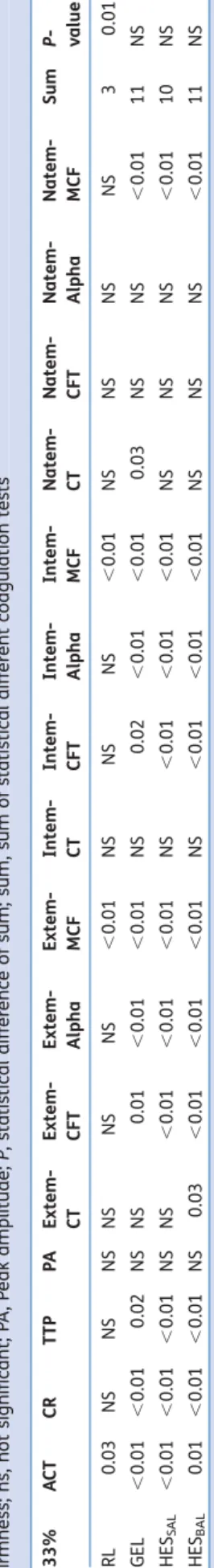
Results.Of 16 parameters measured by the viscoelastic devices, we found three statistically significant differences compared with baseline for RL, but 11 for GEL, 10 for HESSAL, and 11 for
HESBALin the 33% haemodilution group (P¼0.01). Comparing the different solutions, we
observed a significant difference between crystalloids and colloids but none between GEL and HES. In the 66% dilution group, effects on blood coagulation were increased when compared with the 33% dilution group. We found no differences in coagulation impairment between balanced and non-balanced HES products and no differences in the detection of impaired blood coagulation due to haemodilution between the two viscoelastic coagulation tests.
Conclusions.Both ROTEMTMand SONOCLOTTMare sensitive tests for the detection of impaired
blood coagulation due to haemodilution. There are fewer effects on blood coagulation using crystalloids compared with colloids. The effects of GEL and HES are similar. There is no difference between balanced HES 130/0.42 and non-balanced HES 130/0.4.
Accepted for publication: 27 May 2010
During major surgery, administration of synthetic plasma expanders protects the patient from adverse effects of allo-geneic blood transfusions such as transmission of infectious diseases, increases in mortality, major morbidity, transfusion-related acute lung injury, nosocomial infections, increased costs, and immunological effects, including tumour growth promotion.1–6 Colloids and crystalloids influence blood coagulation in different ways. With increasing use, an additional influence on coagulation by the artificial plasma expander itself might become clinically relevant beyond the
effect that coagulation factors and platelets are diluted. In vivo and in vitro, crystalloids show less effect on blood coagulation than colloids, gelatin shows less effect than hydroxyethyl starch (HES), and dextrans show the greatest negative coagulation impact of the colloids.7–11 Third-generation 6% HES 130/0.4 with a lower molecular weight and a lower molar substitution shows a significantly reduced negative impact on blood coagulation in vivo and in vitro compared with early HES products.12–14In addition, some studies suggest less coagulation impairment by HES †Poster presentation at the Annual Meeting of the American Society of Anesthesiologists (ASA) 2008.
plasma-adapted electrolyte concentrations.
The purpose of this study was to assess the effects of in vitro haemodilution on blood coagulation of commonly used volume expanders: 6% HES in a balanced 130/0.42 and non-balanced 130/0.4 solution compared with 4% gelatin solution, and a balanced Ringer’s lactate-crystalloid solution. Apart from standard coagulation variables, we assessed the quality of clot formation using two different viscoelastic devices, SONOCLOTTMand ROTEMTM. In addition,
we evaluated and compared these two point-of-care devices.
Methods
After approval by the institutional ethics committee (Kantonale Ethik-Kommission des Kantons Luzern, protocol number 686) and written informed consent, 10 healthy volunteers (28–52 yr of age) were enrolled. Inclusion criteria for the study were male sex, no history of chronic or acute diseases, no intake of any medication, in particular acetylsalicylic acid or non-steroidal anti-inflammatory agents within 2 weeks before the study, no alcohol or drug abuse, or smoking. Blood samples were obtained from an antecubital vein using an 18 G venous cannula (VasofixTM, B. Braun, Melsungen, Germany). To
exclude blood coagulation activation due to venous stasis, vein puncture, or blood withdrawal, we discarded 5 ml of native blood (whole blood) before 2 ml of native blood for SONOCLOTTManalysis and 4 ml of citrated blood for
thromboe-lastometry were taken for each measurement. The different plasma expander solutions were immediately diluted using polystyrene tubes and the coagulation measurements started within 2 min after having obtained the blood sample. After measurement of an undiluted sample, the blood was diluted 33% and 66% by volume (as in former investigations with similar interrogations),8 9 12 16using as test solutions: an elec-trolyte plasma-adapted, ‘balanced’ Ringer’s solution (contain-ing Na+140 mM, Cl2127 mM, K+4.0 mM, Ca2+2.5 mM, Mg2+
1.0 mM, acetate 24.0 mM, and malate 5.0 mM, RingerfundinTM, B. Braun), a conventional saline-based 4% gelatin solution (con-taining Na+154 mM and Cl2120 mM, Gel 4%TM
, B. Braun), a con-ventional, non-balanced 6% HES 130/0.4/9:1 solution (6%¼concentration/130 kDa¼mean molecular weight/ 0.4¼molar substitution ratio, the average number of hydro-xyethyl groups per unit of glucose/9:1¼C2:C6 ratio, the distri-bution of the hydroxyethyl units between the C2 and C6 positions of the glucose unit) containing Na+154 mM and Cl2
154 mM, VoluvenTM, Fresenius Kabi, Bad Homburg, Germany), and a balanced 6% HES 130/0.42/6:1 solution, based on potato as the biological source (containing Na+140 mM, Cl2
118 mM, K+4.0 mM, Ca2+2.5 mM, Mg2+1.0 mM, acetate 24.0 mM, and malate 5.0 mM, Tetraspan 6%TM, B. Braun).
Routine laboratory tests were done with the automated ana-lysing systems at the central laboratory. All blood samples for routine laboratory tests were obtained at once, whereas native blood samples for SONOCLOTTM and citrated blood
samples for ROTEMTMhad to be obtained with an 18 G venous
cannula shortly before processing. The dilutions were prepared
lysed: haematocrit, haemoglobin concentration, platelet count, fibrinogen, activated partial thromboplastin time (aPTT) and prothrombin time (PT) with international normalized ratio. To rule out a platelet defect, we used the Platelet Function Analyzer PFA-100TM (Dade/Behring, Marburg, Germany) and
investigated the initial steps of platelet-mediated blood coagulation using ADP and epinephrine stimulation.17
Analysis of clot formation was performed with two whole-blood coagulation tests: SONOCLOTTM(Sonoclot II Coagulation
and Platelet Function Analyzer, Sienco Co., Morrison, CO, USA) and activated rotation thromboelastometry (ROTEMTM,
Penta-pharm, Munich, Germany). The principle of these tests is the analysis and graphical presentation of changes in viscoelastic properties of clot formation and retraction and subsequent fibrinolysis. The tests are sensitive to impairment of clot for-mation involving coagulation factors and inhibitors, platelets, and fibrinogen. Technical details of the two coagulation ana-lysers and their parameters have been described previously.18
SONOCLOT
TMTo accelerate the coagulation process, the cuvette contains 5 mg of Celite to activate factor XII at the beginning of the intrinsic pathway. As determined in other SONOCLOTTM
-based trials,8 19we evaluated the following parameters: acti-vated clotting time (ACT), clot rate (CR), peak amplitude (PA), and time to peak (TTP). ACT is the time from test start until the beginning of fibrin formation and corresponds to the con-ventionally measured ACT. Fibrin formation (Gel phase) starts when the viscosity of the sample begins to increase by fibrin formation from fibrinogen and is characterized by the primary slope of the curve (CR). After completion of clot for-mation, PA reflects the fibrinogen concentration. TTP charac-terizes clot retraction and describes the speed and quality of fibrin formation8 18–20(Fig.1).
ROTEM
TMCitrated-anticoagulated blood (0.3 ml) was re-calcified with 20 ml calcium chloride. Activation of coagulation was per-formed as follows: extrinsic thromboelastometry (Extem) with 20 ml of tissue thromboplastin, intrinsic thromboelasto-metry (Intem) with 20 ml of the surface activator partial throm-boplastin, and native thromboelastometry (Natem) without activator, only with re-calcification. We determined the follow-ing parameters: onset of coagulation [coagulation time (CT)], kinetics of clot formation [clot formation time (CFT): the time elapsed between 2 and 20 mm clot firmness], the Alpha angle (angle of a tangent to the clotting curve through the 2 mm point), and maximum clot firmness (MCF). All measure-ments were performed within 2 min after blood withdrawal at 378C using reagents from the manufacturer (Fig.2).
Statistical analysis
Statistical analysis was done using the commercially avail-able software package Statistica Version 7.0 (StatSoft Inc., Tulsa, OK, USA) and StatExact 7.0 (Cytel Software
Corporation, Cambridge, MA, USA). Non-parametric analysis was performed with one-way analysis of variance (ANOVA)
equivalent test procedure with multiple testing and pair characteristic of the data; post hoc comparison with the Bon-ferroni correction was applied to adjust the level of signifi-cance. Subsequently, the Fisher–Freeman–Halton test was applied to analyse the categorical data of inhomogeneous distribution of the coagulation tests by haemodilution.
Because of the obvious effect of the two dilutional steps, the statistical differences are not shown. Of further interest are the differences between the solutions at their various levels of dilution. Data are mean (SD). A P-value ,0.05 was
considered statistically significant.
Results
Ten volunteers with a median body weight (range) of 78 (70 – 86) kg and body height of 181 (175 –187) cm were enrolled. All baseline values including PT, aPTT, fibrinogen,
haematocrit, haemoglobin concentration, and platelet count were within the normal range. Platelet dysfunction was excluded by normal PFA measurements (ADP- and epinephrine-closure time) (Table1). After dilution, fibrinogen, haematocrit, haemoglobin concentration, and platelet count decreased, and PT and aPTT increased, indicating compro-mised blood coagulation (data not shown). Furthermore, all baseline variables measured by SONOCLOTTM and ROTEMTM
were within normal limits as defined by the manufacturers.
SONOCLOT
TMAfter haemodilution, ACT and TTP were prolonged and CR and PA decreased, representing the negative influence of dilution on haemostasis.
33% haemodilution. Compared with baseline, ACT showed a significant prolongation for all agents. Additionally, CR decreased and TTP increased for all colloids (Table2).
0 M I N U T E S M I N U T E S 100 90 80 70 60 50 40 30 20 10 100 90 80 70 60 50 40 30 20 10 100 90 80 70 60 50 40 30 20 10 100 90 80 70 60 50 40 30 20 10 5 10 15 20 SIENCO , INC . SIENCO , INC . 25 30 5 10 15 20 25 30 35 40 B Time (min) Clot signal ACT CR TTP PA Time (min) Clot signal A
Fig 1(A) Normal SONOCLOTTMcurve. (B) SONOCLOTTMcurve after 66% haemodilution with balanced-electrolyte lactated Ringer’s solution (RL).
66% haemodilution. ACT increased and CR decreased sig-nificantly compared with baseline regarding all agents. TTP increased significantly for all colloids compared with baseline (Table3).
ROTEM
TMAfter haemodilution, CT was prolonged, Alpha angle and MCF decreased, and CFT increased.
Table 2 Sta tistical differ ences between agents a t 33% haemodilution compar ed with undiluted baseline. GEL, saline-based gela tin solution; RL, balanced-elec tr olyte lacta ted Ringer’s solution; HES BAL , balanced-elec tr olyte HES solution; HES SA L , saline-based HES solution; A C T, a ctiva ted clotting time; CFT , clot forma tion time; CR, clot ra te; CT , coagula tion time; MCF , maximum clot firmness; ns, not significant; PA , P eak amplitude; P, sta tis tical differ ence of sum; sum, sum of st a tistical differ ent coagulation tes ts 33% A C T C R TTP P A E xtem- CT E xtem- CFT Extem- Alpha E xtem- MCF Intem- CT Intem- CFT Intem- Alpha Intem- MCF Na tem-CT Na tem-CFT Na tem-Alpha Na tem-MCF Sum P - value RL 0.03 NS NS NS NS NS NS , 0.01 NS NS NS , 0.01 NS NS NS NS 3 0.01 GEL , 0.01 , 0.01 0.02 NS NS 0.01 , 0.01 , 0.01 NS 0.02 , 0.01 , 0.01 0.03 NS NS , 0.01 11 NS HES SA L , 0.01 , 0.01 , 0.01 NS NS , 0.01 , 0.01 , 0.01 NS , 0.01 , 0.01 , 0.01 NS NS NS , 0.01 10 NS HES BAL 0.01 , 0.01 , 0.01 NS 0.03 , 0.01 , 0.01 , 0.01 NS , 0.01 , 0.01 , 0.01 NS NS NS , 0.01 11 NS 60 40 20 20 40 60 60 40 20 20 40 60 10 20 30 40 50 10 20 30 40 50 B Clot firmness (mm) Time (min) MCF Alpha Angle CFT CT A Time (min) Clot firmness (mm)
Fig 2 (A) Normal Extem ROTEMTMcurve. (B) Extem ROTEMTMcurve
after 66% haemodilution with balanced-electrolyte lactated Ringer’s solution (RL). CT, coagulation time; CFT, clotting for-mation time; MCF, maximum clot firmness.
Table 1Baseline coagulation and platelet variables of the 10 volunteers. ADP, adenosine diphosphate; aPTT, activated partial thromboplastin time; Hb, haemoglobin; Hct, haematocrit; INR, international normalized ratio; PFA, platelet function analyzer; PT, prothrombin time;SD, standard deviation
Mean (SD) Range Platelet count (103ml21) 220 (41) 175 –289 Hb (g l21) 153 (10) 140 –172 Hct (%) 43 (3) 40 –49 Fibrinogen (g l21) 2.5 (0.5) 1.8 –3.4 PT (%) 98 (10) 85 –118 INR 1.03 (0.05) 0.95 –1.10 aPTT (s) 30 (2) 28 –32
PFA 100-closure time (s)
ADP 88 (12) 65 –110
33% haemodilution. Extem-CT was significantly prolonged compared with baseline only for HESBAL. We found a
sig-nificant Extem-CFT increase and Extem-Alpha decrease between baseline and after dilution by colloids. Extem-MCF decreased significantly for all agents. The col-loids showed a significant Intem-CFT increase and Intem-Alpha decrease compared with baseline. All agents showed a significant decrease in Intem-MCF com-pared with baseline. Natem-CT was only significantly pro-longed between GEL and baseline. Natem-MCF showed a significant decrease to baseline for the colloids (Table2).
66% haemodilution. In Extem-CT, we found a significant prolongation between baseline and HES products. All agents significantly increased Extem-CFT and decreased Extem-Alpha and Extem-MCF compared with baseline. All agents were significantly different from baseline in the Intem measurements (increase in CT and CFT and decrease in Alpha and MCF). In Natem-CT, a significant prolongation was only observed between baseline and HESBAL. Natem-CFT significantly increased and
Natem-Alpha and Natem-MCF decreased between base-line and all agents (Table3).
Summarized results of SONOCLOT
TMand ROTEM
TM We compared the number of significant differences in SONOCLOTTM- and ROTEMTM-parameters (a total of 16values). We found three statistical significant differences compared with baseline values for RL, 11 for GEL, 10 for HESSAL, and 11 for HESBAL (Table 2). The Fisher–Freeman–
Halton test showed a significant difference between the agents listed in Table2(P,0.01). There were no significant differences between the agents in the Freeman–Halton test for the 66% haemodilution group (Table 3). The inter-group analysis showed a pronounced reduction of blood coagulation by the use of colloids compared with crystalloids in both dilution groups (33% and 66%) (Tables A1 and A2).
SONOCLOT
TMvs ROTEM
TMand vs haematological
parameters
Baseline haematological laboratory values were correlated with the readout variables of SONOCLOTTM(ACT, CR, PA, and
TTP) and ROTEMTM (CT, CFT, and MCF with the activation
paths Extem, Intem, and Natem). A statistically significant correlation was found between fibrinogen level and Intem-MCF (r¼0.79) and between platelet count and Natem-MCF (r¼0.73). No correlation could be found between ACT and Extem-, Intem-, or Natem-CT. CR correlated significantly with Extem-CT (r¼0.66) and Intem-CFT (r¼0.74). TTP also correlated significantly with Intem-CT (r¼0.66) and Intem-MCF (r¼0.83). The MCF values as measured with the Extem and Natem activation paths of ROTEMTM also correlated but not with Intem-MCF value. CF
correlated significantly with TTP (r¼0.64) but only with Intem-CFT (r¼0.74). When using a cluster analysis, ACT can most suitably be linked to Extem-CFT and Intem-CFT.
Table 3 Statis tical differ ences betwe en agents a t 66% haemodilution compar ed with undiluted baseline. GEL, saline-based gelatin solution; RL, balanced-elec tr olyte la ct a ted Ringer’s solution; HES BAL , balanced-elec tr olyte HES solution; HES SA L , saline-based HES solution; A C T, a ctiv a ted clotting time; CFT , clot formation time; CR, clot ra te; CT , coagulation time; MCF , maximum clot firmness; NS, not significant; PA , P eak amplitude; P, sta tis tical differ ence of sum; sum, sum of st a tistical differ ent coagulation tes ts 66% A C T C R TTP P A E xtem- CT E xtem-CFT E xtem- Alpha E xtem- MCF Intem- CT Intem- CFT Intem- Alpha Intem- MCF Na tem-CT Na tem-CFT Na tem-Alpha Na tem-MCF Sum P -v alue RL , 0.01 , 0.01 NS NS NS , 0.01 , 0.01 , 0.01 , 0.01 , 0.01 , 0.01 , 0.01 NS , 0.01 , 0.01 , 0.01 12 NS GEL , 0.01 , 0.01 , 0.01 NS NS , 0.01 , 0.01 , 0.01 , 0.01 , 0.01 , 0.01 , 0.01 NS , 0.01 , 0.01 , 0.01 13 NS HES SAL , 0.01 , 0.01 0.01 NS , 0.01 , 0.01 , 0.01 , 0.01 , 0.01 , 0.01 , 0.01 , 0.01 NS , 0.01 , 0.01 , 0.01 14 NS HES BAL , 0.01 , 0.01 0.01 NS 0.02 , 0.01 , 0.01 , 0.01 , 0.01 , 0.01 , 0.01 , 0.01 0.03 , 0.01 , 0.01 , 0.01 15 NS
We found a pronounced inhibitory effect of colloids com-pared with crystalloids on blood coagulation after 33% hae-modilution in vitro. There were no differences in the effects on blood coagulation between the gelatin solution and both HES products, or between the balanced- and the saline-based HES solutions. There was no difference between the colloids in the 66% haemodilution groups. The intergroup comparisons confirmed the pronounced reduction of blood coagulation by the use of colloids compared with crystalloids in both dilution groups.
Intravascular volume expanders inhibit blood coagulation concentration dependently. A mild-to-moderate haemodilu-tion with crystalloids can accelerate blood coagulahaemodilu-tion in vitro8 9and in vivo.21–23An imbalance between decreased
antithrombin III activity and thrombin generation can partly explain this effect.21In contrast, the use of colloids for hae-modilution leads to compromised blood coagulation as shown in most studies. Gelatin solutions might influence the weight and reticular network of fibrin strands and plate-let function by ristocetin and polybrene with decreased von Willebrand factor.24 25 However, other studies have shown only a moderate effect9 26or indicate that blood coagulation might even be accelerated.8 11 HES compromises blood coagulation significantly as in vitro8 9 12 13 and in vivo studies have shown.11 27 28 There are several mechanisms
which influence blood coagulation, for example, an acquired von Willebrand syndrome and decreasing effect on factor VIII and decreased expression and activation of platelet surface GPIIb –IIIa receptor with impaired platelet adhesion and aggregation.13 14A further mechanism appears to be the impaired polymerization of fibrin monomers by HES macro-molecules.11 13 In addition to the quantity of the
adminis-tered HES, the chemical properties of the molecules play an important role in blood coagulation. A high molecular weight seems to be of minor importance compared with a high molar substitution.29 A reduction of the C2/C6 ratio results in faster HES elimination without influence on blood coagulation.30 Low molecular weight, low-substituted 6%
HES 130/0.4 possesses a lower molecular weight and a lower molar substitution, and many trials have observed less negative impact on blood coagulation compared with early HES products.12–14 31 32 When HES is prepared in a balanced solution with physiological electrolyte concen-trations, impaired coagulation is reduced.15 16 33
We did not find evidence for hypercoagulation during hae-modilution with crystalloids. All measured coagulation par-ameters remained unchanged or showed a tendency towards a hypocoagulable condition. Although balanced Ringer’s solution should theoretically compromise coagu-lation only to a low extent due to the additional calcium ions, we could not confirm this hypothesis. The ROTEMTM
device is not suitable for such examinations as the citrated blood samples are re-calcified resulting in a surplus of calcium ions in the sample. In contrast to the ROTEMTM
device, SONOCLOTTM only uses native blood samples and
of a minor influence of balanced volume expanders on hae-modilution compared with saline-based volume expanders in the SONOCLOTTMmeasurements either.
Furthermore, we did not observe significant differences between balanced and non-balanced HES products. Balanced solutions do not possess high and unphysiological concen-trations of sodium and chloride (each 154 mM) as saline-based volume expanders do. Therefore, the risk of developing a hyperchloraemic acidosis after administration of large amounts of such solutions is decreased.34 35Boldt and col-leagues36reported no differences between balanced 6% HES 130/0.42 together with a balanced crystalloid solution and non-balanced 6% HES 130/0.42 together with an unbalanced saline solution in an in vivo study. They found no differences between the two groups regarding Intem-CT, Intem-CFT, and Intem-MCF at baseline and at postoperative day 1. In a follow-up in vitro study, Boldt and colleagues described a more negative effect on coagulation using non-balanced 6% HES 130/0.4 compared with balanced 6% HES 130/0.42. In the 50% dilution group, they found more pronounced changes in Intem-MCF, Intem-CFT, Extem-CT, Extem-CFT, and Extem-MCF when using the non-balanced solution com-pared with the balanced solution, whereas in the 30% dilution group, changes were found in Extem-CFT and Extem-CT.16As mentioned above, ROTEMTMis not a suitable device for
exam-ining the difference between a balanced and a non-balanced volume expander by an in vitro test. In contrast, in our study, we performed an additional viscoelastic coagulation assess-ment and evaluated a total of 16 test parameters of both the ROTEMTMand the SONOCLOTTMdevices.
For both viscoelastic devices, a high sensitivity is required to identify a haemodilution coagulopathy. ACT, CR, and TTP seem to be sensitive parameters for detecting haemodilution coagulopathies of the SONOCLOTTManalysis, whereas ACT is
the most sensitive of all parameters (providing statistical sig-nificant results for all tested agents in our study). Regarding the ROTEMTManalysis, CFT, Alpha, and MCF are sensitive
par-ameters with MCF as the most sensitive one. Extem and Intem measurements are more or less equally sensitive, whereas Natem obviously possesses a lower sensitivity. Natem represents the rotation thromboelastography without activators only with re-calcification. Therefore, it should be very similar to conventional TEG measurements. Although fibrin-polymerization is relevant in dilutional coa-gulopathy and Fibtem is a sensitive test, we decided not to include the Fibtem results in our statistical analysis. Fibtem has only one important parameter (MCF) which showed no difference to the Extem-, Intem-, or Natem-MCF results and had no additional value for our study since we statistically compared significances of the test measurements with the Fisher –Freeman– Halton test—so Fibtem would be the 17th test measurement and would not change our results. Fur-thermore, in 66% haemodilution with colloids, the Fibtem was frequently technically not possible.
Interestingly, the ACT values of SONOCLOTTMand ROTEMTM
complex pattern of correlations between the different vari-ables of the devices shows that the two different viscoelastic methods introduce different ACT analyses. ROTEMTM-CT
describes the time between the beginning of pin rotation until the pin begins to be impaired after fibrin–platelet bonding has linked cup and pin.18The vertical oscillation in the sample of the SONOCLOTTMdevice is sensitive to viscosity
changes during initiation of coagulation. There are further different activators: Celite for SONOCLOTTM, tissue
thrombo-plastin for Extem, partial thrombothrombo-plastin for Intem, or no activator for Natem. The two different procedures and the different activators might explain the lack of statistic corre-lation between the two tests.
A limitation of this in vitro study is the absence of physio-logical reaction during haemodilution including recruitment of additional resources of the coagulation system, changes in plasma ions and acid– base balance, tissue damage, and endothelial injury with their pro-coagulation properties, or interaction between the volume expander and the endothelial system. Further, the different colloid solutions have different half times in vivo (HES.GEL), so that in vivo testing might gen-erate different results compared with in vitro testing. In vivo studies would be necessary to confirm our observations in the clinical setting. Another limitating factor in our study is the difference in the two HES products which are used in our institution. They are from two different companies and possess different molar substitution ratios (0.42 for the balanced HES solution and 0.4 for the saline-based HES ution) and a different C2:C6 ratio (6:1 for the balanced HES sol-ution and 9:1 for the saline-based HES solsol-ution).
In conclusion, haemodilution negatively affected blood coagulation in vitro as measured by the viscoelastic devices SONOCLOTTM and ROTEMTM, with less pronounced changes
when a balanced-electrolyte lactated Ringer’s solution is used when compared with colloidal agents. Saline-based gelatin solution did not show greater influence on haemo-stasis than HES 130/0.4 (only a tendency in intergroup analy-sis). There is no difference between the balanced HES solution and the saline-based HES solution. Both viscoelastic devices, ROTEMTMand SONOCLOTTM, are sensitive to changes
in blood coagulation during haemodilution, with MCF being the most sensitive parameter in ROTEMTM analysis and ACT
in SONOCLOTTManalysis.
Conflict of interest
In the past 5 yr, D.R.S. has received honoraria or travel support for consulting or lecturing from the following companies: Abbott AG, Baar, Switzerland; Alliance Pharmaceutical Corp., San Diego, CA, USA; AstraZeneca AG, Zug, Switzerland; Bayer (Schweiz) AG, Zu¨rich, Switzerland; B. Braun Melsungen AG, Melsungen, Germany; CSL Behring GmbH, Hattersheim am Main, Germany; Fresenius SE, Bad Homburg v.d.H., Germany; Galenica AG, Bern, Switzerland (including Vifor SA, Villars-sur-Glaˆne, Switzerland); GlaxoSmithKline GmbH & Co. KG, Hamburg, Germany; Janssen-Cilag AG, Baar, Switzerland; Novo Nordisk A/S, Bagsva¨rd, Denmark, Octapharma AG,
Lachen, Switzerland; Organon AG, Pfa¨ffikon/SZ, Switzerland; and Roche Pharma (Schweiz) AG, Reinach, Switzerland.
Funding
Institute of Anesthesiology and Intensive Care, Kantonsspi-tal, Lucerne, Switzerland.
Appendix
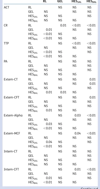
Table A1 Intergroup comparisons of 33% haemodilution with different volume expanders (P,0.05 was considered statistically significant). GEL, saline-based gelatin solution; RL,
balanced-electrolyte lactated Ringer’s solution; HESBAL,
balanced-electrolyte HES solution; HESSAL, saline-based HES
solution; ACT, activated clotting time; CFT, clot formation time; CR, clot rate; CT, coagulation time; MCF, maximum clot firmness; NS, not significant; PA, peak amplitude
RL GEL HESSAL HESBAL
ACT RL NS NS NS GEL NS NS NS HESSAL NS NS NS HESBAL NS NS NS CR RL 0.01 ,0.01 ,0.01 GEL 0.01 NS NS HESSAL ,0.01 NS NS HESBAL ,0.01 NS NS TTP RL NS ,0.01 ,0.01 GEL NS NS NS HESSAL ,0.01 NS NS HESBAL ,0.01 NS NS PA RL NS NS NS GEL NS NS NS HESSAL NS NS NS HESBAL NS NS NS Extem-CT RL NS NS 0.01 GEL NS NS 0.01 HESSAL NS NS NS HESBAL 0.01 0.01 NS Extem-CFT RL NS NS 0.01 GEL NS NS NS HESSAL NS NS NS HESBAL 0.01 NS NS Extem-Alpha RL NS 0.03 ,0.01 GEL NS NS NS HESSAL 0.03 NS NS HESBAL ,0.01 NS NS Extem-MCF RL NS 0.04 ,0.01 GEL NS NS NS HESSAL 0.04 NS NS HESBAL ,0.01 NS NS Intem-CT RL NS NS NS GEL NS NS NS HESSAL NS NS NS HESBAL NS NS NS Intem-CFT RL NS 0.01 ,0.01 GEL NS NS NS HESSAL 0.01 NS NS HESBAL ,0.01 NS NS Continued
References
1 Atzil S, Arad M, Glasner A et al. Blood transfusion promotes cancer progression: a critical role for aged erythrocytes. Anesthesiology 2008; 109: 989– 97
2 Madjdpour C, Spahn DR. Allogeneic red blood cell transfusions: efficacy, risks, alternatives and indications. Br J Anaesth 2005; 95: 33–42
3 Murphy GJ, Reeves BC, Rogers CA, Rizvi SI, Culliford L, Angelini GD. Increased mortality, postoperative morbidity, and cost after red blood cell transfusion in patients having cardiac surgery. Circula-tion 2007; 116: 2544–52
4 Spahn DR, Moch H, Hofmann A, Isbister JP. Patient blood man-agement: the pragmatic solution for the problems with blood transfusions. Anesthesiology 2008; 109: 951– 3
RL GEL HESSAL HESBAL
Intem-Alpha RL ,0.01 ,0.01 ,0.01 GEL ,0.01 NS ,0.01 HESSAL ,0.01 NS NS HESBAL ,0.01 ,0.01 NS Intem-MCF RL 0.01 ,0.01 ,0.01 GEL 0.01 NS NS HESSAL ,0.01 NS NS HESBAL ,0.01 NS NS Natem-CT RL 0.03 NS NS GEL 0.03 NS NS HESSAL NS NS NS HESBAL NS NS NS Natem-CFT RL NS NS NS GEL NS NS NS HESSAL NS NS NS HESBAL NS NS NS Natem-Alpha RL NS NS NS GEL NS NS NS HESSAL NS NS NS HESBAL NS NS NS Natem-MCF RL ,0.01 0.02 ,0.01 GEL ,0.01 NS NS HESSAL 0.02 NS NS HESBAL ,0.01 NS NS
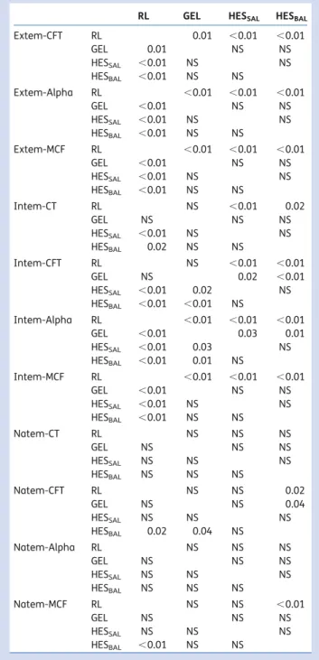
Table A2 Intergroup comparisons of 66% haemodilution with different volume expanders (P,0.05 was considered statistically significant). GEL, saline-based gelatin solution; RL,
balanced-electrolyte lactated Ringer’s solution; HESBAL,
balanced-electrolyte HES solution; HESSAL, saline-based HES
solution; ACT, activated clotting time; CFT, clot formation time; CR, clot rate; CT, coagulation time; MCF, maximum clot firmness; NS, not significant; PA, peak amplitude
RL GEL HESSAL HESBAL
ACT RL ,0.01 NS NS GEL ,0.01 0.03 ,0.01 HESSAL NS 0.03 NS HESBAL NS ,0.01 NS CR RL 0.04 0.02 ,0.01 GEL 0.04 NS NS HESSAL 0.02 NS NS HESBAL ,0.01 NS NS TTP RL 0.01 NS NS GEL 0.01 NS NS HESSAL NS NS NS HESBAL NS NS NS PA RL ,0.01 ,0.01 0.01 GEL ,0.01 NS NS HESSAL ,0.01 NS NS HESBAL 0.01 NS NS Extem-CT RL NS NS NS GEL NS NS NS HESSAL NS NS NS HESBAL NS NS NS Continued
RL GEL HESSAL HESBAL
Extem-CFT RL 0.01 ,0.01 ,0.01 GEL 0.01 NS NS HESSAL ,0.01 NS NS HESBAL ,0.01 NS NS Extem-Alpha RL ,0.01 ,0.01 ,0.01 GEL ,0.01 NS NS HESSAL ,0.01 NS NS HESBAL ,0.01 NS NS Extem-MCF RL ,0.01 ,0.01 ,0.01 GEL ,0.01 NS NS HESSAL ,0.01 NS NS HESBAL ,0.01 NS NS Intem-CT RL NS ,0.01 0.02 GEL NS NS NS HESSAL ,0.01 NS NS HESBAL 0.02 NS NS Intem-CFT RL NS ,0.01 ,0.01 GEL NS 0.02 ,0.01 HESSAL ,0.01 0.02 NS HESBAL ,0.01 ,0.01 NS Intem-Alpha RL ,0.01 ,0.01 ,0.01 GEL ,0.01 0.03 0.01 HESSAL ,0.01 0.03 NS HESBAL ,0.01 0.01 NS Intem-MCF RL ,0.01 ,0.01 ,0.01 GEL ,0.01 NS NS HESSAL ,0.01 NS NS HESBAL ,0.01 NS NS Natem-CT RL NS NS NS GEL NS NS NS HESSAL NS NS NS HESBAL NS NS NS Natem-CFT RL NS NS 0.02 GEL NS NS 0.04 HESSAL NS NS NS HESBAL 0.02 0.04 NS Natem-Alpha RL NS NS NS GEL NS NS NS HESSAL NS NS NS HESBAL NS NS NS Natem-MCF RL NS NS ,0.01 GEL NS NS NS HESSAL NS NS NS HESBAL ,0.01 NS NS
5 Marik PE, Corwin HL. Efficacy of red blood cell transfusion in the critically ill: a systematic review of the literature. Crit Care Med 2008; 36: 2667– 74
6 Hendrickson JE, Hillyer CD. Noninfectious serious hazards of transfusion. Anesth Analg 2009; 108: 759– 69
7 Kozek-Langenecker SA. Influence of fluid therapy on the haemo-static system of intensive care patients. Best Pract Res Clin Anaes-thesiol 2009; 23: 225– 36
8 Konrad C, Markl T, Schuepfer G, Gerber H, Tschopp M. The effects of in vitro hemodilution with gelatin, hydroxyethyl starch, and lac-tated Ringer’s solution on markers of coagulation: an analysis using SONOCLOT. Anesth Analg 1999; 88: 483–8
9 Egli GA, Zollinger A, Seifert B, Popovic D, Pasch T, Spahn DR. Effect of progressive haemodilution with hydroxyethyl starch, gelatin, and albumin on blood coagulation. Br J Anaesth 1997; 78: 684– 9 10 Mittermayr M, Streif W, Haas T et al. Hemostatic changes after crystalloid or colloid fluid administration during major orthopedic surgery: the role of fibrinogen administration. Anesth Analg 2007; 105: 905 –17
11 Innerhofer P, Fries D, Margreiter J et al. The effects of periopera-tively administered colloids and crystalloids on primary platelet-mediated hemostasis and clot formation. Anesth Analg 2002; 95: 858 –65
12 Konrad CJ, Markl TJ, Schuepfer GK, Schmeck J, Gerber HR. In vitro effects of different medium molecular hydroxyethyl starch sol-utions and lactated Ringer’s solution on coagulation using SONO-CLOT. Anesth Analg 2000; 90: 274 –9
13 Kozek-Langenecker SA. Effects of hydroxyethyl starch solutions on hemostasis. Anesthesiology 2005; 103: 654– 60
14 Franz A, Braunlich P, Gamsjager T, Felfernig M, Gustorff B, Kozek-Langenecker SA. The effects of hydroxyethyl starches of varying molecular weights on platelet function. Anesth Analg 2001; 92: 1402– 7
15 Gan TJ, Bennett-Guerrero E, Phillips-Bute B et al. Hextend, a physiologically balanced plasma expander for large volume use in major surgery: a randomized phase III clinical trial. Hextend Study Group. Anesth Analg 1999; 88: 992 –8
16 Boldt J, Wolf M, Mengistu A. A new plasma-adapted hydro-xyethylstarch preparation: in vitro coagulation studies using thrombelastography and whole blood aggregometry. Anesth Analg 2007; 104: 425– 30
17 Kundu SK, Heilmann EJ, Sio R, Garcia C, Davidson RM, Ostgaard RA. Description of an in vitro platelet function analy-zer—PFA-100. Semin Thromb Hemost 1995; 21: 106 –12 18 Ganter MT, Hofer CK. Coagulation monitoring: current techniques
and clinical use of viscoelastic point-of-care coagulation devices. Anesth Analg 2008; 106: 1366– 75
19 Pleym H, Wahba A, Bjella L, Stenseth R. Sonoclot analysis in elderly compared with younger patients undergoing coronary surgery. Acta Anaesthesiol Scand 2008; 52: 28– 35
20 Hett DA, Walker D, Pilkington SN, Smith DC. Sonoclot analysis. Br J Anaesth 1995; 75: 771 –6
21 Ruttmann TG, Lemmens HJ, Malott KA, Brock-Utne JG. The haemodilution enhanced onset of coagulation as measured by the thrombelastogram is transient. Eur J Anaesthesiol 2006; 23: 574 –9
22 Martin G, Bennett-Guerrero E, Wakeling H et al. A prospective, randomized comparison of thromboelastographic coagulation profile in patients receiving lactated Ringer’s solution, 6% hetastarch in a balanced-saline vehicle, or 6% hetastarch in saline during major surgery. J Cardiothorac Vasc Anesth 2002; 16: 441– 6
23 Ng KF, Lam CC, Chan LC. In vivo effect of haemodilution with saline on coagulation: a randomized controlled trial. Br J Anaesth 2002; 88: 475 –80
24 de Jonge E, Levi M. Effects of different plasma substitutes on blood coagulation: a comparative review. Crit Care Med 2001; 29: 1261–7
25 Thaler U, Deusch E, Kozek-Langenecker SA. In vitro effects of gelatin solutions on platelet function: a comparison with hydro-xyethyl starch solutions. Anaesthesia 2005; 60: 554– 9
26 Huttner I, Boldt J, Haisch G, Suttner S, Kumle B, Schulz H. Influ-ence of different colloids on molecular markers of haemostasis and platelet function in patients undergoing major abdominal surgery. Br J Anaesth 2000; 85: 417 –23
27 Knutson JE, Deering JA, Hall FW et al. Does intraoperative hetastarch administration increase blood loss and transfusion requirements after cardiac surgery? Anesth Analg 2000; 90: 801– 7
28 Butwick A, Carvalho B. The effect of colloid and crystalloid pre-loading on thromboelastography prior to Cesarean delivery. Can J Anaesth 2007; 54: 190 –5
29 Madjdpour C, Dettori N, Frascarolo P et al. Molecular weight of hydroxyethyl starch: is there an effect on blood coagulation and pharmacokinetics? Br J Anaesth 2005; 94: 569– 76
30 Schramm S, Thyes C, Frascarolo P et al. Impact of the C2/C6 ratio of high-molecular-weight hydroxyethyl starch on pharmacoki-netics and blood coagulation in pigs. Anesthesiology 2007; 107: 442– 51
31 Van der Linden PJ, De Hert SG, Deraedt D et al. Hydroxyethyl starch 130/0.4 versus modified fluid gelatin for volume expansion in cardiac surgery patients: the effects on perioperative bleeding and transfusion needs. Anesth Analg 2005; 101: 629 –34 32 Haisch G, Boldt J, Krebs C, Kumle B, Suttner S, Schulz A. The
influ-ence of intravascular volume therapy with a new hydroxyethyl starch preparation (6% HES 130/0.4) on coagulation in patients undergoing major abdominal surgery. Anesth Analg 2001; 92: 565– 71
33 Roche AM, James MF, Grocott MP, Mythen MG. Coagulation effects of in vitro serial haemodilution with a balanced electrolyte heta-starch solution compared with a saline-based hetaheta-starch solution and lactated Ringer’s solution. Anaesthesia 2002; 57: 950– 5 34 Wilkes NJ, Woolf RL, Powanda MC et al. Hydroxyethyl starch in
balanced electrolyte solution (Hextend)—pharmacokinetic and pharmacodynamic profiles in healthy volunteers. Anesth Analg 2002; 94: 538– 44
35 Kellum JA. Saline-induced hyperchloremic metabolic acidosis. Crit Care Med 2002; 30: 259 –61
36 Boldt J, Schollhorn T, Munchbach J, Pabsdorf M. A total balanced volume replacement strategy using a new balanced hydoxyethyl starch preparation (6% HES 130/0.42) in patients undergoing major abdominal surgery. Eur J Anaesthesiol 2007; 24: 267– 75