HAL Id: tel-01617531
https://tel.archives-ouvertes.fr/tel-01617531
Submitted on 16 Oct 2017
HAL is a multi-disciplinary open access archive for the deposit and dissemination of sci-entific research documents, whether they are pub-lished or not. The documents may come from teaching and research institutions in France or abroad, or from public or private research centers.
L’archive ouverte pluridisciplinaire HAL, est destinée au dépôt et à la diffusion de documents scientifiques de niveau recherche, publiés ou non, émanant des établissements d’enseignement et de recherche français ou étrangers, des laboratoires publics ou privés.
Development of more precise and efficient antibodies for
cancer targeting : membrane associated form specific
anti-mesothelin antibodies and CAR as an example
Kamal Asgarov
To cite this version:
Kamal Asgarov. Development of more precise and efficient antibodies for cancer targeting : mem-brane associated form specific anti-mesothelin antibodies and CAR as an example. Immunotherapy. Université de Franche-Comté, 2016. English. �NNT : 2016BESA3013�. �tel-01617531�
UNIVERSITE DE FRANCHE-COMTE
UFR SCIENCES MEDICALES ET PHARMACEUTIQUES DE
BESANCON
ECOLE DOCTORALE « ENVIRONNEMENT, SANTE »
Année Universitaire 2015-2016
THESE
Pour l’obtention du Diplôme de Doctorat de l’Université de Franche-Comté Spécialité : Sciences de la Vie et de la Santé
Development of more precise and efficient antibodies for
cancer targeting: Membrane associated form specific
anti-mesothelin antibodies and CAR as an example.
Soutenue le 13 décembre 2016 par
ASGAROV Kamal
Sous la direction du Pr Christophe BORG et Dr Bernard ROYER
Membres du Jury :
Directeur de thèse : Docteur Bernard ROYER Co-directeur de thèse : Professeur Christophe BORG Rapporteurs : Professeur Mohamed Hebbar
Professeur Jan de Mey Examinateurs : Professeur Robin FAHREUS
Docteur Andy CLARK Docteur John Wijdenes
1
Remerciements
Tout d’abord je remercie toute l’équipe de l’UMR 1098, particulièrement Professeur Philippe Saas, Professeur Olivier Adotevi, Docteur Yann Godet, Docteur Christophe Ferrand, tous les étudiants et tout le personnel de m’avoir accueilli et soutenu pendant toutes ces années.
Je tiens à remercier les membres du jury qui m’ont fait l’honneur d’accepter de juger mon travail. Merci en particulier au Professeur Robin Fahreus pour avoir fait le déplacement ainsi qu’au Professeur Mohamed Hebbar et au Docteur Jan de Mey qui ont accepté de lire et d’évaluer la qualité de ce travail effectué lors de cette thèse.
Je tiens à remercier également le Professeur Christophe Borg, qui m’a accueilli au sein de son équipe dès mon Master 2. Je le remercie pour la confiance qu’il m’a accordé, son soutien indéfectible et son enthousiasme. J’ai beaucoup appris à vos côtés, et grâce à vous j’ai beaucoup évolué personnellement et approfondi mes connaissances scientifiques.
Je souhaite remercier tout particulièrement le Docteur Bernard Royer pour ces précieux conseils et sa bonne humeur à toute épreuve.
Merci à John Wijdenes et Andy Clark pour leur soutien particulier pour le projet, et de m’avoir fait confiance pour développer cette technique de production d’anticorps au sein de la plateforme ITAC (Innovative Target Against Cancer). Merci tout particulièrement à John pour ces conseils scientifiques précieux. Merci à Docteur Clark d’avoir pris de son temps pour mon article.
Je n’oublie pas non plus mon ex-camarade Afag Asgarova qui a gradé avant moi. Merci à toi aussi pour ton soutien et ton amitié et ton aide pour l’intégration au sein de l’équipe.
Merci à l’ensemble de mes collègues de la Plateforme ITAC qui m’ont intégré, et permis de travailler avec plaisir et qui m’ont témoigné jusqu’au bout leurs encouragements. Merci à Jérémy pour son aide et ses conseils sur la technique du Phage Display mais surtout pour son amitié et ses blagues originales toujours là où on ne les attend pas. Merci à Charline
2 et Adeline pour leurs précieuses aides sur les techniques de cytométrie et de culture cellulaires qui m’ont permis de finaliser et aboutir à cette thèse. Merci particulièrement à Charline pour ces manips de « compétitions » et Adeline pour toutes les immunisations qu’elle a réalisé et les Immuno-précipitations qu’elle m’a promis. Merci à Tic et Tac pour leur présence joyeuse, leur soutien à fond et leur gentillesse.
Je remercie également mes collègues, Virginie, Vincent, Patricia, Jean Paul, J-R, Lise, Jeanne, Laurent, Walid, Emilie, Marie, Elise, Laurie et Sindy pour leur soutien et leur gentillesse.
A mes amis, en particulier Ilkin pour son soutien moral ; et Hakan pour ses petits plats et ses discussions toujours appréciées.
Je souhaite remercier du fond du cœur toute ma famille qui m’a toujours encouragé. En particulier ma Maman qui m’a toujours soutenu et encouragé mais qui est partie trop tôt pour voir l’aboutissement de ce travail. Merci à mon Papa, ma sœur, et mon frère qui, malgré l’éloignement, m’ont toujours soutenu. Une petite pensée pour mon père et ma grand-mère qui aurait été fier de me voir me hisser au sommet de mon doctorat. Merci aussi à Fatima qui a toujours été présente et surtout qui a gérer les enfants et m’a laissé du temps pour l’écriture de ce manuscrit. Merci à elle pour son soutien sans faille même dans les moments difficiles. Et merci à mes deux enfants, Nilufer et Atilla pour le bonheur et le courage qu’ils m’ont apporté à travers chaque éclat de rire.
Merci à Docteur Stefano Kim et à son épouse Guadalupe Tizon pour leur assistance sur la rédaction en anglais.
3
Contents
REMERCIEMENTS ... 1 ABREVIATIONS ... 8 LIST OF FIGURES ... 14 LIST OF TABLE ... 16 ABSTRACT ... 17 INTRODUCTION ... 19CANCER AND IMMUNOTHERAPY ... 21
I. FDA APPROVED MONOCLONAL ANTIBODIES FOR CANCER TREATMENT ... 22
a. Structure ... 23
b. Antibody engineering approaches ... 26
1. Hybridoma technology ... 26
2. Phage display ... 28
c. Mechanism of action ... 36
1. ADCC and CDC ... 37
d. Pharmacokinetics of antibodies ... 41
e. Targets for therapeutic antibodies ... 43
1. Cell differentiation (CD) antigens: ... 43
2. Growth factors:... 45
3. Immune-checkpoint molecules ... 48
f. Antibody-drug conjugates ... 54
II. CART CELL CLINICAL TRIALS ... 56
a. Introduction... 56
4
c. Different generations ... 59
d. Clinical trials ... 61
IIIMESOTHELIN AS A TARGET FOR CANCER THERAPY ... 63
a. Discovery ... 63
b. Structure and function ... 64
1. Structure: ... 64
2. Biological function ... 65
3. Mesothelin and CA125 interaction ... 67
c. Expression on different cancers ... 68
1. Membrane expression ... 69
2. Soluble mesothelin ... 70
d. Mesothelin as a therapeutic target ... 72
1. Therapeutic antibodies ... 72 2. Recombinant Immunotoxins ... 73 3. Chimeric-antibodies ... 77 4. Antibody-drug conjugates ... 79 5. Vaccine anti-mesothelin ... 81 6. CAR anti-mesothelin ... 82
AIM OF THE THESIS ... 83
RESULTS ... 85
I. ARTICLE IN SUBMISSION ... 87
I. A NEW ANTI-MESOTHELIN ANTIBODY TARGETS SELECTIVELY MEMBRANE ASSOCIATED FORM... 88
ABSTRACT ... 89
KEYWORDS ... 89
INTRODUCTION ... 90
RESULTS ... 93
5
Regrouping Fabs in families following their VH-CDR3 similarities: ... 94
Eliminating soluble mesothelin binding Fabs: ... 94
Competition tests with SMRP in patients’ serum and cell supernatant: ... 95
1H7-hFc specifically binds mesothelin expressing cancer cells ... 97
1H7 does not recognize GPI anchor ... 97
VH-CDR3 sequence comparison: ... 99
DISCUSSION ... 101
ACKNOWLEDGEMENTS ... 103
COMPETING INTERESTS ... 103
MATERIALSANDMETHODS ... 104
Patient samples ... 104
Cell culture ... 104
Immunization of Mice ... 104
RNA extraction and cDNA construction ... 105
Sub-library construction... 105
Library construction ... 105
Panning of library ... 106
Preparation of periplasmic extracts containing soluble Fabs ... 106
Flow cytometry and screening ... 106
Sequencing of antibody genes ... 107
Production and purification of chimeric monoclonal antibodies ... 107
ELISA ... 108
Exosome extraction ... 108
BIBLIOGRAPHY ... 109
FIGURES ... 115
Figure 1. Generation of mouse anti-mesothelin Fabs ... 116
Figure 2. Competition tests with soluble recombinant mesothelin protein ... 118
6
Figure 4. Test on other cancer cell lines ... 122
Figure 5. 1h7-hFc does not bind to GPI ... 124
Figure 6. VH-CDR3 sequence analysis of competing and non-competing Fabs ... 126
SUPPLEMENTARY DATA ... 129
Supplementary Figure 1. ... 130
Supplementary figure 2. ... 132
Supplementary figure 3. ... 134
II. PRELIMINARY RESULTS OBTAINED WITH CAR-MESOTHELIN ... 136
CAR vector construction... 136
Preparation of CAR expressing PG13 cells (packaging cell line) ... 136
Characterization of CAR expression in T cells ... 137
CAR T cells could be activated by mesothelin expressing cells. ... 137
CAR T cells can eliminate mesothelin expressing cells in ‘in vitro’ culture ... 138
MATERIALANDMETHODS ... 139
Cell culture ... 139
Plasmid preparation ... 139
Packaging cell line preparation ... 139
PBMC culture and transduction ... 140
Flow cytometry ... 140
CAR T CD8+ cell degranulation assay ... 140
MTT assay ... 141
Western blotting ... 141
FIGURES ... 143
Figure 1. Retroviral CAR vector construction ... 144
Figure 2. Packaging cell line preparation and validation... 146
Figure 3. PBMC preparation and transduction ... 148
7
Figure 5. ‘In vitro’ validation of functionality of CAR T cells ... 152
DISCUSSION AND PERSPECTIVES ... 155
MESOTHELIN EXPRESSION DURING EPITHELIAL-MESENCHYMAL TRANSITION... 158
MESOTHELIN EXPRESSION ON OXALIPLATIN RESISTANT COLON CANCER CELLS ... 160
SOLUBLE MESOTHELIN IN THE SERUM OF COLON AND PANCREAS CANCER PATIENTS ... 162
BIBLIOGRAPHY ... 172
ANNEXE 1. TRIALS USING SECOND OR THIRD GENERATION CAR T CELLS IN THE TREATMENT OF MALIGNANT DISEASES AND REGISTERED AT HTTP://CLINICALTRIALS.GOV AS OF MAY 2016 ARE LISTED ... 203
8
ABREVIATIONS
A____________________________________________________________________ ADC : Antibody-drug conjugate
ADCC : antibody dependent cell cytotoxicity ADP : adenosine diphosphate
APC : Antigen Presentating Cell
B____________________________________________________________________ BV : brentuximab-vedotin
C____________________________________________________________________ CAR : chimeric antigen receptor
CD- : cell differentiation antigen
CDC : complement dependent cytotoxicity CDR : complementarity-determining region CH : heavy chain constant domain
CLL : chronic lymphocytic leukemia CM : central memory
9 D____________________________________________________________________
DNA : deoxyribonucleic acid DLT : dose-limiting toxicity
DTH : delayed-type hypersensitivity
E____________________________________________________________________ EF2 : elongation factor 2
EGF : epithelial growth factor
EGFR : epithelial growth factor receptor ELISA : enzyme linked cell sorbent assay EM : effector memory
EMA : European Medicines Agency
F____________________________________________________________________ FACS : fluorescence-activated cell sorting
Fab : fragment antigen-binding Fc : Fragment crystallizable FcR : Fc binding receptor
FDA : United states food and drug administration Fv : Fragment variable
10 G____________________________________________________________________
GM-CSF : granulocyte-macrophage colony-stimulating factor GPI : glycosyl-phosphatidyl-inositol
H____________________________________________________________________ HSV-tk : Herpes simplex virus thymidine kinase
HLA : human leucocyte antigen
I____________________________________________________________________ iCasp9 : inducible caspase 9
IFN-γ : interferon gamma IgG1 : Immunoglobulin G1
IkB : nuclear factor of kappa light polypeptide gene enhancer in B-cells inhibitor IKK : IkB kinase
IL- : interleukin
L____________________________________________________________________ LAG3 : lymphocyte activation gene 3 protein
LB : lysogeny broth medium
11 mAb : monoclonal antibody
MACS : magnetic-activated cell sorting MHC : major histocompatibility complex MMAE : Monomethyl auristatin E MPF: megakaryocyte-potentiating factor MPM : malignant pleural mesothelioma MTD : maximum tolerated dose
N___________________________________________________________________ NF-κB : nuclear factor kappa-light-chain-enhancer of activated B cells
NK : natural killer
NPT : 2,4,6-trinitrophenyl
P____________________________________________________________________ PCR : polymerase chain reaction
PD-1 : programmed cell death protein 1
PD-L1 : programmed cell death protein 1 ligand 1 PD-L2 : programmed cell death protein 1 ligand 2 PE : pseudomonas exotoxin
PE 24 : pseudomonas exotoxin 24 kDa PEG : poly-ethylene glycol
12 PI-PLC : GPI specific phospholipase C
PKC : protein kinase C PLC : phospholipase C
PVDF : Polyvinylidene difluoride
R____________________________________________________________________ RI : recombinant immunotoxin
RNA: ribonucleic acid
RT-PCR : reverse transcription PCR
S____________________________________________________________________ Sc-Fv : single chain fragment variable
SDS : sodium dodecylsulfate
SMRP : soluble mesothelin related peptides
SPECT-CT : single photon emission computed tomography-computed tomography
T____________________________________________________________________ TACE : TNF-α converting enzyme
TCR : T cell receptor
TIM3 : T cell membrane protein 3 TNF-α : tumor necrosis factor alpha
13 TME : tumor micro-environment
Treg : regulator T lymphocyte
V____________________________________________________________________ VEGF : vascular endothelial growth factor
VEGFR : vascular endothelial growth factor receptor VH : variable domain of heavy chain
VHH : nanobodies; specific antibodies comprising only heavy chain variable domain VL : variable domain of light chain
14
List of Figures
Figure 1. Antibody structure (Vincent J et al. 2011). ... 23
Figure 2. Chimeric and humanized antibodies (Patrick Chames et al., 2009). ... 24
Figure 3. Schematic illustration of hybridoma technology (Abdullah Farhan et al. 2016). .... 26
Figure 4. Phage library construction. ... 28
Figure 5. Schematic illustration of filamentous bacteriophage M13. ... 29
Figure 6. Different fragments of antibodies. ... 31
Figure 7. Phage panning and screening on ELISA (http://axiomxinc.com) ... 33
Figure 8. Selection of phages by antigen expressing cells. ... 34
Figure 9. Mechanism of CDC and ADCC (Katherine K. Matthay et al. Clin Cancer Res 2012;18:2740-2753). ... 36
Figure 10. EGFR signaling pathway (Morten Bjerklund). ... 46
Figure 11. Immune check-point regulation (Pardoll et al. 2012). ... 49
Figure 12. Interactions of different immune checkpoints between T-cells, APCs, and cancer cells that can be exploited in cancer therapeutics (El-Osta et al., 2016). ... 50
Figure 13. Antigen targets for ADCs in solid tumors. (www.bjcancer.com) ... 54
Figure 14. Antigen binding with normal T cell receptor and chimeric antigen receptor (Kroemer et al. 2015). ... 57
Figure 15. Structure of different generations of chimeric antigen receptors (Kroemer et al. 2015). ... 59
15
Figure 17. Mesothelin/CA125 interaction. ... 67
Figure 18. Mesothelin expression in human tumors (Hassan R., et al., 2008). ... 69
Figure 19. SS1P acting mechanism (Pastan I. et. Al 2006). ... 73
Figure 20.Mesothelin staining on A549 and A549 treated with TNFa+TGFb. ... 159
Figure 21. MTT assay after 3 days of exposition to different doses of oxaliplatin. OxaR: Oxaliplarin resistant cell lines; OxaR nttt pd 72h: resistant cells which were not threated with oxaliplatin during 72 hours before the test. ... 160
Figure 22. Mesothelin expression on colon cancer cells. ... 161
Figure 23. Mesothelin concentration in the serum of healthy donors and highly metastatic colon and pancreas cancer patients. ... 162
16
List of Table
Table 1. FDA approuved monoclonal antibodies for cancer therapy
17
ABSTRACT
Antibody based immune treatment is a promising component of cancer therapy. To date there are more than 30 approved monoclonal antibodies for cancer therapy. More than 350 antibodies are also in different phases of clinical development. Mesothelin is one of the most promising targets for immunotherapy. It is present at relatively low levels in mesothelial cells of the pleura, peritoneum and pericardium of healthy individuals, but is highly expressed in a number of different cancers, including mesotheliomas, stomach cancers, squamous cell carcinomas, as well as prostate, pancreatic, lung, and ovarian cancers. Mesothelin is a glycosylphosphatidylinositol (GPI)-linked glycoprotein synthesized as a 69 kDa precursor and proteolytically processed into a 30 kDa NH2-terminal secreted form (formerly referred to as Megakaryocyte Potentiating Factor (MPF)) and a 40 kDa membrane-bound form. Besides that it can be cleaved by a protease leading to the production of a soluble, shedded, form of mesothelin. It has already been shown that this soluble form of mesothelin acts as a ligand and neutralizes the mesothelin targeting therapeutic antibodies. Therefore antibodies could not reach cancer cells and remained inefficient. In our work we decided to develop discriminating antibodies specific to a membrane associated form so as to overcome the antagonism produced by soluble forms of mesothelin. To this aim we used a novel method of mouse immunization, in which we first tolerized the mouse with soluble mesothelin before immunization with mesothelin expressing cells. By using phage display technology we obtained nearly 150 mesothelin recognizing clones in 34 VH-CDR3 families, among which we identified only 2 families that bind membrane mesothelin with high affinity and do not recognize any other soluble form of mesothelin. Here we suggest that this Fab can be effective candidates to be used for mesothelin expressing cancer therapy being allowed to pass through the soluble mesothelin barrier. To show their efficacy for therapeutic use we constructed a CAR with the sc-Fv of a membrane-form discriminating antibody.
19
21
Cancer and immunotherapy
Cancer is the second cause of death worldwide, after cardio-vascular diseases and represents a significant public health problem. A great majority of patients are diagnosed at an advanced stage, requiring the use of heavy therapies such as radiotherapy and chemotherapy. Unfortunately, these therapies lack specificity and are therefore very toxic. The development of targeted therapies, more effective and less toxic has long been a major challenge in cancer research. The progressive understanding of the molecular mechanisms involved in tumor progression has given place to the recent development of new therapeutic approaches targeting signal transduction, tumor angiogenesis, cell cycle or inducing tumor apoptosis.
Cancer treatment has been evolving in the past decades from the era of chemotherapy and radiotherapy to the development of targeted immune based therapies.
Immunotherapy is a treatment strategy which uses different biological or chemical agents as a vector to target a specific signaling pathway (monoclonal antibodies) or as agents modulating established cancer immunity. These include:
- Monoclonal antibodies
- Blockade of immune checkpoint inhibitors - Cancer vaccines
22
I.
FDA approved monoclonal antibodies for cancer treatment
Monoclonal therapeutic antibodies (mAbs) are the molecules which target specific antigens to inhibit cancer development. After the identification of specific cancer markers expressed on cell surface, thanks to their high specificity, antibodies were engineered to target these proteins and eliminate the cancer cells. The use of mAbs as therapeutic tools for cancer treatement started in the early 1980s. Antibodies were first produced in murine species and engineered by hybridoma technology for targeting hematologic malignancies. Although murine antibodies revealed successful results, the human immune system developed rapidly an immunogenic response leading to the production of anti-murine antibodies. By the development of antibody engineering tools, chimeric, humanized and also full human antibodies were constructed to avoid the undesirable reactivity of the human immune system. Antibodies or their engineered forms (Fab, sc-Fv, etc.) were also modified by conjugating them with several drugs or toxins to increase their therapeutic efficacy. During the last years, after the discovery of inhibitory proteins which block the human T cell activation and produce the depletion of immune response, another application of mAbs was also implemented in therapy by targeting these molecules. Currently, there are more than 30 therapeutic antibodies and their derivate for targeting both hematologic and solid tumors approved by US food and drug administration (FDA), European Medicines Agency (EMA), and a number of other national regulatory agencies for clinical treatments as well as more than 350 mAbs in different phases of clinical trials.
23
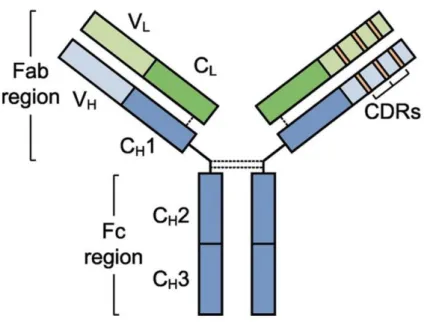
a. Structure
The most used form of antibodies in therapeutics is IgG1. An IgG1 molecule consists of two identical heavy and two identical light chains. The heavy chain contains three constants (CH1, CH2, CH3) and a variable domain (VH), while the light chain includes only one constant (CL) and one variable (VL) domains. Each variable domain consists of three complementarity-determining regions (CDR1, CDR2 and CDR3). CH2-CH3 represents together a Fc (constant fragment) portion and CH1-VH and CL-VL represents a Fab portion of an antibody. Heavy chains attached to each other and to light chains with the number of disulfide bonds. Research has shown that the specificity of an antibody depends on its variable domains, especially on the sequence of VH-CDR3 (Figure 1).
Even if the variable fragment (Fv) is necessary for antigen recognition, the constant fragment (Fc) of antibodies serves to exhibit
biological effector
functionality by interacting with complement and Fc-receptors expressed on different blood cells.
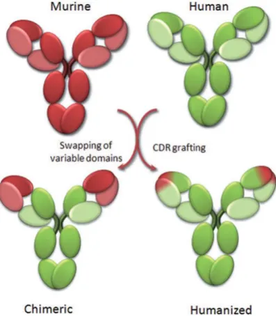
Monoclonal antibodies are classified according to their production and engineering methods (Figure 2):
24 - Full murine antibody: non-engineered and produced in murine species and
containing only murine IgG1 constant and variable regions.
- Full human antibody: Engineered and obtained from human cells and only human IgG1 constant and variable regions.
- Chimeric antibody: Variable fragments are produced in murine species and linked to human constant fragment.
- Humanized antibody: It is the most advanced format of chimeric antibody. It contains only CDRs from murine and the rest belongs to a human antibody.
Figure 2. Chimeric and humanized antibodies (Patrick Chames et al.,
2009).
Murine sequences are depicted in red and human sequences in
25 Administration of a therapeutic antibody can lead to an anti-antibody response (AAR). Consequently, to resolve this problem, a certain type of antibodies has been engineered to have a much similar structure to human antibodies. In 1984 Boulianne et al. have demonstrated the feasibility of functional chimeric mouse/human antibody (1). Then by substitution of CDR regions of human IgG1 with the same region of antigen specific animal antibodies humanized antibodies have also been developed. The similar affinity of the humanized antibodies has been observed comparingthem to their animal homologues (2,3). In 1991 Padlan EA has shown that the humanized antibodies are less antigenic (4). William
Ying et al. have published a review of reported AAR to murine, mouse–human chimeric, and
humanized antibodies (5). They have demonstrated that it is very rare for a murine mAb to show negligible immunogenicity, and that it is very common for nearly the 100% of patients to produce human anti-mouse antibodies (HAMA). However, in contrast they have found that patients treated with chimeric antibodies produce 40% human anti-chimeric antibodies (HACA). This response has been diminished to only 9% with humanized antibodies. Another study has also confirmed by administering the mouse anti-human TNF antibody and its humanized homolog to healthy voluntaries that the humanized antibody reduces the immunogenicity by nearly 90 % (6).
26
b. Antibody engineering approaches
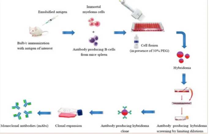
1. Hybridoma technology
To produce the antibodies for therapeutic aims, animals are immunized by different methods with antigens in accordance to the desired antibody characteristics. Kohler and
Milstein have demonstrated in 1975 that the cell lines made by the fusion of mouse myeloma
cells and mouse spleen cells from an immunized animal can produce appropriate antibodies. The authors of this work which described a method of antibody production based on hybridoma technology received the Nobel Prize in Physiology or Medicine in 1984. Hybridoma technology is the most used method to produce antibodies from an immunized animal. In this method the extracted B cells from the spleen or lymph nodes of an animal are
fused with immortalized myeloma cells by using polyethylene-glycol (PEG) as a fusion
27 promoting agent to produce hybridoma cells. Myeloma cell lines selected in this technology are deficient for a hypoxanthine-guanine phophoribosyl-transferase (HGPRT-) enzyme which participates in the neo-synthesis of nucleotides. Because of this molecular deficiency, myeloma cells use another pathway of nucleotide neo-synthesis which can be blocked using a chemical agent (most commonly aminopterin and thymidine). The fusion of a myeloma cell with a spleen cell enables them to acquire a functional HGPRT pathway. Also a medium containing aminopterin select myeloma cells that had actually been acquired by fusion (complementation) HGPRT pathway and which are therefore capable of activating the alternative pathway of nucleotide production (7). These cells are distributed in cell culture plates as single cell colonies and they are let to produce the antibodies. Antibody production and the presence of a specific candidate can be screened by different techniques such as enzyme linked cell sorbent assay (ELISA) and analytical tools like fluorescence-activated cell sorting (FACS) to find the target-specific antibodies (Figure 3).
The possibility to produce full human antibody by generating hybridoma cells from human lymphocytes has also been investigated (8,9). Antigen-specific human B cells are rarely present in circulation, and display low fusion efficiency. Steinitz et al. showed that the infection and transformation of B cells by Epstein-Barr virus (EBV) substantially increases the fusion efficiency and development of mAbs (10). EBV transformation of B cells isolated from peripheral blood of two individuals with asymptomatic human immunodeficiency virus type 1 (HIV-1) has been successfully used for the production of human mAbs against HIV-1 (11).
Even though hybridoma technology allows producing murine antibodies and regarding the poor reproducibility related to hybridoma cell cultures, this technology could not be
28 successfully used for other animals. Moreover, immunization with proteins which have a high similarity with its murine homologue results in the generation of low-affinity or non-specific antibodies. Altogether, these limitations necessitated development of new antibody producing technologies.
2. Phage display
The development in molecular biological applications in antibody production conducted to use phage display technology to generate a diverse number of antibodies. In 1985 Smith demonstrated that proteins, together with cloned antigens can be displayed on the phage surface (12). Then Mccafferty et al. have shown the possibility of expression of the
immunoglobulin variable genes on phage surfaces after the extraction and amplification from hybridomas or directly from splenocytes or B cells using the polymerase chain reaction, and cloning them into expression vectors (13). They have also described affinity chromatography for specific phage isolation. One year later the same team prepared the phage library from
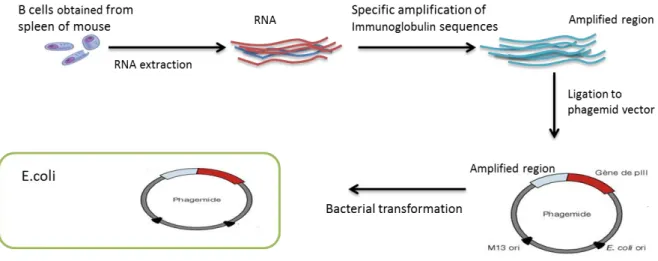
Figure 4. Phage library construction.
B cells are obtained from mouse spleen, total RNA is extracted and converted to cDNA by reverse transcription. Immunoglobulin sequences are specifically amplified by PCR. To obtain the phage library, amplified sequences are cloned to phagemid vector and transformed to E.coli cells.
29 peripheral blood lymphocytes (PBLs) of unimmunized donors by using the VH and VL genes and showed the use of this library for isolation of antigen or hapten specific antibodies (14). Preparation and use of phage libraries from seropositive individuals has also been demonstrated in the early 1990s (15).
To produce antibodies by phage display technology, in the first step, RNA extracted from murine splenocytes or human peripheral blood lymphocytes is converted to DNA by reverse-transcription reaction. Then the antibody coding sequences are amplified by using specific primers and cloned into phage vector to create a library. After the transformation of phage library to E.coli, several panning (selection) rounds are performed to select specific phages. Selected phages are eluted by using acid shock or enzymatic cleavage and then retransformed to E.coli strain. Single colonies are isolated by spreading the bacteria onto LB-agar plate. Finally to identify specific mAbs or antibody fragments, supernatant of monoclonal bacterial culture is screened by using ELISA or FACS (Figure 4) (16).
Phages are viruses which infect bacteria and could replicate its DNA inside the bacteria. M13 filamentous phage which has the advantage to multiply at extremely high titers (1012-1013 pfu/ml) and possesses a simple DNA genome is commonly used for surface displaying of banks of random peptides and antibodies (17,18) (Figure 5). Its genome is a simple circular DNA strand of about 6.4 kb
PV
III
PV
III
Figure 5. Schematic illustration of filamentous bacteriophage M13.
30 coding for 10 proteins. Protein pIII, pVI, pVII, pVIII and pIX form the structure of the capsid. The pIII protein is anchored to the extremity of phage with its C- terminal domain, and its N-terminal domain is responsible for the attachment to the pili of E. coli. pII, pV and pX are involved in replication of the phage DNA, while the pI and PIV form a channel in the membrane of E. coli, allowing the release of newly produced phage particles (19). The number of copies of protein pVIII is variable and serves to form the capsid of phage. The size of the viral tube consisting of the pVIII protein is variable and can be extended depending on the length of the DNA to be packaged. This flexibility allows the insertion of large fragments of exogenous DNA into the viral genome and rends M13 ideal for displaying antibodies or peptides. Another advantage of M13 phage that it does not induce lysis of bacteria which differs it from other phages (17).
Generally, pIII and pVIII proteins are used for the presentation of peptides or antibody fragments by M13 phage. The pVIII protein is the most abundant, but, it may present only short size peptides (6-8 amino acids) due to the size restrictions imposed by the geometric assembly constraints of pVIII subunits to form the capsid (20). Displaying multiple copies of an antigen could also result in the selection of low affinity clones (21). In contrast, pIII protein is presented with only 5 copies on the phage surface which allows to display high molecular weight peptides and to select the clones with the highest affinity (22).
Although pIII allows to present high molecular weight peptides, a full length IgG expression in E. coli is generally problematic (23). As a matter of fact, IgG expression and isolation in E. coli has been demonstrated by some groups investigating phage display technology, it was not described clearly (24–26). Alternatively, smaller antibody variants as Fabs, sc-Fvs, etc. can be successfully displayed by pIII (Figure 6). The advantage of these
31 small fragments is that, they can target the occluded epitopes on the cell surface which otherwise are not accessible by full antibodies.
Figure 6. Different fragments of antibodies.
IgG1: full antibody immunoglobulin 1; Fab: fragment antigen binding; Sc-Fv: single chain variable fragment; VL: variable light chain; VH: variable heavy chain.
The main aim of phage display is to obtain libraries containing the largest diversity of antibody or antibody fragments. The probability of selecting molecules of interest with a high affinity is proportional to the size of the library, as well as the diversity of the molecular repertoire. In general, libraries are composed of 107-108 clones (27), and they could be divided into four main types which include (28):
- Naive antibody libraries: Genetic information coding for antibodies obtained from non-immunized animal B cells or splenocytes. Marks et al. have constructed for
32 the first time the naïve libraries from human peripheral blood B cells with 107
clones (14).
- Immunized antibody libraries: Libraries constructed from the genetic materials obtained from different animals which are selectively immunized with a desired antigen or from seropositive humans (29–32).
- Synthetic antibody libraries: Construction of synthetic libraries involves rearranging VH and VL gene segments in vitro and introducing artificial complementarity determining region (33–35).
- In-vivo recombined antibody libraries: Phage vectors obtained from a synthetic library were transfected to cells and the phage population produced from this primary library was then allowed to infect bacteria. This method allowed the exchange between different VH-VL vectors to create more diverse libraries. Additionally, these libraries have been shown to have the diversity of 1010 and 1011
33
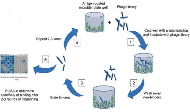
The next step of this technology is dedicated to the selection of high affinity antibodies using several selection rounds. The principle of affinity selection is based on the reversible bond between an antibody library and its target. The recombinant phages were thus selected for their ability to bind to a selected target. The selections are often made using the purified molecule of interest, and are generally composed of the following steps: i) fixing of the target phage, ii) washing to eliminate the less specific phage, iii) eluting of specific phages iv) and finally infecting E. coli bacteria where the selected phages will be amplified. The repetition of these binding-elution steps allows to highly enrich the proportion of high affinity phages to target. Usually 2-3 rounds of selection in increasing stringency conditions are required to
34
isolate high affinity antibodies for the target (38) (Figure 7).
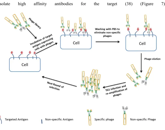
Figure 8. Selection of phages by antigen expressing cells.
Targeted antigen expressing cells are incubated with antibody phage library. Non-binding phages are eliminated by several wash steps and then binder phages are eluted. E. coli cells are infected with eluted phage and amplified. Then the next round of selection is started. 2-3 rounds are necessary to identify specific binders.
Selection strategies are usually dependent on the desired characteristics of the antibody. For therapeutic uses, antibodies must recognize the naturally expressed form of antigens on cell surfaces. Selection rounds can be performed on the targeted antigen expressing cells to identify natural-form binders (39) (figure 8). Cell selection method could be performed successfully to select cell surface antigens targeting phages.
Zhu et al. have described for the first time the therapeutic use of an antibody obtained
35 antibody phage display library constructed from spleen cells of mice immunized with a soluble form of a human vascular endothelial growth factor (VEGF) receptor, could block VEGF binding to kinase insert domain-containing receptor (KDR). In vitro cell culture assays showed that these antibodies could significantly suppress the mitogenic response of human umbilical vein endothelial cells to recombinant human VEGF in a dose-dependent manner, and reduce VEGF-dependent cell proliferation by 60% and 40% (41). An in vivo analysis of these recombinant antibodies in a rat cornea angiogenesis model revealed that both of the two antibodies could suppress the development of new corneal vessels (42).
Recently another antibody engineering approach was also developed to generate antibodies from isolated single B cells which allowed to study the expressed antibody repertoire in humans (43). Based on surface marker expression, individual cells at different stages of human B cell development are isolated by fluorescence-activated cell sorting. For each cell Ig heavy and corresponding Ig light chain gene transcripts are amplified and cloned into eukaryotic expression vectors to produce monoclonal human antibodies of the same specificity in vitro. This method of cloning and express human monoclonal antibodies is unbiased, highly efficient, requires only small cell numbers and the recombinant antibodies allow direct conclusions on the frequency of specific human B cells in a diverse repertoire.
36
c. Mechanism of action
In therapy, antibodies are used as full monoclonal antibodies or conjugated with different drugs, immunotoxins, and cytokines. Therapeutic function of antibodies depends on the specificity of the paratope leading to antigen recognition and neutralizing or agonistic properties. The nature of the Fc fragment, or its vectorization using antibody-drug conjugation technology might also be used to develop a functional mAb-based therapy.
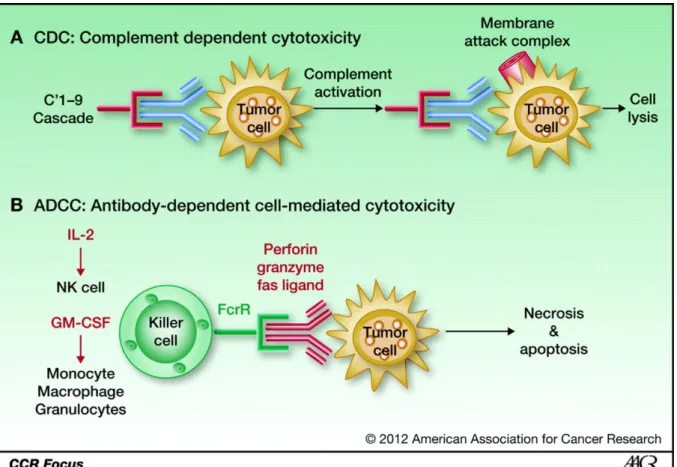
Figure 9. Mechanism of CDC and ADCC (Katherine K. Matthay et al. Clin Cancer Res
2012;18:2740-2753).
The mAb binds to the receptor and initiates the complement cascade, which results in the formation of a membrane attack complex that makes a hole within the cell membrane, causing cell lysis and death. B, ADCC: The Fc fragment of the monoclonal antibody binds the Fc receptors on monocytes, macrophages, granulocytes, and natural killer cells (NK). These cells in turn engulf the bound tumor cell and destroy it. Natural killer cells secrete cytokines that lead to cell death. GM-CSF, granulocyte macrophage colony-stimulating factor; IL, interleukin.
37
1. ADCC and CDC
Antibody dependent cell cytotoxicity (ADCC) represents the killing of target cells by effector cells that are activated by binding to the Fc portion of the monoclonal antibody molecule (44) (figure 6). Monocytes, macrophages granulocytes and natural killer (NK) cells express Fc-receptors (FcR) which bind to the Fc part of the antibody and trigger the cellular activation. Activated effector cells can make the synapse with the tumor cell and eliminate it.
NK cell cytotoxic activity was linked to cancer risk in 2000 by Imai et al. (45). They have demonstrated after the 11 year of follow-up survey of 3625 healthy persons that the in 154 cases cancer was developed and the low cytotoxic activity of peripheral blood cells was associated with a high cancer risk. NK cell activation is controlled by the balance of inhibitory (KIR family receptors) and activator signals triggered by NKG2D, DNAM-1, and 2B4 and the natural cytotoxicity receptors including NKp46, NKp44, and NKp30 (46–49).
Additionally, Fc receptors play a crucial role for an antibody dependent cellular cytotoxicity of NK cells. The human FcyR family contains six known members in three subgroups, including FcyRI (CD64), FcyRIIa,b,c (CD32a,b,c) and FcyRIIIa,b (CD16a,b), expressed by various effector cells of the immune system, including macrophages, neutrophils, dendritic cells and natural killer (NK) cells (50). These cells are recruited by antibodies and then the synapses are created with targeted cells, these synapses lead to the release of perforin/granzyme and the establishment of the Fas/FasL interaction, both leading to apoptosis of the target cells. Except FcyRIIb receptor which recognizes human IgG1 and IgG3 with low affinity, all of the other FcyRs are activator receptors (51,52). FcγRI (CD64) binds to human IgG1 and IgG3 with high affinity, the FcγRIIa binds human IgG1, IgG2, and IgG3. FcγRIIIb is expressed only by neutrophils and may play a role in neutrophil activation.
38 CD56dim CD16+ NK cells highly express an activator low affinity FcγRIIIa (CD16) receptor which mediates ADCC and do not express inhibitory receptor FcRIIb. Activation of FcyRIIIa depends on the affinity to the Fc part of an antibody. Recognition of the Fc part of an antibody by FcyRIIIa triggers a strong activating signal which impairs the inhibitory signals and results in both cytotoxicity and the cytokine secretion (53,54). Hubert et al. have demonstrated the role of FcyR in ADCC by using a mouse model. They showed that the cytotoxicity of a Tn antigen-specific chimeric mAb against murine breast cancer was diminished in FcyR deficient mice (55). Another study has also confirmed the role of inhibitory and activator FcyR in ADCC. The inhibitory receptor FcyRIIb deficient mice showed much more ADCC after the treatment by transtuzumab and Herceptin (both anti-Her2 mAbs) when compared with mice deficient for activator receptors (56).
The principal and most encountered problem with full, unconjugated, antibodies is their low affinity to Fcy receptors (FcyR) to generate ADCC (57). The polymorph variant of FcyRIIIa which is expressed in 20 % of the population and contains a valine at position 158 instead of phenylalanine in 80 % of the population has been identified (58,59). A 5 times higher affinity to IgG1 Fc of FcyRIIIa-158V compared with FcyRIII-158F that leads to a more efficient in vivo ADCC in peripheral blood mononuclear cells (PBMCs) or purified NKs. Musolino et al. demonstrated with fifty-four consecutive patients with Her2/neu-amplified breast cancer receiving trastuzumab (anti-Her2 antibody) plus taxane that PBMCs with FcyRIIIa-158V/V or/and FcyRIIa-131H/H genotypes had a significantly higher trastuzumab-mediated cytotoxicity than PBMCs harboring different genotypes (60).
Monoclonal antibodies are used mostly in second line therapies in patients who have already received several cycles of chemotherapeutic agents which hamper their immune
39 systems; their effector immune cells are therefore not really functional. For this reasons, several clinical trials are performed in order to use the mAbs in first line therapies with or without the combination with chemo-or radio-therapy (61–67). This hypothesis could be associated with the confusing results of several studies using rituximab (anti-CD20 mAb) to evaluate the role of the polymorphism of FcyRIIIa or FcyRIIb. Cartron et al. have demonstrated in 49 patients having received rituximab for a previously untreated follicular non-Hodgkin lymphoma that the FcyRIIIa-158V/V genotype was correlated to a better rituximab efficacy (58). Interestingly all of other studies on first line administration of rituximab agreed that the FcyRIIIa-158V/V genotype was the single favorable parameter associated with clinical and molecular responses (59,68–70). In contrast these results have not been confirmed in second line studies (71–73). Prochazka et al. with a large cohort of 102 patients have demonstrated that the FcγRIIIA receptor genotype does not influence an outcome in patients with follicular lymphoma treated with risk-adapted immune-chemotherapy. Similar confusing results in favor of first line therapy which shows considerable high ADCC with FcyRIIIa-158V/V genotype have been published for transtuzumab, cetuximab (anti-EGFR mAb) and anti-GD2 antibodies (74,75).
It has also been reported that the glycosylation of the CH2 domain (Asn 297) of the Fc portion has a negative effect on ADCC (76). The presence of fucose residues in the carbohydrate part has been shown to decrease the affinity of an antibody to the FcRyIII, therefore impairing ADCC efficiency (77,78). Glycosylation of antibodies has been demonstrated as a result of the method of production, especially depending on an antibody producing cell line (79).
40 Another limitation for a ADCC reaction is the competing effect by naturally occurring IgGs in human blood for the binding on FcyR receptors. A study to evaluate a role of naturally occurring IgGs on inhibition of fully human monoclonal IgG1 against epithelial cell adhesion molecule (Ep-CAM) and trastuzumab induced ADCC has been conducted (80). The results showed that the adding of the human serum with physiological levels of IgG1 inhibits the ADCC induced by those therapeutic antibodies and could be reversed by eliminating the IgG1 by affinity chromatography.
The Fc part of antibodies can also enhance the complement dependent cell death (CDC) by binding the C1q. C1q binding triggers the complement cascade which leads to formation of membrane attack complex (C5b to C9) at the surface of target cell, as a result of the classical pathway of complement activation. All therapeutic mAbs couldn’t activate the complement cascade, because it has been already shown that to induce CDC, an antibody must bind an epitope near the cell surface. This mechanism of cytotoxicity has been largely studied for anti-CD20 antibodies. Cragg et al. showed that the complement depletion using cobra venom factor markedly reduced the efficacy of rituximab and 1F5 (anti-CD20 mAb) in 2 lymphoma xenograft models (81). Another study has also demonstrated that the anti-CD20 antibody ofatumumab induces considerably higher levels of CDC (82). Based on a higher CDC activity novel types of anti-CD20 mAbs were developed and they showed to be more performant in ‘in vitro’ assays (83). Using murine models, antibodies which induce ADCC and CDC at the same time have been shown to be more suitable for cancer treatment (84).
41
d. Pharmacokinetics of antibodies
Pharmacokinetic properties of mAbs differ markedly from those of non-antibody-type drugs, and these properties can have important clinical implications (85). Antibodies and endogenous immunoglobulins are protected from degradation by binding to the neonatal Fc-receptor (FcRn), which explains their long elimination half-lives (57). FcRn was discovered as a responsible of the transmission of maternal antibodies from mother to pup (86,87). In addition, FcRn protects IgG from degradation, thereby explaining the long half-life of this class of antibody in the serum (88,89). Because of the β2microglobulin association the structure of FcRn, is related to MHC class I (90). FcRn binds the Fc portion of IgGs. A major site of expression of FcRn is vascular endothelial cells where FcRn functions to extend the serum persistence of IgG by recycling internalized IgG back to the surface. Akilesh et al demonstrated by using FcRn deficient mice that in addition to its expression in the vascular endothelium of several organs, FcRn was highly expressed in bone marrow-derived cells and professional APCs in different tissues. Experiments using bone marrow chimeras showed that FcRn expression in these cells acted to significantly extend the half-life of serum IgG indicating that in addition to the vascular endothelium, bone marrow-derived phagocytic cells are a major site of IgG homeostasis (91,92). Another study confirmed the expression of FcRn in monocytes, intestinal macrophages, and dendritic cells (93). FcRn bound human IgG at different pHs which indicates that IgG enter cells through non-specific, fluid-phase pinocytosis (94). Endocytosed IgG is trafficked along the endosomal pathway and encounters FcRn in the early endosome where the acidic conditions favors a productive IgG-FcRn interaction (95).
42 With the aim of increasing the serum half-life of the therapeutic IgGs several studies have been performed. Ghetie et al. have used the random mutagenesis of Thr252, Thr254, and Thr256 followed by selection using bacteriophage displays and obtained the mutant of FcRn with the highest affinity and a significantly longer serum half-life than the wild type fragment. (96). Another study showed that the mutagenesis of His-436-Ala decreased the activity of the Fc hinge fragments in both ’in vivo’ and ‘in vitro’ assays (97). The several mutations in the Fc portion of antibodies also suggested the better binding to FcRn and resulted with an increased half-life (98). Ulrich et al have also described a variable number of tandem repeats polymorphism influencing the transcriptional activity of the neonatal FcRn promoter (99).
43
e. Targets for therapeutic antibodies
The first and the most important step to generate an antibody for therapeutic aims is the choice of the target antigen. The expression or the functional pathway of the antigen must be restricted for tumor cells. For unconjugated antibodies, it is desirable to target cell surface antigens which are not internalized when they bind antibody. In contrast internalization is desirable for the antibodies conjugated with toxins or drugs (100).
Different antigens and cellular pathways for approved antibodies for clinic use described below (Table 1):
1. Cell differentiation (CD) antigens:
The therapeutic objective here is to identify an antigen whose expression is selectively distributed in the organism in order to specifically target a tissue or a cell while controlling the specific distribution of the mAb in the whole organism. An example of such development is illustrated by anti-CD20 mAb.
CD20 is a cell differentiation antigen, expressed from early pre-B cells to later in differentiation, but it is absent on terminally differentiated plasma cells. Rituximab was the first chimeric antibody approved for therapeutic use which targets CD20 (101). It is active in a variety of human lymphomas and chronic lymphocytic leukemia (66,102,103). Rituximab induces cell death through ADCC and CDC (104). McLaughlin et al. have published the results of 166 patients with relapsed low grade or follicular lymphoma with rituximab of which 48 % responded (105). Rituximab can induce CDC and ADCC as well as direct programmed cell death (106,107). Even if it is widely used for lymphoma therapy alone or in combination regimens mainly for relapsed and refractory lymphomas (108,109), the efficacy
44 Table 1. FDA approuved monoclonal antibodies for cancer therapy (www.mycancergenome.org).
45 of rituximab is modest and often variable especially when used for CLL treatment which results with an objective response rates ranged between 25% and 35% (110,111). Second generation mAbs designed as humanized or fully human with unmodified Fc domain and compared with rituximab in clinical trials. Among them, Ocrelizumab showed higher ADCC and lower CDC and ofatumumab showed higher CDC and less immunogenicity as compared with rituximab (112–114). The third generation humanized mAbs are modified mAbs in the Fc region. The Fc domain was engineered with the glycol or protein. The main goal of this modification is to improve the therapeutic efficacy in all patients; particularly among patients in which the expression with low affinity version of the Fc receptor is found on their effector immune cells (115). More recently other three, tositumomab, ofatumomab, obinutuzomab, anti-CD20 targeting antibodies have also been approved for treatment of diverse range of B-cell malignancies.
Other cell differentiation antigens targeted by alemtuzumab and daratumumab are CD52 and CD38 respectively. Like rituximab, these antibodies are effective in hematological malignancies (116–119).
2. Growth factors:
Family of growth factor receptors and ligands, because of their large expression in tumor cells are the promising targets for the treatment of solid cancers by monoclonal antibodies.
Epithelial growth factor receptors (EGFR) and vascular endothelial growth factor receptors (VEGFR) and their ligands have already been targeted by monoclonal antibodies which were approved for clinical use.
46 It was demonstrated in the late 1980’s and the early 1990’s that the EGF family proteins are highly expressed in solid malignancies and correlates with a poor prognosis
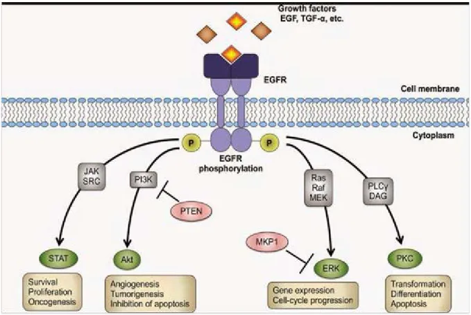
(120,121), which makes them attractive candidates for antibody targeting. Activation of EGF receptors leads to different signaling pathways which increase cell proliferation and impair tumor cell apoptosis. It has also been demonstrated that the activation of EGFR by binding its ligand triggers tumorigenic processes, as cell survival, cell cycle progression, angiogenesis and inhibition of apoptosis (122,123) (Figure 10). Cetuximab is the first approved chimeric antibody for EGFR inhibition. Panitumumab is another fully human antibody which impairs EGFR signaling and thus leading to cell death. Panitumumab has been shown to be effective for colorectal cancer patients either as a single agent or combined with chemotherapy (124–
Figure 10. EGFR signaling pathway (Morten Bjerklund).
Binding to its ligand activates EGF receptor and leads to auto phosphorylation. This activation triggers different signaling pathways which increase cell proliferation, angiogenesis, and abolishes apoptosis.
47 126). The role of KRAS status have been investigated to compare the panitumumab monotherapy to best supportive care by detecting KRAS mutations in tumor sections collected in a phase III mCRC trial. The results of this study suggested that the wild type (WT) KRAS patients had a longer overall survival (127). Similar results have also been obtained for the cetuximab treatment in favor of non-mutated KRAS status (128). Several clinical studies have also confirmed that the patients whose tumors were KRAS wild type, respond to these anti-EGFR antibodies, but in some cases an acquired resistance to cetuximab have also been observed. (129–132). To identify the mechanisms of acquired cetuximab resistance, Montagut et al. established cetuximab-resistant cells from the highly sensitive human mCRC cell line DiFi (which is wild-type for KRAS, BRAF and PI3K and has an amplification of EGFR). These cells were treated and rendered resistant to cetuximab. Without detecting neither the mutations in KRAS, BRAF nor PIK3CA or loss of PTEN expression, they identified an EGFR ectodomain mutation (S492R) in cetuximab resistant cells. Furthermere, they have also identified this mutation within two of ten subjects with disease progression after the cetuximab treatment. They also showed that the subject with cetuximab resistance harboring the S492R mutation responded to treatment with panitumumab (133).
Another EGF receptor overexpressed in breast and gastric cancers is HER2/EGFR2. It is targeted by transtuzumab and pertuzumab. Neutralizing antibodies targeting other variants of EGFRs are in clinical evaluation stages (134,135). The results of two trials that compared adjuvant chemotherapy with or without concurrent trastuzumab in women with surgically removed HER2-positive breast cancer demonstrated that trastuzumab combined with paclitaxel after doxorubicin and cyclophosphamide treatement improves outcomes (136). Several studies have confirmed the efficacy of transtuzumab or pertuzumab against HER2
48 overexpressing invasive breast cancers in both locoregional and advanced breast cancers (137,138). Although HER2 overexpressing cancers are sensitive to the antibody treatment, but soluble form of HER2 has been shown as an indicator of poor prognosis in HER2+ metastatic breast cancer and impairs the antibody efficacy (139,140).
VEGF receptors and ligands are also targeted by therapeutic antibodies in solid tumors. This family of proteins has an essential role in tumor angiogenesis. Therefore blocking the ligand or the receptor of VEGFs alters the tumor progression. The most utilized anti-VEGF antibody is bevacizumab (avastin) which neutralizes VEGF and impairs its binding to the receptor as well as triggering a signaling pathway of angiogenesis. Since it has no direct effect in tumor eradication, it is combined with different chemotherapies to obtain the better results (61,62,141,142). In a clinical trial conducted with 813 patients with previously untreated metastatic colorectal cancer it has been demonstrated that the addition of bevacizumab to fluorouracil-based combination chemotherapy results in statistically significant and clinically meaningful improvement in survival among patients with metastatic colorectal cancer (143). The study on 1,401 patients with metastatic colorectal cancer has revealed that the addition of bevacizumab to oxaliplatin-based chemotherapy significantly improved PFS in first-line therapy (144).
3. Immune-checkpoint molecules
A better understanding of the role of the immune system provides great insight into tumor microenvironment (TME) hence enabling the development of novel and highly promising immune modulators (145). Effector T cells (CD8+) can be activated and they can play a crucial role in TME for cancer cell elimination. Activation of T cells passes through the recognition of the antigen presented by major histocompatibility complex (MHC) class I by
49 its T Figure 11. Immune check-point regulation (Pardoll et al. 2012).
50 cell receptors (TCR) but also needs a second costimulatory signal modulated by B7 family members CD28 or CD80 (146–149). or There are also inhibitory molecules expressed by tumor cells to impair the T cell activation (150). Tumor cell death depends on the modulation of these inhibitor and activator molecules (Figure 11 and 12). The checkpoint molecules, CTLA-4, programmed death-1 (PD1), and its ligand (PD-L1), etc. are the inhibitory signals
that serve to cancer cells to evade the immune system (151). Even if the activation signal by TCR and the co-stimulation receptors (145,145,152–154) are present, interaction between the ligand and receptor of checkpoint molecules can generate negative feedback signals that
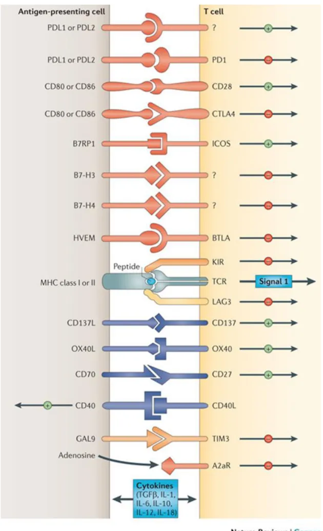
Figure 12. Interactions of different immune checkpoints between T-cells, APCs, and cancer cells that can be exploited in cancer therapeutics (El-Osta et al., 2016).
Abbreviations: APC, antigen-presenting cell; KIR, killer immunoglobulin like receptor; LAG-3, lymphocyte activation gene-3; PD-1, programmed death-1; PD-L1, programmed death ligand 1; MHC1, major histocompatibility complex 1; NK, natural killer; TCR, T-cell receptor; 4-1BB, CD137; 4-1BBL, 4-1BB ligand.
51 dampen the immune response. Therefore the blockade of checkpoint molecules by monoclonal antibodies is a great avenue for cancer immunotherapy.
CTLA4 is the first immune-checkpoint receptor targeted by therapeutic antibodies. It regulates early T cell activation and it is a co-inhibitor of CD28. CD28 binds to its ligand CD80 or CD86 and strongly amplifies the activator signal of T cells received after MHC class I and TCR interaction. CTLA4 binds to the same ligands stronger than CD28, and acting as a competitor antagonist which impairs the T cell activation (155–158). The principal role of CTLA4 has been described to be the down-modulation of T helper cell activity and enhancement of regulatory T (159,160) cells which have been described as a poor prognostic for cancer progression (161,162). These characteristics of CTLA4 made it to become a crucial target for immune therapy. CTLA4 blockade has been shown to enhance the immunologic response, which ultimately can induce tumor regression. Also in the case of poorly immunogenic tumors, in a murine model which does not induce substantial endogenous immune responses, the combination of a granulocyte–macrophage colony-stimulating factor (GM-CSF)-transduced cellular vaccine and a CTLA4 antibody could induce a strong enough immune response to slow tumor growth and in some cases eliminate established tumors (163). Ipilimumab, an anti-CTLA4 antibody showed in clinical trials showed survival benefits for melanoma patients and as a matter of fact it was approved by FDA in 2010 for clinical use (164).
PD1 is another immune-checkpoint receptor expressed in the cell surface of T-cells. PD1 expression is induced when the cells are activated (165). PD1 can bind two ligands (PD-L1 and PD-L2) expressed on cancer cells. The interaction between PD1 and its ligands blockades the T cell activation by recruiting the phosphatase SHP2 (166). PD1 is also highly
52 expressed on Treg cells, where it may enhance their proliferation after binding to its ligand. Blockade of the PD1 pathway may also enhance anti-tumor immune responses by diminishing the number and/or suppressive activity of intra-tumoral Treg cells.
PD-L1 is expressed in melanoma, ovarian, lung and many other cancer cells (167– 169). PD-L1 expression was also observed in tumor infiltrating myeloid cells as myeloid derived suppressor cells which correlates with a poor prognosis of tumors. PD-L1 expression is regulated by different factors in different tumors. In some tumors like glioblastoma, the PD-L1 expression appears as a result of an oncogenic pathway activation which is called innate immune resistance (170). The alternative mechanism (adaptive immune resistance) for PDL1 upregulation on tumors is the response of tumor cells to interferons (IFNs)- predominantly IFNγ (171–173). Interestingly, the interaction between PD-L1 and CD80 (in this case expressed as a receptor on T cells) has been shown to induce an inhibitory signaling pathway in T cells (174).
Anti-PD1 and anti PD-L1 antibodies have been demonstrated to be effective for various cancers (152,154,168,175). The mechanisms of the blockade with PD1 and anti-L1 antibodies are different, because even though anti-PD1 antibodies block both PD-L1/PD1 and PD-PD-L1/PD1 interactions, they could not target PD-L1/CD80 interaction. However anti-PD-L1 antibodies neutralize the PD-L1/CD80 and PD-L1/PD1 interactions but not the binding of PD-L2 to PD1.
Lymphocyte activation gene 3 (LAG3; also known as CD223), 2B4 (also known as CD244), B and T lymphocyte attenuator (BTLA; also known as CD272), T cell membrane protein 3 (TIM3; also known as HAVcr2), adenosine A2a receptor (A2aR) and the family of killer inhibitory receptors, B7 family inhibitory ligands — in particular B7-H3 (also known as
53 CD276) and B7-H4 (also known as B7-S1, B7x and VCTN1) have been identified as immune-checkpoint blockaders in animal models and are targeted by monoclonal antibodies which are under clinical investigations (176–182).
54
f. Antibody-drug conjugates
Antibody-drug conjugates (ADC) combine the selectivity of an antibody and the toxicity of chemotherapy. An antibody portion of ADC binds selectively with the target cell and is internalized by endocytosis. Then the cytotoxic drug kills the target cell. Antigen selection is important for ADCs; i) it must be highly expressed and has to be selective for
tumor cells. It must have a limited expression in normal tissues, ii) it could be internalized after binding to ADC for a successful delivering of the toxic agent, iii) it must not be shed or downregulated after the ADC treatment. There are more than 25 antigens investigated to be targeted by ADCs in different phases on clinical trials (Figure 13). These include i) overexpressed targets on cancer cells as NCAM, CD70, Mesothelin, CEACAM5, Mucin 1, etc. ii) Antigens acting in angiogenesis; PSMA, ROBO4, VEGFR2, etc. iii) antigens presented in tumor stroma; collagen IV, periostin, etc (Figure 10).
55 Two main groups of drugs, microtubule inhibitors and DNA-damaging drugs are used for ADC development. The linkage between the antibody and the drug is also important, because it must be stable and must not be cleaved in patients’ blood till the delivery of ADC to the target cell. It must as well comprise a cleavage site for lysosomal enzymes for the release of drug once ADC is internalized.
ADCs were firstly designed as therapeutic agents for second line therapy in solid tumors or hematological malignancies to circumvent resistance to chemotherapy. To date, only two ADCs are approved by the FDA for cancer therapy, T-DM1 and brentuximab-vedotin (BV). T-DM1 is composed of trastuzumab, a humanized IgG1 anti-HER-2 antibody linked with a stable non-cleavable linker to the maytansinoid DM1, a potent tubulin inhibitor (183). T-DM1 has been approved in the second line setting by the FDA for HER-2-positive patients who had previously received treatment with trastuzumab and taxane chemotherapy. It has also been investigated in clinical trials as a single agent (184,185).
Brentuximab vedotin comprises an anti-CD30 mAb connected to the highly potent tubulin inhibitor Monomethyl auristatin E (MMAE) (186). It targets CD30, a tumor necrosis factor (TNF) family member and triggers cell death once it binds with the antigen. BV has gained approval for the treatment of patients with relapsed or refractory CD30+ Hodgkin lymphoma following autologous stem cell transplant (ASCT) or patients not legible for ASCT who have failed at least two other chemotherapy treatments (187,188). BV has also been approved for patients with anaplastic large cell lymphoma (ALCL) as a second line treatment (189,190).