HAL Id: hal-03121844
https://hal.archives-ouvertes.fr/hal-03121844
Submitted on 19 Mar 2021
HAL is a multi-disciplinary open access
archive for the deposit and dissemination of sci-entific research documents, whether they are pub-lished or not. The documents may come from teaching and research institutions in France or abroad, or from public or private research centers.
L’archive ouverte pluridisciplinaire HAL, est destinée au dépôt et à la diffusion de documents scientifiques de niveau recherche, publiés ou non, émanant des établissements d’enseignement et de recherche français ou étrangers, des laboratoires publics ou privés.
and hnRNPM during Myoblast Differentiation
Nadera Ainaoui, Fransky Hantelys, Edith Renaud-Gabardos, Morgane Bunel,
Frédéric Lopez, Françoise Pujol, Rémi Planès, Elmostafa Bahraoui, Carole
Pichereaux, Odile Burlet-Schiltz, et al.
To cite this version:
Nadera Ainaoui, Fransky Hantelys, Edith Renaud-Gabardos, Morgane Bunel, Frédéric Lopez, et al.. Promoter-Dependent Translation Controlled by p54nrb and hnRNPM during Myoblast Differentiation. PLoS ONE, Public Library of Science, 2015, 10 (9), pp.e0136466. �10.1371/journal.pone.0136466�. �hal-03121844�
Promoter-Dependent Translation Controlled
by p54
nrb
and hnRNPM during Myoblast
Differentiation
Nadera Ainaoui1☯, Fransky Hantelys1☯, Edith Renaud-Gabardos1☯, Morgane Bunel1,
Frédéric Lopez2, Françoise Pujol1, Remi Planes3, Elmostafa Bahraoui3,
Carole Pichereaux4, Odile Burlet-Schiltz4, Angelo Parini5, Barbara Garmy-Susini5,
Anne-Catherine Prats1*
1 TRADGENE, UPS (EA4554), Toulouse, France, 2 UMR U1037-CRCT, Inserm, UPS, Toulouse, France, 3 UMR U1043-CPTP, Inserm, UPS, Toulouse, France, 4 UMR U5282-IPBS, CNRS, UPS, Toulouse, France, 5 UMR U1048-I2MC, Inserm, UPS, Toulouse, France
☯ These authors contributed equally to this work. *Anne-Catherine.Prats@inserm.fr
Abstract
Fibroblast growth factor 1 (FGF1) is induced during myoblast differentiation at both tran-scriptional and translational levels. Here, we identify hnRNPM and p54nrb/NONO present in protein complexes bound to the FGF1 promoter and to the mRNA internal ribosome entry site (IRES). Knockdown or overexpression of these proteins indicate that they cooperate in activating IRES-dependent translation during myoblast differentiation, in a promoter-depen-dent manner. Importantly, mRNA transfection and promoter deletion experiments clearly demonstrate the impact of the FGF1 promoter on the activation of IRES-dependent transla-tion via p54nrband hnRNPM. Accordingly, knockdown of either p54 or hnRNPM also blocks endogenous FGF1 induction and myotube formation, demonstrating the physiological rele-vance of this mechanism and the role of these two proteins in myogenesis. Our study dem-onstrates the cooperative function of hnRNPM and p54nrbas regulators of IRES-dependent translation and indicates the involvement of a promoter-dependent mechanism.
Introduction
Gene expression in eukaryotes is regulated at multiple levels. Transcription, as well as post-transcriptional processes such as mRNA splicing, polyadenylation, degradation and transla-tion, require a wide range of multi-component cellular machines in order to finely control protein production. For a long time, these steps have been considered to be a simple linear assembly line. Then, it has become apparent that gene expression, including steps such as tran-scription, capping, splicing, polyadenylation, RNA export and degradation, is coordinated in a complex and extensively coupled network [1,2]. However, coordination resulting from the co-transcriptional loading of mRNA processing proteins by the C-terminal domain of RNA poly-merase II seemed to exclude translational regulatory complexes [3].
OPEN ACCESS
Citation: Ainaoui N, Hantelys F, Renaud-Gabardos E, Bunel M, Lopez F, Pujol F, et al. (2015) Promoter-Dependent Translation Controlled by p54nrband
hnRNPM during Myoblast Differentiation. PLoS ONE 10(9): e0136466. doi:10.1371/journal.pone.0136466 Editor: Yoon Ki Kim, Korea University, REPUBLIC OF KOREA
Received: April 17, 2015 Accepted: August 4, 2015 Published: September 2, 2015
Copyright: © 2015 Ainaoui et al. This is an open access article distributed under the terms of the
Creative Commons Attribution License, which permits unrestricted use, distribution, and reproduction in any medium, provided the original author and source are credited.
Data Availability Statement: All relevant data are within the paper and its Supporting Information files. Funding: This work has been funded by Association Française contre les Myopathies (grant 16966), Association pour la Recherche sur le Cancer (grant PJA-20131200230), Ligue pour la Recherche Contre le Cancer, Cancéropole Grand Sud-Ouest, Institut National du Cancer (grant RIBOCAN no. 2008-039), Fondation de l’Avenir (grant ET0-583), Région Midi-Pyrénées (grant 11052677), Fonds Européen du Développement Régional (grant REFBIO-VEMT). There was no commercial funder. The funders had no
The fibroblast growth factor 1 (FGF1) gene provides an attractive system to decipher a mechanism of coupling between transcription and translation. Indeed, we have shown that this growth factor is induced by a transcription-translation coupling mechanism during myoblast differentiation [4]. The FGF1 gene structure has been well documented in human. Transcrip-tion occurs from four promoters A, B, C and D, which are either tissue specific or inducible [5,
6]. Promoters A, B and C are conserved in mouse [5]. The promoter A is active in heart, skele-tal muscle and kidney while the promoter B is brain specific [4,7]. Promoters C and D are inducible and considered as markers of cell proliferation [8–10]. The only FGF1 promoter to be activated during myoblast differentiation is the promoter A whereas the other promoters remain silent [4].
Transcription from each promoter generates alternative splicing of exon 1, leading to four mRNAs with distinct 5' untranslated regions (5'UTR), each of them expressing the same pro-tein, FGF1. Two of these 5' UTRs contain internal ribosome entry sites (IRES) [11]. IRESs have been described in several mRNAs containing long structured 5' UTRs. Most of these mRNAs code for proteins involved in the control of gene expression [12]. IRES structures have been determined in several mRNAs by chemical and enzymatic methods, allowing identification of stem loops responsible for the IRES activity [11,13,14]. As regards the FGF1 IRES, the IRES structural domain is conserved in mammals [11]. However this IRES structure is different from the IRES structures found in other mRNAs. In particular, it clearly differs from the IRES structure of another member of the FGF family, FGF2, suggesting distinct regulations of these growth factors by the IRES-dependent mechanism [13]. IRES-mediated translation is involved in the major translational process in conditions when the classical mechanism of cap-depen-dent translation is blocked [14–16]. We have previously shown that FGF1 is translationally induced via the IRES present in the promoter A transcribed mRNA, while cap-dependent translation is blocked during the first steps of myoblast differentiation [4]. FGF1 IRES-medi-ated translation is concomitant with transcriptional activation at promoter A. Furthermore IRES activity is drastically enhanced when transcription is controlled by promoter A, com-pared to the cytomegalovirus (CMV) promoter [4].
The concept of a nuclear event governing formation of the“IRESome”, the ribonucleopro-tein complex responsible for activation of IRES-dependent translation, has been reported in the literature for c-myc and Smad1 IRESs, although the cell factors responsible for this process have not been identified [17,18]. Furthermore, most ITAFs are nuclear proteins primarily identified as splicing regulators. The first to be identified, pyrimidine tract binding protein (PTB), was described as an intron-binding protein before being characterized as an ITAF of encephalomyocarditis virus (EMCV) IRES [19,20]. Several years after being identified as an ITAF for EMCV IRES, PTB has been shown to control several cellular IRESs such as APAF-1, BAG-1 or Bip [21–23]. Other splicing regulators, such as hnRNPK, hnRNPC1/2, hnRNPA1 or RBM4, have been identified as ITAFs [24–28]. HnRNPA1 has been described as an ITAF of several IRESs including FGF2, XIAP and c-myc IRESs [24,29,30]. It has been recently shown that this ITAF couples nuclear export and translation of several IRES-containing mRNAs [31].
Although the FGF1 IRES A has been reported to be stimulated during myoblast differentia-tion and muscle regeneradifferentia-tion, nothing was known about ITAFs responsible for such reguladifferentia-tion [4]. Biomolecular interaction analysis (BIA) uses a surface plasmon resonance phenomenon to characterize macromolecular interactions and has been recently optimized to enable the recov-ery and identification of interacting molecules by coupling BIA with mass spectrometry (MS) [32]. BIA-MS has proven successful in the identification of the partners in protein-protein interactions as it provides a high sensitivity compared to classical affinity chromatography approaches and permits identification from small quantities of bound ligands. A very repro-ducible "microelution" is now possible with the BIACORE 3000 instruments. This novel
role in study design, data collection and analysis, decision to publish, or preparation of the manuscript. Competing Interests: The authors have declared that no competing interests exist.
technology was selected in the present study to search for proteins bound to the FGF1 pro-moter A and IRES A during myoblast differentiation to find candidate ITAFs and transcription factors involved in the transcription-translation coupling mechanism.
In the present report, we identified hnRNPM and p54nrb/NONO bound (directly or indi-rectly) to FGF1 promoter A and IRES A in differentiating myoblasts (C2C12). Knockdown and overexpression approaches revealed that p54nrband hnRNPM are required to activate the IRES-dependent translation in a promoter-dependent manner. Furthermore, p54nrband/or hnRNPM knockdown inhibited myotube formation. Altogether, this study identifies a novel regulation of FGF1 gene expression, implying a cooperation between promoter and transla-tional regulators to promote the time controlled process of myoblast differentiation.
Materials and Methods
Plasmids
The bicistronic plasmids with the CMV promoter and FGF1 IRES A (pCRF1AL2) or EMCV IRES (pCREL2), with the FGF1 promoter A and IRES A (pP1ARF1AL2) were previously described [4]. Human hnRNPM4 and p54nrbcDNAs were the gifts of M. Swanson and A. Krai-ner, respectively. cDNAs were PCR-amplified and introduced into the pTRIP-DU3-MCS vec-tor (pTRIP-p54 and pTRIP-HM) [33–35]. The bicistronic vector pTRIP-p54iHM contains the two cDNAs separated by the FGF1 IRES. Plasmid construction details are available upon request.
Cell culture and transfection
C2C12 myoblasts (European Collection of Cell Culture ECACC No 91031101) were main-tained in Dulbecco’s modified Eagle’s medium (DMEM) with 20% fetal calf serum in 100-mm diameter dishes at 37°C with 5% CO2. For differentiation, cells were changed into fusion medium (DMEM with 5% horse serum). Transient transfections were performed in 6-well dishes using 1μg plasmid with FuGene-6 (Biochemichals) and OptiMEM (Gibco-BRL, Invitrogen).
Small interference RNAs were from Thermoscientific ONE TARGET plus SMARTpool tar-geting hnRNP M (siM) cat L-013452-01 lot 100508 or p54 cat L-007756-01 lot 100809 (sip54) or siGENOME non-targeting siRNA (sic). Only inS3 File, different siRNAs were used: siM-2 (5’-CAUUGGAAUGGGAAACCUATT-3’) and sip54-2 (5’-GCUGAAUUUGCUCCAAAU ATT-3’) (Sigma). C2C12 cells were transfected with 50nM siRNA with Hyperfect transfection reagent (Qiagen). mRNA transfection, were performed using Lipofectamine 2000 (Invitrogen) according to the manufacter’s recommendations, with 2μg of each bicistronic mRNA. Cells were incubated at 37°C for 12h before harvesting and analysis.
Cell fractionation
C2C12 cells on 14 cm culture dishes were rinsed twice in cold phosphate-buffered saline (PBS 1X). Cells were scraped with a rubber policeman in 1 ml PBS 1X and pelleted by centrifugation at 2500 g for 10 min at 4°C. The pellet was resuspended in the 400μl buffer A (10 mM HEPES pH7.9; 10 mM KCl; 0,1 mM EGTA; 1 mM DTT; 1X proteases inhibitors). The cells were allowed to swell on ice for 15 min. 25μl the buffer B (100 mM HEPES; 100 mM KCL2; 10 mM EGTA; 500 mM MgCL2; 10% NP40) was added and the tube was vigorously vortexed for 10 sec. The homogenate was centrifuged for 30 sec; the supernatant containing cytoplasmic extract was recovered. The nuclear pellet was resuspended in 50μl the ice-cold buffer C (20 mM HEPES, 400 mM NaCl; 1mM EGTA; 1 mM DTT; 1% proteases inhibitors) and vigorously rocked at 4°C
for 15 min on a shaking platform. The nuclear extract was centrifuged for 5 min at 4°C and the supernatant recovered.
Preparation of biotinylated RNA and DNA, and of capped and
polyadenylated mRNA
The FGF1 5’UTR cDNA containing the IRES A sequence obtained by PCR was cloned in the pCR 4Blunt-TOPO plasmid (Invitrogen) downstream the T3 sequence [11]. The IRES T3 pro-moter fragment was cut with NcoI and transcription was performed with the MEGAscript T3 (Ambion), as per the manufacturer’s protocol, in the presence of UTP-16-biotin (Roche Diag-nostics GmbH), and the newly synthesized RNA was purified using an RNeasy column (Qia-gen). The FGF1 promoter A was amplified from the bicistronic plasmid pP1AF1AL2 with the 5’ biotin primers purchased from Invitrogen [4]: primer 5’biotin forward: 5'agcttaggtgag-gagcctttcca-3', reverse: 5'accctgaaaggcagatgtgg-3'.
The purification PCR products were performed by the Nucleospin Extract II kit (Macherey-Nagel).
Capped and polyadenylated bicistronic mRNAs containing the FGF-1 IRES-A or the EMCV IRES were obtained using as templates PCR fragments amplified from bicistronic plas-mids pCRF1AL2 and pCREL2, respectively, using primers (oligo(dt) included in the 3’ primer) purchased from Sigma [4]. Transcription in vitro was performed with mMessage mMachine T3 Ultra kit (Ambion) according to the manufacter’s recommendations.
BIA-MS experiments
SPR experiments were performed on a Proteon XPR36 (Biorad) and BIAcore 3000 (GE Health-care) apparatus. The biotinylated targets RNA and DNA were immobilized on NLC sensorchip (Biorad) or carboxymethylated dextran sensor chips coated with streptavidin (SA sensorchip BIAcore) prepared according to the manufacturer’s instructions. All RNA and DNA samples were prepared in HBS-EP buffer (0.01 M HEPES, pH 7.4; 15 M NaCl; 3 mM EDTA; 0.005% 20 surfactant). The proteins were injected at 20μl/min at 20°C, at 500 μg/mL concentration across the sensor surface in this buffer. The regeneration of the RNA or DNA coated surface was achieved at 90 sec with a solution (20mM TEA; 0.5 M Urea).
Eluted protein samples (6μL) were digested by the addition of 10μL of a solution of modi-fied trypsin in 25 mM NH4HCO3(20 ng/μL, sequence grade, Promega) at 37°C for 4h. The peptides mixtures were analyzed by nanoLC-MS/MS using a nanochromatography system (Ulmitate, Dionex) coupled to a Q-Star (Applied Biosystems, Altringham, USA) or an LTQ-Orbitrap (Thermo Fisher Scientific) mass spectrometer. Five microliters of each sample were loaded on a C18 precolumn (300μm inner diameter x5 mm; Dionex) at 20 μL/min in 5% acetonitrile, 0.05% trifluoroacetic acid. After 5 min of desalting, the precolumn was switched on line with the analytical C18 column (75-μm inner diameter x 15 cm; PepMap C18, Dionex) equilibrated in 95% solvent A (5% acetonitrile, 0.2% formic acid) and 5% solvent B (80% aceto-nitrile, 0.2% formic acid). Peptides were eluted using a 5–50% gradient of solvent B during 80 min at a 300 nl/min flow rate. The mass spectrometer was operated in data-dependent acquisi-tion mode. MS spectra were acquired on the 300–2000 m/z range and the three most intense ions were then selected for CID fragmentation. Dynamic exclusion was used within 60 s to pre-vent repetitive selection of the same peptide.
Database search and data analysis
The Mascot Daemon software (version 2.3.2, Matrix Science, London, UK) was used to per-form database searches in batch mode with all the raw files acquired on each sample. Data
were searched against all entries in the SwissProt Human Mouse_20090127 protein database (36322 sequences; 19974433 residues). Oxidation of methionine was set as a variable modifica-tion for all Mascot searches. Specificity of trypsin digesmodifica-tion was set for cleavage after Lys or Arg except before Pro, and one missed trypsin cleavage site was allowed. The mass tolerances in MS and MS/MS were set to 5 ppm and 0.6 Da, respectively. Mascot results were parsed with the in-house developed software Mascot File Parsing and Quantification (MFPaQ) version 4.0 and protein hits were automatically validated if they satisfied a false discovery rate (FDR) of 1% with peptides of a minimal length of 8 amino acids [36].
ChIP
C2C12 cells on 14 cm culture dishes (10 millions cells per dish) were treated 10 min at 25°C with cross-linking buffer (buffer A, ChIP Diagenode kit) at a final concentration of 1% formal-dehyde. The cross-linking reaction was quenched by addition of glycine at 0.125 M (5 min at 25°C) and the cross-linked cells were washed with the ice-cold PBS and treated with 500μl of lysis buffer (buffer B, Diagenode kit). Lysates were centrifuged for 5 min at 4°C, pellets were resuspended in 50μL of buffer C containing protease inhibitors and incubated for 10 min at 4°C with gentle mixing. DNA was adjusted at a concentration of 10ng/μL as recommended by the supplier. The chromatin samples were sonicated for 15 cycles of 30 sec“ON”/30 sec “OFF” using the Bioruptor from Diagenode, then subjected to immunoprecipitation using antibodies anti-hnRNPM (1/D8) (Santa Cruz Biotechnologies), or anti-p54nrb/NONO (BD Bio-sciences), or without antibody for the“mock” control. The immunoprecipitates or the input without immunoprecipitation were qPCR-amplified using according to the manufacturer's instruc-tions. The TBP (TATA binding protein) gene was used as a reference gene.
RNA immunoprecipitation (RIP)
RNA-binding protein immunoprecipitation (RIP) is the RNA analog of the more well-known ChIP application and used to identify specific RNA molecules (of many types) associated with specific nuclear or cytoplasmic binding proteins. C2C12 cells on 14 cm culture dishes were treated for 10 min at 25°C with cross-linking buffer containing formaldehyde at 1% final con-centration (buffer A, Magna RIP kit from Millipore). The cross-linking reaction was quenched for 5 min at 25°C with addition of glycine to 0.125 M. The cross-linked cells were rinsed twice in cold phosphate-buffered saline (PBS 1X). Cells were scraped with a rubber policeman in 10 mL PBS 1X and pelleted by centrifugation at 1500g for 5 min at 4°C. Pellets were resus-pended in 500μL of RIP lysis buffer (1% NP40, 10 mM HEPES, 100 mM KCl, 5 mM MgCl2, protease inhibitor cocktail 1x, RNases inhibitors, Magna RIP kit, Millipore).
50μL of magnetic beads protein A/G was washed four times in 500μL of RIP wash buffer (0.05M Tris, 0.15M NaCl, pH 7.5) then incubated for 30 min at room temperature with 10μg of antibody anti-hnRNPM (1D8 Santa Cruz Biotechnologies), anti-p54nrb/NONO (BD Bio-sci-ences) or anti-IgG. The beads-antibody complex was washed twelve times in 500μL of RIP wash buffer before adding 900μL of immunoprecipitation buffer (RIP wash buffer: 0.5 M EDTA, 5μL RNase inhibitor). The cell lysate was centrifuged at 14000 rpm for 10 min at 4°C, 100μL of the supernatant was added to each sample of beads-antibody complex and incubated at 4°C overnight. Immunoprecipitates were washed six times before protein and RNA recovery. Proteins were recovered with 50μl of SDS-PAGE loading buffer and analyzed by Western Blot. RNA was recovered by incubation of the immunoprecipitate for 30 min at 55°C in 150μl of proteinase K buffer containing 117μL of RIP wash buffer, 15 μL of SDS 10% and 18 μL of pro-teinase K at 10 mg/mL. The immunoprecipitates were analyzed by RT-PCR using the High
Capacity cDNA Reverse Transcription kit from Applied Biosystems and qPCR-amplified according to the manufacturer’s instructions.
Immunoprecipitation
C2C12 cells were harvested in 1 mL PBS (1X), pelleted and resuspended in 100μl of lysis buffer (RIPA). 100μL Protein G-plus or Protein A was precoated with mouse anti-hnRNPM (1D8 Santa Cruz Biotechnologies) or anti-p54 antibody (BD Biosciences), and was incubated with 70μL of the cell lysate. After 5 times washing with IP buffer plus 0.5% BSA, proteins were eluted in 100μL elution buffer and 50μL was loaded on SDS-PAGE gel for Western analysis using rabbit antibodies against hnRNPM (Sigma AV40620), p54 (Sigma N8789). Mock experi-ments were performed similarly but without primary antibody.
Western blotting
C2C12 were harvested in lysis buffer (50 mM Tris-HCL pH8, 150mM NaCl, 1mM EDTA, 1% NP40, 20μL protease inhibitor mixture (Roche) and clarified by centrifugation at 13000 rpm for 10 min at 4°C. Proteins supernatant were quantified using the Bradford method (Biorad). Primary antibodies were mouse monoclonal antibodies against hnRNPM (Santa Cruz Bio-technologies, 1/400), p54nrb (BD Bio-sciences, 1/400), myogenin (Santa Cruz Biotechnologies 1/400), Glyceraldehyde 3-phosphate deshydrogenase (GAPDH, Santa Cruz biotechnologies 1/ 10000) and the rabbit polyclonal FGF1 (F5521, sigma, 1/400). Secondary antibodies were per-oxidase-conjugated-AffiniPure Donkey Anti-Rabbit IgG and AffiniPure Donkey Anti-Mouse IgG (Jackson ImmunoResearch, 1/10000).
Protein detection was carried out using Supersignal West Pico Cheluminescent Substrat and ECL western blotting substrate (ThermoScientific).
RNA extraction and real time RT-qPCR
Total RNA was isolated from C2C12 cells using Nucleospin RNA II kit (Macherey-Nagel). After DNase I treatment (DNase I Amplification Grade, Invitrogen), reverse transcription was performed with the High capacity cDNA Archive Kit (Applied Biosystems, Foster City, CA). As an internal control, ribosomal 18S (m18S) RNA was used.
Quantitative PCR was performed on a Stepone sequence detection system (Applied Biosys-tems) using Sybr Green PCR Master Mix (Applied BiosysBiosys-tems) for detection of LucF, LucR, FGF1 and 18S transcripts.
Primer sequences are available upon request.
Reporter activity assay
Protein from C2C12 cells were extracted with reporter Passive Lysis buffer (Promega) and pro-tein concentration was quantified by Bradford Standard Assay. Quantification of biolumines-cence was performed with a luminometer (Centro LB960, Berthold) using the Dual-Luciferase Reporter Assay (Promega France).
Ethics statements
Experiments have been conducted with the genetically modified organisms agreement n°496 from the French Technologies High Committee.
Authors comply with best practices in publication ethics, specifically regarding authorship, dual publication, plagiarism, figure manipulation, and competing interests.
Results
BIA-MS identification of p54
nrband hnRNPM bound to the FGF1
promoter and IRES
BIA-MS technology was used to identify proteins bound to the FGF1 promoter A and IRES A. Biotinylated promoter DNA and IRES RNA were (separately) immobilized on biacore strepta-vidin sensorchips (Fig 1A). This method is not denaturing and allows identification of proteins present in the complex bound to DNA or RNA. Binding to nucleic acids may be direct or indi-rect. Total or nuclear cell extracts, from proliferating or differentiating C2C12 cells (day 2), were injected into the BIACORE 3000 apparatus to obtain the association phase (Fig 1B). Inter-estingly, a significant association was obtained with extracts of differentiating cells, but not with proliferating cell extracts. Using nuclear extracts an important binding activity was obtained with both FGF1 IRES RNA and promoter DNA. Bound proteins were recovered and identified by nanoLC-MS/MS after tryptic digestion (Fig 1CandS1 File). Proteins from total extracts bound to the IRES included: 1) ribosomal proteins and elongation factor eEF1A1, expected to be involved in translation and 2) nuclear proteins including several histones, nucleophosmin, and the splicing protein hnRNPM (Fig 1C, Table A inS1 File) [37–39]. Using nuclear extracts, we identified IRES interactions with the splicing proteins hnRNPA3, U2AF2, SFR2 and U5 snRNP component S1 as well as p54nrb, a protein involved in both splicing and transcription (Fig 1C, Table B inS1 File) [27,28]. Interestingly, none of these proteins bound the EMCV IRES, used as a control (Table C inS1 File). Several DNA binding proteins were identified as interacting (directly or indirectly) with the FGF1 promoter: Poly(ADP-ribose) polymerase (PARP), transcriptional activator Purβ, ATP-dependent DNA helicases KU70 and KU86 and p54nrb, which was also bound to the IRES (Fig 1C, Table D inS1 File). The dual presence of p54nrbpromoted it as the most interesting candidate. Furthermore, its interaction with hnRNPM and co-localization within defined nuclear structures incited us to focus on the role of these two proteins in the control of FGF1 expression during myoblast differentiation [40].
P54
nrband hnRNPM bind to FGF1 promoter and IRES in differentiating,
but not in proliferating myoblasts
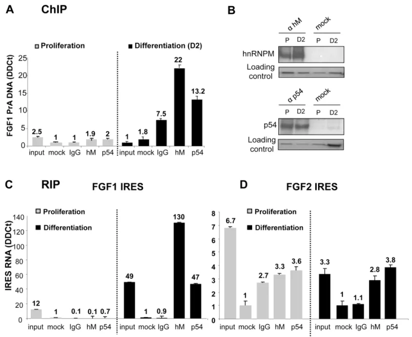
Interaction of hnRNPM and p54nrbwith the promoter was analyzed by chromatin immuno-precipitation (ChIP), using DNA extracts either from proliferating or differentiating myoblasts (Fig 2A). We checked by immunoprecipitation that the two proteins are expressed in prolifer-ating as well as in differentiprolifer-ating cells (Fig 2B). However no interaction was detected with either of the two proteins in proliferating cells, whereas significant interaction was observed with both proteins in differentiating myoblasts (Fig 2A). RNA immunoprecipitation (RIP) was also performed using anti-hnRNPM or anti-p54 antibodies revealing that the two proteins interact with the FGF1 IRES RNA in differentiating, but not in proliferating, myoblasts (Fig 2C). These data demonstrated that hnRNPM and p54nrbinteract (directly or indirectly) with FGF1 pro-moter A and IRES A only in differentiating myoblasts. In contrast the two proteins very poorly interact with the FGF2 IRES, and such interaction did not increase during differentiation (Fig 2D).
Thus, hnRNPM and p54nrbbinding to FGF1 promoter and IRES correlates with the previ-ously shown induction during differentiation [4]. Binding and activity are very weak during proliferation and induced by day 2 of differentiation. This suggests that these proteins may be involved in the specific activation of FGF1 mRNA accumulation and IRES-mediated
p54
nrband hnRNPM knockdown silences the FGF1
promoter-dependent accumulation of mRNA during myoblast differentiation
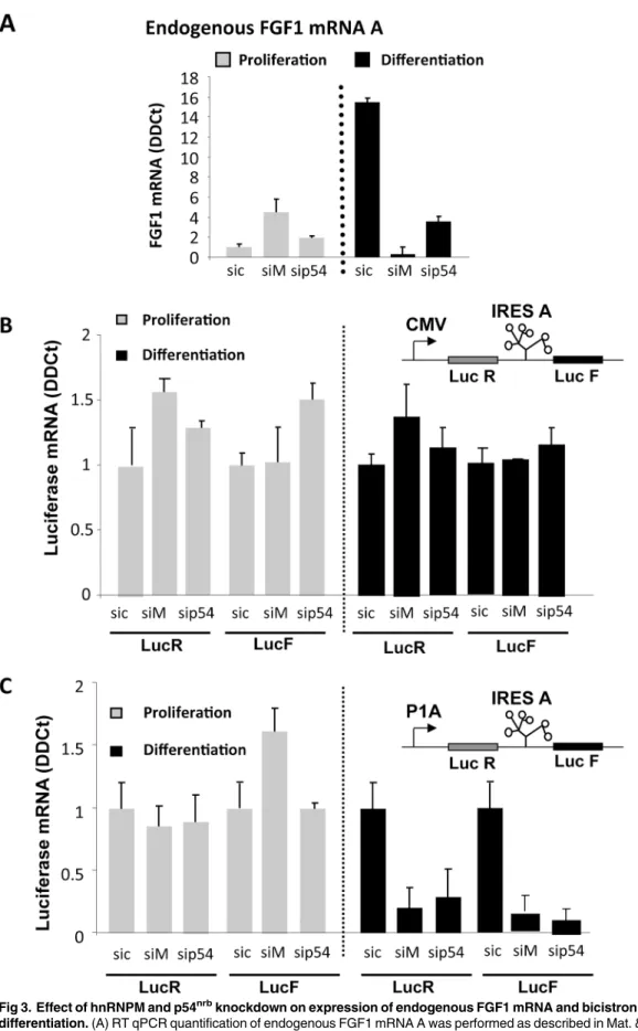
The role of p54nrband hnRNPM on FGF1 expression was studied by a knockdown approach using siRNA smartpools targeting either p54nrb(sip54) or hnRNPM (siM) (S2 File). Myoblasts were co-transfected with bicistronic vectors containing the FGF1 IRES and siRNAs. The bicis-tronic cassette, coding forrenilla luciferase (LucR) and firefly luciferase (LucF) separated by the FGF1 IRES, was under the control of either the CMV promoter or the FGF1 promoter A (Fig 3). Endogenous FGF1 mRNA was quantified by RT qPCR, and expression of the bicistro-nic mRNA under the control of either the CMV or FGF1 promoter was compared.
RT qPCR quantification of endogenous FGF1 mRNA clearly showed, as we have previously reported, a strong induction of FGF1 mRNA accumulation at day 2 of differentiation [4]. This induction was abolished upon transfection with siRNA against either hnRNPM or p54nrb (Fig 3AandS2 File).
Bicistronic mRNA levels produced from the CMV or FGF1 promoter were quantified with LucF and LucR couples of primers. No variation of mRNA amount was observed with the CMV promoter in response to siRNA knockdown of hnRNPM or p54nrb(Fig 3B). In contrast, the level of bicistronic mRNA transcribed from the FGF1 promoter was strongly downregulated by hnRNPM or p54nrbknockdown in differentiating myoblasts (Fig 3C). In addition, the two cis-trons LucR and LucF were present in equal amounts in all experiments, ruling out any effect of hnRNPM or p54nrbon a putative cryptic splicing site or promoter in the IRES (Fig 3B and 3C). These data showed, together with the ChIP data (Fig 2A), that hnRNPM and p54nrb/NONO binding to the FGF1 promoter A is responsible for FGF1 mRNA accumulation during myo-blast differentiation, suggesting an effect of these proteins on the FGF1 mRNA transcription or stability.
p54
nrband hnRNPM knockdown silences the FGF1 IRES during
myoblast differentiation
The knockdown approach was also used to evaluate the effect of hnRNPM and p54nrbon trans-lation mediated by the FGF1 IRES A. In the bicistronic construct, LucR activity reflects the mRNA level and cap-dependent translation while LucF reflects the IRES activity. Luciferase activities were measured in myoblasts co-transfected with bicistronic vectors and siRNAs tar-geting hnRNPM and/or p54nrb(see above, andS2 File). Under the CMV promoter, FGF1 IRES activity decreased by 5.6 or 7 fold following knockdown of hnRNPM or p54nrb, respectively (Fig 4A, left panel). A double knockdown resulted in a 13.5 fold inhibition (Fig 4A, right panel). These effects were observed in differentiating but not proliferating myoblasts. When the bicistronic cassette was under the control of the FGF1 promoter A, the IRES activity was 39
Fig 1. Identification of RNA and DNA binding proteins by BIA-MS. (A) BIACORE 3000 analysis using surface plasmon resonance. Left: the ligand corresponding to biotinylated RNA or DNA is immobilized by streptavidin coupled to the sensorchip gold surface. On the other side of the sensorchip, a light source is directed to the sensorchip through a prism and is reflected in all angles except one at which it is absorbed into the gold surface in the form of an evanescent wave. The binding of the analyte corresponding to RNA or DNA binding proteins to the ligand results in a change in the index of refraction proportional to the number of bound molecules. This generates a shift in the absorption angle that is recorded by the detector and appears on a sensogram. Right: Sensogram generated during a binding cycle followed by protein recovery. The response appears in resonance units (RU). The association phase lasts from the start to the end of analyte injection (400 s). Protein recovery is achieved at 800 s and theΔRU indicates the efficiency of dissociation of the bound proteins. (B) C2C12 myoblast protein binding and recovery. Total extracts of proliferating or differentiating C2C12 myoblasts (left) or nuclear extracts of differentiating C2C12 (right) were injected as analytes in several BIACORE 3000 channels after immobilization of ligands corresponding to FGF1 IRES A RNA (nt 1 to 442) or promoter A DNA (distal part nt 1 to 391). Bound proteins were recovered as described in Mat. & Meth. (C) Mass spectrometry analysis of proteins recovered from the BIACORE 3000 experiments. For each BIA-MS experiment, 6 recovery cycles were pooled to obtain a sufficient RU quantity (about 2000 RU). The mass spectrometry analysis was performed as described in Mat. & Meth. The most significant proteins (RNA and DNA binding proteins) are listed here, whereas the complete list is provided inS1 File. hnRNPM and p54nrbhave been selected as the most interesting candidates. doi:10.1371/journal.pone.0136466.g001
fold higher than with the CMV promoter (as published previously) and more strongly affected by hnRNPM or p54nrbknockdown (8 and 11 fold, respectively,Fig 4B, left panel) [4]. In addi-tion, the double knockdown drastically inhibited the IRES activity 54 fold (Fig 4B, right panel). Additional siRNAs targeting p54nrbor hnRNPM, but not contained in the smartpools, were
Fig 2. Interaction of hnRNPM and p54nrbproteins with FGF1 promoter and IRES. (A) ChIP assays were performed as described in Mat. & Meth. with genomic DNA purified and fragmented from C2C12 extracts in proliferation (left panel) or differentiation 2 days after serum-starvation treatment (right panel). Immunoprecipitation experiments were performed with anti-hnRNPM (1/D8), anti-p54nrbantibodies, control IgG provided by the manufacturer (IgG) or
without antibodies (mock), respectively. qPCR quantification was achieved with primers specific to the 1–391 FGF1-A promoter fragment or with primers specific to the TBP gene used as the reference gene. The values are expressed relatively to the reference gene. Experiments were performed in biological triplicates and repeated three times. A representative experiment is shown (mean +- standard deviation). (B) HnRNPM and p54nrbprotein levels were
analyzed by immunoprecipitation followed by Western blotting in proliferating (P) or differentiating (day 2 after serum-starvation, D2) C2C12 cell lysates, using anti-hnRNPM or antip54 antibodies. The loading control was checked by Ponceau Red staining. (C, D) RIP assays were performed as described in Mat. & Meth. with cell lysates from proliferating or differentiating myoblasts. Immunoprecipitation was achieved as in (A). RNAs present in the
immunoprecipitated complexes were quantified by RT qPCR using primers specific to the FGF1 (C) or FGF2 IRES (D). The reference gene was 18S RNA, reflecting non specific RNA binding. The values are expressed relatively to the control (mock) without antibody. Experiments were performed in biological triplicates and repeated three times. A representative experiment is shown (mean +- standard deviation).
Fig 3. Effect of hnRNPM and p54nrbknockdown on expression of endogenous FGF1 mRNA and bicistronic mRNAs during myoblast
differentiation. (A) RT qPCR quantification of endogenous FGF1 mRNA A was performed as described in Mat. & Meth. during C2C12 myoblast proliferation or day 2 differentiation after transfection with siRNA ONE TARGET PLUS smartpool against hnRNPM (siM), p54nrb(sip54) or siRNA control (sic). mRNA
assessed, confirming that the effect of p54 or hnRNPM knockdown on FGF1 IRES activity is specific to these targets (S3 File).
To check the specificity of FGF1 IRES regulation by hnRNPM and p54nrb, knockdown was performed as above using bicistronic contructs with the EMCV or FGF2 IRES. Results showed that the activities of the EMCV and FGF2 IRESs remained completely unaffected by either knockdown (Fig 4C and 4D).
These data suggested that hnRNPM and p54nrbparticipate in the IRESome complex responsi-ble for FGF1 IRES A activation during myoblast differentiation, but are unaresponsi-ble to activate EMCV or FGF2 IRESs. In addition, the presence of the FGF1 promoter A stimulated the IRES activating function of the two proteins, suggesting that the recruitment of hnRNPM and p54nrbon the pro-moter by a direct or indirect interaction might enhance their recruitment in the IRESome.
P54
nrbis sufficient to stimulate the FGF1 promoter whereas it
cooperates with hnRNPM to activate the FGF1 IRES
To evaluate the double activity of p54nrband hnRNPM on FGF1 promoter and IRES, C2C12 myoblasts were co-transfected by the bicistronic FGF1 IRES-containing dual luciferase plasmid and by plasmids coding either p54nrbor hnRNPM, or both proteins (Fig 5A and 5B). Measure-ment of the LucR activity showed significant mRNA accumulation in response to p54nrb over-expression (Fig 5C). The IRES activity reflected by the ratio LucF/LucR was enhanced by p54nrbalone, but more strongly with the combination of p54nrband hnRNPM (Fig 5D and 5E). Surprisingly, overexpression of hnRNPM alone inhibited both mRNA accumulation and IRES activities, suggesting that the ratio of the two proteins may be important for hnRNPM function (Fig 5C–5E). The effects of protein overexpression were stronger in proliferating myoblasts, which could be expected, because promoter and IRES are in a basal inactive state in proliferat-ing cells, while they are activated by the endogenous p54nrband hnRNPM in differentiating cells. These results suggested that p54nrbis sufficient to enhance mRNA accumulation, whereas it cooperates with hnRNPM to stimulate the IRES. Such a cooperative effect confirmed the data of the knockdown experiments (Figs3and4).
Regulation of IRES activity by hnRNPM and p54
nrbis
promoter-dependent
To analyse the impact of the promoter on activation of IRES-dependent translation by hnRNPM and p54nrb, C2C12 cells were transfected with either bicistronic plasmid DNAs or in vitro transcribed capped and polyadenylated bicistronic mRNAs (Fig 6). DNA transfection resulted (as already shown inFig 4) in a high FGF1 IRES activity, strongly induced by C2C12 differentiation, and downregulated by the knockdown of hnRNPM or p54nrb(Fig 6AandS4 File). In contrast, basal FGF1 IRES activity was 500 times lower following RNA transfection, and no induction by either hnRNPM or p54 was observed (Fig 6BandS4 File). These features were not observed with the EMCV IRES, whose activity was low after DNA or RNA transfec-tion and was not affected by hnRNPM or p54 knockdown (Fig 6C and 6D). As a negative
quantification was standardized with RNA 18S. Experiments were performed in biological triplicates and repeated three times. A representative experiment is shown (mean +- standard deviation). The knockdown efficiency, analyzed by Western blot, is shown inS2 Fileand was also checked by RT qPCR (not shown). (B, C) C2C12 cells co-transfected with bicistronic plasmids and 48h later with siRNA siM, sip54 or sic as above.Renilla luciferase (LucR, left) and firefly luciferase (LucF, right) mRNA levels were quantified by RT qPCR during C2C12 myoblast proliferation (grey histograms) and differentiation (black histograms), standardized to 18S RNA. The bicistronic cassette contains either the CMV promoter (B) or the FGF1 promoter A (C). For each experiment, values are shown relatively to the siRNA control. Experiments were performed in biological triplicates and repeated at least three times. A representative experiment is shown (mean +- standard deviation).
control, we used a construct containing a hairpin between the two cistrons instead of an IRES. The LucF/LucR ratios in the absence of an IRES were very low for both DNA and RNA trans-fections and were not affected by hnRNPM or p54 knockdown (Fig 6E and 6F). These results suggested that a nuclear step may be a prerequisite to IRES induction by the hnRNPM and p54nrb.
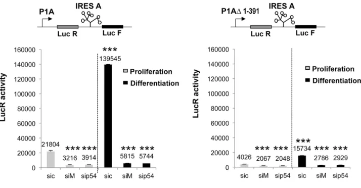
To go further with this hypothesis, we transfected C2C12 with a construct containing a deleted form of promoter F1A, P1AΔ1–391 (Fig 7). Measurements of LucR activity showed that this promoter is about ten times less efficient than the wild type promoter, whereas it was still sensitive to hnRNPM and p54 knockdown (Fig 7A). In contrast, in the presence of the deleted form of the promoter, IRES activity was no longer induced and was very mildly altered by the knockdown of hnRNPM or p54nrb(Fig 7B).
These two sets of data showed that FGF1 IRES induction by hnRNPM and p54nrbdepends on a sequence present in the promoter and involves multifunctional roles of these proteins.
p54
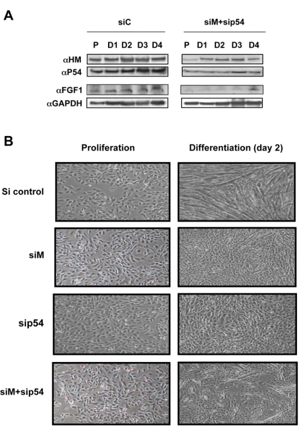
nrband hnRNPM are required for FGF1 induction and myotube
formation
To determine whether p54nrband hnRNPM are responsible for endogenous FGF1 induction during differentiation and if these proteins exhibit a physiological role in the development of muscle fiber, simple and double knockdowns of hnRNPM and/or p54nrbwere performed (Fig 8A). Expression of FGF1, analyzed by Western Blot, was strongly inhibited by the simple or double knockdown (Fig 8Aand data not shown). Furthermore, formation of myotubes was drastically affected by knockdown of p54nrbor hnRNPM, and even more strongly inhibited by the double knockdown (Fig 8B). Decrease of endogenous FGF1 expression upon treatment with siRNA against p54nrband/or hnRNPM confirmed that these two proteins are involved in the control of FGF1 expression and consequently in its induction during differentiation. In addition, these data also revealed that p54nrband hnRNPM are required for differentiation of myoblasts into myotubes, revealing the important role of these proteins in myogenesis.
Discussion
The present data reveal that FGF1 IRES activity is promoter-dependent and that this process is governed by two multifunctional trans-acting factors, p54nrband hnRNPM. P54nrband hnRNPM also appear as crucial actors of myoblast differentiation, demonstrating the physio-logical relevance of this new mechanism.
Nuclear formation of the IRESome
The present data suggest that IRESome formation occurs in the nuclear compartment. The reg-ulating features of p54nrband hnRNPM on both promoter and IRES activities are not
Fig 4. Effect of hnRNPM and/or p54nrbknockdown on the regulation of FGF1 IRES A activity during myoblast differentiation. C2C12 cells were first transfected with bicistronic plasmids and 48h later with siRNAs targeting hnRNPM (siM), p54nrb(sip54) or control (sic), (seeFig 3). Luciferase activities were measured as described in Mat. & Meth two days after siRNA transfection, from C2C12 myoblasts maintained in proliferation (grey histogram) or serum-starved to induce differentiation (day 2, black histogram). The bicistronic cassette contains LucR and LucF reporter genes, separated by different IRESs (FGF1, FGF2 or EMCV). LucR and LucF activities reflect the cap-dependent and IRES dependent translation, respectively [4]. The knockdown efficiency was checked by Western blot (S2 File). (A, B) FGF1 IRES activities were measured in C2C12 myoblasts in proliferation (grey histogram) and differentiation two days after serum-starvation treatment (black histogram). The bicistronic constructs are schematized. The bicistronic cassette is under the control of either the CMV promoter (A) or the FGF1 promoter A (B). Single and double knockdowns are shown in left and right panels, respectively. IRES activities are represented as LucF/LucR ratios. (C, D). Absence of effect of hnRNPM and/or p54nrbknockdown on the regulation of EMCV and FGF2 IRESs during myoblast differentiation. Activities of EMCV (C) and FGF2 (D) IRESs were measured as above. For all panels, experiments were performed in biological triplicates and repeated at least three times. A representative experiment is shown (mean +- standard deviation).
independent events, as these two translational regulators do not work when the promoter is absent (RNA transfection) or inactivated (promoter mutation).
The importance of ITAF subcellular localization in the regulation of their activity appeared a few years ago, with the first observation that a nuclear event is required in the control of c-myc IRES activity [18]. Later, it has been shown that shuttling of ITAFs, such as hnRNP A1, RBM4, PTB and PCBP1, between the nucleus and the cytoplasm influences their activity [24,
27,30,41]. Two different mechanisms have been proposed [42]: 1) the nuclear localized ITAFs may associate with their target in this compartment, resulting in the sequestration of the mRNA in the nucleus until an appropriate signal occurs, allowing cytoplasmic export and translation, 2) the ITAFs may be primarily located in the nucleus to keep them separate from their target IRESs and await a signal that warrants their accumulation in the cytoplasm. These two models could apply to cellular and viral IRESs, respectively, as explained by Semler & Waterman who argue that nuclear versus cytoplasmic synthesis of IRES-containing mRNAs is a major determinant in assembling different RNA–protein complexes [43]. The synthesis of uncapped picornaviral mRNAs occurs in the cytoplasm, in contrast to cellular mRNAs, which are transcribed in the nucleus and then exported to the cytoplasm. Our study not only rein-forces the hypothesis that the IRESome is constituted in the nucleus, but further elaborates this model by showing that its formation is influenced by the promoter activity.
HnRNPM and p54
nrbcooperate in activation of IRES-dependent
translation
The IRES-regulating function of hnRNPM discovered in our study represents a novel finding, as well as its cooperative activity with p54nrb. Interestingly, nuclear interaction of hnRNPM and p54nrbhas been previously reported [40]. Like other hnRNPs, hnRNPM has been first identified as a splicing regulatory protein, associated with early spliceosomes where it modu-lates the alternative splicing pattern of specific mRNAs including the FGF receptor [39,40]. Interestingly, hnRNPM has also been reported to influence the localization ofnanos mRNA by binding to its 3' UTR [44]. The additional function of hnRNPM shown in our study confirms that hnRNPs are multifunctional, depending on their interacting partners.
p54nrb/NONO is also a multifunctional protein. It has been previously shown to associate with 5' splicing sites in large transcription-splicing complexes, providing a direct role for this protein in linking transcription to splicing [3,45]. P54nrbalso behaves as a specific transcrip-tional activator, binding to the intracisternal A particle proximal enhancer element, and as an RNA binding protein, preferentially binding to the sequence UAGGGA/U identified by SELEX [37]. In addition, p54nrbhas been identified as an ITAF of the myc family IRESs (c-, L- and N-myc), and has also been shown to stimulate the APAF-1 IRES [46]. Here we demonstrate that p54nrbis able to cooperate with hnRNPM, suggesting that the two proteins may activate the FGF1 IRES as a complex. Unexpectedly, hnRNPM overexpression is inhibitory, whereas knockdown experiments clearly showed it is required for both IRES and promoter activation. We hypothesize that excess of hnRNPM might inhibit p54nrbfunctions on FGF1 gene
Fig 5. Cooperative effect of p54nrband hnRNPM on FGF1 IRES activation. (A) Schema of the constructs used for C2C12 cell co-transfection. The target plasmid is the bicistronic dual luciferase vector with the FGF1 promoter and IRES. The effector plasmids express p54nrb(p54) or hnRNPM (HM), or co-express p54nrband hnRNPM. The latter plasmid is a bicistronic contruct containing the FGF1 IRES. (B) Western blots of transfected proliferating C2C12 cell
extracts using antibodies against p54(αHM), hnRNPM (αHM) of GAPDH as a control (αGAPDH). (C-E) Luciferase activities measurement of co-transfected proliferating or differentiating C2C12 cell extracts. LucR activity reflects the FGF1 mRNA promoter activity (as cap-dependent translation does not significantly vary, as shown by RNA transfection, see Table B inS4 File) (C). LucF reflects IRES-dependent translation but is also dependent on mRNA amount (D). LucF/LucR ratio reflects the IRES activity normalized to mRNA amount, expressed relatively to the control (co-transfection with empty vector) (E). Experiments were performed in biological triplicates and repeated three times. The statistical test used is the Student test. (mean +- standard deviation, *p<0.05, **p<0.01, ***<0.001).
expression, presumably by preventing p54nrbbinding to FGF1 DNA and RNA due to an inade-quate stoechiometry of the two proteins.
Although the proteins are expressed in proliferating cells, they are not found in the complex with the IRES or the promoter A. This suggests that the proteins might carry different post-translational modifications. Alternatively these proteins could join the complex through the interaction with another protein, which is absent in the proliferating cells and remains to be discovered. This could also explain that overexpressed hnRNPM is not functional.
Very few proteins were bound to the EMCV IRES, which may be explained by the low effi-ciency of the EMCV IRES in myoblasts. However, we were also surprised that no transcription factor was bound to promoter DNA. This indicates that BIA-MS does not provide an exhaus-tive view of all proteins bound to RNA or DNA and that one can miss important proteins. This also explains that we have missed hnRNPM present in the promoter-bound complex.
Although the direct interaction of p54nrband hnRNPM with RNA has not been demon-strated, the present study strongly suggest that the two proteins are ITAFs involved in FGF1 IRES activation. However The BIA-MS data provides a list of additional proteins bound to the FGF1 IRES, including histones and splicing factors. These proteins may participate in the IRE-Some ribonucleoprotein.
Promoter-dependent activation of the FGF1 IRES may involve
transcription or mRNA stabilization
Promoter-dependent translation controlled by hnRNPM and p54nrbasks the question of how a promoter is able to impact on translation, which is in principle cytoplasmic.
One among several possible models, one can propose transcription/translation coupling: hnRNPM and p54nrbmight be first recruited to the promoter, where they would contribute to transcription activation. This process would also facilitate hnRNPM and p54nrbbinding onto mRNA IRES and subsequently activate IRES-dependent translation. A recent study supports the hypothesis of a co-transcriptional ITAF binding, by showing that the translation elongation factor eEF1A1 couples transcription to translation during heat shock response [47]. These authors have demonstrated that eEF1A1 activates HSP70 transcription, then associates with RNA polymerase II, binds the mRNA 3’UTR, stabilizes it and facilitates its nuclear export to active ribosomes. hnRNPM and p54nrbmay well have a similar mode of action, except that they bind to the mRNA 5’UTR and directly activate IRES-dependent translation by their ITAF function.
Other hnRNPs have been previously described for their involvement in transcription: hnRNPK, reported as a transcription factor interacting with the RNA polymerase II machinery in the c-myc promoter, and hnRNP A1, involved in the transcription of the KRAS proto-onco-gene [48,49]. More recently, hnRNPC has been shown to regulate transcription in osteoblasts [50]. Interestingly, hnRNP A1 and-K also exhibit an ITAF function, although no coupling between transcriptional and translational functions has been mentioned [24,25,30].
An alternative model to explain promoter-dependent regulation of translation by hnRNPM and p54nrbmight be their effect on mRNA stability. Binding of these proteins to the FGF1 pro-moter would not have an effect on transcription but would facilitate their binding to the
Fig 6. Comparison of IRES activities following DNA or RNA transfection. (A, C, E) C2C12 cells were transfected with bicistronic plasmids containing either the FGF1 IRES, or EMCV IRES or a hairpin (control without IRES) and with siRNA siM, sip54 or sic, and IRES activities measured as inFig 4B, 4D and 4FC2C12 cells were transfected with siRNA siM, sip54 or sic and 24h later with bicistronic mRNAs containing either the FGF1 IRES, or the EMCV IRES, or a hairpin as above. mRNAs were transcribed in vitro, capped and polyadenylated, as described in Mat. & Meth. The LucF/LucR activity ratio was measured 12h post-transfection. Experiments were performed in biological triplicates and repeated three times. The Student test was used. A representative experiment is shown (mean +- standard deviation,*p<0.05, **p<0.01, ***<0.001). For A and B, the Luc R and Luc F values are shown inS4 File. doi:10.1371/journal.pone.0136466.g006
Fig 7. Influence of transcription level on the activity of the FGF1 IRES A. C2C12 cells were transfected with bicistronic plasmids containing either the complete promoter 1A or a deleted promoter lacking nucleotides 1 to 391. Transfected cells were treated 24h later with siRNA siM, sip54 or sic, and luciferase activities were measured as inFig 6. (A) mRNA expression in the presence of promoter 1A and 1AΔ1–391 reflected by the LucR activities. (B) Activity of FGF1 IRES A in the presence of promoter 1A and 1AΔ1–391, reflected by the Luc F/ LucR ratio as above. Experiments were performed in biological triplicates and repeated three times. The statistical test used is the Student test. (mean +- standard deviation,*p<0.05, **p<0.01, ***<0.001). doi:10.1371/journal.pone.0136466.g007
Fig 8. Role of hnRNPM and p54nrbin myoblast differentiation. (A) Effect of hnRNPM and p54nrbknockdown on expression of endogenous FGF1. C2C12
cells were transfected with siRNA, siM+sip54 or sic. Western blot was performed as above using cell extracts of proliferating (P) and differentiating myoblasts (D1 to D4). GAPDH was used as a normalization control. These data correspond to a representative experiment (repeated at least three times). (B) Effect of hnRNPM and/or p54nrbknockdown on myotube formation. C2C12 cells were transfected with siRNA siM, sip54, siM+sip54 or sic and myotube formation was
followed by phase contrast microscopy. Data are shown for day 2 cells compared to proliferative cells. doi:10.1371/journal.pone.0136466.g008
nascent mRNA, resulting in mRNA stabilization, as has been shown for the yeast protein Dbf2P [51]. These authors demonstrate that Dbf2p is recruited to the SW15 and CBL2 promot-ers, and co-transcriptionally deposited onto the SW15 and CLB2 mRNAs. Once they are exported into the cytoplasm, the two mRNAs are protected from degradation by Dbf2p. According to this model, hnRNPM and p54nrbbinding to mRNA might have the double effect of stabilizing the FGF1 mRNA and activating the IRES. Anyway, further investigation is needed to determine whether the promoter-dependent function of hnRNPM and p54nrbinvolves tran-scription enhancement or RNA stabilization.
Promoter-dependent translation, a physiologically relevant mechanism
during myogenesis
An important feature of our study is the physiological relevance of p54nrband hnRNPM func-tion in myoblast differentiafunc-tion. It clearly appears that p54nrband hnRNPM are important for myotube formation and such a role is novel for these two proteins. We have previously shown that FGF1 is required for myoblast differentiation [4]. Thus the effect of hnRNPM and/or p54 knockdown on myoblast differentiation may be a direct consequence of FGF1 downregulation. Alternatively, hnRNPM and p54nrbmay act on the expression of additional IRES-containing genes, including Smad5 and utrophin A IRESs whose activites have been reported in myoblasts [17,52,53]. It will be of great interest to investigate the possible role of hnRNPM and p54nrbin the promoter-dependent regulation of such IRESs during muscle development.
As seen with the coordination of transcription and mRNA processing, such as splicing, whose regulators often exhibit the additional function of ITAFs (i.e. hnRNPs), one can expect that the promoter-dependent translation mechanism described in our study will extend to other IRES-containing mRNAs. Such a mechanism becomes more significant for genes that are induced in response to conditions that block cap-dependent translation, such as stress. Cou-pling of IRES activation with the promoter guarantees that the induction of a given gene in response to a specific stimulus will result in the synthesis of that gene product.
Supporting Information
S1 File. Identification of RNA and DNA binding proteins by BIA-MS.Total or nuclear extracts of differentiating C2C12 myoblasts were injected (seeFig 1) as analytes in several BIA-CORE 3000 channels after immobilization of ligands corresponding to FGF1 IRES A, EMCV IRES, FGF1 promoter A or CMV promoter. Bound proteins were recovered as described in Mat. & Meth. and identified by mass spectrometry. For each BIA-MS experiment, 6 recovery cycles were pooled to obtain a sufficient RU quantity (about 2000 RU). Mass spectrometry analysis was performed as described in Mat. & Meth. Bound proteins (RNA and DNA binding proteins) are listed here. (Table A) FGF1 IRES RNA, C2C12 total extracts, (Table B) FGF1 IRES RNA, C2C12 nuclear extracts, (Table C) EMCV IRES RNA, C2C12 total extracts, (Table D) FGF1 promoter A DNA, C2C12 nuclearFGF1 extracts.
(DOC)
S2 File. Knockdown of hnRNPM and/or p54nrb.C2C12 cells were first transfected with bicis-tronic plasmids and 48h later with siRNAs targeting hnRNPM (siM), p54nrb(sip54) or control (sic). RNA levels and luciferase activities are presented in Figs3and4, respectively, from C2C12 myoblasts maintained in proliferation or at day 2 of differentiation. p54nrband hnRNPM expression was analysed by Western blot following single knockdown with siRNA siM (Fig A) or sip54 (Fig B) or double knockdown with the two siRNAs together (Fig C). (TIF)
S3 File. Effect of hnRNPM and/or p54nrbknockdown on the regulation of FGF1 IRES A using siRNAs different from the smartpool.C2C12 cells co-transfected with bicistronic plas-mids and 48h later with a siRNA against hnRNPM (siM), p54nrb(sip54) or siRNA control (sic). SiM and siP54 corresponded to sequences different from that of the siRNA smartpools used inFig 4. (Fig A) Ratio of the luciferase activities measured as inFig 4, two days after siRNA transfection, from differentiating C2C12 myoblasts (day 2). (Fig B) The knockdown was checked by Western blot as inS2 File.
(TIF)
S4 File. Effect of hnRNPM and p54 knockdown on FGF1 IRES activity after DNA or RNA transfection.(Table A) C2C12 cells were transfected with bicistronic plasmids containing the FGF1 promoter and IRES (seeFig 6) and with siRNA siM, sip54 or sic. (Table B) C2C12 cells were transfected with siRNA siM, sip54 or sic and 24h later with bicistronic mRNA containing the FGF1 IRES (seeFig 6). mRNAs were transcribed in vitro, capped and polyadenylated, as described in Mat. & Meth. For Tables A and B, firefly andrenilla luciferase activities were mea-sured. Values are presented as well as the LucF/LucR (F/R) ratio representing the IRES activity. Experiments were performed in biological triplicates and repeated three times. The Student test was used (mean +- standard deviation).
(DOC)
Acknowledgments
We thank J.J. Maoret (Genotoul quantitative transcriptomics facility), B. Montsarrat (Genou-toul Proteomics facility), P. Billon, N. Fenie, J.B. Mauroux, J. Mermet and M. Serrero for tech-nical assistance. This work was supported by Association Française contre les Myopathies (AFM), Association pour la Recherche sur le Cancer, Ligue pour la Recherche Contre le Can-cer, Cancéropole GSO, INCA, Fondation de l’Avenir, Région Midi-Pyrénées, European fund-ing (FEDER). N.A. and E.R.G. had a fellowship from AFM, F.H. from the Région Midi-Pyrénées.
Author Contributions
Conceived and designed the experiments: EB AP BGS ACP. Performed the experiments: NA FH ERG MB FL FP RP CP. Analyzed the data: NA FH ERG MB RP EB CP OBS BGS ACP. Contributed reagents/materials/analysis tools: ACP. Wrote the paper: NA FH ERG AP BGS ACP.
References
1. Braunschweig U, Gueroussov S, Plocik AM, Graveley BR, Blencowe BJ. Dynamic integration of splic-ing within gene regulatory pathways. Cell. 2013 Mar 14; 152(6):1252–69. PMID:23498935. Pubmed Central PMCID: 3642998. Epub 2013/03/19. eng. doi:10.1016/j.cell.2013.02.034
2. Maniatis T, Reed R. An extensive network of coupling among gene expression machines. Nature. 2002 Apr 4; 416(6880):499–506. PMID:11932736.
3. Pandit S, Wang D, Fu XD. Functional integration of transcriptional and RNA processing machineries. Curr Opin Cell Biol. 2008 Jun; 20(3):260–5. PMID:18436438. doi:10.1016/j.ceb.2008.03.001
4. Conte C, Ainaoui N, Delluc-Clavieres A, Khoury MP, Azar R, Pujol F, et al. Fibroblast growth factor 1 induced during myogenesis by a transcription-translation coupling mechanism. Nucleic Acids Res. 2009 Sep; 37(16):5267–78. PMID:19561198. doi:10.1093/nar/gkp550
5. Madiai F, Hackshaw KV, Chiu IM. Characterization of the entire transcription unit of the mouse fibro-blast growth factor 1 (FGF-1) gene. Tissue-specific expression of the FGF-1.A mRNA. J Biol Chem. 1999 Apr 23; 274(17):11937–44. PMID:10207015.
6. Myers RL, Chedid M, Tronick SR, Chiu IM. Different fibroblast growth factor 1 (FGF-1) transcripts in neural tissues, glioblastomas and kidney carcinoma cell lines. Oncogene. 1995 Aug 17; 11(4):785–9. PMID:7544453.
7. Myers RL, Payson RA, Chotani MA, Deaven LL, Chiu IM. Gene structure and differential expression of acidic fibroblast growth factor mRNA: identification and distribution of four different transcripts. Onco-gene. 1993 Feb; 8(2):341–9. PMID:7678925.
8. Chotani MA, Chiu IM. Differential regulation of human fibroblast growth factor 1 transcripts provides a distinct mechanism of cell-specific growth factor expression. Cell Growth Differ. 1997 Sep; 8(9):999– 1013. PMID:9300182.
9. Chotani MA, Payson RA, Winkles JA, Chiu IM. Human fibroblast growth factor 1 gene expression in vascular smooth muscle cells is modulated via an alternate promoter in response to serum and phorbol ester. Nucleic Acids Res. 1995 Feb 11; 23(3):434–41. PMID:7533902. Pubmed Central PMCID: 306694. Epub 1995/02/11. eng.
10. Payson RA, Chotani MA, Chiu IM. Regulation of a promoter of the fibroblast growth factor 1 gene in prostate and breast cancer cells. The Journal of steroid biochemistry and molecular biology. 1998 Aug; 66(3):93–103. PMID:9719443. Epub 1998/08/27. eng.
11. Martineau Y, Le Bec C, Monbrun L, Allo V, Chiu IM, Danos O, et al. Internal ribosome entry site struc-tural motifs conserved among mammalian fibroblast growth factor 1 alternatively spliced mRNAs. Mol Cell Biol. 2004 Sep; 24(17):7622–35. PMID:15314170.
12. Baird SD, Lewis SM, Turcotte M, Holcik M. A search for structurally similar cellular internal ribosome entry sites. Nucleic Acids Res. 2007; 35(14):4664–77. PMID:17591613.
13. Bonnal S, Schaeffer C, Creancier L, Clamens S, Moine H, Prats AC, et al. A single internal ribosome entry site containing a G quartet RNA structure drives fibroblast growth factor 2 gene expression at four alternative translation initiation codons. J Biol Chem. 2003 Oct 10; 278(41):39330–6. PMID:12857733. 14. Morfoisse F, Kuchnio A, Frainay C, Gomez-Brouchet A, Delisle MB, Marzi S, et al. Hypoxia induces
VEGF-C expression in metastatic tumor cells via a HIF-1alpha-independent translation-mediated mechanism. Cell reports. 2014 Jan 16; 6(1):155–67. PMID:24388748. Epub 2014/01/07. eng. doi:10. 1016/j.celrep.2013.12.011
15. Braunstein S, Karpisheva K, Pola C, Goldberg J, Hochman T, Yee H, et al. A hypoxia-controlled cap-dependent to cap-incap-dependent translation switch in breast cancer. Mol Cell. 2007 Nov 9; 28(3):501–12. PMID:17996713.
16. Holcik M, Sonenberg N. Translational control in stress and apoptosis. Nat Rev Mol Cell Biol. 2005 Apr; 6(4):318–27. PMID:15803138.
17. Shiroki K, Ohsawa C, Sugi N, Wakiyama M, Miura K, Watanabe M, et al. Internal ribosome entry site-mediated translation of Smad5 in vivo: requirement for a nuclear event. Nucleic Acids Res. 2002 Jul 1; 30(13):2851–61. PMID:12087169. Pubmed Central PMCID: 117063. Epub 2002/06/28. eng. 18. Stoneley M, Subkhankulova T, Le Quesne JP, Coldwell MJ, Jopling CL, Belsham GJ, et al. Analysis of
the c-myc IRES; a potential role for cell-type specific trans-acting factors and the nuclear compartment. Nucleic Acids Res. 2000 Feb 1; 28(3):687–94. PMID:10637319.
19. Garcia-Blanco MA, Jamison SF, Sharp PA. Identification and purification of a 62,000-dalton protein that binds specifically to the polypyrimidine tract of introns. Genes Dev. 1989 Dec; 3(12A):1874–86. PMID:
2533575.
20. Jang SK, Wimmer E. Cap-independent translation of encephalomyocarditis virus RNA: structural ele-ments of the internal ribosomal entry site and involvement of a cellular 57-kD RNA-binding protein. Genes Dev. 1990 Sep; 4(9):1560–72. PMID:2174810.
21. Kim YK, Hahm B, Jang SK. Polypyrimidine tract-binding protein inhibits translation of bip mRNA. J Mol Biol. 2000 Nov 24; 304(2):119–33. PMID:11080450.
22. Mitchell SA, Spriggs KA, Coldwell MJ, Jackson RJ, Willis AE. The Apaf-1 internal ribosome entry seg-ment attains the correct structural conformation for function via interactions with PTB and unr. Mol Cell. 2003 Mar; 11(3):757–71. PMID:12667457.
23. Pickering BM, Mitchell SA, Evans JR, Willis AE. Polypyrimidine tract binding protein and poly r(C) bind-ing protein 1 interact with the BAG-1 IRES and stimulate its activity in vitro and in vivo. Nucleic Acids Res. 2003 Jan 15; 31(2):639–46. PMID:12527772.
24. Bonnal S, Pileur F, Orsini C, Parker F, Pujol F, Prats AC, et al. Heterogeneous nuclear ribonucleopro-tein A1 is a novel internal ribosome entry site trans-acting factor that modulates alternative initiation of translation of the fibroblast growth factor 2 mRNA. J Biol Chem. 2005 Feb 11; 280(6):4144–53. PMID:
25. Evans JR, Mitchell SA, Spriggs KA, Ostrowski J, Bomsztyk K, Ostarek D, et al. Members of the poly (rC) binding protein family stimulate the activity of the c-myc internal ribosome entry segment in vitro and in vivo. Oncogene. 2003 Sep 11; 22(39):8012–20. PMID:12970749.
26. Holcik M, Gordon BW, Korneluk RG. The internal ribosome entry site-mediated translation of antiapop-totic protein XIAP is modulated by the heterogeneous nuclear ribonucleoproteins C1 and C2. Mol Cell Biol. 2003 Jan; 23(1):280–8. PMID:12482981.
27. Lin JC, Hsu M, Tarn WY. Cell stress modulates the function of splicing regulatory protein RBM4 in translation control. Proc Natl Acad Sci U S A. 2007 Feb 13; 104(7):2235–40. PMID:17284590. 28. Spriggs KA, Bushell M, Mitchell SA, Willis AE. Internal ribosome entry segment-mediated translation
during apoptosis: the role of IRES-trans-acting factors. Cell Death Differ. 2005 Jun; 12(6):585–91. PMID:15900315.
29. Jo OD, Martin J, Bernath A, Masri J, Lichtenstein A, Gera J. Heterogeneous nuclear ribonucleoprotein A1 regulates cyclin D1 and c-myc internal ribosome entry site function through Akt signaling. J Biol Chem. 2008 Aug 22; 283(34):23274–87. PMID:18562319. Pubmed Central PMCID: 2516998. Epub 2008/06/20. eng. doi:10.1074/jbc.M801185200
30. Lewis SM, Veyrier A, Hosszu Ungureanu N, Bonnal S, Vagner S, Holcik M. Subcellular relocalization of a trans-acting factor regulates XIAP IRES-dependent translation. Mol Biol Cell. 2007 Apr; 18(4):1302– 11. PMID:17287399.
31. Roy R, Durie D, Li H, Liu BQ, Skehel JM, Mauri F, et al. hnRNPA1 couples nuclear export and transla-tion of specific mRNAs downstream of FGF-2/S6K2 signalling. Nucleic Acids Res. 2014 Nov 10; 42 (20):12483–97. PMID:25324306. Pubmed Central PMCID: 4227786. Epub 2014/10/18. eng. doi:10. 1093/nar/gku953
32. Lopez F, Pichereaux C, Burlet-Schiltz O, Pradayrol L, Monsarrat B, Esteve JP. Improved sensitivity of biomolecular interaction analysis mass spectrometry for the identification of interacting molecules. Pro-teomics. 2003 Apr; 3(4):402–12. PMID:12687608.
33. Datar KV, Dreyfuss G, Swanson MS. The human hnRNP M proteins: identification of a methionine/argi-nine-rich repeat motif in ribonucleoproteins. Nucleic Acids Res. 1993 Feb 11; 21(3):439–46. PMID:
8441656.
34. Dong B, Horowitz DS, Kobayashi R, Krainer AR. Purification and cDNA cloning of HeLa cell p54nrb, a nuclear protein with two RNA recognition motifs and extensive homology to human splicing factor PSF and Drosophila NONA/BJ6. Nucleic Acids Res. 1993 Aug 25; 21(17):4085–92. PMID:8371983. Pubmed Central PMCID: 310009. Epub 1993/08/25. eng.
35. Prats AC, Van den Berghe L, Rayssac A, Ainaoui N, Morfoisse F, Pujol F, et al. CXCL4L1-fibstatin cooperation inhibits tumor angiogenesis, lymphangiogenesis and metastasis. Microvascular research. 2013 Jun 4. PMID:23747987. Epub 2013/06/12. Eng.
36. Bouyssie D, Gonzalez de Peredo A, Mouton E, Albigot R, Roussel L, Ortega N, et al. Mascot file pars-ing and quantification (MFPaQ), a new software to parse, validate, and quantify proteomics data gener-ated by ICAT and SILAC mass spectrometric analyses: application to the proteomics study of
membrane proteins from primary human endothelial cells. Molecular & cellular proteomics: MCP. 2007 Sep; 6(9):1621–37. PMID:17533220. Epub 2007/05/30. eng.
37. Basu A, Dong B, Krainer AR, Howe CC. The intracisternal A-particle proximal enhancer-binding protein activates transcription and is identical to the RNA- and DNA-binding protein p54nrb/NonO. Mol Cell Biol. 1997 Feb; 17(2):677–86. PMID:9001221.
38. Emili A, Shales M, McCracken S, Xie W, Tucker PW, Kobayashi R, et al. Splicing and transcription-associated proteins PSF and p54nrb/nonO bind to the RNA polymerase II CTD. Rna. 2002 Sep; 8 (9):1102–11. PMID:12358429.
39. Hovhannisyan RH, Carstens RP. Heterogeneous ribonucleoprotein m is a splicing regulatory protein that can enhance or silence splicing of alternatively spliced exons. J Biol Chem. 2007 Dec 14; 282 (50):36265–74. PMID:17959601.
40. Marko M, Leichter M, Patrinou-Georgoula M, Guialis A. hnRNP M interacts with PSF and p54(nrb) and co-localizes within defined nuclear structures. Exp Cell Res. 2010 Feb 1; 316(3):390–400. PMID:
19874820. doi:10.1016/j.yexcr.2009.10.021
41. Dobbyn HC, Hill K, Hamilton TL, Spriggs KA, Pickering BM, Coldwell MJ, et al. Regulation of BAG-1 IRES-mediated translation following chemotoxic stress. Oncogene. 2008 Feb 14; 27(8):1167–74. PMID:17700523.
42. Lewis SM, Holcik M. For IRES trans-acting factors, it is all about location. Oncogene. 2008 Feb 14; 27 (8):1033–5. PMID:17767196.
43. Semler BL, Waterman ML. IRES-mediated pathways to polysomes: nuclear versus cytoplasmic routes. Trends Microbiol. 2008 Jan; 16(1):1–5. PMID:18083033.
44. Jain RA, Gavis ER. The Drosophila hnRNP M homolog Rumpelstiltskin regulates nanos mRNA locali-zation. Development. 2008 Mar; 135(5):973–82. PMID:18234721. doi:10.1242/dev.015438
45. Kameoka S, Duque P, Konarska MM. p54(nrb) associates with the 5' splice site within large transcrip-tion/splicing complexes. Embo J. 2004 Apr 21; 23(8):1782–91. PMID:15057275.
46. Cobbold LC, Spriggs KA, Haines SJ, Dobbyn HC, Hayes C, de Moor CH, et al. Identification of internal ribosome entry segment (IRES)-trans-acting factors for the Myc family of IRESs. Mol Cell Biol. 2008 Jan; 28(1):40–9. PMID:17967896.
47. Vera M, Pani B, Griffiths LA, Muchardt C, Abbott CM, Singer RH, et al. The translation elongation factor eEF1A1 couples transcription to translation during heat shock response. eLife. 2014; 3:e03164. PMID:
25233275. Pubmed Central PMCID: 4164936. Epub 2014/09/19. eng. doi:10.7554/eLife.03164
48. Michelotti EF, Michelotti GA, Aronsohn AI, Levens D. Heterogeneous nuclear ribonucleoprotein K is a transcription factor. Mol Cell Biol. 1996 May; 16(5):2350–60. PMID:8628302.
49. Xodo L, Paramasivam M, Membrino A, Cogoi S. Protein hnRNPA1 binds to a critical G-rich element of KRAS and unwinds G-quadruplex structures: implications in transcription. Nucleic Acids Symp Ser (Oxf). 2008 (52: ):159–60. PMID:18776302.
50. Lisse TS, Vadivel K, Bajaj SP, Chun RF, Hewison M, Adams JS. The heterodimeric structure of hetero-geneous nuclear ribonucleoprotein C1/C2 dictates 1,25-dihydroxyvitamin D-directed transcriptional events in osteoblasts. Bone research. 2014; 2. PMID:25506471. Pubmed Central PMCID: 4261231. Epub 2014/12/17. Eng.
51. Trcek T, Larson DR, Moldon A, Query CC, Singer RH. Single-molecule mRNA decay measurements reveal promoter- regulated mRNA stability in yeast. Cell. 2011 Dec 23; 147(7):1484–97. PMID:
22196726. Pubmed Central PMCID: 3286490. Epub 2011/12/27. eng. doi:10.1016/j.cell.2011.11.051
52. Miura P, Coriati A, Belanger G, De Repentigny Y, Lee J, Kothary R, et al. The utrophin A 5'-UTR drives cap-independent translation exclusively in skeletal muscles of transgenic mice and interacts with eEF1A2. Hum Mol Genet. 2010 Apr 1; 19(7):1211–20. PMID:20053670. doi:10.1093/hmg/ddp591
53. Miura P, Thompson J, Chakkalakal JV, Holcik M, Jasmin BJ. The utrophin A 5'-untranslated region con-fers internal ribosome entry site-mediated translational control during regeneration of skeletal muscle fibers. J Biol Chem. 2005 Sep 23; 280(38):32997–3005. PMID:16061482.