HAL Id: hal-02565818
https://hal.archives-ouvertes.fr/hal-02565818
Submitted on 23 Sep 2020HAL is a multi-disciplinary open access archive for the deposit and dissemination of sci-entific research documents, whether they are pub-lished or not. The documents may come from teaching and research institutions in France or abroad, or from public or private research centers.
L’archive ouverte pluridisciplinaire HAL, est destinée au dépôt et à la diffusion de documents scientifiques de niveau recherche, publiés ou non, émanant des établissements d’enseignement et de recherche français ou étrangers, des laboratoires publics ou privés.
Antimicrobial peptide arrays for wide spectrum sensing
of pathogenic bacteria
Éric Pardoux, Agnès Roux, Raphaël Mathey, Didier Boturyn, Yoann Roupioz
To cite this version:
Éric Pardoux, Agnès Roux, Raphaël Mathey, Didier Boturyn, Yoann Roupioz. Antimicrobial peptide arrays for wide spectrum sensing of pathogenic bacteria. Talanta, Elsevier, 2019, 203, pp.322-327. �10.1016/j.talanta.2019.05.062�. �hal-02565818�
Antimicrobial peptide arrays for wide
1
spectrum sensing of pathogenic
2
bacteria
3 4
Éric Pardouxa,b, Agnès Rouxa, Raphaël Matheya, Didier Boturynb, Yoann Roupioza,* 5
aUniv. Grenoble Alpes, CNRS, CEA, INAC-SyMMES, 38000 Grenoble, France 6
bUniv. Grenoble Alpes, CNRS, DCM, 38000 Grenoble, France 7
*Corresponding author: yoann.roupioz@cea.fr 8
9
Abstract
10
Fast detection of bacteria in samples presumed to be un-contaminated, such as blood, is of great 11
importance. Indeed, rapid diagnosis allows the set-up of appropriate antibiotic treatment. Besides clinical 12
issues, there are many other domains, such as food processing or drug manufacturing, where the strict 13
absence of any bacteria has to be assessed. Because the bacterial load found in most contaminated 14
samples is often below the limit of detection for currently validated assays, a preliminary enrichment step 15
is required to allow bacterial multiplication before proceeding to the analysis step, whatever it might be – 16
cultural, immunological or molecular methods. In this study, we describe the use of a biosensor for single-17
step bacteria detection. The whole analysis is performed in less than 20 hours, during the growth phase of 18
the micro-organisms, using an array of antimicrobial peptides (AMPs) coupled with a surface plasmon 19
resonance imager (SPRI). A wide range of bacterial strains are assayed, showing differentiated affinity 20
patterns with the immobilized peptides, which are confirmed by multivariate analysis. This work 21
establishes the evidence that antimicrobial peptides, mostly used so far in the antibiotic drug industry, are 22
suited for the wide-spectrum detection of unknown bacteria in samples, even at very low initial loads. 23
Moreover, the small set of AMPs that were assayed provided a specific affinity profile for each pathogen, 24
as confirmed by multivariate analyses. Furthermore, this work opens up the possibility of applying this 25
method in more complex and relevant samples such as foodstuff, urine or blood. 26 27 28
Keywords
29Antimicrobial Peptides; Bacteria Detection; Pathogens; Surface Plasmon Resonance imaging 30
32
Introduction
33
The presence of bacteria in a normally sterile environment is a huge issue for human health, 34
since it can cause severe infections. Whether they originate in foodstuffs or bodily fluids such as 35
blood or urine, the rapid and sensitive detection of pathogenic microorganisms is thus of great 36
importance. Although fast diagnosis is vital during an infection, the early detection of pathogens 37
is made difficult because of their low concentrations. For this reason, all samples drawn from 38
patients are first diluted and cultured in a liquid medium to increase the bacterial concentration, 39
before proceeding to the analysis of the enriched samples. So far, culture-based techniques 40
have remained the gold standard in the process of diagnosing bacterial infections, though 41
several alternative methods have emerged in recent decades to devise faster and more 42
sensitive ways to achieve bacterial detection [1]. Assays based on DNA amplification have 43
paved their way to a large use in clinics [2]. In spite of their faster response, these assays are 44
still time-consuming, quite expensive, and require a high level of skills. Moreover, such methods 45
may give false-positive results due to the presence of residual DNA, released from killed 46
pathogenic bacteria in some samples, for instance after antibiotic treatment. Mass spectrometry 47
has also become widely used for the precise identification of strains [3]. However, it requires 48
isolated colonies of the pathogen to be grown on a solid medium and thus can only be 49
performed several days after enrichment of the sample. Owing to these considerations, new 50
technologies enabling faster diagnostics are still emerging, promising simple and low-cost on-51
field implementation [4,5]. Amongst other alternatives under research, biosensors able to detect 52
whole viable cells directly from a suspect sample are very promising. Nevertheless, each of 53
these exciting alternatives still require a preliminary enrichment step to increase the initial 54
bacterial concentration above their limit of detection (10! to 10! CFU.ml-1, depending on the 55
method). Surprisingly, only a few approaches couple the enrichment step with the detection 56
step. From this perspective, surface plasmon resonance imaging (SPRI) is a promising method. 57
Indeed, this technique is based on the optical detection of interactions at the surface of a 58
metallic layer, thus enabling the monitoring of molecular interactions without labels. Although 59
SPRI-based molecular detections are less sensitive than ELISA for biomolecular interaction 60
studies, SPRI has been used in versatile applications from molecular studies to whole cell or 61
living bacteria detection, taking advantage of its real-time and label-free multiplexing abilities 62
[6,7]. This optical method has already proven its ability to successfully detect and identify 63
pathogens starting from food matrices or blood spiked with low bacterial concentrations (few 64
CFU.ml-1), using arrays of proteins and antibodies [8–11]. Such one-step approach enables 65
sensitive detection during the enrichment phase, with a wide range of probes used in parallel 66
that hence allow a high selectivity. However, although antibodies or other protein probes have 67
been proven to have high efficiency as ligands (enabling strain or even serotype identification) 68
they are known to present several difficulties such poor stability, difficulty of production, high 69
cost, and/or burdensome handling. It is the reason why new sets of probes have been benched 70
by researchers, the most prominent being aptamers, molecularly imprinted polymers, and 71
peptides [12]. Aptamers and molecularly imprinted polymers usually show high specificity and 72
reliability [13,14]. Nevertheless, to achieve wide spectrum detection, the multiplexing of large 73
series of probing ligands remains an issue, which limits the extent of wide spectrum detection. 74
Antimicrobial peptides (AMPs) are a subset of peptides presenting outstanding bactericidal 75
activity [15,16]. Their binding to bacteria mainly relies on physico-chemical interactions, due to 76
their polycationic or amphiphilic characteristics. Hence, they can easily attach to negatively 77
charged lipopolysaccharides (LPS) anchored in the membrane of Gram-negative bacteria via 78
electrostatic interactions [17,18]. Conformational changes of their structure can also mediate the 79
attachment to bacterial outer walls [19]. Although AMPs have so far mainly been investigated to 80
design new drugs or antimicrobial applications [20], they are also promising ligands as potential 81
alternatives to antibodies as large spectrum recognition elements towards bacteria [21,22]. 82
Indeed, they have naturally evolved to interact with a wide range of pathogenic bacteria and they 83
are not only easy to synthesize but also easier to handle than antibodies for instance, since they 84
can resist harsh chemical conditions and air-drying for storage. 85
Surprisingly, the use of AMPs as recognition elements has only rarely been commented on in 86
the literature [23–26]. Those studies relied on the specific recognition of particular groups of 87
bacteria, for instance targeting either a bacterial genus or a Gram class. Herein, we demonstrate 88
the use of AMPs in a multiplexed fashion, with several different peptidic probes exposed to a 89
contaminated sample. Thanks to the development of antimicrobial peptides arrays, we expand 90
the spectrum of detectable bacterial strains in a single one-step assay. Wide-spectrum sensing 91
of bacterial pathogens would hence be achieved. A set of AMPs inspired by previous works on 92
biosensors (see Table 1), has been produced by solid-phase synthesis and arrayed on the gold 93
surface of SPRI prisms. Such arrays were then assayed with a series of samples spiked with 94
pathogenic bacteria at low levels. Those strains are representative of major species encountered 95
in both foodborne infections and bacteremia. We were thus able to assess the performance of 96
such peptide-based sensors for wide-spectrum detection of bacteria. 97
Material and methods
98
Reagents 99
Phosphate Buffered Saline (PBS), Dimethyl Sulfoxide (DMSO), Bovine Serum Albumin (BSA), 100
glycerol and Tryptic Soy Broth (TSB) culture medium were purchased from Sigma-Aldrich (Saint 101
Quentin Fallavier, France). The solid culture medium Tryptone Soy Agar (TSA) was bought from 102
bioMérieux (Lyon, France). Ultra-pure water (18,2 !" of resistivity) was obtained from an ELGA 103
PURELAB flex dispenser (Veolia Water, France). 104
Peptides 105
106
Name Sequence Targets Technique Ref.
Clavanin A VFQFLGKIIHHVGNFVHGFSHVF–spacer–C–NH2 E. coli, S. aureus
& S. Typhimurium
Electrochemical impedance
[27] Magainin 1 GIGKFLHSAGKFGKAFVGEIMKS-spacer–C–NH2 E. coli & S.
Typhimurium Fluorescence [28] E. coli & Salmonella Electrochemical capacitance [24] E. coli Fluorescence [29] E. coli Field-effect transistor [30]
monocytogenes
PGQ GVLSNVIGYLKKLGTGALNAVLKQ–spacer–C–NH2 E. coli Fluorescence [32]
Leucocin A 24 C–spacer–SVNWGEAFSAGVHRLANGGNGFW–OH Listeria monocytogenes & E. coli Electrochemical impedance [25, 33] Control peptide C–spacer–RGEWFWGNLVVSAASFGNHNAGG–OH Scrambled version of Leucocin A 24 (newly
introduced)
Table 1. Set of arrayed antimicrobial peptides. Sequences are listed along with the bacteria they 107
can detect, the detection techniques that they were used in and literature references for each. 108
Spacer is corresponding to AEEA. 109
110
All peptides were synthesized by Smart Bioscience (Saint Égrève, France) through standard 111
Fmoc solid-phase method. A (2-(2-(amino)ethoxy)ethoxy)acetic acid (AEEA) spacer was 112
inserted between the peptide sequence and the additional terminal cysteine. When this cysteine 113
amino acid was added at the C-terminus, the supplier amidated this latter to achieve higher 114
synthesis yields, otherwise the terminal amino acid was left free. The sequences of the chosen 115
peptides are listed in Table 1. The certificates of analysis furnished by Smart Bioscience are 116
given in the Electronic Supplementary Information to confirm peptide purities and molecular 117
weights. 118
Bacterial strains 119
The Salmonella enterica subspecies enterica serovar Typhimurium (S. Typhimirium), 120
CIP104474, was obtained from the Pasteur Institute (Paris, France). Listeria monocytogenes 121
strain belonging to the molecular serotype IVc was acquired from the Institut Scientifique 122
d’Hygiène et d’Analyse (ISHA, Massy, France). It had been isolated from chicken meat. The 123
methicillin-resistant Staphylococcus aureus subspecies aureus (MRSA), ATCC43300, the 124
Staphylococcus epidermidis strain, ATCC12228, and the Escherichia coli serovar O1:K1:H7
125
(isolated from urine), ATCC11775, were all purchased from the American Type Culture 126
Collection (Manassas, Virginia, USA). 127
Culture conditions 128
Prior to each experiment, an individual bacterial colony was isolated on a TSA plate and 129
resuspended in 4 mL of sterile TSB. This bacterial culture was then incubated for 18 hours at 130
37°C under constant agitation (180 rpm). Ten-fold serial dilutions of the bacterial suspension 131
were then performed in TSB. 100 !" of one of the ten-fold dilution series was used for sample 132
spiking. The 10!! and 10!! dilutions were plated (100 !" on TSA plates, in triplicates) to 133
determine the initial bacterial concentration through manual colony counting. For each assay, 134
sterility controls were performed. 135
Peptide arraying 136
Lyophilized peptides were resuspended in DMSO at 1 !". !!!! and stored at -80C. Each 137
peptide sequence was diluted at 100 !" in PBS 1x, with 5% (v/v) of glycerol ahead of arraying 138
biochips. SPRi-biochips bought from Horiba Scientific (Palaiseau, France) consisted of glass 139
prisms coated with a thin gold layer for direct functionalization using thiol moieties (see fig. S1 in 140
the Electronic Supplementary Information for more details). Peptide biochips were prepared 141
using a sciFLEXARRAYER (Scienion, Berlin, Germany), a piezo-dispenser allowing arraying of 142
4 !" droplets per spot. Each array resulted in 12 spots with a 450 !" diameter, and a 800 !" 143
pitch between each spot. All peptides were systematically arrayed in duplicate. This step was 144
performed at room temperature under a humid atmosphere (75% relative humidity). After 145
arraying, prisms were incubated for 18 hours at 25°C, under 94% humidity, to allow the 146
complete formation of self-assembled monolayers of peptides onto gold. Chips were then rinsed 147
with ultra-pure water, dried under an argon flow for a few seconds, and stored up to several 148
weeks at +4°C until experiments. Effective surface functionalization was assessed by AFM 149
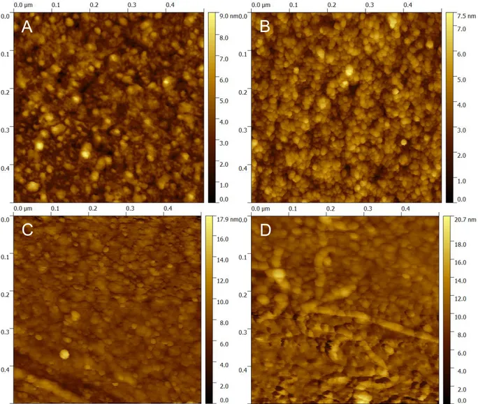
comparison with bare gold surfaces (supplemental figure S2). 150
Biochip conditioning and sample processing 151
Prisms were systematically incubated with a sterile solution of PBS+1% BSA before each 152
experiment to mimic interfering proteins of complex media such as blood, and to block non-153
specific interactions with gold. The surface of the prism was then rinsed with 3 mL of sterile PBS, 154
before loading the biochip in the Surface Plasmon Resonance imager. 900 !" of sterile TSB 155
were then injected in the culture chamber containing the AMP-array, followed by 100 !" of the 156
bacterial dilution used for spiking. Interactions of bacteria on the surface of the prisms were 157
observed in real-time with the SPRi-Lab+ system (Horiba Scientific, Palaiseau, France), at 37°C. 158
The AMP-array was positioned so that its surface was vertical, on the sidewall of the culture 159
chamber. This avoids any non-specific signal from sedimenting bacteria. Sensors were 160
systematically discarded after use. 161
SPR data analysis 162
Real-time monitoring of the SPR signal began when the bacteria were spiked into the sample. 163
Regions of interest (ROI) were defined on each duplicate of AMP spots, in order to monitor the 164
temporal change of reflectivity. Kinetics data were directly collected and processed in R 165
programming language. Duplicate spots of each peptide were averaged, corrected with respect 166
to the reference signal, and plotted. Peptide-free gold ROIs, coated with BSA, were chosen as 167
reference signals for non-specific interactions. Reflectivity shifts as a function of time for each 168
ROI allowed the assessment of the presence of bacteria in samples without further processing of 169
the data, by the observation of a temporal response shift. Kinetics data were used to generate a 170
database of interaction patterns between peptides and bacteria. For each spot, the first 171
derivative of the smoothed temporal data was calculated. The maximum value of this derivative, 172
as well as the corresponding smoothed reflectivity shift, were recorded. Unsupervised 173
multivariate analysis was performed on this database thanks to the FactomineR package, 174
embedded in the R software [34]. Names of targeted bacteria were used as a descriptive 175
variable, and therefore did not affect the results of the Principal Components Analysis (PCA). As 176
data were heterogeneous, they were centered and reduced. On one hand, PCA score plots were 177
used to distinguish results from one bacterial strain to another. On the other hand, they also 178
permitted a high level of repeatability of the process from different duplicates of the same assay. 179
Hierarchical Clustering on Principal Components (HCPC) was also performed on the outcome of 180
the PCA. This statistical method ensured that targeted bacteria were indeed discriminated from 181
each other, based on the Euclidean distance separating the replicates in the PCA results. 182
183
Results and discussion
184
Bacterial growth monitoring by SPRI 185
The set of five bacterial strains to be detected with the peptide array has been chosen to 186
represent the diversity of pathogens frequently involved in bloodstream or urinary tract 187
infections, as well as food poisoning. The set also reflects microbial morphological diversity, with 188
both Gram-positive and negative species, including bacilli and cocci. 189
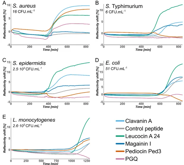
Growth kinetics monitored by SPRI of the bacteria are depicted in fig. 1, datasets retrieved from 190
those results allow defining characteristic detection times of bacteria (Supplemental Table S1) 191
and affinity patterns with the peptides. 192
193
194
Figure 1. SPR kinetic data of the bacterial growth for five different bacterial strains cultured in
195
tryptic soy broth at 37°C. Each curve is the average of duplicate spots for a given peptide
196
arrayed on the sensor, with subtraction of the reference signal taken on peptide free gold.
AMP-based arrays have permitted the detection of pathogens present at low initial concentration 198
in all samples, at most in 19 hours, and in even less than 12 hours in the majority of cases. All 199
culture conditions being the same, variations of detection signal can come from dissimilarities in 200
generation times of the different strains. Shortest detection times were for S. aureus and S. 201
epidermidis, in about 6 to 7 hours. Similar conclusions with solid based culture methods are
202
hitherto obtained in at least 24 hours. Compared to other biosensing methods using AMPs to 203
analyze pre-enriched samples, we obtained a very low limit of detection of virtually one live 204
bacterium per sample. The coupling of the enrichment step in our one-step process, not only 205
improved the safety for the operator, but also significantly improved both the overall processing 206
time and the limit of detection, by comparison to the two-steps methods described so far. 207
Naturally, the lower limit of detection comes with a longer detection time to get bacterial 208
concentrations sufficient for SPRI detection. 209
Individual peptide responses for bacterial pathogens detection 210
Peptides showed very distinct profiles of response depending on the targeted bacterial strains. 211
Individual examination of their performances is thus interesting to understand underlying 212
interactions with bacteria. Interestingly, Leucocin A 24 interacted strongly with all tested bacterial 213
strains. Nonetheless, the scrambled version of Leucocin A 24, here used as a control peptide, 214
displayed only weak to no interactions at all with any bacterial pathogen, suggesting that 215
interactions of bacteria with tethered peptides are not only governed by physico-chemical 216
mechanisms but more probably by structural interactions between peptides and bacterial 217
membranes. Such observation has already been described for therapeutic peptides [35]. 218
Clavanin A functionalized surfaces exhibited stronger SPRI responses with Staphylococci spp., 219
which are Gram-positive bacteria, although this peptide also enabled to successfully detect 220
Salmonella Typhimurium and E. coli, although with weaker interactions. With our method,
221
Magainin I only displayed mild interactions with all the tested bacteria, whatever their Gram 222
coloration (except for S. aureus which gave a very low shift). On the contrary, Ped3, a Pediocin 223
fragment, exhibited a moderate ability to detect Listeria monocytogenes, as previously described 224
[31], but surprisingly Ped3 also displayed a potent interaction with MRSA in our assay. In the 225
meantime it did not interact with S. epidermidis, although they belong to the same genus. This 226
selective response might be caused by an association of the peptide with a peculiar receptor 227
specific to the membrane of MRSA. PGQ displayed interactions with E. coli, which is consistent 228
with the literature [32]. However it detected S. aureus, which is a Gram-positive strain, and had 229
no apparent interactions with Salmonella, another Gram-negative bacterium, that also displays 230
LPS, which is a hint that the interactions between PGQ and bacteria is not only mediated by 231
interacting with the latter. 232
These results tend to show that AMPs keep a wide spectrum of affinity towards bacteria after 233
tethering on a surface. Moreover, association of AMPs enabled to obtain various kinetic profiles 234
depending on the bacterial strain. Interestingly, our SPRI detection method allowed to unravel 235
interactions which remained unobserved with other methods [25]. This could be explained by the 236
establishment of weaker interactions between bacteria and AMPs thanks to their longer 237
exposure time to the cultured bacteria. 238
Multivariate analyses of kinetic results 239
Multivariate analyses were performed to objectively assess the discrimination values of our 240
method. To do so, PCA was performed based on reflectivity shift and sensitivity values at the 241
maximum of the first derivative for each peptide (Figure 2A). Each bacterial strain was 242
processed once on an AMP micro-array, giving 64 sets of values, from all possible combinations 243
of peptide duplicates responses at the surface. This combinatorial analyzing method ensured 244
that statistical variability of the overall response was totally represented, and that cherry picking 245
of data was avoided. Experiments on different bacterial strains with the same set of AMPs gave 246
clustered results in the PCA 2D-score plot. Moreover, those clusters of data points were well 247
segregated from each other according to the monitored pathogen: results were repeatable 248
regardless of the set of replicates in the same monitoring. The first two principal components 249
represented only 70.3% of the total variance. This meant that the data were of high 250
dimensionality, thus explaining why such a good discrimination between bacterial strains could 251
be obtained. However, those first two components were sufficient in our case to clearly 252
distinguish between distinct bacterial strains. Such results underlined potential identification 253
ability of the method, although more experiments would be required to create an even more 254
exhaustive database. Results of the PCA were also used to perform a Hierarchical Clustering on 255
Principal Components (fig. 2B), leading again to a satisfying separation of the different strains in 256
separate clusters. Such discriminated profiles came from the variety of affinities displayed by the 257
peptides towards the tested bacteria. Due to different physicochemical properties and structures, 258
each peptide interacted differently to Gram or genera, hence the overall combination 259
discriminated the tested strains. Multivariate analyses with appropriate databases thus allow 260
pathogen identification methods to be derived from the usual key-lock principle and enable 261
cross-reactivity sensors as demonstrated by [36]. 262
263
264
Figure 2. Multivariate data analyses for 5 bacterial strains processed on AMP-arrays. (A)
2D-265
score plot of the database generated from the kinetics of SPRI bacterial growth monitoring.
266
Ellipses were annotated to represent the 95% confidence interval. (B) Hierarchical Clustering on
267
Principal Components (HCPC) performed on the data from Fig. 2a. Distances between points
268
are Euclidean.
269 270
In this work, we demonstrated the suitability of AMP-arrays as powerful ligands of bacterial 271
pathogens. Traditionally studied in the field of pharmacology and antimicrobial applications, 272
AMPs were revealed to be potent ligands for pathogens in biosensing applications. Not only they 273
are well-adapted for the detection of bacteria in a standard culture medium such as tryptic soy 274
broth, but compared to antibodies, the biochip functionalization and operation were less tedious, 275
thanks to peptide resistance to air-drying without loss of activity in the process. 276
The use of AMPs in a multiplexed fashion allowed a fast and simple assessment of the presence 277
of pathogens even at low concentrations. Detection times ranged from 6 to 19 hours, in a one-278
step assay. This is shorter than standard methods in blood or foodstuffs, which require 279
enrichment times of usually 24 hours before confirmation of the culture positivity. Each tested 280
bacterial strain resulted in different kinetic responses on the part of the AMP-array. Ped3 even 281
exhibited a near-specific behavior, interacting almost uniquely with MRSA. Further studies could 282
assess this point, which would be of importance in the detection of this prominent pathogen. Our 283
results, for instance for Leucocin A24 and its scrambled version, showed that the recognition of 284
bacteria by AMPs is not uniquely driven by physico-chemical interactions, but also by their 285
secondary structuration. Moreover, tethering of the AMPs on the surface of our sensor did not 286
seem to alter this interaction with bacteria in solution. The study of the whole set of AMPs 287
interacting with live bacteria gave clearly different affinity profiles. This combined approach was 288
preferred to a biomolecular study to enable the identification of several bacterial pathogens, with 289
a single AMP set. The response to polymicrobial infections, which represent only 10% of 290
bloodstream infections [37,38], is still to be investigated to check how AMPs responses might be 291
deciphered to identify two, or more, pathogenic strains present in the same sample. 292
Furthermore, the small set of AMPs we used provided very distinct affinity profiles towards 293
different pathogens, as multivariate analysis confirmed. Arrays presenting a larger collection of 294
peptides could hence enable the possibility to create interaction profile libraries and therefore the 295
identification of bacterial genus, species or strain of the infecting pathogen in a sample. 296
To the extent of our knowledge, the system we described is the first one-step biosensor assay 297
using different multiplexed AMPs and enabling detection of several viable bacterial strains in 298
biomedically relevant concentrations along with a discrimination capacity in a label-free assay. In 299
the near future, this approach could be applied for the detection of any pathogen in samples 300
such as foodstuff, urine or blood without any pre-requisite knowledge of the threat. 301
If these auspicious results are confirmed on a larger scale, it could permit the development of a 302
one-step assay – not only able to detect common pathogens overnight – but also to identify 303
them in reference to a database of affinity patterns. 304
305
Color should be used for any figures in print
306307 308
Acknowledgements
309
EP thanks the Labex ARCANE and CBH-EUR-GS program (ANR-17-EURE-0003) for his 310
doctoral fellowship. 311
312
Competing interests statement
313
The authors declare no financial or non-financial competing interests. 314
315
References
316
[1] A. van Belkum, G. Durand, M. Peyret, S. Chatellier, G. Zambardi, J. Schrenzel, D. Shortridge, 317
A. Engelhardt, W.M. Dunne, Rapid clinical bacteriology and its future impact, Ann. Lab. Med. 33 318
(2013) 14, https://doi.org/10.3343/alm.2013.33.1.14. 319
[2] O. Opota, K. Jaton, G. Greub, Microbial diagnosis of bloodstream infection: Towards 320
molecular diagnosis directly from blood, Clin. Microbiol. Infec. 21 (2015) 323–331, 321
https://doi.org/10.1016/j.cmi.2015.02.005. 322
[3] A.E. Clark, E.J. Kaleta, A. Arora, D.M. Wolk, Matrix-assisted laser desorption ionization-time 323
of flight mass spectrometry: A fundamental shift in the routine practice of clinical microbiology, 324
Clin. Microbiol. Rev. 26 (2013) 547–603, https://doi.org/10.1128/cmr.00072-12. 325
[4] M. Guido Marcello, M.R. Tumolo, A. De Donno, T. Verri, F. Serio, F. Bagordo, A. Zizza, In 326
vitro diagnosis of sepsis: A review, Pathol. Lab. Med. Int. (2016) 1, 327
https://doi.org/10.2147/plmi.s49800. 328
[5] D.C. Vanegas, C.L. Gomes, N.D. Cavallaro, D. Giraldo-Escobar, E.S. McLamore, Emerging 329
biorecognition and transduction schemes for rapid detection of pathogenic bacteria in food, 330
Compr. Rev. Food Sci. F. (2017), https://doi.org/10.1111/1541-4337.12294. 331
[6] E. Suraniti, E. Sollier, R. Calemczuk, T. Livache, P.N. Marche, M.-B. Villiers, Y. Roupioz, 332
Real-time detection of lymphocytes binding on an antibody chip using SPR imaging, Lab Chip. 7 333
(2007) 1206, https://doi.org/10.1039/b708292d. 334
[7] J.B. Fiche, J. Fuchs, A. Buhot, R. Calemczuk, T. Livache, Point mutation detection by surface 335
plasmon resonance imaging coupled with a temperature scan method in a model system, Anal. 336
Chem. 80 (2008) 1049–1057, https://doi.org/10.1021/ac7019877. 337
[8] S. Bouguelia, Y. Roupioz, S. Slimani, L. Mondani, M.G. Casabona, C. Durmort, T. Vernet, R. 338
Calemczuk, T. Livache, On-chip microbial culture for the specific detection of very low levels of 339
bacteria, Lab Chip. 13 (2013) 4024, https://doi.org/10.1039/c3lc50473e. 340
[9] L. Mondani, Y. Roupioz, S. Delannoy, P. Fach, T. Livache, Simultaneous enrichment and 341
optical detection of low levels of stressed Escherichia coli O157:H7 in food matrices, J. Appl. 342
Microbiol. 117 (2014) 537–546, https://doi.org/10.1111/jam.12522. 343
[10] A. Morlay, A. Duquenoy, F. Piat, R. Calemczuk, T. Mercey, T. Livache, Y. Roupioz, Label-344
free immuno-sensors for the fast detection of listeria in food, Measurement. 98 (2017) 305–310, 345
https://doi.org/10.1016/j.measurement.2016.06.038. 346
[11] V. Templier, T. Livache, S. Boisset, M. Maurin, S. Slimani, R. Mathey, Y. Roupioz, Biochips 347
for direct detection and identification of bacteria in blood culture-like conditions, Sci. Rep. 7 348
(2017), https://doi.org/10.1038/s41598-017-10072-z. 349
[12] V. Templier, A. Roux, Y. Roupioz, T. Livache, Ligands for label-free detection of whole 350
bacteria on biosensors: A review, TrAC Trend. Anal. Chem. 79 (2016) 71–79, 351
https://doi.org/10.1016/j.trac.2015.10.015. 352
[13] R. Singh, M.D. Mukherjee, G. Sumana, R.K. Gupta, S. Sood, B. Malhotra, Biosensors for 353
pathogen detection: A smart approach towards clinical diagnosis, Sensor. Actuat. B-Chem. 197 354
(2014) 385–404, https://doi.org/10.1016/j.snb.2014.03.005. 355
[14] M. Shahdordizadeh, S.M. Taghdisi, N. Ansari, F.A. Langroodi, K. Abnous, M. Ramezani, 356
Aptamer based biosensors for detection of Staphylococcus aureus, Sensor. Actuat. B-Chem. 357
241 (2017) 619–635, https://doi.org/10.1016/j.snb.2016.10.088. 358
[15] M. Zasloff, Antimicrobial peptides of multicellular organisms, Nature. 415 (2002) 389–395, 359
https://doi.org/10.1038/415389a. 360
[16] H.G. Boman, Antibacterial peptides: Basic facts and emerging concepts, J. Intern. Med. 254 361
(2003) 197–215, https://doi.org/10.1046/j.1365-2796.2003.01228.x. 362
[17] M.R. Yeaman, Mechanisms of antimicrobial peptide action and resistance, Pharmacol. Rev. 363
55 (2003) 27–55, https://doi.org/10.1124/pr.55.1.2. 364
[18] L.T. Nguyen, E.F. Haney, H.J. Vogel, The expanding scope of antimicrobial peptide 365
structures and their modes of action, Trends Biotechnol. 29 (2011) 464–472, 366
https://doi.org/10.1016/j.tibtech.2011.05.001. 367
[19] M. Torrent, J. Valle, M.V. Nogués, E. Boix, D. Andreu, The generation of antimicrobial 368
peptide activity: A trade-off between charge and aggregation?, Angew. Chem. Int. Edit. 50 369
(2011) 10686–10689, https://doi.org/10.1002/anie.201103589. 370
[20] A.K. Marr, W.J. Gooderham, R.E. Hancock, Antibacterial peptides for therapeutic use: 371
Obstacles and realistic outlook, Curr. Opin. Pharmacol. 6 (2006) 468–472, 372
https://doi.org/10.1016/j.coph.2006.04.006. 373
[21] R.R. Silva, K.Y.P.S. Avelino, K.L. Ribeiro, O.L. Franco, M.D.L. Oliveira, C.A.S. Andrade, 374
Optical and dielectric sensors based on antimicrobial peptides for microorganism diagnosis, 375
Front. Microbiol. 5 (2014), https://doi.org/10.3389/fmicb.2014.00443. 376
[22] M. Hoyos-Nogués, F.J. Gil, C. Mas-Moruno, Antimicrobial peptides: Powerful biorecognition 377
elements to detect bacteria in biosensing technologies, Molecules. 23 (2018) 1683, 378
https://doi.org/10.3390/molecules23071683. 379
[23] N. Kulagina, K. Shaffer, F. Ligler, C. Taitt, Antimicrobial peptides as new recognition 380
molecules for screening challenging species, Sensor. Actuat. B-Chem. 121 (2007) 150–157, 381
https://doi.org/10.1016/j.snb.2006.09.044. 382
[24] M.S. Mannoor, S. Zhang, A.J. Link, M.C. McAlpine, Electrical detection of pathogenic 383
bacteria via immobilized antimicrobial peptides, P. Natl. Acad. Sci USA. 107 (2010) 19207– 384
19212, https://doi.org/10.1073/pnas.1008768107. 385
[25] H. Etayash, L. Norman, T. Thundat, K. Kaur, Peptide-bacteria interactions using engineered 386
surface-immobilized peptides from class IIa bacteriocins, Langmuir. 29 (2013) 4048–4056, 387
https://doi.org/10.1021/la3041743. 388
[26] C.A. Andrade, J.M. Nascimento, I.S. Oliveira, C.V. de Oliveira, C.P. de Melo, O.L. Franco, 389
M.D. Oliveira, Nanostructured sensor based on carbon nanotubes and clavanin A for bacterial 390
detection, Colloid. Surface. B. 135 (2015) 833–839, 391
https://doi.org/10.1016/j.colsurfb.2015.03.037. 392
[27] A.G.S. Junior, M.D. Oliveira, I.S. Oliveira, R.G. Lima-Neto, S.R. Sá, O.L. Franco, C.A. 393
Andrade, A simple nanostructured impedimetric biosensor based on clavanin A peptide for 394
bacterial detection, Sensor. Actuat. B-Chem. 255 (2018) 3267–3274, 395
https://doi.org/10.1016/j.snb.2017.09.153. 396
[28] N.V. Kulagina, M.E. Lassman, F.S. Ligler, C.R. Taitt, Antimicrobial peptides for detection of 397
bacteria in biosensor assays, Anal. Chem. 77 (2005) 6504–6508, 398
https://doi.org/10.1021/ac050639r. 399
[29] M.-S. Chang, J.H. Yoo, D.H. Woo, M.-S. Chun, Efficient detection of Escherichia coli 400
O157:H7 using a reusable microfluidic chip embedded with antimicrobial peptide-labeled beads, 401
Analyst. 140 (2015) 7997–8006, https://doi.org/10.1039/c5an01307k. 402
[30] Y. Chen, Z.P. Michael, G.P. Kotchey, Y. Zhao, A. Star, Electronic detection of bacteria using 403
holey reduced graphene oxide, ACS Appl. Mater. Inter. 6 (2014) 3805–3810, 404
https://doi.org/10.1021/am500364f. 405
[31] S. Azmi, K. Jiang, M. Stiles, T. Thundat, K. Kaur, Detection of Listeria monocytogenes with 406
short peptide fragments from class IIa bacteriocins as recognition elements, ACS Comb. Sci. 17 407
(2015) 156–163, https://doi.org/10.1021/co500079k. 408
[32] S. Arcidiacono, P. Pivarnik, C.M. Mello, A. Senecal, Cy5 labeled antimicrobial peptides for 409
enhanced detection of Escherichia coli O157:H7, Biosens. Bioelectron. 23 (2008) 1721–1727, 410
https://doi.org/10.1016/j.bios.2008.02.005. 411
[33] H. Etayash, L. Norman, T. Thundat, M. Stiles, K. Kaur, Surface-conjugated antimicrobial 412
peptide leucocin A displays high binding to pathogenic Gram-positive bacteria, ACS Appl. Mater. 413
Inter. 6 (2014) 1131–1138, https://doi.org/10.1021/am404729c. 414
[34] S. Lê, J. Josse, F. Husson, FactoMineR: An R package for multivariate analysis, J. Stat. 415
Softw. 25 (2008), https://doi.org/10.18637/jss.v025.i01. 416
[35] C.D. Fjell, J.A. Hiss, R.E.W. Hancock, G. Schneider, Designing antimicrobial peptides: Form 417
follows function, Nat. Rev. Drug Discov. (2011), https://doi.org/10.1038/nrd3591. 418
[36] J.R. Carey, K.S. Suslick, K.I. Hulkower, J.A. Imlay, K.R.C. Imlay, C.K. Ingison, J.B. Ponder, 419
A. Sen, A.E. Wittrig, Rapid identification of bacteria with a disposable colorimetric sensing array, 420
J. Am. Chem. Soc. 133 (2011) 7571–7576, https://doi.org/10.1021/ja201634d. 421
[37] M. Pavlaki, G. Poulakou, P. Drimousis, G. Adamis, E. Apostolidou, N.K. Gatselis, I. Kritselis, 422
A. Mega, V. Mylona, A. Papatsoris, A. Pappas, A. Prekates, M. Raftogiannis, K. Rigaki, K. 423
Sereti, D. Sinapidis, I. Tsangaris, V. Tzanetakou, D. Veldekis, K. Mandragos, H. Giamarellou, G. 424
Dimopoulos, Polymicrobial bloodstream infections: Epidemiology and impact on mortality, J Glob 425
Antimicrob Re. 1 (2013) 207-212, https://doi.org/10.1016/j.jgar.2013.06.005. 426
[38] C. Royo-Cebrecos, C. Gudiol, C. Ardanuy, H. Pomares, M. Calvo, J. Carratalà, A fresh look 427
at polymicrobial bloodstream infection in cancer patients, PLoS One, 12 (2017), 428
https://doi.org/10.1371/journal.pone.0185768. 429
Supplementary Information:
Antimicrobial peptide arrays for wide
spectrum sensing of pathogenic
bacteria
Éric Pardouxa,b, Agnès Rouxa, Raphaël Matheya, Didier Boturynb, Yoann Roupioza,* aUniv. Grenoble Alpes, CNRS, CEA, INAC-SyMMES, 38000 Grenoble, France
bUniv. Grenoble Alpes, CNRS, DCM, 38000 Grenoble, France *Corresponding author: yoann.roupioz@cea.fr
Table S1. Detection times of the presence of the different bacterial strains are determined by the timestamp of the kinetics first derivative highest value. It thus corresponds to the middle of the jump between the baseline signal and the plateau. It is given for the shortest time for each bacterial strain, associated with the peptide giving this result. Only peptides giving a strong positive signal are included in the analysis. Times are given as the average over duplicates of each peptide. Bacterial strain Initial concentration (CFU.mL-1) Shortest detection time (min) Peptide E. coli ATCC11775 51 677 Clavanin A S. Typhimurium CIP104474 6 537 Magainin I S. aureus ATCC43300 16 433 Leucocin A 24 S. epidermidis ATCC12228 2.5·103 374 Leucocin A 24 L. monocytogenes IVc 2.6·103 1160 Leucocin A 24
Figure S1. (A) Scaled scheme showing characteristic dimensions of the SPRi-biochip, including a representation of the observable field of view with the Lab+. (B) Picture of a SPRi-biochip right after a spotting.
Figure S2. AFM images of monolayers of peptides prepared in the same conditions as SPRI biochips: (A) bare gold as a reference; (B) Magainin I; (C) Leucocin A 24; (D) Control peptide. They were recorded in air using the peak force mode of a Dimension Icon AFM (from Bruker, Santa Barbara, CA). The cantilevers were triangular and using a force contact of 0.1 N/m at a 70 kHz frequency and a 0.5 Hz scan rate. Image processing was performed using the Gwyddion microscopy software.
A
C
B
Date Batch
Product name Catalog #
2658,14 g/mol
TFA
1 mg/mL
Not defined
2657,407 Da Theoritical monoisotopic mass 2656,38 Da Certificate of analysis 14/03/2016 849_s1p4f2 Magainin 1-Ncys Product specifications AA sequence C-AEEA-GIGKFLHSAGKFGKAFVGEIMKS-OH Disulfide bond 0 Formula C121H193N31O32S2
Appearance White powder
Theoritical average weight CAS number
Handling & Storage
Storage Shipped at ambient temperature under lyophilized powder. Store at -20°C (-4°F). Do not freeze-thaw.
Aliquot sample if required and store at -80°C (-112°F). Expiry date
Source Synthetic
Counterion
Solubility (recommendation) 10% acetonrile solution
Handling and use restriction Use with caution. Might be toxic. Product intented for research use only, not for use in diagnostic or therapeutic procedures.
Analytical results
Observed purity rate 97,60% Observed monoisotopic mass [M+H+]+
Chromatogram ( +33 (0) 456 520 869 7 +33 (0) 456 520 868 Smartox Biotechnology 570 rue de la chimie 38400 St Martin d'Hères
MS Spectra
( +33 (0) 456 520 869 7 +33 (0) 456 520 868
Smartox Biotechnology 570 rue de la chimie 38400 St Martin d'Hères
Date Batch
Product name Catalog #
2914,39 g/mol
TFA
1 mg/mL
Not defined
2913,41 Da
Theoritical monoisotopic mass 2912,50 Da
Certificate of analysis 21/03/2016 847_s1p1f1 clavaninA CAS number Product specifications AA sequence VFQFLGKIIHHVGNFVHGFSHVF-AEEA-C-NH2 Disulfide bond 0 Source Synthetic Counterion
Solubility (recommendation) 50% of Acetonitrile solution Formula C140H201N37O30S
Appearance White powder
Theoritical average weight
Handling and use restriction Use with caution. Might be toxic. Product intented for research use only, not for use in diagnostic or therapeutic procedures.
Analytical results
Observed purity rate 93,47% Observed monoisotopic mass [M+H+]+
Handling & Storage
Storage Shipped at ambient temperature under lyophilized powder. Store at -20°C (-4°F). Do not freeze-thaw.
Aliquot sample if required and store at -80°C (-112°F). Expiry date
Chromatogram ( +33 (0) 456 520 869 7 +33 (0) 456 520 868 Smartox Biotechnology 570 rue de la chimie 38400 St Martin d'Hères
MS Spectra ( +33 (0) 456 520 869 7 +33 (0) 456 520 868 Smartox Biotechnology 570 rue de la chimie 38400 St Martin d'Hères
Date Batch
Product name Catalog #
2681,89 g/mol
TFA
1 mg/mL
Not defined
2681,17 Da
Theoritical monoisotopic mass 2680,22 Da
Analytical results
Observed purity rate 90,49% Observed monoisotopic mass [M+H+]+
Handling & Storage
Storage Shipped at ambient temperature under lyophilized powder. Store at -20°C (-4°F). Do not freeze-thaw.
Aliquot sample if required and store at -80°C (-112°F). Expiry date
Handling and use restriction Use with caution. Might be toxic. Product intented for research use only, not for use in diagnostic or therapeutic procedures.
Source Synthetic
Counterion
Solubility (recommendation) 50 % of Acetonitrile solution Formula C119H194N36O34S
Appearance White powder
Theoritical average weight CAS number Product specifications AA sequence C-AEEA-RGEWFWGNLVVSAASFGNHNAGG-OH Disulfide bond 0 Certificate of analysis 22/03/2016 850_s2p1r1 peptide Contrôle Smartox Biotechnology
Chromatogram ( +33 (0) 456 520 869 7 +33 (0) 456 520 868 Smartox Biotechnology 570 rue de la chimie 38400 St Martin d'Hères
MS Spectra ( +33 (0) 456 520 869 7 +33 (0) 456 520 868 Smartox Biotechnology 570 rue de la chimie 38400 St Martin d'Hères
Date Batch
Product name Catalog #
2657,16 g/mol
TFA
1 mg/mL
Not defined
2656,39 Da
Theoritical monoisotopic mass 2655,40 Da
Observed purity rate 95,61% Observed monoisotopic mass [M+H+]+
Storage Shipped at ambient temperature under lyophilized powder. Store at -20°C (-4°F). Do not freeze-thaw.
Aliquot sample if required and store at -80°C (-112°F). Expiry date
Handling and use restriction Use with caution. Might be toxic. Product intented for research use only, not for use in diagnostic or therapeutic procedures.
Analytical results
Source Synthetic
Counterion
Solubility (recommendation) 10% acetonrile solution
Handling & Storage
Formula C121H194N32O31S2
Appearance White powder
Theoritical average weight CAS number Product specifications AA sequence GIGKFLHSAGKFGKAFVGEIMKS-AEEA-C-NH2 Disulfide bond 0 Certificate of analysis 21/03/2016 851_s1p1f1 Magainin 1-Ccys Smartox Biotechnology
Chromatogram ( +33 (0) 456 520 869 7 +33 (0) 456 520 868 Smartox Biotechnology 570 rue de la chimie 38400 St Martin d'Hères
MS Spectra ( +33 (0) 456 520 869 7 +33 (0) 456 520 868 Smartox Biotechnology 570 rue de la chimie 38400 St Martin d'Hères
Date Batch
Product name Catalog #
2704,23 g/mol
TFA
1 mg/mL
Not defined
2703,59 Da
Theoritical monoisotopic mass 2702,55 Da
Observed purity rate 97,92% Observed monoisotopic mass [M+H+]+
Storage Shipped at ambient temperature under lyophilized powder. Store at -20°C (-4°F). Do not freeze-thaw.
Aliquot sample if required and store at -80°C (-112°F). Expiry date
Handling and use restriction Use with caution. Might be toxic. Product intented for research use only, not for use in diagnostic or therapeutic procedures.
Analytical results
Source Synthetic
Counterion
Solubility (recommendation) Water
Handling & Storage
Formula C121H211N33O34S
Appearance White powder
Theoritical average weight CAS number Product specifications AA sequence GVLSNVIGYLKKLGTGALNAVLKQ-AEEA-C-NH2 Disulfide bond 0 Certificate of analysis 21/03/2016 852_s1p1f2 peptide PGQ Smartox Biotechnology
Chromatogram ( +33 (0) 456 520 869 7 +33 (0) 456 520 868 Smartox Biotechnology 570 rue de la chimie 38400 St Martin d'Hères
MS Spectra ( +33 (0) 456 520 869 7 +33 (0) 456 520 868 Smartox Biotechnology 570 rue de la chimie 38400 St Martin d'Hères
Date Batch
Product name Catalog #
2681,89 g/mol
TFA
1 mg/mL
Not defined
2681,12 Da
Theoritical monoisotopic mass 2680,22 Da
Certificate of analysis 22/03/2016 853_s1p1r2 Leucocin A 24 Product specifications AA sequence C-AEEA-SVNWGEAFSAGVHRLANGGNGFW-OH Disulfide bond 0 Formula C119H170N36O34S
Appearance White powder
Theoritical average weight CAS number
Source Synthetic
Counterion
Solubility (recommendation) 50% of Acetonitrile solution
Handling & Storage
Storage Shipped at ambient temperature under lyophilized powder. Store at -20°C (-4°F). Do not freeze-thaw.
Aliquot sample if required and store at -80°C (-112°F). Expiry date
Handling and use restriction Use with caution. Might be toxic. Product intented for research use only, not for use in diagnostic or therapeutic procedures.
Analytical results
Observed purity rate 90,34% Observed monoisotopic mass [M+H+]+
Chromatogram ( +33 (0) 456 520 869 7 +33 (0) 456 520 868 Smartox Biotechnology 570 rue de la chimie 38400 St Martin d'Hères
MS Spectra ( +33 (0) 456 520 869 7 +33 (0) 456 520 868 Smartox Biotechnology 570 rue de la chimie 38400 St Martin d'Hères