HAL Id: halsde-01022002
https://hal.archives-ouvertes.fr/halsde-01022002
Submitted on 27 May 2020
HAL is a multi-disciplinary open access
archive for the deposit and dissemination of
sci-entific research documents, whether they are
pub-lished or not. The documents may come from
teaching and research institutions in France or
abroad, or from public or private research centers.
L’archive ouverte pluridisciplinaire HAL, est
destinée au dépôt et à la diffusion de documents
scientifiques de niveau recherche, publiés ou non,
émanant des établissements d’enseignement et de
recherche français ou étrangers, des laboratoires
publics ou privés.
Evolutionary and ecological perspectives of Late
Paleozoic ferns. Part III. Anachoropterid ferns
(including Anachoropteris, Tubicaulis, the Sermayaceae,
Kaplanopteridaceae and Psalixochlaenaceae)
Jean Galtier, Tom L. Phillips
To cite this version:
Jean Galtier, Tom L. Phillips. Evolutionary and ecological perspectives of Late Paleozoic ferns. Part
III. Anachoropterid ferns (including Anachoropteris, Tubicaulis, the Sermayaceae, Kaplanopteridaceae
and Psalixochlaenaceae). Review of Palaeobotany and Palynology, Elsevier, 2014, 205, pp.31-73.
�10.1016/j.revpalbo.2014.02.012�. �halsde-01022002�
Evolutionary and ecological perspectives of Late Paleozoic ferns. Part III.
Anachoropterid ferns (including Anachoropteris, Tubicaulis, the
Sermayaceae, Kaplanopteridaceae and Psalixochlaenaceae)
Jean Galtier
a,⁎
, Tom L. Phillips
baUMR AMAP, CIRAD, TA-A51/PS2, Boulevard de la Lironde, 34398 Montpellier cedex 5, France b
Department of Plant Biology, University of Illinois, 265 Morrill Hall, 505 South Goodwin Ave., Urbana, IL 61801, USA
a b s t r a c t
a r t i c l e i n f o
Article history: Received 7 October 2013
Received in revised form 13 January 2014 Accepted 26 February 2014
Available online 25 March 2014
Keywords: Filicalean fern Evolution Paleoecology Permineralization Carboniferous–Permian
The anachoropterid ferns, previously assigned to the family Anachoropteridaceae, are a group of anatomically preserved late Paleozoicfilicalean ferns characterized by a C-shaped foliar xylem with abaxially recurved arms (inversicatenalean anatomy) and two main protoxylem strands. The variously curved to strongly inrolled foliar xylem certainly reflects different evolutionary trends within the morphogenus Anachoropteris. The occurrence of two groups of Tubicaulis is supported by differences in cauline and foliar anatomy and the presence vs. absence of precocious pinnae. Tubicaulis with solid protostele bears petioles which are not of the Anachoropteris type. Protostelic, rarely siphonostelic, cauline structures corresponding to several types of epiphyllous shoots are well documented on rachides of several Anachoropteris species and in the genus Kaplanopteris. These shoots, borne on dominant scrambling fronds, are a common means of vegetative propagation, similar to those known in the contemporaneous botryopterid ferns. This contrasts with the highly branched rhizomatous cauline system of Psalixochlaena (a whole plant reconstruction is provided) and the erect stems, of tree-ferns type, known in some Tubicaulis and the probably related Grammatopteris. A hemi-epiphytic habit characterized some Anachoropteris and Tubicaulis. This group of ferns therefore exhibited an important diversity of habits. Informa-tion on the distal regions of fronds, i.e. on pinnule morphology and fertile parts, is unfortunately missing in the majority of taxa. Where known, the pinnules are small and dissected, and sporangia, grouped in sori, have a lat-eral annulus. However, differences in soral and sporangial morphology support the recognition of the families Sermayaceae, Kaplanopteridaceae and Psalixochlaenaceae. The discovery of new fertile anachoropterid pinnae with adaxially borne branched soral receptacles will justify the distinction of a new family. Finally, there is no well supported anatomical evidence of fertile frond compressions belonging to anachoropterid ferns.
© 2014 Elsevier B.V. All rights reserved.
1. Introduction
In thefirst two parts of this review we recognized the Zygopteridales as an extinct group of true ferns known from the late Devonian to the early Permian (Phillips and Galtier, 2005) while Ankyropteris and otherfilicalean ferns of the family Tedeleaceae may have originated from the clepsydroid zygopterid clade (Phillips and Galtier, 2011). These two groups are distinct from all the other ferns by the possession of a phyllophore-type of petiole and, additionally, by a second kind of (small) leaf, known as vascularized aphlebiae, which cloak the stem. The present review concerns the filicalean anachoropterid ferns, based on anatomically preserved taxa, characterized by a petiolar
xylem strand with abaxial curvature/concavity and typically two groups of protoxylem on the adaxial face. These ferns are sometimes referred to as“inversicatenaleans” due to the inverted orientation of their C-shaped foliar xylem strand in comparison to the commonly adaxially concave foliar strand of modernfilicaleans.
The morphogenus Anachoropteris shows the maximum specific diversity with several evolutionary trends in curved to strongly inrolled foliar xylem (Galtier and Phillips, 1996). Our knowledge of Anachoropteris has long been restricted to their foliar structures. Thefirst evidence of a cauline member was presented byDelevoryas and Morgan (1954)for Anachoropteris clavata (now transferred to the genus Kaplanopteris
Tomescu et al., 2006). Many anachoropterid ferns exhibited epiphyllous shoots borne either laterally or adaxially, or resulting from dichotomy or trifurcation of the rachis (Phillips, 1974; Holmes, 1979). New data are provided in the present paper on these different types of shoots. These developmental strategies represent effective means of vegetative ⁎ Corresponding author.
E-mail address:jean.galtier@cirad.fr(J. Galtier).
http://dx.doi.org/10.1016/j.revpalbo.2014.02.012
0034-6667/© 2014 Elsevier B.V. All rights reserved.
Contents lists available atScienceDirect
Review of Palaeobotany and Palynology
propagation which are well known in botryopterid ferns but absent in the contemporaneous zygopterid and ankyropterid ferns.
Tubicaulis was originally established for large erect stems with a solid protostele and C-shaped petiolar xylem which is different from the Anachoropteris-type. We document the occurrence of two groups of Tubicaulis differing in stelar and petiolar anatomy and presence vs absence of precocious pinnae. However, interconnections of some Tubicaulis showing a vitalized protostele with some Anachoropteris are now well established. Related genera included in this study are Psalixochlaena with a well documented dominant role of the dichoto-mous rhizome, the small protostelic to siphonostelic Apotropteris, and Grammatopteris, with erect stem and bar-shaped foliar xylem, rather similar to Tubicaulis. The broad diversity in size, branching and habit of anachoropterid ferns is discussed.
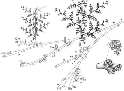
A few species are well known as whole plants, including laminate foliage and fertile parts with small annulate sporangia. They support the recognition of severalfilicalean families (Sermayaceae, Psalixochlaenaceae and Kaplanopteridaceae) but the radiation of the group remains to be documented in more detail. We provide a re-construction of the whole plant Psalixochlaena based on the detailed studies byHolmes (1977, 1981a). There are no well established com-pression assemblages of plants assignable to anachoropterid ferns.
In this review it is not our intent to taxonomically revise or establish new taxa but rather to complement the published observations and explore ecological and evolutionary implications.
2. Materials and methods
Fossil plants from the following collections and institutions have been studied and illustrated with these designated abbreviations in the explanations: MNHNP, Muséum National d'Histoire Naturelle, Paris; MHNA, Muséum d'Histoire Naturelle, Autun; NHM, Natural History Museum, London; NMP, National Museum, Prague; SMNHS, Swedish Museum Natural History, Stockholm; UI, University of Illinois, Champaign-Urbana; ULG, University de Liège; UM2, Université de Montpellier; and USTL, Université de Lille. In addition, materials from the Museum für Naturkunde, Berlin and Museum für Naturkunde, Chemnitz have been examined but not illustrated with the exception of pictures of Tubicaulis solenites (Plate XII, 1–3) and Grammatopteris freitasii (Plate XVII, 5–8) kindly provided by R. Rössler.
Additional information has been obtained from new preparations re-alized for the purpose of this study in our laboratories in Champaign-Urbana and Montpellier, using the peel technique (Galtier and Phillips, 1999).
3. Ferns with Anachoropteris-type petiole 3.1. Occurrences
The genus Anachoropteris was established byCorda (1845)for ana-tomically preserved, isolated rachides with involuted vascular strand. Subsequently more than ten species have been attributed to this genus, ranging from the Namurian C (Early Pennsylvanian) to the Late Permian. This group of ferns, and the related Tubicaulis, Sermaya, Donnegia, Kaplanopteris, Psalixochlaena, Apotropteris, and Grammatopteris, are therefore mainly restricted to the Pennsylvanian with their latest repre-sentatives in the Permian (Table 1). A single taxon, represented by isolat-ed petioles of Grammatopteris bertrandii, was attributisolat-ed to the Early Carboniferous but its Visean age (Corsin, 1937) needs to be confirmed. As a result, the anachoropterid ferns have a shorter and more recent evo-lutionary history than the zygopterids which showed successive radia-tions in early and late Carboniferous times (Phillips and Galtier, 2005).
The oldest undisputable representative is an isolated rachis of Anachoropteris sp. illustrated byRemy and Remy (1977,fig. 49) from the Namurian C of Essen-Werden (Germany). This specimen shows a
C-shaped xylem strand with short abaxial“arms” and two widely sepa-rated adaxial protoxylem strands (Fig. 1A).
During Early Pennsylvanian (Langstettian) time, the anachoropterids show an important specific diversity reflected in the shape and degree of involuteness of the foliar xylem, in transverse section, which is either: massive with very short arms (Fig. 1B), C-shaped to nearly closed (Fig. 1C), involute (Fig. 1D), or revolute (Fig. 1E). However, this diversification/rapid radiation may be the result of collecting bias due to the rich fossil–plant assemblages of this age preserved in early Westphalian European coal balls (Galtier, 1997).
During the Late Pennsylvanian and Permian times the same variability is observable and one may suggest several morphological/ evolutionary trends:
1. “gillotii–radnicensis group” with short and thick foliar xylem and very short arms (Fig. 1B1 to B3;Plate I, 5);
2.“robusta group” with curved to C-shaped to nearly closed xylem (Fig. 1C1 to C7;Plate I, 3–4);
3. Anachoropteris williamsonii and other with involute xylem (Fig. 1D1–2;
Plate I, 1);
4.“pulchra–involuta group” with revolute xylem (Fig. 1E1 to E7;
Plate I, 2).
Even if it is tempting to consider the simple anatomy of thefirst group as primitive and the most complex one of the last group as derived, we have no evidence of this.
Considering that we have illustrated, inFig. 1, specimens showing the maximum size known for each taxon one may recognize an increase in size within the“robusta” and the “pulchra–involuta” groups both of which extend until the Early Permian. It must be noted that the largest in the recorded data (Fig. 1C4, C7, E5–7) were preserved in cherts (clastic substrates) in contrast to most of the others from coal-ball peats. In addition to foliar xylem shape, some precise anatomical charac-ters are of taxonomic and evolutionary interest:
– The general shape of the xylem strand may be expressed in ratios of radial width/tangential width of the xylem strand.
– The distance between the two protoxylem strands (arrows,Plate I) on the rectilinear median region (“apolar bar”) is variable; as a result, the ratio of this distance (= apolar length)/tangential width of the xylem strand also proved to be taxonomically significant (Corsin, 1937).
– The xylem thickness is generally uniform but xylem is sometimes thinner near the protoxylem strands (Plate I, 1, 2, 6). On the other hand, distal enlargement of the arms is uncommon, either just perceptible (Plate I, 4) or quite marked, resulting in club-shaped arms (Plate I, 6) in Anachoropteris clavata. Interestingly this taxon is now separated, for independent reasons, in a different genus, Kaplanopteris.
– The diameter of metaxylem tracheids is also variable and this is without relation to the xylem-strand size.
– The pinna trace is either in the form of a solid oval strand (gillotii and robusta groups) or of a U-shaped strand (williamsonii and pulchra– involuta groups, with very rare exceptions).
– Finally, distinct foliar to cauline types of branching proved to be characteristic of some groups as demonstrated in this paper. In the text below, the different“groups” are considered with empha-sis on their vegetative morphology, including new data on branching. Information on fertile parts, ecology and habit follows.
3.2. The Anachoropteris pulchra–involuta group
The earliest known representative of this group with very involute/ revolute xylem arms is an unpublished specimen,“Anachoropteris laveinei” (Fig. 1E1), from the basal Langsettian/early Pennsylvanian of
Table 1
Stratigraphic range of Anachoropteris (1–13), Tubicaulis (17–27) and related genera:
(1) Anachoropteris williamsonii (a, Truebano, Spain; b, Lancashire (First, Union) and Yorkshire, England; Bouxharmont, Belgium; Finefrau, The Netherlands and Germany; c, Katharina, Ruhr, Germany; d, Aegir, The Netherlands. see stratigraphic references inGaltier, 1997).
(2–7) Anachoropteris “pulchra–involuta group”
(2) A. pulchra (a, Radnitz, Czech Republic,Corda, 1845; b, Parker Coal, IN; c, Calhoun Coal, IL,Phillips, 1974, 1980; d, Grand-Croix, France,Corsin, 1937; e, Autun, France,Renault, 1868; f, Sardinia, Italy,Galtier et al., 2011; g, Chemnitz, Germany,Rössler, 2001).
(3) A. involuta (a, Parker Coal, IN; b, Friendsville Coal, IL; c, Duquesne, OH,Phillips, 1980; d, Calhoun Coal, IL (type specimen)Hoskins, 1930; e, Grand-Croix, France,Corsin, 1937). (4) Anachoropteris sp. with adaxial shoots (a, Upper Path Fork Coal, EKY; b, Murphysboro equivalent Coal, IN; c, Fleming Coal, KS; d, Herrin Coal, IL, WKY; e, Baker Coal, WKY;Phillips,
1974, 1980).
(5) A.“laveinei” (Truebano, Spain,Beckary, 1988). (6) A. gigas (Autun, France,Corsin, 1937). (7) A. ovata (Autun, France,Corsin, 1937).
(8a–c) A. gillotii (a, Truebano, Spain,Beckary, 1988; b, Bouxharmont,Holmes, 1979; c, Grand-Croix, France,Corsin, 1937). (9) A. radnicensis (Radnitz, Czech Rep.,Corda, 1845).
(10a–c) A. robusta (a, Fleming Coal, KS; b, Calhoun Coal, IL; c, Grand-Croix, France,Corsin, 1937).
(11a–f) A. pautetii (a, Murphysboro equivalent Coal, IN; b, Herrin Coal, IL; c, Baker Coal, WKY; d, Parker Coal, IN; e, Calhoun Coal, IL,Phillips, 1980; f, Grand-Croix, France,Corsin, 1937). (12) A. circularis (Autun, France,Corsin, 1937).
(13) Anachoropteris sp. (a, A. sp.1. Essen-Werden, Germany,Remy and Remy, 1977; b, A. sp.2 = A. robusta? and c, A. sp.3 Bouxharmont, fromHolmes and Fairon-Demaret, 1984; d, A. sp.4 Xuanwei formation, Guizhou prov. China,Hilton et al., 2004).
(14) Anachoropteris clavata (a, Parker Coal, IN; b, Friendsville Coal IL; c, Duquesne Coal, OH; d, Calhoun Coal, IL;Graham, 1935; Delevoryas and Morgan, 1954; Phillips, 1980). Kaplanopteris clavata, Duquesne Coal, OH,Tomescu et al, 2006.
(15) Sermaya biseriata (Calhoun Coal, IL,Eggert and Delevoryas, 1967). (16) Doneggia complura (Duquesne Coal, OH,Rothwell, 1978). (17–27) Tubicaulis
(17) T. sutcliffii (Shore, England,Stopes, 1906).
(18) Tubicaulis sp. (a, T. sp.1. Union, England, new data; b, T. sp.2. Carbon Hill Mine, Iowa,Hall, 1961; c, T. sp.3. Murphysboro equivalent Coal, IN, new data; d, T. sp.4. Herrin, Paradise, WKY, from dichotomy of rachis,Galtier and Phillips, 1996).
(19) T. multiscalariformis (Fleming Coal, KS,Delevoryas and Morgan, 1952). (20) T. grandeuryi (Grand-Croix, France,Galtier and Holmes, 1984). (21) Tubicaulis sp. (Grand-Croix, France,Galtier and Holmes, 1984). (22) T. scandens (Calhoun Coal, IL,Mamay, 1952).
(23) T. stewartii, Calhoun Coal, IL,Eggert, 1959).
(24) T. berthieri (Autun, France,Bertrand and Bertrand, 1911). (25) T. cf. berthieri (Chemnitz, Germany,Rössler, 2001). (26) T. solenites (Chemnitz, Germany,Cotta, 1832); (27) T. africanus (Tanganyika,Holden and Croft, 1962).
(28) Psalixochlaena cylindrica (Truebano, Spain,Beckary, 1988; New Castle Coal Bed, Walker County, Alabama,Winston and Phillips, 1991; Union, England; Bouxharmont, Belgium,
Holmes, 1977).
(29) Apotropteris minuta (Calhoun Coal, IL,Morgan and Delevoryas, 1954). (30–33) Grammatopteris
(30) G. rigollotii (Autun, France,Renault, 1891).
(31) G. baldaufii (Chemnitz, Germany,Beck, 1920; Rössler and Galtier, 2002). (32) G. freitasii (Pedra de Fogo Fm, Tocantins, Brazil,Rössler and Galtier, 2002). (33) G. bertrandii (Esnost, France,Corsin, 1937).
Mississipian Pennsylvanian Permian
Visean Serpukhov Bashkirian Moscovian Kasimovian Gzelian Early Late Chesterian Morrowan Atokan Desmoines Missourian Virgilian
Visean Namur A Namur B Namur C Langsettian Duckmant Bolsovian Westphal D Cantabr Baruelian Stephan B Stephan C Asselian
Anachoropteris 1 a-d * * * * 2 a * b-d * * * e-g * * * 3 a-e * * * * * 4 a * b-e * * * * 5 * 6 * 7 * 8 a-b * * c * 9 * 10 a * b-c * * 11 a-c * * * d-f * * * 12 * 13 a-c * * * d * Kaplanopteris 14 a-d * * * * Sermaya 15 * Donnegia 16 * Tubicaulis 17 * 18 a * b-d * * * 19 * 20 * 21 * 22 * 23 * 24 * 25 * 26 * 27 * Psalixochlaena 28 a-c * * * Apotropteris 29 * Grammatopteris 30 * 31 * 32 * * 33
Spain (Beckary, 1988). Several species, based on rachides showing a similar anatomy, are long known from middle Pennsylvanian to early Permian time.
3.2.1. Anachoropteris pulchra Corda, the type species
The specimens described byCorda (1845)come from the Whetstone horizon which represents a complex of volcaniclastics in the Radnice group of coals (Czech Republic); they are considered as Middle Pennsyl-vanian (Bolsovian) in age (Oplustil et al., 2009). One small rachis (about 2.7 mm diameter) was described and, very accurately, illustrated by
Corda (1845, Plate 56,figs. 1–2). This section (one polished surface) showing the characteristic strongly involute xylem anatomy, dense cortex and presence of hairs, is illustrated on ourPlate II,Fig. 1. Another section of the same rachis (Plate II, 2) is above the departure of sub-opposite pinna trace; the trace on the left is O-shaped.Kubart (1916, Plate VII,figs. 49–51) illustrated other sections, unfortunately not found, showing earlier stages of incipient pinna trace. We agree with
Corsin (1937)to consider this O-shaped trace as a feature of taxonomic significance. Smaller rachides, about 1.5 mm in diameter showing a C-shaped, just involuted, xylem strand (Plate II, 3), have been described by Corda under the name of Anachoropteris rotundata, but they probably correspond to secondary rachides of Anachoropteris pulchra. It is worth mentioning that Chorionopteris lamina with synangia was considered
as fertile parts of A. pulchra (Kubart, 1916); however, the reinvestigation of the original material did not allow us to recognize any organic con-nection. It is suggested that Chorionopteris represents reproductive parts of scolecopterids belonging to one of the associated marratialean Psaronius plants.
Following Corda, Renault (1868) attributed to Anachoropteris pulchra some rachides from the Early Permian of Autun possessing a similarly involute xylem strand; the same identification was proposed byCorsin (1937),Phillips (1974, 1980),Rössler (2001)andGaltier et al. (2011)for rachides from, respectively, the Late Pennsylvanian of France and the U.S.A., and the Early Permian of Germany and Sardinia (Table 1: 2). All these rachides are younger and much broader (up to 5–8 mm in diameter) than the A. pulchra type from Radnice. Some of them possess a ratio of radial/tangential width of xylem strand about 0.8, similar to that of the type species. However, in most cases this ratio is smaller and the shape of the revolute xylem strand is significantly different; therefore, such specimens are distinguished as Anachoropteris involuta and Anachoropteris ovata. The last species is characterized by a departing O-shaped pinna trace resulting from the separation of a prominent loop of the lateral arm (Plate I, 2), and by tracheids smaller than those of A. involuta (Plate II, 4). The Early Permian A. ovata may be considered as a derived member of the “pulchra–involuta group”.
Fig. 1. Diversification of foliar anatomy in the Anachoropteridaceae. The different letterings correspond to suggested morphological trends: B1–3/radnicensis–gillotii group; C1–7/robusta group; D–E/involuta–pulchra–williamsonii group. The taxa are broadly arranged in stratigraphic sequence:
A. Anachoropteris sp. (redrawn fromRemy and Remy, 1977) Namurian C, Essen-Werden, Germany.
B1. A. gillotii (Holmes, 1979); C1. Anachoropteris sp. (Holmes and Fairon-Demaret, 1984); D1. Anachoropteris williamsonii, all from Bouxharmont, Belgium. E1. Anachoropteris“laveinei” Truebano, Spain (Beckary, 1988). F. Psalixochlaena cylindrica (Holmes, 1981a), Union, England. All Early Westphalian A–Langsettian, Early Pennsylvanian.
B2. Anachoropteris radnicensis; E2. A. pulchra (Corda, 1845) both from Radnice, Czech Rep., Westphalian C–Bolsovian/Early Desmoinesian equivalent, Middle Pennsylvanian.
C2. Anachoropteris robusta Fleming Coal, West Mineral, KS (new data); C3. Anachoropteris cf. pautetii Freeman Orient Mine 5, IL (new data); D2. Anachoropteris sp. Herrin Coal, Shawneetown, IL (new data; E3. Anachoropteris sp. twin branching, Murphysboro Coal, Cayuga, IN (new data). All Westphalian D/Desmoinesian equivalent, Mid-dle Pennsylvanian.
B3. Anachoropteris gillotii (type); C4. Anachoropteris robusta (type); C5. Anachoropteris pautetii (type), all redrawn fromCorsin (1937), Grand-Croix, France; C6. Anachoropteris robusta (new data) Calhoun Coal, IL; E4. Anachoropteris involuta (type), Calhoun, IL (redrawn fromHoskins, 1930); E5. A. involuta, Grand-Croix, France. G. Kaplanopteris (Anachoropteris) clavata (type, redrawn fromDelevoryas and Morgan, 1954), Calhoun Coal, Berryville, IL. All Stephanian A/Missourian equiv-alent, Late Pennsylvanian.
C7. Anachoropteris circularis; E7. Anachoropteris gigas; E6. Anachoropteris ovata (types, redrawn fromCorsin, 1937), all from Autun, France, Autunian/Asselian equivalent, Early Permian.
3.2.2. Late Pennsylvanian Anachoropteris involuta, with lateral shoot Anachoropteris involuta was founded by Hoskins (1930) on a rachis, 6.5–7 mm in diameter, from a coal ball from the Calhoun Coal, Illinois (Late Pennsylvanian, Missourian, Barruelian equivalent). Re-examination of the type material by one of us (TLP) confirmed the rather poor preservation mentioned by Hoskins. However, the slide sequence showed stages in lateral extra-marginal development of pinna-trace formation that is similar to those reconstructed byCorsin (1937,figs. 34–35)and later illustrated byPhillips (1974,figs. 44–45). Therefore, A. involuta differs from Anachoropteris pulchra in its pattern of emission of an U-shaped pinna trace and in the smaller ratio of radial/tangential width of xylem strand; these are arguments to segre-gate the two species (compareFigs. 4 and 5with 1 and 2 onPlate II).
Our understanding of Anachoropteris involuta has been based on
Corsin's (1937)detailed investigation of material from cherts of the Late Pennsylvanian (Barruelian equivalent) of Grand-Croix, France. Most of the rachides/petioles examined by Corsin are 6–8 × 3–4 mm in diameter with a xylem strand about 4 × 2 mm. The departing primary pinna trace is U- then C-shaped (PT,Plate II, 4–5); detached primary pinna rachides are 2.5 × 1.5 mm wide, with a C-shaped xylem strand; and supposed secondary pinna rachides are a little more than 1 mm broad with a small C-shaped xylem. The same author also described precocious traces (“sorties hâtives”) inserted at the base of primary pin-nae and therefore homologous to secondary pinpin-nae. They are solid traces (250–300 μm) constituted of very small tracheids. The first is de-tached from the outside (catadromic side) of the very proximal region of the U-shaped pinna trace; the second trace is detached higher up to the inside (anadromic).Corsin (1937, Plate 28: 2, 4)demonstrated
that thefirst trace was entering an organ detached from the pinna base, extending in a horizontal plane, and recurved towards the adaxial face of the main rachis. Due to this strange morphology, and by analogy with those organs found in a similar position at the base of pinnae in some zygopterids and ankyropterids, Corsin designated them as “aphlebiae”; however, he did not reconstruct the morphology of these organs. We have no evidence of laminate pinnules belonging to this species.
At least three distinct branching types in the development of shoots on fronds have been mentioned by Phillips (1974) within the Anachoropteris involuta group but only one is known for the Late Penn-sylvanian members. Pinnately compound fronds of A. involuta bearing protostelic shoots laterally have been illustrated byHall (1961)and
Phillips (1974)for late Pennsylvanian rachides from Berryville, Illinois. Such a rachis shows a small cauline stele (white arrow,Plate II,Fig. 6) still attached to a U-shaped pinna trace in the form of a common trace. Right above this trace the cellular continuity of the rachis cortex with an adjacent protostelic stem (S,Plate II, 6) is visible. The last organ represents a recurved shoot bearing immediately many large roots. One must note the spectacular increase in diameter of the cauline stele by comparison with the most proximal cauline component of the departing common trace. Furthermore, there is evidence of one incipi-ent petiole trace (IP,Plate II, 6) borne on this shoot.
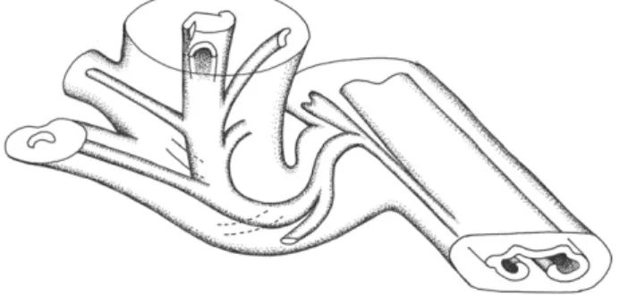
It is significant that the same type of foliar to cauline branching was described in a contemporaneous Late Pennsylvanian Anachoropteris involuta from France (Galtier and Holmes, 1984). These authors provid-ed a reconstruction (reproducprovid-ed hereFig. 2) of the shoot laterally borne on the rachis and quickly recurved. In this case also the cauline stele was Plate I. Anatomical diversity of abaxially curved foliar xylem strands in anachoropterids. Arrows pointing the two main protoxylem strands adaxially situated on the rectilinear median “bar”. All transverse sections and scale bars = 1 mm.
1. Involuted xylem strand of Anachoropteris sp. Note the large tracheid diameter and the solid elliptical pinna trace at left. Middle Pennsylvanian, Baker Coal, Providence, WKY. UI40201EBOT-52.
2. More involuted (= revolute) strand of Anachoropteris ovata. Note the small tracheid diameter and the“loop” of the lateral arm, at left, corresponding to incipient pinna trace. Early Permian, Autun. MNHNP 2237 Renault 105.
3. C-shaped xylem of Anachoropteris cf. robusta. Note the uniform thickness, short abaxial arms with a sharp tip and solid pinna trace at right. Middle Pennsylvanian, Freeman Orient Mine 5, IL. UI3217CTOP-62.
4. C-shaped xylem strand of Anachoropteris circularis. Note the very long xylem arms with slightly enlarged tips. Early Permian, Autun. MHNA 94 DLC.
5 Massive xylem strand of Anachoropteris gillotii. The strand is slightly abaxially bent without typical xylem arms. Early Pennsylvanian, Bouxharmont, Belgium. ULG BX695A1ABOT 96.
6. U-shaped xylem strand of Kaplanopteris (Anachoropteris) clavata. The xylem is very thin in the median bar but quickly enlarges along club-shaped arms. Late Pennsylvanian, Calhoun Coal, Berryville, Illinois. UI1168CTOP-7.
proximally attached to a pinna trace. The stem, followed over 2 cm length, increased in diameter up to 12 mm while the cauline protostele was described as a vitalized protostele withfiles of xylem parenchyma. Very numerous roots occur all along the stem and the emission of sev-eral helically arranged leaf traces has been documented. The leaf trace, initially bar-shaped, becomes abaxially curved. In the free petiole the fo-liar xylem strand is in the form of an open C-shape about 1.6 mm wide, i.e. much smaller and very different from the involuted xylem of the parent rachis. Considering similarities in stelar organization, leaf trace emission, petiole anatomy, and overall size and organization, this epiphyllous shoot was interpreted as conforming to the diagnosis of Tubicaulis stewartiiEggert (1959)from the Late Pennsylvanian of Illinois.
3.2.3. Middle Pennsylvanian involute Anachoropteris, with adaxial shoots The diversity of Middle Pennsylvanian age Anachoropteris (C2–3, D2, E3,Fig. 1) is documented only from American coal-ball material. This variability includes rare rachides with very involuted/revoluted xylem (Plate III, 1–2) which are similar to Anachoropteris involuta, particularly in the pattern of U-shaped pinna-trace formation (comparePlate III, 1 with A. involuta onPlate II, 4). In this example there are paired pinna traces and then strictly opposite primary pinnae (PP,Plate III, 2) with a trace becoming C-shaped. This section is of particular interest in showing also vascular strands and oblique sections of the base of two “precocious” secondary pinnae (SP) situated on the acroscopic side. If these secondary pinnae are thefirst borne this is a difference with A. involuta whereCorsin (1937)demonstrated that thefirst secondary pinna (“aphlebia”) was catadromous. However, there is the possibility that thefirst precocious pinna was born on the basiscopic side and was either missed or destroyed.
Most Middle Pennsylvanian Anachoropteris rachides are once invo-luted (i.e. not revoinvo-luted like Anachoropteris involuta). They produce U-shaped primary pinna traces like those illustrated onPlate III,fig. 3 but more rarely they possess a solid oval pinna trace (Plate I, 1). Foliar branching has been observed in detail. The large rachis, at left on
Plate III,fig. 3, shows the base of one primary pinna with U-shaped trace (arrow) and one small trace (SPT) to a secondary pinna situated on the acroscopic side. In opposite position there is a short expansion of the pinna base cortex suggestive that thefirst secondary pinna was actually attached here, i.e. in basiscopic (catadromous) position. By anal-ogy with Corsin's observation it is suggested that the distal portion of the aphlebia-like secondary pinna corresponds to the small rachides (SP) ex-tending along the adaxial side of the main rachis. One free primary pinna rachis (PP,Plate III, 3) is up to 3 mm in diameter; its xylem strand re-mains U-shaped and shows swollen arms, two characters distinguishing it from the primary pinna of A. involuta. The upper region of this rachis shows one recurved expansion interpreted as the base of a secondary pinna; this is supported by the two small traces (SPT, TPT) which are the probable vascular strands of secondary and tertiary pinnae. One iso-lated, probable secondary rachis (SR, at the bottom of thefigure), shows branching traces but evidence of lamina is missing. Partial reconstruction of the basal region of such a secondary rachis (recurved and aphlebia-like) is proposed on the left part ofFig. 3.
Asfirst mentioned byPhillips (1974)these Middle Pennsylvanian Anachoropteris rachides commonly bear adaxial protostelic shoots. This feature is well documented in the specimen onPlate III,fig. 4 that shows, adaxially from the median region of the rachis, the buildup of tracheids into a cauline trace. The obliquely sectioned cortex of the shoot, with outgoing roots (R), is visible just above. OnPlate III,fig. 5 the main foliar member is seen in perfect transverse section while the cauline trace departure is shown in longitudinal section with roots (R) right from the base; serial sections indicate that the shoot isfirst borne perpendicularly to the parent rachis and then markedly reflexed. The rachis is also emitting a primary pinna trace laterally (PT,Plate III, fig. 5). The reconstruction inFig. 3is based on this specimen showing, in the forefront, xylary organization of the rachis bearing a pinna trace at left, as well as an adaxial shoot becoming recurved and bearing peti-ole traces. In the background the cortical tissues of the rachis are repre-sented with the traces of the primary pinna and of the two basal secondary pinnae. The resulting small (aphlebia-like) secondary pinnae arch over the adaxial face of the main rachis. This arching over of sec-ondary pinnae across the adaxial face of the rachis is common and may represent a means of hooking and support for leaning or climbing fronds. For simplicity all adventitious roots have been omitted from the drawing. Another example of a rachis bearing both a pinna trace and a recurved shoot is shown onPlate IV, 1.
In addition, even small foliar members bear shoots, as illustrated by the rachis ofPlate IV,figs. 2–3. This rachis is interpreted as a primary pinna, considering its small size and the U-shaped xylary configuration with swollen arms similar to those shown in the primary pinna of Anachoropteris sp. (e.g. PP,Plate III, 3). The incipient adaxial cauline trace is seen in C,Plate IV,fig. 2 while on a subsequent section (Plate IV, 3) the stele of the small stem, curved upward, is twice sectioned.
Another branching type in the development of shoots on fronds is il-lustrated onPlate IV, 4–7 and reconstructed on figs. 4–5. The specimen onPlate IV, 4 is a transverse section of a distorted rachis showing prolif-eration of small tracheids (S) in the median region of the involuted xylem strand and separation (arrow) of the xylem arm on the left side. The section higher up (Plate IV, 5) differs by the increasing prolif-eration of the cauline tracheids in the median region. More distally
Plate II. Rachides of the“pulchra–involuta group” showing the characteristic very involute/revolute xylem strand. All transverse sections and scale bars = 1 mm.
1. Anachoropteris pulchra, type specimen illustrated by Corda (1845) (Taf. LVI, 1–2). The xylem strand shows a relatively high ratio of radial/tangential width; note the narrow cortex and hairs (H). NMP, Corda coll. E212aBOT.
2. Anachoropteris pulchra, type specimen, another section of the same rachis illustrated by Kubart (1916) (Taf. VII, 51, 55). The pinna trace at left (PT) is O-Shaped. NMP, Corda coll. E212aTOP.
3. Anachoropteris pulchra, probable second order rachis with less involute xylem strand. NMP, Corda coll. E214cTOP.
4–5. Anachoropteris involuta, the ratio of radial/tangential width of xylem strand is smaller; the pinna trace (PT) is still attached and U-shaped in 4, and it is free and C-shaped in 5. Late Pennsylvanian, Grand-Croix, France. 4 = UM2 GC514AT01; 6 = SMNHS Florin coll. 3426.
6. Anachoropteris involuta, the pinna trace (PT) has an incipient secondary trace (white arrow) which is the most proximal part of a cauline trace which diverges higher up into the coalescent stem (S). The diameter of the cauline protostele quickly increased, showing a departing petiole trace (IP). Several large roots (R) are detached from the very proximal region of the shoot. Late Pennsylvanian, Calhoun Coal, Berryville, IL. UI22931 1301LTOP-1.
Fig. 2. Reconstruction of an epiphyllous shoot borne laterally on a rachis of Anachoropteris involuta from the Late Pennsylvanian of Grand-Croix, France. The strongly recurved proto-stelic stem bears numerous C-shaped, small petiole traces.
(Plate IV, 6) this rachis seems to divide into two lateral foliar members (F1 and F2) and one median cauline siphonostele (S). At this stage there is still a common cortex cloaking the two foliar organs. The foliar member F1 was greatly compressed with its xylem obliterated while the xylem of foliar member F2 is in the form of a“ring” clearly derived from the right side inrolled arm of the original anachoropterid strand. This circular xylem strand is actually thicker abaxially with a double row of large tracheids interpreted as the result offlattening of the pre-viously inrolled tip of the xylem arm; on the opposite adaxial surface small protoxylem tracheids are visible. Another specimen, shown on
Plate IV,fig. 7, is interpreted as a little more distal region of the same type of branching. The siphonostelic cauline trace is departing upward on the adaxial side and both foliar members show well preserved xylary strands with bilateral symmetry and protoxylem groups on the adaxial side. However, F1 shows an internal bar separated from the xylem ring while in F2 the bar is contiguous with the lower part of the ring; the last anatomy is very similar to that of F2 inPlate IV,fig. 6. At this stage both foliar members still possess a common cortex. Another example of a most distal section (Plate V, 1) was beyond the recurved cauline stele, and foliar members are fully separated; anatomy of one foliar member (arrow at left) appears unchanged while there are two involuted xylary strands (a dichotomy) at right (double arrows). Finally, this foliar to cauline branching type may be interpreted as a trifurcation (two close branchings) of the involute foliar member producing one median siphonostelic cauline shoot, departing adaxially, and two lateral foliar members with initially circular xylem strands. The reconstruction in
Fig. 4(not taking in account thefirst branching, i.e. the separation of the compressed foliar member F1) shows the second branching, i.e. the separation of the cauline siphonostele and of foliar member F2. The reconstruction inFig. 5tracks all 3 members (2 foliar and the medi-an cauline) in a more distal region of the trifurcation.
The last branching type in the development of shoots on fronds is interpreted as a dichotomy of the main involuted foliar member resulting in a cauline siphonostelic trace (Plate V, 2–3). As the cauline stele (S,Plate V, 2) begins to form petiolar traces, the remaining half of foliar xylem (F, Plate V, 2) progressively recurves toward a C to invo-lute shape. At this stage both xylem strands are within a common cortex. Higher up (Plate V, 3) the siphonostelic cauline stele enlarges and its pith shows ground tissue similar to that inside the petiolar traces. 3.3. Early Pennsylvanian Anachoropteris williamsonii
This fern wasfirst described byWilliamson (1878)under the name of Rachiopteris gleiche from British coal balls, then as Rachiopteris rotundata byFelix (1886)from Westphalia. Subsequently, it was attrib-uted to the genus Anachoropteris byScott (1920)and distinguished as a new species, Anachoropteris williamsonii, byKoopmans (1928).Corsin (1937)provided a detailed specific diagnosis.Holmes (1981b), studying new British and Belgian material,first described the evidence of foliar to cauline branching in this species.
Anachoropteris williamsonii is of relatively rare occurrence and re-stricted to Early Pennsylvanian coal balls from west Europe (line 1,
Table 1) ranging from the basalmost Langsettian of Spain, England, Belgium, The Netherlands and Germany up to the basal Duckmantian of The Netherlands.
The main rachis is small, about 4–5 × 3 mm in transverse section, and the maximum diameter of 8 mm mentioned byCorsin (1937)
seems greatly overestimated. The primary xylem strand, about 2 × 1 mm (Plate VI, 1–3), is in the form of a slender involute arc only 1 or 2 tracheids thick. Metaxylem tracheids are up to 150–200 μm diameter; they exhibit reticulate thickenings to multiseriate scalariform pitting. There are two protoxylem strands on the median rectilinear adaxial face, separated by more than 1 mm. The successive stages of pinna-trace formation may be followed onPlate VI,figs. 3 and 2; they consist of marginal development of U-shaped traces. This is conforming to ob-servation byHolmes (1981b)whileCorsin (1937)erroneously de-scribed a massive pinna trace. Primary pinnae are borne alternately (Plate VI, 3) to sub-oppositely (Plate VI, 2). In the pinna base the U-shaped xylem strand is about 300 × 150μm broad and it shows the pre-cocious emission of a small cylindrical trace (arrow,Plate VI, 5) less than 100μm diameter; this is the trace of the first second order pinna borne on the outer (catadromic) side which is becoming free (SP,Plate VI, 1) as a structure recurved towards the adaxial side of the main rachis. The primary pinna rachis is about 1 mm diameter at its base and it quick-ly divides (PP,Plate VI, 1 and 4) giving rise, on the inner (anadromic) side, to another secondary pinna rachis (SP,Plate VI, 1 and 4). The latter small rachis is cylindrical, about 0.7 mm in diameter, with a broad homogeneous cortex and a tiny apparently cylindrical xylem strand. We have not obtained information on either more distal branching of these secondary pinnae or evidence of lamina, but this is suggestive that the Anachoropteris williamsonii frond was at least tri-pinnate.
The occurrence of shoots on foliar members of Anachoropteris williamsonii wasfirst mentioned byHolmes (1981b)from two speci-mens illustrated here. One Belgian specimen of Anachoropteris shows the initial enlargement of the xylem strand median region due to the proliferation of small tracheids (arrow,Plate VII, 1); higher up a trifurca-tion results into a middle trace, in the form of a solid, more or less cylin-drical, protostele (S,Plate VII, 2), and two lateral, circular xylem strands (F1–2,Plate VII, 2), each corresponding to one of the previous enrolled Plate III. Rachides of the“pulchra–involuta group” from the North American Middle Pennsylvanian. Main foliar members showing pinna traces and departures and/or adaxial shoots, all in cross sections. Scale bar = 1 mm.
1–2. Anachoropteris sp. petiole with“twin” foliar branching. Murphysboro Coal, Cayuga, IN. 1: The two opposite U-shaped pinna traces are just detached. 2: More distal section of the same showing the bases of primary pinnae (PP) and of truncated secondary pinnae (SP) with their small trace. UI25760DBOT-2 and 25760ETOP-54.
3. Anachoropteris sp. Two main foliar members and two primary pinnae, one attached (U-shaped xylem with swollen arms, arrowed) and one separate (PP). Note curvature of secondary pinna (SP) extending over adaxial surface of main rachis at left. SR, detached secondary pinna rachis; SPT, TPT, secondary and tertiary pinna traces. Baker Coal, Providence, WKY. UI8222BBOT-52.
4–5. Anachoropteris sp. main foliar members showing adaxial shoot origins. Herrin Coal, Shawneetown, IL. 4: Shows the buildup of small tracheids (arrow) into a cauline trace and roots (R) above in shoot cortex. UI3123DBOT-30. 5: Shows the cauline trace departure in longitudinal section with roots (R), the rachis is also emitting a pinna trace (PT) laterally. UI slide 4133-7966BTOP-10.
Fig. 3. Reconstruction of Anachoropteris sp. foliar member with protostelic adaxial shoot from the American Middle Pennsylvanian. The rachis is also emitting a lateral pinna trace which is shown with paired small lateral secondary pinna traces. Note how the resulting small pinna arches over the adaxial face.
xylem arms. The median bundle is about 1 mm in diameter and the two lateral ones are twice as large. At this stage the three xylem strands are still within a common ground tissue and higher up (Plate VII, 3) they
become free. The median strand, interpreted as a cauline stele, has been followed over a distance of 9 cm; its diameter rapidly increased, then remained constant around 1.7 mm. The protostele consists only
of tracheids, about 100μm in diameter with some broader in the central area. The cauline stele shows the emission of several helically arranged petiolar strands which are initially bar-shaped then slightly curved abaxially (PT,Plate VII, 3). Two protoxylem strands are visible on the ad-axial face of one incipient bar-shaped leaf trace (IPT,Plate VII, 3). The stem cortex is incompletely preserved but broad cells are commonly distributed within the middle parenchymatous region. One of the two lateral organs resulting from the trifurcation (F2,Plate VII, 3) is well preserved and illustrated in detail onPlate VI, 6. It is circular in cross sec-tion, about 3.5 mm diameter with a cortex of very small cells at the pe-riphery and larger ones in the middle region. The circular xylem strand (1.4 × 1.2 mm) is slightly dorsiventral with two groups of small tra-cheids (probable protoxylem strands) on the adaxial face (arrows,
Plate VI, 6). The other tracheids, up to 200μm broad, are similar to those of the initial Anachoropteris trace. The central area is occupied by small sclerotic cells which correspond histologically to the inner ground tissue seen within the involute arm of Anachoropteris. Interestingly, roots (R,Plate VI, 6) are emitted in the proximal region of these organs. These strange structures, interpreted as foliar, are only preserved for a very short distance. Therefore, this Belgian specimen clearly documents the trifurcation of a rachis of A. williamsonii resulting in the production of a middle epiphyllous shoot in the form of a protostelic cauline organ arising adaxially as reconstructed inFig. 6.
A second specimen of the same age, found in a British coal ball (Union Seam, Burnley, Lancashire) confirms this branching. In this example the most proximal region is very poorly preserved; however, in sections just beyond the trifurcation (Plate VII, 4 and 5) one can rec-ognize the massive cauline protostele (S)flanked by the two circular xylem strands (F1–2). The cauline stele is recurved (Plate VII, 4) and cut twice in another section with the evidence of afirst petiole trace de-parture (Plate VII, 5). The protostele is preserved for more than 12 cm; its diameter increases from 1 to 1.6 mm and parenchyma cells become intermixed with tracheids in the distal region (Plate VII, 6). Numerous massive leaf traces are borne helically on the stem and they soon ac-quire a slight abaxial curvature. Unfortunately, the stem cortex is mostly destroyed. As in the Belgian specimen, it was not possible to follow, be-yond a few mm, the two organs with circular xylem resulting from the trifurcation; in one of them (F1 on left,Plate VII, 4) the xylem is 1 mm in diameter and thicker on one side with protoxylem strands on the other side, suggesting a bilateral symmetry of foliar nature. The cortex is rela-tively well preserved with scattered large cells and the free organ is about 4 mm in diameter, as in the Belgian specimen.
In conclusion, we have the evidence of epiphyllous shoots borne, in both cases, on an Anachoropteris williamsonii rachis. According to
the anatomy of their protostele and of their massive, rectangular then C-shaped leaf traces, these stems conform to the genus Tubicaulis. They represent a new example of shoots borne on foliar members of anachoropterid ferns. As reconstructed inFig. 6, in A. williamsonii the shoot results from a trifurcation of the rachis into two lateral foliar members and a median adaxial stem, as in the American Middle Pennsylvanian Anachoropteris described just above; differences concern the protostelic versus siphonostelic cauline trace and the absence versus presence of internal bar in the circular xylem of the two resulting foliar members. This may be an argument to consider the Middle Pennsylvanian anachoropterids as derived members of the same group as the Early Pennsylvanian A. williamsonii.
The Tubicaulis-type of stems borne on Anachoropteris williamsonii rachides are comparable to several species of Tubicaulis previously described from the Pennsylvanian. The stem protostele is solid in the Belgian specimen but it is ranging from solid to mixed in the English shoot and this difference may be ontogenetic. As a result, thefirst one was considered byHolmes (1981b)as similar to Tubicaulis scandens (Mamay, 1952) while the second was compared to Tubicaulis stewartii (Eggert, 1959) and Tubicaulis multiscalariformis (Delevoryas and Morgan, 1952). One unnamed species of Tubicaulis from the early Westphalian of England was mentioned byMillay (1970)who noted similarities with T. stewartii and T. multiscalariformis. This stem is illus-trated for thefirst time onPlate VII,fig. 7. This isolated stem with a mixed protostele probably represents the distal part of the same taxon represented inFigs. 4 to 6of the same plate. Of interest, this specimen shows free petioles (P,Plate VII, 7) with a C-shaped but not enrolled xylem strand, therefore distinct from the A. williamsonii rachis anatomy. Isolated rachides with the same anatomy occur in Bouxharmont coal balls and they have been illustrated byHolmes and Fairon-Demaret (1984, Plate 3,fig. 7)under the name of Anachoropteris sp.2; they are referred as 13c in ourTable 1.
3.4. The Anachoropteris gillotii–radnicensis group
As stated above (Section 3.1. andFig. 1B1–B3) these ferns are characterized by a short and thick foliar xylem with very short arms justifying the erection of the new group based on foliar anatomy. This separation from the“robusta group” is further supported by differences in cauline branching (seeSection 3.5).
Anachoropteris gillotiiCorsin (1937)type specimen from Grand-Croix (Late Pennsylvanian), shows a uniformly thick and slightly recurved xylem strand. Holmes (1979) attributed older Belgian specimens, from Early Pennsylvanian coal balls, to the same species Plate IV. Other examples of shoots borne on North American Middle Pennsylvanian Anachoropteris rachides. All from Herrin Coal, Illinois. Scale bar = 1 mm.
1. Anachoropteris sp. The rachis, in cross section, shows both a pinna trace (PT) and small cauline tracheids adaxially as well as partial longisection of the recurved shoot (S). Shawneetown, IL. UI9033DTOP-52.
2–3. Anachoropteris sp. Example of small foliar member, here one pinna, bearing shoot. Shawneetown, IL. 2: Primary pinna with U-shaped xylem and swollen arms showing incipient (C) cauline trace. UI8266ABOT-8. 3: the same showing the recurved adaxial shoot (CX) with expanded twice sectioned xylem. UI8266ABOT.
4–5. Large foliar member of an involute Anachoropteris, with lateral foliar strand at left (arrow) and proliferation of tracheids (S) in the median region. Note the continuity of the common cortical tissue. 5. Shows the increasing proliferation of cauline tracheids (S) in the median region. Shawneetown, IL. UI2927ITOP-41 and 46.
6. More distal section of the same, at this level the xylem is divided into two lateral foliar strands (F1, very compressed at left, and F2 at right) and a median siphonostelic cauline trace (S). The foliar xylem F2 obviously derived from the closing of the inrolled right arm of the initial anachoropterid strand shown in 4. Shawneetown, IL. UI2927ITOP-63.
7. Another specimen, interpreted as a more distal level, with central siphonostele departing adaxially and better preserved xylem as closed rings in foliar members (F1, F2), still within a common cortex. In foliar xylem F1 there is a“bar” of xylem internal to the closed ring while in F2 the internal bar appears contiguous with the lower region. This represents only a slight change from the xylem anatomy of F2 in 6. Protoxylem groups of small tracheids are on the adaxial face of foliar strands. Nashville, IL. UI Slide 3973, 2524B-7.
Plate V. Other shoots borne on North American Middle Pennsylvanian Anachoropteris rachides. Scale bar = 1 mm. (see on page 42)
1. Detached foliar member (arrow) with typical xylem ring and internal bar and two involute xylary strands (double arrows, upper right corner) resulting from a dichotomy. Nashville, IL. UI Slide 4065, 1578BBOT-6.
2–3. One specimen of involute Anachoropteris sp. with the main foliar member at stages of dichotomy resulting in an ephemeral siphonostelic trace to a Tubicaulis type stem. Middle Pennsylvanian, Herrin Coal, Illinois.
2. Section above dichotomy: the remaining half of foliar xylem (F) recurves toward a C- to involute-shape while the cauline strand (S) begins to form petiolar traces. UI slide 6136-136.
considering that no significant differences can be found with Corsin's figures. Comparison of sections of one French rachis (Plate VIII, 1) and of the Belgian specimen (Plate VIII, 2) actually supports Holmes' asser-tion. In transverse section the foliar member has a rounded contour,
up to 8 mm diameter in the type specimen; xylem strands range from 0.4 mm × 1 mm to 0.8 mm × 2 mm. They are slightly concave on the ab-axial side, and forming an arc of about one third of a circle. The central apolar region is uniformly 3–5 tracheids thick, while adaxial arms are
very short with rounded tips. In both specimens (Plate VIII, 1–2) there is an outer cortex of small elongate cells and an inner cortex composed of wider often isodiametric cells. Very large cells (120μm wide and more than 800μm long) are scattered through the inner cortex; they are conspicuous in both transverse (Plate VIII, 1–2) and longitudinal (Plate VIII, 11) sections and also characterize high order foliar members. Such cells are present in the cortex of other species (e.g. Anachoropteris involuta and Anachoropteris williamsonii) and they are often considered to have a secretory function. Foliar branching is documented in the Belgian A. gillotii with pinna base (PI,Plate VIII, 2) showing primary pinna trace and departing basiscopic secondary pinna trace. Distal regions of the frond are unknown.
One must consider that the foliar xylem of Anachoropteris gillotii with very slight abaxial curvature and small arms (B1, B3,Fig. 1) shows the simplest xylem configuration recorded for the genus, togeth-er with the poorly known Anachoropttogeth-eris radnicensisCorda (1845)from the Middle Pennsylvanian (Bolsovian) of Radnice, Czech Republic (B2,Fig. 1); this was confirmed by re-examination of the type material of the last species (Plate VIII, 3, 4, 12). The rachis (3.8 × 5 mm diameter) of A. radnicensis has a massive xylem strand (1.1 mm × 2.2 mm), still less concave abaxially and with shorter arms than A. gillotii. One pinna trace (0.2 × 0.3 mm) is shown onPlate VIII, 4. The cortex is similar to that of A. gillotii with scattered large and long cells with black content (arrows,plate VIII, 3 and 12). Another specimen from Grand-Croix (Plate VIII, 5) shows a slender xylem strand (0.7 mm × 1.8 mm) with very slight curvature, intermediate in characters between A. gillotii and A. radnicensis. Considering their overlap in age, the identity of the two species is probable.
Anachoropteris gillotii is of particular interest in a type of trifurcation of a foliar member which gives rise to two foliar organs and a median,
slightly adaxial cauline strand which shows the initiation of a petiole trace, as described and nicely reconstructed byHolmes (1979,Fig. 1). This was observed in two Belgian specimens; at the most proximal ex-tremity the foliar xylem (1 mm × 2 mm) shows a slight abaxial curva-ture and three protoxylem strands (arrows,Plate VIII, 6). Higher up, the right-hand protoxylem divides in two, then one third of the foliar xylem mass departs to the right as an oval shaped strand (TF1,Plate VIII, 7) with two adaxial protoxylem groups. At this stage the larger strand, tF2, is curved on both sides with two adaxial protoxylem poles; both strands are bound by the same cortex with typical scattered large cells. A little higher, a buildup of small tracheids (TS,Plate VIII, 8) occurs around the right-hand protoxylem of tF2 and several roots are seen to depart from this region. Finally the addition of tracheids con-tinues to form the stem stele which separates (TS,Plate VIII, 9). At this level the foliar member at right (F1) is free while the stem stele and left-hand foliar member strand TF2 are bound in a common cortex. At higher levels the three organs become free; the stem stele is circular with a diameter of 0.8 mm and one centrarch proxylem strand. It then prepares to emit a bar-shaped petiole trace (PT,Plate VIII, 10). The sides of the“bar” remain attached while the center is separated by the formation of a lacuna (L,Plate VIII, 10) interpreted byHolmes (1979)
as a probable decurrent parenchymatous strip adaxial to petiole strand. Information is unfortunately missing on more distal regions of these specimens.
This type of foliar to cauline branching is a distinctive morphological feature that is characteristic of Anachoropteris gillotii, where one rachis divides unequally to form two new foliar members, the largest of which produces a stem; this is without an exact parallel with the situa-tion described above in Anachoropteris williamsonii (Section 3.3) and Middle Pennsylvanian American specimens (Section 3.2.3) where di-chotomy of the rachis occurs simultaneously with the production of a central adaxial stem and two foliar organs with a circular xylem strand distinct from the involute parent rachis. In A. gillotii the xylem strands of the two resulting rachides reproduce the initial configuration. 3.5. The Anachoropteris robusta group
This group was established byCorsin (1937)for species characterized by a uniformly thick xylem strand with more or less recurved but not inrolled arms, including Anachoropteris robusta, Anachoropteris pautetii, Anachoropteris circularis, Anachoropteris gillotii and Anachoropteris Fig. 5. Xylary reconstruction corresponding to a“trichotomy” cloaked by a common cor-tex, like that illustrated onPlate IV, 7. The reconstruction shows the median siphonostelic stem forming an incipient leaf trace. Note the rectangular foliar xylem with bar in the mid to lower part of the xylem supply and the adaxial ridges of protoxylem. The foliar xylem to the right shows a progressive conversion to an involute pattern with pinna trace depar-ture. On the left side the foliar xylem undergoes an unequal dichotomy with each member resuming a closed xylary configuration.
Fig. 4. Composite reconstruction of another type of shoot origin. The lower drawing corre-sponds to the specimen illustrated onPlate IV, 4–6. It shows the adventious shoot connect-ed to the foliar xylem on the right while the compressconnect-ed left foliar side (F1,Plate IV, 5) is omitted on the reconstruction. The adventitious shoot has a circular siphonostele, and the first leaf trace also has an O-shaped xylem. On the right side the separating foliar xylem strand progressively incurls to form a closed oval with a small inner xylem bundle.
radnicensis. In the present work the last two species have been treated as a separate group (Section 3.4) even if they are considered as more closely related to the Anachoropteris of the robusta group than to the involute forms.
3.5.1. The Anachoropteris robusta group
The oldest member of this group, and the oldest known Anachoropteris (Fig. 1A), was illustrated byRemy and Remy (1977,fig. 49)under the name of Anachoropteris sp. from coal balls of the Namurian C of Essen-Werden, Germany. According to theirfigure, this rachis was about 6 × 3 mm in diameter; its broad xylem strand (3.8 mm × 1.2 mm) was of uni-form thickness, with a long median“apolar” region and rather short arms with a sharp tip. These characters allow distinguishing this Anachoropteris from those of the gillotii–radnicensis group where the xylem strands pos-sess a shorter median region and very small arms with round tips (com-pareFig. 1A withFig. 1B1–B3). However, this Anachoropteris may be considered as basal to the“robusta group” (Fig. 1C1–C7) where the xylem strands show variously long arms.
The type of Anachoropteris robustaCorsin (1937)from the Late Pennsylvanian of Grand-Croix (Fig. 1C4) shows a C-shaped xylem strand (up to 4.7 × 2.7 mm) with recurved and tapered (horn-like) arms, and protoxylem strands protruding adaxially as crescentic cusps (arrows,
Plate IX, 1). The departing pinna trace is initially oval-shaped.Corsin (1937)distinguished Anachoropteris pautetii (Fig. 1C5) for smaller rachi-des with proportionally longer xylem arms.Holmes and Fairon-Demaret (1984, Plate 3, 5)illustrated, as Anachoropteris sp.1 (Fig. 1C1), a rachis from Bouxharmont which may be attributed to A. robusta but showing proportionally longer xylem arms (Plate IX, 3). Additional specimens, with xylem strand and cortex more similar to those of A. robusta, suggest the occurrence of this species already in the Early Pennsylvanian. Fur-thermore we found evidence of associated small rachides (Plate IX, 2) that we interpret as free pinnae, or 2d and 3d order rachides. The prima-ry pinna or second order rachis (PP) is 1.6 × 1 mm broad with a small bent xylem strand and departing pinna trace; the free tertiary rachis (SP) is about 0.5 mm in diameter. However, we did notfind evidence of pinnules. The species A. robusta is now also recorded from the Middle Pennsylvanian (Fig. 1C2–3;Plate I, 3 andPlate IX, 4–5) and the Late Pennsylvanian (Fig. 1C6;Plate IX, 6) of the U.S.A. The American rachides show some variability in the length of the xylem arms and of the median “apolar” region but in all cases, as in A. robusta, the xylem strand is uni-formly thick and the similar cortical tissues include scattered large cells in the outer cortex; when preserved, the central cortex (inside the xylem concavity) shows thickened cells. Massive oval to circular lateral
traces have been observed in the American rachides (Plate I, 3;Plate IX, 5–6); they are interpreted as probable pinna traces.
Anachoropteris circularis was founded byCorsin (1937)on a single specimen, about 6 mm diameter, from Early Permian cherts of Autun.
Holmes (1981b)studied the variability of this species in rachides rang-ing from 2 to 9 mm diameter with a circular to oval contour (Plate IX, 7–11), a xylem strand 1 to 4.5 mm wide, and homogeneous ground cortex extending inside the xylem concavity even in small rachides (Plate IX, 7–10). Incipient lateral oval traces (LT, Pl. IX, 7) are similar to pinna traces in Anachoropteris robusta. One large rachis shows a rounded primary pinna base (Plate IX, 11) with a trapezoidal pinna trace (PPT) showing two adaxial protoxylem strands; in addition, one secondary pinna (SP) is seen arching across the adaxial face of the rachis while the trace for another secondary pinna (SPT) is departing to the right. The recurved secondary pinna rachis is rather thick but evidence of further branching or of laminate pinnules is lacking. The morphology of this arching pinna is similar to that known in several involute anachoropterids (e.g.Plate III, 3 andPlate VI, 1) and designated as “aphlebiae” byCorsin (1937).
Comparison of two large-sized specimens of Anachoropteris circularis and Anachoropteris robusta supports their taxonomic distinction: the xylem strand has long arms with rounded and eventually slightly en-larged tips in A. circularis (Plate IX, 10) instead of shorter tapered arms and proportionally longer median region in A. robusta (Plate IX, 1). This is expressed by differences in the ratios a = xylem strand radial width/ tangential width and b = length of median apolar region/tangential width, with always aN b in A. circularis while it is about a = b in A. robusta. However, in some rachides from Autun the xylem arms are proportionally shorter and with sharper tips (Plate IX, 11) and these are similar to Anachoropteris pautetii. Actually, some Belgian (Plate IX, 3) and American (Plate IX, 4) rachides attributed to A. robusta show a ratio aN b similar to that in A. circularis but none possessed enlarged and rounded tips. In contrast, the oldest Anachoropteris sp. from Germany (Fig. 1A) shows very low values with ab b. It is not our purpose in this paper to solve the taxonomic problem of probable synonymies, and we consider the species of the“robusta group” (Fig. 1C1–7) as representing an evolutionary continuum from the Early Pennsylvanian to the Early Permian.
3.5.2. Lateral shoots in Anachoropteris of the“robusta group”
Thefirst example of such foliar to cauline branching was described byHolmes (1981b)in a rachis, from the Late Pennsylvanian of Grand-Croix, illustrated here onPlate IX, 13. This rachis (6 mm broad with a
Plate VI. Foliar members of the Early Pennsylvanian Anachoropteris williamsonii from European coal balls of Belgium and England. Scale bars = 1 mm, except in 4 and 5 = 0.5 mm.
1. Transverse section of main rachis showing, at right, departing pinna trace (TPP) and recurved basiscopic secondary pinna (SP). On the left, detached primary pinna (PP) and acroscopic secondary pinna (SP). Bouxharmont, Belgium. ULG BX 220 EB12.
2. Same rachis, more proximal section showing detail of the sub-opposite pinna traces (arrows) corresponding to the two pinnae shown in 1. ULG BX220 FB03. 3. Anachoropteris williamsonii rachis from British coal ball showing an initial stage in one pinna trace formation (arrow). Burnley, Lancashire. UM2 B141 A 01. 4. Detail of detached primary (PP) and secondary (SP) pinna rachides. Note the U-shaped xylem strand of R2 and the small circular strand of R3. ULG BX 220 EB10. 5. Detail of pinna base with the broad U-shaped pinna xylem and the precocious emission of the small trace (arrow) to thefirst basiscopic secondary pinna (shown free and
recurved in 1). ULG BX220 FT01.
6. Transverse section of one foliar structure resulting from the trifurcation of a rachis (= detail of F2, Plate VII, 3). Note the circular xylem strand with two groups of small tracheids (arrows) and central small cells similar to those inside inrolled xylem arms of 2. One root (R) is departing at left. ULG BX595 EeB01.
Plate VII. Shoots resulting from trifurcation of the Early Pennsylvanian Anachoropteris williamsonii. All scale bars = 1 mm. (see on page 46)
1–3: Trifurcation of a probable Anachoropteris williamsonii rachis resulting in a median adaxial shoot, Bouxharmont, Belgium.
1. Proximal oblique transverse section of the rachis showing the proliferation of small tracheids (arrow) in the median region, and the strongly inrolled arms. ULG BX595EB01. 2. Section just above the xylem trifurcation showing the median, slightly adaxial, cauline xylem strand (S) and the two lateral O-shaped xylem bundles (F 1–2); all three are
still within a common cortex. ULG BX595EB07.
3. Distal section showing, below, the two foliar organs (F 1–2) with their O-shaped xylem and, above, the stem with its solid protostele (S), an incipient petiole trace (IPT) and free petiole traces (PT). The outer stem cortex is missing. ULG BX595EeB01.
4–6: Similar epiphyllous shoot from Union Seam, England.
4. Section above the trifurcation showing the median recurved xylem of the shoot (S) and the two O-shaped xylem strands of foliar structure (F 1–2). UM2 B36TB 31. 5. Section showing the stem xylem twice sectioned (S); the foliar structures (F 1–2) are less well preserved. UM2 B36TB 55.
6. Distal section, detail of the stem protostele with evidence of parenchyma cells and two petiole traces (PT). UM2 B36 ET01.
7. Tubicaulis sp. Stem from a Lancashire coal ball described by Millay (1970). The outer cortex is preserved; the cauline protostele and petiole traces are similar to those of shoots illustrated in 3 and 6. Note the free petiole (P) with a slightly recurved xylem. UM2 LAN2700EB02.
xylem strand 3.2 × 1.9 mm) was attributed with some reservations to Anachoropteris robusta because its xylem strand was of irregular thick-ness, as in Anachoropteris pautetii, with the arms thicker than the medi-an region medi-and less recurved thmedi-an those of A. robusta. Considering that
this rachis is twice larger than the type specimen of A. pautetii, this dif-ference may be of taxonomic value and not an ontogenetical one. How-ever, the main interest of this specimen concerns the occurrence of a departing trace (ST,Plate IX, 13) in the form of a cylindrical protostele,