HAL Id: hal-01813975
https://hal.archives-ouvertes.fr/hal-01813975
Submitted on 9 Nov 2018
HAL is a multi-disciplinary open access
archive for the deposit and dissemination of
sci-entific research documents, whether they are
pub-lished or not. The documents may come from
teaching and research institutions in France or
abroad, or from public or private research centers.
L’archive ouverte pluridisciplinaire HAL, est
destinée au dépôt et à la diffusion de documents
scientifiques de niveau recherche, publiés ou non,
émanant des établissements d’enseignement et de
recherche français ou étrangers, des laboratoires
publics ou privés.
Proteobacteria from the human skin microbiota:
Species-level diversity and hypotheses
C. Cosseau, S. Romano-Bertrand, H. Duplan, O. Lucas, I. Ingrassia, Christel
Hénocq-Pigasse, Christine Roques, E. Jumas-Bilak
To cite this version:
C. Cosseau, S. Romano-Bertrand, H. Duplan, O. Lucas, I. Ingrassia, et al.. Proteobacteria from
the human skin microbiota: Species-level diversity and hypotheses. One Health, Elsevier, 2016, 2,
pp.33-41. �10.1016/j.onehlt.2016.02.002�. �hal-01813975�
OATAO is an open access repository that collects the work of Toulouse
researchers and makes it freely available over the web where possible
Any correspondence concerning this service should be sent
to the repository administrator:
tech-oatao@listes-diff.inp-toulouse.fr
This is a publisher’s version published in:
http://oatao.univ-toulouse.fr/20496
To cite this version:
Cosseau, Céline and Romano, Sara and Duplan, Hélène and
Lucas, O. and Ingrassia, Isabelle and Hénocq-Pigasse,
Christel and Roques, Christine and Bilak, Estelle
Proteobacteria from the human skin microbiota: Species-level
diversity and hypotheses. (2016) One Health, 2. 33-41. ISSN
2352-7714
Official URL:
https://doi.org/10.1016/j.onehlt.2016.02.002
Proteobacteria from the human skin microbiota: Species-level
diversity and hypotheses
C. Cosseau
a,1, S. Romano-Bertrand
b,c,⁎
,1, H. Duplan
d, O. Lucas
a, I. Ingrassia
a, C. Pigasse
a,
C. Roques
a, E. Jumas-Bilak
b,ca
Université de Toulouse 3, UPS, UMR 5503, Laboratoire de Génie Chimique, Faculté des Sciences Pharmaceutiques, 35 chemin des maraîchers, 31062 Toulouse Cedex 9, France
b
Université Montpellier 1, UMR5569, équipe Pathogènes Hydriques, Santé Environnements, Faculté de Pharmacie, 15, Avenue Charles Flahault, BP 14491, 34093 Montpellier Cedex 5, France
c
Département d'Hygiène Hospitalière, CHRU de Montpellier, 778 rue de la croix verte, 34000 Montpellier, France
d
Centre R&D Pierre Fabre, Pierre Fabre Dermo-Cosmétique, 3 Avenue Hubert Curien, 31035 Toulouse Cedex1, France
a b s t r a c t
a r t i c l e i n f o
Article history: Received 21 April 2015
Received in revised form 5 December 2015 Accepted 2 February 2016
Available online 4 March 2016
The human skin microbiota is quantitatively dominated by Gram-positive bacteria, detected by both culture and metagenomics. However, metagenomics revealed a huge variety of Gram-negative taxa generally considered from environmental origin. For species affiliation of bacteria in skin microbiota, clones of 16S rRNA gene and col-onies growing on diverse culture media were analyzed. Species-level identification was achieved for 81% of both clones and colonies. Fifty species distributed in 26 genera were identified by culture, mostly belonging to Actinobacteria and Firmicutes, while 45 species-level operational taxonomic units distributed in 30 genera were detected by sequencing, with a high diversity of Proteobacteria. This mixed approach allowed the detection of 100% of the genera forming the known core skin Gram-negative microbiota and 43% of the known diversity of Gram-negative genera in human skin. The orphan genera represented 50% of the current skin pan-microbiota. Improved culture conditions allowed the isolation of Roseomonas mucosa, Aurantimonas altamirensis and Agrobacterium tumefaciens strains from healthy skin. For proteobacterial species previously described in the en-vironment, we proposed the existence of skin-specific ecotypes, which might play a role in the fine-tuning of skin homeostasis and opportunistic infections but also act as a shuttle between environmental and human microbial communities. Therefore, skin-associated proteobacteria deserve to be considered in the One-Health concept connecting human health to the health of animals and the environment.
© 2016 The Authors. Published by Elsevier B.V. This is an open access article under the CC BY-NC-ND license
(http://creativecommons.org/licenses/by-nc-nd/4.0/). Keywords: Proteobacteria Skin microbiota Species-level identification Culture 16S rRNA gene 1. Introduction
Various human microbiota are now deciphered in depth thanks to metagenomics and new generation DNA sequencing (NGS)[1,2]. These methods allowed comparative microbial ecology by examining the influence of environmental factors, body sites and pathology on the diversity of microbiome[3–12]. Compared to culture-based ap-proaches, NGS has generally extended the range of microbial diversity of complex communities and detected yet-to-be-described bacterial taxa. However, culture-based approaches detect living microorganisms and may be more sensitive for the detection of minority bacteria able to
grow on artificial media in monoculture[13,14]. Polyphasic studies associating molecular and culture-based analyses remained scarce
[15,16] despite the development of high-throughput methods named culturomics[13].
Human skin is colonized by a complex microbial community, consid-ered for a long time as dominated by Gram-positive bacteria such as staphylococci, micrococci, corynebacteria, Propionibacterium spp., Brevibacterium spp., and members of the genus Acinetobacter being the most frequently encountered Gram-negative bacteria in human skin microbiota. These bacteria belong to the long-term resident microbiota, based on the frequency with which they have been detected
[2,4,15,17–21]. Beside these well-described bacteria, culture-independent approaches demonstrate that Gram-negative bacteria, particularly Proteobacteria, represent an important component of the skin microbiota[2,12,19–22]. Despite their detection in numerous metagenomic studies and diverse physio-pathological conditions, cuta-neous Proteobacteria remained poorly described, mainly because iso-lates were not available and sequences generated by NGS were generally too short to obtain an accurate species affiliation.
⁎ Corresponding author at: Université Montpellier 1, UMR5569, équipe Pathogènes Hydriques, Santé Environnements, Faculté de Pharmacie, 15, Avenue Charles Flahault, BP 14491, 34060 Montpellier Cedex 5, France. Tel.: +33 4 11 75 94 30; fax: +33 4 11 75 94 63.
E-mail address:sara.romano-bertrand@univ-montp1.fr(S. Romano-Bertrand).
1
CC and SRB contributed equally.
http://dx.doi.org/10.1016/j.onehlt.2016.02.002
2352-7714/© 2016 The Authors. Published by Elsevier B.V. This is an open access article under the CC BY-NC-ND license (http://creativecommons.org/licenses/by-nc-nd/4.0/).
Contents lists available atScienceDirect
One Health
This study aimed to precise the phylogenetic relationships and taxonomy of Proteobacteria from healthy human skin microbiota by analysis of 16S rRNA gene sequences of more than 800 bp and by strain cultivation. Phylotypes and isolates will be described to the species and/ or genotype level in order to compare skin-associated Proteobacteria with related environmental ecotypes.
2. Materials and Methods
2.1. Cutaneous samples, isolates and clones
The present study was an ancillary study proposed beside clinical study on atopy (Institut de Recherche Pierre Fabre, unpublished data). Briefly, in the main study, donors are tertiary workers (no healthcare workers) in urban areas without particular exposure to animals and soil. They took a shower using mild soap between 4 and 6 h before sam-pling. For each donor, one sample was taken from the inner forearm protected by clean personal garments until sampling. Sampling was performed according to the method described by Fleurette using a transferfluid able to maintain the viability and to avoid proliferation of the microbiota[23]. Briefly, the open end of a sterile glass cylinder, with an area of 3.14 cm2 was manually placed on the skin. Two milliliters of RTF medium[23]sterilized by 0.22μm filtration was used to collect skin microbiota. Four successive spots were realized each dur-ing 1 min with the same liquid or a total forearm skin area of 12.56 cm2. One tenth of the liquid was used for microbial culture, the remaining was stored at−20 °C for molecular analysis.
A total of 311 isolates and 278 16S rRNA gene clones obtained from two healthy-donors included in the clinical study were analyzed herein. Isolates were obtained by culture on Columbia agar supplemented with 5% sheep blood (Biomérieux) incubated under aerobic and anaerobic (Anoxomat) conditions at 37 °C for 5 days. One colony of each morphotype observed was harvested, sub-cultured and stored at −20 °C in cryopreservative medium (Eugon broth +10% glycerol). A selective isolation of Gram-negative bacilli was performed at 30 °C for 5 days using culture media implemented by vancomycin (7.5 mg/L): R2A agar (Pronadisa), Schaedler agar (Difco) and Chocolat agar (Difco) for 8 samples from 2 other healthy donors.
The 16S rRNA clones library had been obtained after total DNA extraction directly from samples (MagNA Lyser Green beads, Roche Mo-lecular Biochemicals), and purification (QIAamp DNA micro purification kit, Qiagen, Germany). The amplification of a 863 bp-sequence of 16SrRNA gene was performed using the universal primers, Universel1 (5′ AGCAGCCGCGGTRATWC 3′) and Universel2 (5′ ACGGGCGGTGTG TAC 3′)[24,25]. The purified amplicons (QIAquick PCR Purification kit, Qiagen) were ligated into the plasmid vector pGEM®-T Easy, then
trans-formed into JM109 High Efficiency competent cells using the pGEM®-T
Easy Vector Systems kit (Promega). JM109 transformed cells were streaked onto Luria–Bertani agar plates containing 100 μg/mL ampicil-lin, 40μg/mL X-gal, 0.5 mM IPTG for blue/white screening as previously described[15]. The insert of each selected white clone was amplified and sequenced (ABI PRISM 3130 Genetic Analyzer, Applied Biosystems, USA) using T7 and SP6 primers as previously described[26].
2.2. Bacterial identification
Each colonial morphotype was submitted to identification by molec-ular methods based on 16S rRNA gene sequencing[15]. For members of the genus Staphylococcus, ITS 16S–23S and tuf gene sequencing were used for species affiliation[27,28].
2.3. Phylogenetic analysis and taxon affiliation
Sequences used for further sequence analysis corresponded to high-quality sequences, i.e. presenting less than 0.5% undetermined positions. Nucleotide sequences were analyzed using the Blast program
in NCBI and Greengenes database. Chimera was detected using Bellorophon software in the Greengenes website (greengenes.lbl.gov). For database comparison, we retained the stringent threshold value of 98.7% of similarity with a fully defined cultured strain (type or non-type) as recommended for bacterial species delineation[29], for the af-filiation of a clone to a species- or a genus-level operational taxonomic unit (OTU). When a similarity level of more than 98.7% was obtained for an uncultured bacterial clone only, the sequence was classified as 16S rRNA gene clone and affiliated to a genus according Greengenes da-tabase. Beside sequences identified herein, the sequences used to recon-struct phylogenies were chosen by Blast analysis as follows. For each clone sequence, we included the most related deposited sequence and the most related sequences corresponding to (1) validated species and (2) human skin clone. The dataset of sequences was aligned using ClustalW software[30]. The most appropriate substitution model deter-mined according to Akaike information criterion calculated with Modeltest (v.3.7) was GTR plus gamma distribution, plus invariant sites[31]. ML phylogenetic analysis was performed using PHYML v2.4.6, gamma shape parameter being estimated from the dataset
[32]. ML bootstrap support was computed using PhyML after 100 reiterations.
The Shannon-Wiener (H′) and Simpson (D) diversity indexes were calculated for each phylum and according to the type of method (culture or uncultured approaches)[33,34].
3. Results
3.1. Taxonomic diversity in the skin microbiota
Table 1shows species-level identification of the bacterial isolates and clones.Fig. 1summarizes the qualitative and quantitative diversity repartition of the skin microbiota according to phylum and type of cell wall structure. Our approach allowed species-level identification for 81% of both clones and colonies. The culture detected 50 species-level OTUs including 39 taxonomic species distributed in 26 genera among 311 colonies tested. The sequencing of 278 clones allowed the identi fi-cation of 45 species-level OTUs in 30 genera, including 26 taxonomic species, 6 pairs of undifferentiated taxonomic species, 7 groups of relat-ed species and 5 unaffiliated OTUs (Table 1).
Gram-positive bacteria belonging to Firmicutes and Actinobacteria represented 90.3% of the colonial morphotypes studied. Genera belong-ing to Firmicutes (175 isolates) were Bacillus, Streptococcus, Enterococ-cus, Gemella, Eubacterium and Staphylococcus. The latter was the most diverse genus of the cultivable skin microbiota, with 15 different species identified. Actinobacteria appeared more diverse to the genus level since the 106 isolates affiliated to this phylum belonged to 10 different genera (Table 1;Fig. 1). Uncultured clones affiliated to Firmicutes and Actinobacteria were minority (Table 1;Fig. 1) compared to clones belonged to Gram-negative bacterial phyla (Table 1): Proteobacteria of the alpha (27.0%), beta (23.9%) and gamma (47.2%) subdivisions and Bacteroidetes (1.9%). Most of the Gram-negative genera identified by sequencing were not detected in culture, except for Acinetobacter, Pseudomonas and Sphingomonas. Indeed, only 30 of the total of 311 bac-terial colonies tested (9.7%) corresponded to Gram-negative bacteria. Fourteen isolates belonged to Proteobacteria (Enterobacteriaceae and non-fermentative bacilli) (Table 1), and 16 isolates of Gram-negative anaerobes were represented by one unique species, Prevotella buccae in the phylum Bacteroidetes.
Fig. 1revealed that the distribution of clones and colonies by phylum and type of cell wall structure differed markedly. Cultivating Gram-positive bacteria were quantitatively the most represented (175 colonies,Fig. 1) but by contrast their taxonomic diversity was low (21 species, H′ and D indexes of 0.3236 and 0.0057 respectively) (Fig. 1;
Table 2). The ratio Gram-positive versus Gram-negative varied clearly according to the method used: the Gram-positive bacteria were mostly detected by culture (ratio of 9.37) but only partially by the molecular
approach (ratio of 0.89) and vice versa for Gram-negative bacteria (Fig. 1). Moreover, most clones had no cultivable counterparts. These re-sults showed major discrepancy between positive and
Gram-negative diversities assessed by each approach. According to these results, the diversity was higher for Gram-negative bacteria retrieved by molecular approach than by culture, in particular for Proteobacteria
Table 1
Bacterial species identified after culture by phenotypic and/or molecular methods, and genera or species-level OTUs obtained by 16S rRNA gene sequencing.
Phyla Firmicutes Actinobacteria Proteobacteria Bacteroidetes Species or
genus
Identification after culture Bacillus sp.a
Actinomyces sp.a
Acinetobacter radioresistens Chryseobacterium indologenes Bacillus sphaericus Brevibacterium sp. Enterobacter sp.a
Prevotella buccae Enterococcus faecalis Cellulomonas sp.a Enterobacter aerogenes
Eubacterium lentum Cellulosimicrobium cellulans Klebsiella oxytoca Gemella morbillorum Corynebacterium sp. Moraxella osloensis Staphylococcus sp.a
Corynebacterium amycolatum Pasteurella sp.a
Staphylococcus aureus Corynebacterium riegelii Pseudomonas aeruginosa Staphylococcus auricularis Dermabacter hominis Serratiaficaria Staphylococcus capitis Kocuria sp.a
Sphingomonas mucosissima Staphylococcus caprae Kocuria kristinae
Staphylococcus cohnii Micrococcus sp. Staphylococcus epidermidis Micrococcus luteus Staphylococcus
haemolyticus
Micrococcus muciloginosus
Staphylococcus hominis Propionibacterium sp.a
Staphylococcus lugdunensis Propionibacterium acnes Staphylococcus schleiferi Propionibacterium avidum Staphylococcus sciuri Propionibacterium granulosum Staphylococcus warnerii Rothia dentocariosa Staphylococcus xylosus Streptococcus sp.a Streptococcus oralis No. of isolates 175 106 14 16 No. of species 21 18 9 2 No. of genera 6 10 8 2
Identification by 16S rRNA gene sequencing
Bacillus sp. Corynebacterium sp.c Acidovorax delafieldii
Sphingobacterium siyangense Staphylococcus epidermidis groupa Detzia papillomatosis/Detzia cinnameab Acinetobacter lwoffii
Staphylococcus hominis Micrococcus yunnanensis Acinetobacter junii/Acinetobacter baumanniia
Streptococcus sp. Propionibacterium acnes Acinetobacter johnsonii
Streptococcus infantis Propionibacterium granulosum Aeromonas salmonicida and relateda
Veillonella rogosae Pseudonocardia chlorethenivorans Agrobacterium tumefaciens/Rhizobium pusensea Alcanivorax dieselolei Alcanivorax venustensisb Aurantimonas coralicida Bradyrhizobium elkanii Escherichia coli Haemophilus parainfluenzae Halomonas neptunia/Halomonas alkantarticaa Halomonas aquamarina Idiomarina loihiensis Imtechium assamiensisb Marinobacter hydrocarbonoclasticus Neisseria perflava/Neisseria subflavaa
Paracoccus haeundaensis Paracoccus seriniphilus Paracoccus yeeii Paracoccus sp. 1c Paracoccus sp. 2c Pelomonas puraquae Pseudomonas putida groupa
Pseudomonas stutzeri groupa
Pseudomonasfluorescens groupa
Rasbo bacteriumc Sphingomonas asaccharolytica groupa Sphingomonas sp.c Sphingomonas aquatilis Stenotrophomonas maltophilia No. of clones 81 33 127 1
No. of species level OTUs 6 6 32 1
No. of genera 4 5 20 1
a
A group or a pair of described species that could not be discriminated by the markers used.
b
Taxonomic name not validly published.
(H′ = 0.3384 vs 0.1396 and D = 0.0088 vs 0.0769 for uncultured and culture approaches, respectively) (Table 2).
To address the lack of growing of cutaneous Proteobacteria on blood agar medium, we tested a posteriori their growth on Gram-negative se-lective media containing vancomycin. We tested the R2A medium, a medium developed to study bacteria that will not readily grow on rich and complex organic media[35]such as bacteria from water or other poor environments, as well as enriched media, chocolate agar and Scheadler broth. In this purpose, 8 additional skin samples were obtained from 2 additional healthy donors volunteers from the research team. Five samples were positive for Protebacteria: Roseomonas mucosa (n = 2), Agrobacterium tumefaciens (n = 2), Acinetobacter johnsonii (n = 1), Acinetobacter lwoffii (n = 1), Aurantimonas altamirensis (n = 2) and Pseudomonas psychrotolerans (n = 1). All strains were found onto chocolate plus vancomycin medium except Roseomonas mucosa that only grew in Schaedler medium. No growth was observed onto R2A agar plates.
3.2. Phylogenetic taxonomy of Proteobacteria in skin microbiota
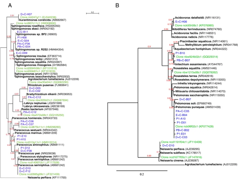
Proteobacterial sequences were classified according to alpha, beta and gamma subdivisions that contained 11, 4 and 17 OTUs representing 43, 38 and 75 different clones, respectively. The phylogenetic trees showed the repartition of the clone sequences in the known proteobacterial diversity (Fig. 2A–C).
The Alphaproteobacteria subdivision was represented by the genus Paracoccus (Rhodobacterales) (34.9%), and the orders Sphingomonadales (44.2%) and Rhizobiales (20.9%). The phylogenetic analysis (Fig. 2A) allowed affiliation to the species Aurantimonas coralicida, A. tumefaciens/
Rhizobium pusense, Bradyrhizobium elkanii, Paracoccus yeeii, Paracoccus haeundaensis, Paracoccus seriniphilus and Paracoccus marinus. Clones af fil-iated to the genus Sphingomonas were related but at less than 98% identity with several species not discriminated by 16S rRNA gene sequencing in-cluding Sphingomonas aquatilis (Fig. 2A). In the Paracoccus clade, two clones (PA+C_C06 and PA+C_C07) corresponded to a yet undescribed species. In Rhizobiales, one clone was related to“Rasbo bacterium”, an undescribed species detected in plasma samples during acute sepsis
[36]. In all cases, a clone detected in metagenomic studies on skin micro-biota was related to the clone detected herein (Fig. 2A).
The Betaproteobacteria subdivision was dominated by Pelomonas, which represented 70% of the clones of this sub-division with mainly the species Pelomonas puraquae. Other clones grouped with the non val-idate species Imtechium assamiensis. Published cutaneous clones grouped together with this non validate species but differed from Aquabacterium fontiphilum and other related species (Fig. 2B). Other clones grouped in Acidovorax spp. and Neisseria spp. but identification to the species level was not accurate for these genera. The most related but undistinguished species were indicated in the tree (Fig. 2B).
The gamma subdivision was mainly represented by the genera Acinetobacter (26.7%), Idiomarina (16%), Alcanivorax (12%) and Pseudomonas (9.4%) and the species Escherichia coli (20%). In the genus Acinetobacter, the clones were distributed among the species A. lwoffii, Acinetobacter baumanii, Acinetobacter junii and A. johnsonii. Seven clones were distributed among pseudomonads (Fig. 2C) mainly in Pseudomonas putida, Pseudomonas stutzeri and Pseudomonas fluorescens groups.
Finally, the phylogeny confirmed that four phylotypes (notedcin
Table 1) corresponded to undescribed species of Alphaproteobacteria. 3.3. Core and pan Gram-negative skin microbiota
The recent studies of molecular ecology have enriched our knowl-edge about bacteria associated with the human skin. The Gram-negative diversity described in these studies and herein is compared inTable 3. The genera Acidovorax, Acinetobacter, Pseudomonas and Stenotrophomonas were isolated from independent donors and from different sampling sites (forearm, forehead, inner elbow and back) in all of the 6 studies or in all studies but one. Therefore, one can hypoth-esize that these bacteria may belong to the permanent core skin micro-biota. If we consider the bacteria detected in at least 50% of the studies
Fig. 1. Bacterial diversity of the skin microbiota according to phylum and type of cell wall structure. Number of colonies and clones assessed the quantitative representation of each phylum. Number of cultured species and number of uncultured OTUs assessed the species diversity in each phylum.
Table 2
Phylum diversity indexes for culture and uncultured approaches. H′: Shannon-Wiener index, D: Simpson index.
Culture approach Uncultured approach
H′ D H′ D Firmicutes 0.323556959 0.005747126 0.366337084 0.0125 Actinobacteria 0.366860144 0.00952381 0.271695022 0.03125 Gram positive 0.091653203 0.003571429 0.354596189 0.008849558 Proteobacteria 0.139582952 0.076923077 0.338360873 0.007936508 Bacteroidetes 0.152653592 0.066666667 0.022681561 0 Gram negative 0.225587993 0.034482759 0.336876674 0.007874016
namely core50skin microbiota, ten genera are highlighted (in bold in
Table 3). Our approach coupling culture and molecular approach detect-ed 100% of the genera forming the current core50 skin microbiota
(Table 3). Moreover, we detected from only 2 subjects 43% of the known diversity of Gram-negative genera in human skin (Table 3). 4. Discussion
Current metagenomics based on NGS explores in depth the bacterial diversity and avoids the bias of cultivability. It has changed our vision of human microbiota but several limitations worth to be underlined. Par-ticularly, NGS presents a lack of sensitivity for the detection of minority OTUs present at less than 106bacteria per sample[37]. The size of
se-quenced DNA fragments varied greatly among techniques and studies rending comparison hard to perform and explaining some discrepancies
[38]. In the case of the gut microbiota, the relative abundance of the phyla Bacteroidetes and Firmicutes depends on the 16S rRNA gene hy-pervariable region analyzed[39]. These discrepancies observed to the phylum level are probably magnified when genera or species are con-sidered. Generally, in NGS studies, the quality of taxonomic affiliation is low and limited to the genus due to the size of generated sequences. Herein, we used classical methods such as Sanger sequencing and
culture with the aim to affiliate to taxonomic species a collection of clones and bacterial colonies isolated from healthy skin. This combined approach was targeted on Proteobacteria that present high diversity in skin microbiota but remain scarcely described to the species-level in previous studies on skin metagenome.
Dekio et al. in 2005 described for thefirst time the presence of Pseu-domonas, Stenotrophomonas, Acidovorax, Bradyrhizobium and Neisseria as proteobacteria inhabiting the skin microbial ecosystem[15]. Then, Gao et al. (2007) and Grice et al. (2008) reported two major molecular analyses confirming that Gram-negative bacteria are common residents and not contaminants from environment or other microbiota[19,22]. They reported also the presence of bacteria usually found in the envi-ronment such as Methylobacterium, Sphingobium, Diaphorobacter, Enhydrobacter, Serratia, Pedomicrobium, Paracoccus, Halomonas and Delftia. Proteobacteria appears qualitatively the more diverse phylum
[22]even if the quantitative predominance of the Gram-positive genera classically detected in culture is confirmed[20]. Recently, Probst et al. characterized another underestimated biodiversity, the Archaea of the human skin microbiota using the cloning of 16S rRNA gene PCR products[40].
Our study, limited to the forearm of two subjects, confirmed that the skin bacterial community is dominated in diversity by Gram-negative
D+C-H07
Clone nbt247g11 (EU540238)
Aurantimonas coralicida (AB682667)
Clone ncd907c11c1 (HM308990) Sphingomonas roseiflava (HG005350) E+C-B11 P1-H08 E+C-G04 PA+C-F06 PB+C-E04 Sphingomonas sp. R2S2 (HM484354) Sphingomonas leidyi (JN591312) Sphingomonas aquatilis (NR024997) E+C-D09 Clone ncd2694h08c1 (KF101818) Clone ncd269d01c1 (KF101818) Sphingomonas pruni (NR113760) Rhizobium pusense (FJ969841) Clone nbw30e04c1 (GQ062290) D+C-D05 E+C-C02 PA+C-C12 Clone nbw305b01c1 (GQ087694)
Sphingomonas insulae (EF363714)
Labrys neptuniae (JQ291599) Labrys okinawensis (AB236169) Rasbo bacterium (AF007948)
PA+C-C12 Clone nbw572e08c1 (GQ105250) Paracoccus homiensis (NR043733) PA+C-C06 PA+C-C07 Clone ncd875b11c1 (HM298280) Paracoccus aestuarii (NR044342) Paracoccus marinus (AB681209)
P1-E10 P1-H10
Clone ncd313g11c1 (HM312214)
Paracoccus aminophilus (AB681111)
P1-D12 D+C-F08
Paracoccus yeei (NR029038) Paracoccus stylophorae (NR117275) Paracoccus seriniphilus (AB681242) Paracoccus seriniphilus (AB681242) Clone ncd1406f10c1 (JF117528)
E+C-D10 D+C-F12
Clone ncd2685g09c1 (JF221459)
Neisseria perflava (KF111700) Agrobacterium tumefaciens (AJ012209)
Bradyrhizobium elkanii (NR036953) Sphingomonas rhizogenes (AY962684) Sphingomonas sp. BF2 (X8905) Sphingomonas abaci (NR042192) Sphingomonas asaccharolytica (NR02932) 0.91 0.92 0.93 0.88 0.65 0.78 0.90 0.87 0.81 0.90 0.79 0.77 0.86 0.92 0.81 0.99 0.8 0.82 0.63 0.95 0.96 0.67
Imtechium assamiensis (AY544767) Aquabecterium fontiphilum (NR044322) Acidovorax facilis (NR1148551) Mitsuaria chitosanitabida (NR114070) P1-B10 Clone nbw924b02c1 (GQ029314) Albidiferax ferrireducens (NR074760) Acidovorax delafieldii (NR116131) Clone nck95h08c1 (KF076980)
Roseateles aquatilis (AM501446) Clone nbw1215e06c1 (GQ078262) Roseateles terrea (NR042616) Roseateles depolymerans (NR115055) Pelomonas saccharophila (NR115050) Inhella inkyongensis (NR114244) Pelomonas aquatica (NR042614) D+C-B07
Pelomonas soli (EF660749) PA+C-C05
E+C-B04
Pelomonas puraquae (AM501439)
P1-A10 P1-D01 PB+C-B02 Clone nck96f02c1 (KF077428) P1-E01 D+C-C03 Clone ncd1191f04c1 (JF110439) D+C-D10
Neisseria perflava (AJ239295) Neisseria subflava (KC178517)
Clone ncd1677f05c1 (JF147816) Neisseria cinerea (AJ239287)
Agrobacterium tumefaciens (AJ012209) PB+C-B07 Methylibium petroleiphilum (NR041768) Acidovorax radicis (NR117776) E+C-H06 Piscinibacter aquaticus (NR114061) 0.61 0.75 0.94 0.74 0.85 0.9 0.87 0.79 0.85 0.78 0.89 0.88 0.89 0.81 0.96 0.75 0.83 0.95 0.7
A
B
Fig. 2. Phylogenetic tree showing the relationship of the 16S rRNA gene sequences. ML phylogenetic tree showing the 16S rRNA gene sequence relationships of the clones obtained in this study (in blue) with cultured and uncultured members of Alphaproteobacteria (A), Betaproteobacteria (B) and Gammaproteobacteria (C). The sequences used to reconstruct this tree were obtained from the GenBank database (accession numbers are indicated in brackets). Sequences from uncultured bacteria obtained from skin human samples were in green. Sequences of strains of validate species and some not validate but published species were in black bold type. Neisseria perflava was used as outgroup for Alphaproteobacteria, Agrobacterium tumefaciens C58 for Betaproteobacteria and Pelomonas aquatica for Gammaproteobacteria. The scale bar indicates substitutions per nucleotide position. Numbers given at the nodes represent bootstrap percentages.
species, which participate to the skin pan-microbiota expanse. In this study, orphan genera represented 50% of the current pan-microbiota. Similarly, they represented 58% of the pan-microbiota in the study of Grice[22]. Moreover, about 70% (with 84.4% of Proteobacteria) of the genera detected by Gao et al. on the forearm were specific to an individ-ual donor[19]. This high variability among subjects and studies could suggest that members of Proteobacteria are temporary environmental contaminants of the human skin. The question if Proteobacteria are tran-sient or resident on human skin is not definitively settled in this pilot study performed on two subjects without longitudinal follow-up and without comparison according environmental conditions. However, the consistency of results between methodologically different studies suggests that very diverse Proteobacteria actually reside in skin microbi-ota and that beside the 10 genera found to belong to the core50
microbiota, Gram-negative diversity participates probably to the inter individual and inter body site variations[41].
By the approaches proposed herein, we detected all the members of the core50 negative skin microbiota and 43% of the
Gram-negative skin pan-microbiota. These data validate our strategy, in spite of the lack of NGS data. It is particularly noteworthy that only a magni-tude of 2 was observed between the 87 skin-associated bacterial genera detected in Human Metagenomic Project (4 cutaneous site and 242 subjects)[2]versus 48 genera detected by cloning and culture from one cutaneous site from 2 subjects in this study.
Since long ago, Gram-positive bacteria (Firmicutes and Actinobacteria) are recognized as the major component of the culturable skin microbiota
[17]. When blood agar culture at 37 °C was used, we detected 92.0% of Gram-positive bacteria. Conversely, the same culture conditions yielded only few Gram-negative bacteria whereas most of those that had been detected by sequencing are considered to be culturable. The richness of the medium and the incubation temperature could be the causes of growth defect. The incubation at 30 °C of rich and poor media with van-comycin that inhibited Gram-positive bacteria led to the growth of R. mucosa, A. tumefaciens, A. altamirensis, A. johnsonii, A. lwoffii and P. psychrotolerans. Growth is observed only for rich media but not onto R2A whereas these strains belong to taxa that generally grow onto poor media such as R2A medium (Bergey's manual). This result sug-gests that the species or strains associated to skin microbiota may pres-ent particular requirempres-ents. Therefore, the existence of particular ecotypes of Proteobacteria specialized in human skin mutualism might be hypothesized.
Sequencing and phylogeny reconstruction detected many Proteobacteria considered to be environmental belonging to the orders Rhizobiales, Sphingomonadales, Burkholderiales, Oceanospirillales, Alteromonadales and Pseudomonadales. However, lifestyle of donors, that are tertiary workers in urban area, excluded regular or recent expo-sure to animals, plants, fresh or marine waters and soil. Most clones as-sumed to belong to environmental species have closest relatives sequences detected previously from the human skin (clones in green inFig. 2) rather than from other ecosystems[4,19,22]. These clones being detected in independent analysis, one can hypothesize again that some of these sequences represent specific ecotypes in the human cutaneous microbiota. The metagenomic study of Mathieu et al. demonstrated functions that clearly illustrate the unique life style of the skin microbial communities and reinforce the hypothesis skin-specific ecotypes[42]. Microbiota description at the species-level completed by strain isolation is a way to study these ecotypes.
Some phenotypic and metabolic traits appear common to several species detected in skin microbiota and could help to define the proteobacterial skins ecotypes. Mainly, several clones correspond to halo-philic genera and species of Gammaproteobacteria isolated in marine envi-ronments: Alcanivorax dieseoli, Alcalinivorax venustensis, Halomonas aquamarina, Halomonas neptunia or Halomonas alkantartica, Marinobacter hydrocarbonoclasticus and Idiomarina loihiensis. Halophily is consistent with the salt rich environment of the skin. Some of halophilic species in Gammaproteobacteria are also hydrocarbonoclastic: Alcanivorax dieseoli, Halomonas alkantartica, and Marinobacter hydrocarbonoclasticus. Such proprieties are also described for the genera Acinetobacter and Pseudomo-nas. Other clones grouped with the non validate species Imtechium assamiensis were described as forming biofilms on polychlorinated biphenyls surfaces[43]. Out of gammaproteobacteria Pseudonocardia chlorethenivorans and Sphingomonas spp. are other alkane-degrading bacteria detected herein[44–46].
The cutaneous microbiota is a major bacterial reservoir involved in opportunistic infections, particularly in health-care associated infections (HAI). Consequently, hand washing and skin antisepsis are now considered the most important interventions to prevent the spread of HAI agents [47]. Among Proteobacteria detected in this study, some species such as Stenotrophomonas, Aeromonas, Pseudomonas and Acinetobacter have been associated with opportunistic infections Acinetobacter baumanii (NR074737)
Clone ncd2626h11c1 (KF101645)
D+C-G08 E+C-G02
Acinetobacter junii (AB860303)
P1-E11 D+C-E05 D+C-D07
Acinetobacter iwoffii (AB859068)
Clone ncm62a09c1 (KF107790) Clone ncd2731d09c1 (JF235906) Acinetobacter johnsonii (KJ788662) D+C-C06 PB+C-C04 P1-E02 PA+C-D08 PB+C-C03 P1-D07 PA+C-F02 Clone nck337f10c1 (EU536675) E+C-H03 P1-G08 E+C-G06b E+C-G06 Clone nck348d04c1 (KF110622) PB+C-B08 PA+C-B04 PA+C-G01 Escherichia coli (HF584706) Clone ncd1962d10c1 (KF100545) P1-D09 Aeromonas salmonicida (X60405) Clone ncd1601a03c1 (JF129813) P1-D08 P1-D10
Haemophilus parainfluenzae (EU083530)
Clone ncd2588c07c1 (JF226961) D+C-D06 Halomonas aquamarina (KJ466006) E+C-D03 Clone nbt36b02 (FJ893830) P1-B04
Halomonas alkantartica (AJ564880)
Clone nbt35a01 (FJ893881)
Halomonas neptunia (AF212202)
D+C-C05 P1-H02 P1-B07 Alcanivorax dieselolei (NR074734) Clone nbt17c02 (FJ893106) E+C-E05 PA+C-E04 E+C-C04 Alcanivorax venustensis (NR025145) Clone nbt249b03 (EU536675) Alcanivorax hongdengensis (NR044499) Pseudomonas monteilii (NR114224) PA+C-D03 Clone nbt246g05 (EU540187)
Marinobacter hydrocarbonoclasticus (AB019148)
E+C-F05
Clone nbw775d06c1 (GQ009349)
Pseudomonas putida (AF094745)
Clone ncd1197f12c1 (JF066709)
P1-A11
Pseudomonas xanthomarina (HF679141) Pseudomonas stutzeri (U26416) Pseudomonas chloritidismutans (AY017341)
Clone nbt235e08 (EU538713)
Pseudomonas teessidea (AM419154) Pseudomonas veronii (NR112075)
P1_E07
Clone nbt84f09 (EU538300) Clone nbt26e06 (EU535522)
Pseudomonas fluorescens (AJ308308)
Clone nbt210f06 (EU534765)
Pseudomonas brenneri (HQ825004) Pseudomonas collierea (AM421016) D+C-A07
Clone nbw231f09c1 (GQ069682)
Pseudomonas fluorescens (FN666563) Pseudomonas trivialis (AJ492831) Pseudomondas azotoformans (NR113600) E+C-B09 Pseudomonas palleroniana (NR029050) Clone ncd132c09c1 (HM250031) D+C-B08 Clone ncd116e02c1 (HM257907) Clone ncd132f11c1 (HM249682) Clone ncd2658a09c1 (JF220920)
Pseudomonas_extremorientalis (AF405328) Pseudomonas tolaasii (NR115613)
E+C-H05 P1-C08
P1-G02
Idiomarina loihiensis (AF288370) Idiomarina sp. (EF409427)
Clone nbt71e11 (EU537245)
Pelomonas aquatica (NR042614) P1-C09 Clone nbt98c02 (EU539095) Stenotrophomonas maltophilia (KJ748603) 0.91 0.84 0.78 0.89 0.84 1 1 0.97 0.86 0.92 1 1 0.84 0.78 0.85 0.67 0.97 0.90 0.99 0.84 0.78 0.95 0.83 0.91 0.87 0.79 0.84 1 0.89 0.99 0.07
C
Fig. 2 (continued).[48–50]. To our knowledge, the opportunistic pathogen Aeromonas has never been detected in the normal skin microbiota so far. Beside these well-known opportunistic pathogens, bacteria assumed to be environ-mental are more and more described in opportunistic infections. For instance, A. altamirensis was described as part of the microbial commu-nity that produces deleterious colonization of Paleolithic paintings in Altamira Cave[51]and since then, it has been mainly described in human infections[52]. For A. tumefaciens, a well-known phytopathogen agent, the population structure showed a genetic sub-population asso-ciated with human beings and involved in infections, clearly apart from environmental and plant-associated strains[53]. Finally, the genus Roseomonas gathers species mainly isolated from environment but R. mucosa and Roseomonas gilardii are frequently described in human infections[54]. By analogy with the main lifestyle observed for most members of Roseomonas spp., the source of human infections caused by R. mucosa is searched into environment and not among endogen microbial community[54,55]. These cases are particularly em-blematic of the need of species-level identification in human microbiota in order to assess infectious risk and prevent opportunistic infections.
The association of known Gram-positive species to disease or healthy states has been previously established, for example, acne and atopic dermatitis correlated with the prevalent colonization of Propionibacterium acnes and Detzia maris, respectively [10,56]. Concerning Gram-negative bacteria, Proteobacteria seem more abun-dant in biopsies of psoriasis lesions and in chronic wounds than in healthy skin, without knowing whether this skin microbiota disequilib-rium is a cause or a consequence of these dermatologic disorders[7,9, 12]. Considering the variations in diversity among subjects and body sites of Proteobacteria in skin pan-microbiota, it is probable that the di-versity by itself is not directly correlated to dermatologic disorders[41]. However, the Human Metagenome Project showed that metabolic var-iations among individuals might indicate pathways for facing personal-ized immune and environmental or behavioral exposures. The wide Gram-negative bacteria repertory and its interindividual variations in skin microbiome might participate to these metabolic variations. 5. Conclusion
Inter-ecosystem comparisons suggest that the human skin commu-nities possess strong capacities for interacting with their environment
Table 3
Data comparison with the 5 main published studies that characterized healthy skin micro-biota by molecular approaches.
This table compiles all the genus of Gram-negative bacteria that have been detected on the skin. Data are obtained from 5 published studies and this study. Studies were based on molecular methods in combination or not with culture analysis. D for Dekio et al. [15], G for Gao et al. [19], Gr for Grice et al. [22], H for Human Microbiome Project Consortium [2], Z for Zeeuwen et al. [12], and C for this study. The color intensity is function of the fre-quency of detection. The core50
microbiota corresponded to genera found in at least 50% of the published studies. Genera belonged to the core50
are in bold face. % of Gram negative skin pan-microbiota is the proportion of genera identified in each study among total num-ber of known genera indentified on the skin in the 6 studies, namely skin pan-microbiota (62 genera). % Gram-negative orphan is the proportion of orphan genera identified in each study among the skin pan-microbiota. % of Gram negative genera of the core50
microbiota is the proportion of genera belonging to the core50
in each study among the current core50
[42]. Beyond their large diversity, skin-associated Proteobacteria slightly differed from their environmental counterpart. The genetic relatedness between skin and environmental ecotypes in the same proteobacterial species might act as shuttles linking environmental and human micro-bial communities. In this context, virulence or resistance genes ex-change could be of great concern in the emergence of multiresistant pathogenic bacteria.
The One Health concept recognizes that the health of humans is con-nected to the health of animals and environment. Microbiota and partic-ularly skin microbiota probably have a pivotal role in the continuity between human-associated bacteria and other microbial communities in animals, plants and environments. Therefore, microbiota relation-ships and overlaps with other communities need to be explored in the One Health approach beside other means of pathogenic exchanges such as vectors and direct transmission of pathogens.
Acknowledgments
This work was supported by the CERPER, Pierre Fabre Dermo-Cosmétique, the French Research Ministry and ADEREMPHA associa-tion. The authors are grateful to Pr. Devine from Leeds Dental Institute for critical reading of this manuscript.
References
[1] NIH HMP Working Group, J. Peterson, S. Garges, M. Giovanni, P. McInnes, L. Wang, et al., The NIH Human Microbiome Project, Genome Res. 19 (2009) 2317–2323. [2] Human Microbium Project Consortium, Structure, function and diversity of the
healthy human microbiome, Nature 486 (2012) 207-4.
[3] M.E. McBride, W.C. Duncan, J.M. Knox, The environment and the microbial ecology of human skin, Appl. Environ. Microbiol. 33 (1977) 603–608.
[4] N. Fierer, M. Hamady, C.L. Lauber, R. Knight, The influence of sex, handedness, and washing on the diversity of hand surface bacteria, Proc. Natl. Acad. Sci. 105 (2008) 17994–17999.
[5] E.K. Costello, C.L. Lauber, M. Hamady, N. Fierer, J.I. Gordon, R. Knight, Bacterial com-munity variation in human body habitats across space and time, Science 326 (2009) 1694–1697.
[6] E.A. Grice, H.H. Kong, S. Conlan, C.B. Deming, J. Davis, A.C. Young, et al., Topograph-ical and temporal diversity of the human skin microbiome, Science 324 (2009) 1190–1192.
[7] A. Han, J.M. Zenilman, J.H. Melendez, M.E. Shirtliff, A. Agostinho, G. James, et al., The importance of a multifaceted approach to characterizing the microbialflora of chronic wounds, Wound Repair Regen. 19 (2011) 532–541.
[8] T. Staudinger, A. Pipal, B. Redl, Molecular analysis of the prevalent microbiota of human male and female forehead skin compared to forearm skin and the influence of make-up, J. Appl. Microbiol. 110 (2011) 1381–1389.
[9] A. Fahlén, L. Engstrand, B.S. Baker, A. Powles, L. Fry, Comparison of bacterial micro-biota in skin biopsies from normal and psoriatic skin, Arch. Dermatol. Res. 304 (2012) 15–22.
[10] H.H. Kong, J. Oh, C. Deming, S. Conlan, E.A. Grice, M.A. Beatson, et al., Temporal shifts in the skin microbiome associated with diseaseflares and treatment in children with atopic dermatitis, Genome Res. 22 (2012) 850–859.
[11] L.K. Ursell, J.C. Clemente, J.R. Rideout, D. Gevers, J.G. Caporaso, R. Knight, The inter-personal and intrainter-personal diversity of human-associated microbiota in key body sites, J. Allergy Clin. Immunol. 129 (2012) 1204–1208.
[12] P.L. Zeeuwen, J. Boekhorst, E.H. van den Bogaard, H.D. de Koning, P.M. van de Kerkhof, D.M. Saulnier, et al., Microbiome dynamics of human epidermis following skin barrier disruption, Genome Biol. 13 (2012) R101.
[13] G. Greub, Culturomics: a new approach to study the human microbiome, Clin. Microbiol. Infect. Off. Publ. Eur. Soc. Clin. Microbiol. Infect. Dis. 18 (2012) 1157–1159.
[14]O.A. Alexeyev, Bacterial landscape of human skin: seeing the forest for the trees, Exp. Dermatol. 22 (2013) 443–446.
[15] I. Dekio, H. Hayashi, M. Sakamoto, M. Kitahara, T. Nishikawa, M. Suematsu, et al., De-tection of potentially novel bacterial components of the human skin microbiota using culture-independent molecular profiling, J. Med. Microbiol. 54 (2005) 1231–1238.
[16] A. Mathieu, T.M. Vogel, P. Simonet, The future of skin metagenomics, Res. Microbiol. 165 (2014) 69–76.
[17]J.J. Leyden, K.J. McGinley, K.M. Nordstrom, G.F. Webster, Skin microflora, J. Invest. Dermatol. 88 (3) (1987) 65s–72s.
[18] K. Chiller, B.A. Selkin, G.J. Murakawa, Skin microflora and bacterial infections of the skin, J. Investig. Dermatol. 6 (2001) 170–174.
[19] Z. Gao, C. Tseng, Z. Pei, M.J. Blaser, Molecular analysis of human forearm superficial skin bacterial biota, Proc. Natl. Acad. Sci. 104 (2007) 2927–2932.
[20] Z. Gao, G.I. Perez-Perez, Y. Chen, M.J. Blaser, Quantitation of major human cutaneous bacterial and fungal populations, J. Clin. Microbiol. 48 (2010) 3575–3581.
[21] I. Dekio, M. Sakamoto, H. Hayashi, M. Amagai, M. Suematsu, Y. Benno, Characteriza-tion of skin microbiota in patients with atopic dermatitis and in normal subjects using 16S rRNA gene-based comprehensive analysis, J. Med. Microbiol. 56 (2007) 1675–1683.
[22] E.A. Grice, H.H. Kong, G. Renaud, A.C. Young, G.G. Bouffard, R.W. Blakesley, et al., A diversity profile of the human skin microbiota, Genome Res. 18 (2008) 1043–1050. [23] J. Fleurette, M.J. Transy, Estimation of the activity of an antiseptic agent on skin mi-crobialflora under various conditions, Ann. Biol. Clin. (Paris) 34 (1976) 203–210. [24]N. Khammar, L. Malhautier, V. Degrange, R. Lensi, J.-J. Godon, J.-L. Fanlo, Link
be-tween spatial structure of microbial communities and degradation of a complex mixture of volatile organic compounds in peat biofilters, J. Appl. Microbiol. 98 (2005) 476–490.
[25] E.A. Trafny, K. Kozłowska, M. Szpakowska, A novel multiplex PCR assay for the de-tection of Salmonella enterica serovar Enteritidis in human faeces, Lett. Appl. Microbiol. 43 (2006) 673–679.
[26] J.U. Jeung, S.K. Cho, K.S. Shim, S.H. Ok, D.S. Lim, J.S. Shin, Construction of two pGEM 7Zf(+) phagemid T-tail vectors using AhdI-restriction endonuclease sites for direct cloning of PCR products, Plasmid 48 (2002) 160–163.
[27] F. Martineau, F.J. Picard, P.H. Roy, M. Ouellette, M.G. Bergeron, Species-specific and ubiquitous-DNA-based assays for rapid identification of Staphylococcus aureus, J. Clin. Microbiol. 36 (1998) 618–623.
[28] F. Martineau, F.J. Picard, D. Ke, S. Paradis, P.H. Roy, M. Ouellette, et al., Development of a PCR assay for identification of staphylococci at genus and species levels, J. Clin. Microbiol. 39 (2001) 2541–2547.
[29]E. Stackebrandt, E. Brambilla, S. Cousin, W. Dirks, R. Pukall, Culture-independent analysis of bacterial species from an anaerobic mat from Lake Fryxell, Antarctica: prokaryotic diversity revisited, Cell. Mol. Biol. 50 (2004) 517–524.
[30]J.D. Thompson, D.G. Higgins, T.J. Gibson, CLUSTAL W: improving the sensitivity of progressive multiple sequence alignment through sequence weighting, position-specific gap penalties and weight matrix choice, Nucleic Acids Res. 22 (1994) 4673–4680.
[31] D. Posada, K.A. Crandall, MODELTEST: testing the model of DNA substitution, Bioinforma. Oxf. Engl. 14 (1998) 817–818.
[32] S. Guindon, O. Gascuel, A simple, fast, and accurate algorithm to estimate large phy-logenies by maximum likelihood, Syst. Biol. 52 (2003) 696–704.
[33]T.C.J. Hill, K.A. Walsh, J.A. Harris, B.F. Moffett, Using ecological diversity measures with bacterial communities, FEMS Microbiol. Ecol. 43 (2003) 1–11.
[34] G.P. Gafan, V.S. Lucas, G.J. Roberts, A. Petrie, M. Wilson, D.A. Spratt, Statistical analy-ses of complex denaturing gradient gel electrophoresis profiles, J. Clin. Microbiol. 43 (2005) 3971–3978.
[35] D.J. Reasoner, J.C. Blannon, E.E. Geldreich, Rapid seven-hour fecal coliform test, Appl. Environ. Microbiol. 38 (1979) 229–236.
[36] G. Blomqvist, L. Wesslén, C. Påhlson, E. Hjelm, B. Pettersson, T. Nikkilä, et al., Phylo-genetic placement and characterization of a new alpha-2 proteobacterium isolated from a patient with sepsis, J. Clin. Microbiol. 35 (1997) 1988–1995.
[37] J.-C. Lagier, F. Armougom, M. Million, P. Hugon, I. Pagnier, C. Robert, et al., Microbial culturomics: paradigm shift in the human gut microbiome study, Clin. Miicrobiol. Infect. 18 (2012) 1185–1193.
[38] L. Liu, Y. Li, S. Li, N. Hu, Y. He, R. Pong, et al., Comparison of next-generation sequenc-ing systems, J. Biomed. Biotechnol. 2012 (2012) 251364.
[39] M.J. Claesson, Q. Wang, O. O'Sullivan, R. Greene-Diniz, J.R. Cole, R.P. Ross, et al., Com-parison of two next-generation sequencing technologies for resolving highly com-plex microbiota composition using tandem variable 16S rRNA gene regions, Nucleic Acids Res. 38 (2010), e200.
[40] A.J. Probst, A.K. Auerbach, C. Moissl-Eichinger, Archaea on human skin, PLoS One 8 (2013), e65388.
[41] E.A. Grice, J.A. Segre, The skin microbiome, Nat. Rev. Microbiol. 9 (2011) 244–253. [42] A. Mathieu, Delmont TO, T.M. Vogel, P. Robe, R. Nalin, P. Simonet, Life on human
sur-faces: skin metagenomics, PLoS One 8 (2013), e65288.
[43] A.J. Macedo, K.N. Timmis, W.-R. Abraham, Widespread capacity to metabolize polychlorinated biphenyls by diverse microbial communities in soils with no signif-icant exposure to PCB contamination, Environ. Microbiol. 9 (2007) 1890–1897. [44] P. Kämpfer, U. Kohlweyer, B. Thiemer, J.R. Andreesen, Pseudonocardia
tetrahydrofuranoxydans sp. nov, Int. J. Syst. Evol. Microbiol. 56 (2006) 1535–1538. [45]H. Zhang, Y.K. Lee, W. Zhang, H.K. Lee, Culturable actinobacteria from the marine sponge Hymeniacidon perleve: isolation and phylogenetic diversity by 16S rRNA gene-RFLP analysis, Antonie Van Leeuwenhoek 90 (2006) 159–169.
[46]A.E.F. Little, C.R. Currie, Symbiotic complexity: discovery of afifth symbiont in the attine ant–microbe symbiosis, Biol. Lett. 3 (2007) 501–504.
[47] A.M. Milstone, C.L. Passaretti, T.M. Perl, Chlorhexidine: expanding the armamentar-ium for infection control and prevention, Clin. Infect. Dis. 46 (2008) 274–281. [48] W.Y. Teo, M.Y. Chan, C.M. Lam, C.Y. Chong, Skin manifestation of Stenotrophomonas
maltophilia infection—a case report and review article, Ann. Acad. Med. 35 (2006) 897–900.
[49] K.R. Pardesi, S.P. Yavankar, B.A. Chopade, Plasmid distribution & antimicrobial sus-ceptibility patterns of Acinetobacter genospecies from healthy skin of a tribal popu-lation in western India, Indian J. Med. Res. 125 (2007) 79–88.
[50]S.P. Yavankar, K.R. Pardesi, B.A. Chopade, Species distribution and physiological characterization of Acinetobacter genospecies from healthy human skin of tribal population in India, Indian J. Med. Microbiol. 25 (2007) 336–345.
[51] V. Jurado, J.M. Gonzalez, L. Laiz, C. Saiz-Jimenez, Aurantimonas altamirensis sp. nov., a member of the order Rhizobiales isolated from Altamira Cave, Int. J. Syst. Evol. Microbiol. 56 (2006) 2583–2585.
[52] M.-L. Luong, S. Békal, D.C. Vinh, D. Lauzon, V. Leung, G.N. Al-Rawahi, et al., First re-port of isolation and characterization of Aurantimonas altamirensis from clinical samples, J. Clin. Microbiol. 46 (2008) 2435–2437.
[53]F. Aujoulat, E. Jumas-Bilak, A. Masnou, F. Sallé, D. Faure, C. Segonds, et al., Multilocus sequence-based analysis delineates a clonal population of Agrobacterium (Rhizobium) radiobacter (Agrobacterium tumefaciens) of human origin, J. Bacteriol. 193 (2011) 2608–2618.
[54]J.D. Bard, J.G. Deville, P.H. Summanen, M.A. Lewinski, Roseomonas mucosa isolated from bloodstream of pediatric patient, J. Clin. Microbiol. 48 (2010) 3027–3029.
[55] S.-F. Tsai, C.-H. Chen, K.-H. Shu, M.-J. Wu, Peritonitis caused by Roseomonas in a pa-tient undergoing automated peritoneal dialysis: case report and literature review, Intern. Med. 51 (2012) 1721–1724.
[56] C. Dessinioti, A.D. Katsambas, The role of Propionibacterium acnes in acne pathogenesis: facts and controversies, Clin. Dermatol. 28 (2010) 2–7.