HAL Id: hal-01353372
https://hal.inria.fr/hal-01353372
Submitted on 27 Nov 2018
HAL is a multi-disciplinary open access
archive for the deposit and dissemination of
sci-entific research documents, whether they are
pub-lished or not. The documents may come from
teaching and research institutions in France or
abroad, or from public or private research centers.
L’archive ouverte pluridisciplinaire HAL, est
destinée au dépôt et à la diffusion de documents
scientifiques de niveau recherche, publiés ou non,
émanant des établissements d’enseignement et de
recherche français ou étrangers, des laboratoires
publics ou privés.
Post–Myocardial Infarction Ventricular Tachycardia
Ablation During Long-Term Follow-Up
Seigo Yamashita, Hubert Cochet, Frédéric Sacher, Saagar Mahida, Benjamin
Berte, Darren Hooks, Jean-Marc Sellal, Nora Al Jefairi, Antonio Frontera,
Yuki Komatsu, et al.
To cite this version:
Seigo Yamashita, Hubert Cochet, Frédéric Sacher, Saagar Mahida, Benjamin Berte, et al.. Impact of
New Technologies and Approaches for Post–Myocardial Infarction Ventricular Tachycardia Ablation
During Long-Term Follow-Up. Circulation. Arrhythmia and electrophysiology, Lippincott Williams
& Wilkins, 2016, 9 (7), �10.1161/CIRCEP.116.003901�. �hal-01353372�
1
C
atheter ablation is an effective technique for the management
of post–myocardial infarction ventricular tachycardia
(post-MI VT).
1–3The role of substrate-based VT ablation techniques
is expanding.
4–7In this context, elimination of local abnormal
ventricular activities (LAVA) has emerged as an important end
point for VT ablation.
7During the past decade, the emergence
of innovations, such as three-dimensional electroanatomic
map-ping (3D-EAM),
8multipolar catheters for high-density
map-ping,
7and real-time integration of structural VT substrate from
imaging, have enhanced our ability to identify ablation targets.
9However, the impact of these technological innovations on
abla-tion outcome has not been systematically studied.
1,10–14The
pur-pose of this study is to identify the impact of these innovations
on ablation outcome for post-MI VT.
Methods
Study Population
From January 2008 to November 2013, consecutive patients under-going catheter ablation for post-MI VT were enrolled. Inclusion cri-teria were history of MI and drug-resistant sustained VT. Exclusion
© 2016 American Heart Association, Inc.
Circ Arrhythm Electrophysiol is available at http://circep.ahajournals.org DOI: 10.1161/CIRCEP.116.003901
Background—During the past years, many innovations have been introduced to facilitate catheter ablation of post–myocardial
infarction ventricular tachycardia. However, the predictors of outcome after ablation were not thoroughly studied.
Methods and Results—From 2009 to 2013, consecutive patients referred for post–myocardial infarction ventricular
tachycardia ablation were included. The end point of the procedure was complete elimination of local abnormal ventricular
activities (LAVA) and ventricular tachycardia (VT) noninducibility. The predictors of outcome with primary end point of
VT recurrence were assessed. A total of 125 patients were included (age: 64±11 years; 7 women) for 142 procedures. The
left ventricle was accessed via transseptal, retrograde aortic, and epicardial approaches in 87%, 33%, and 37% of patients,
respectively. Three-dimensional electroanatomical mapping system was used in 70%, multipolar catheter in 51%, and
real-time image integration in 38% (from magnetic resonance imaging in 39% and multidetector computed tomography in
93%) of patients. Before ablation, VT was inducible in 75%, and endocardial/epicardial LAVA were present in 88%/75%.
After ablation, complete LAVA elimination was achieved in 60%, and VT noninducibility in 83%. During a median
follow-up of 850 days (interquartile range, 439–1707), VT recurrence was observed in 36%. Multivariable analysis
identified 3 independent outcome predictors: the ability to achieve complete LAVA elimination (R
2=0.29; P<0.0001; risk
ratio=0.52 [0.38–0.70]), the use of real-time image integration (R
2=0.21; P=0.0006; risk ratio=0.49 [0.33–0.74]), and the
use of multipolar catheters (R
2=0.08; P=0.05; risk ratio=0.75 [0.56–1.00]).
Conclusions—Achievement of complete LAVA elimination and use of scar integration from imaging and multipolar
catheters to focus high-density mapping are independent predictors of VT-free survival after catheter ablation for
post–myocardial infarction ventricular tachycardia. (Circ Arrhythm Electrophysiol. 2016;9:e003901. DOI: 10.1161/
CIRCEP.116.003901.)
Key Words: catheter ablation
◼ heart failure ◼ infarction ◼ myocardial infarction ◼ ventricular tachycardia
Received January 12, 2016; accepted June 14, 2016.
From the Department of Cardiac Electrophysiology (S.Y., F.S., S.M., B.B., D.H., J.-M.S., N.A.J., A.F., Y.K., H.S.L., S.A., A.D., N.D., M.H., M.H., P.J.) and Department of Cardiovascular Imaging, (H.C., F.L., M.M.), Hôpital Cardiologique du Haut-Lévêque–CHU de Bordeaux, Pessac, France; IHU LIRYC ANR-10-IAHU-04, Equipex MUSIC ANR-11-EQPX-0030, Université de Bordeaux-Inserm U1045, Pessac, France (H.C., F.S., A.D., N.D., F.L., M. Hocini, M. Haïssaguerre, M.M., P.J.); and Inria, Asclepios Team, Sophia Antipolis, France (M.S.).
Correspondence to Seigo Yamashita, MD, PhD, Department of Cardiac Electrophysiology, Hôpital Cardiologique du Haut-Lévêque, Ave de Magellan, Bordeaux-Pessac, 33604, France. E-mail seigoy722@yahoo.co.jp
Impact of New Technologies and Approaches for
Post–Myocardial Infarction Ventricular Tachycardia
Ablation During Long-Term Follow-Up
Seigo Yamashita, MD, PhD; Hubert Cochet, MD, PhD; Frédéric Sacher, MD, PhD;
Saagar Mahida, MBChB, PhD; Benjamin Berte, MD; Darren Hooks, MD, PhD;
Jean-Marc Sellal, MD; Nora Al Jefairi, MD; Antonio Frontera, MD; Yuki Komatsu, MD;
Han S. Lim, MBBS, PhD; Sana Amraoui, MD; Arnaud Denis, MD; Nicolas Derval, MD;
Maxime Sermesant, PhD; François Laurent, MD; Mélèze Hocini, MD;
Michel Haïssaguerre, MD, PhD; Michel Montaudon, MD, PhD; Pierre Jaïs, MD, PhD
criteria were presence of intracardiac thrombus, New York Heart Association class IV heart failure, and cardiac surgery within the past 2 months (unless VT was incessant). VT storm was defined as ≥3 VT episodes in 24 hours.
All patients underwent catheter ablation with the same end point, that is, complete LAVA elimination during sinus rhythm (SR)7 and
noninducibility of any VT, but with various methods according to the evolution of EP technology during the course of the study. The use of 3D-EAM, multipolar catheters, and real-time image integra-tion was solely based on the availability of the technology and was independent of patient characteristics (except for contraindications to contrast-enhanced multidetector computed tomography [MDCT] and magnetic resonance imaging [MRI]). This study was approved by the Institutional Review Board, and all patients gave informed consent.
Electrophysiological Mapping Study
Procedures were performed under conscious sedation. A 5F, steer-able, quadripolar/decapolar catheter (Xtrem; Sorin, France, or Dynamic; BostonScientific, Inc, Cambridge, MA) was placed in the right ventricular apex or coronary sinus. The left ventricle was ac-cessed by a transseptal (BRK Needle, Agilis Sheath; St Jude Medical, St Paul, MN) or retrograde aortic approach. Epicardial mapping (Tuohy Needle, Agilis Sheath; St Jude Medical) was performed15 in
cases with suspected epicardial VT origin (from 12-lead ECG and/or absence of endocardial LAVA and/or failure of endocardial ablation). Contraindications to epicardial access included previous cardiac sur-gery and pericardial adhesions. After left ventricle access, a 50 U/kg heparin bolus was administered (activated clotting time maintained at >250 seconds). Twelve-lead ECG and intracardiac electrograms were recorded continuously (LabSystem Pro; Bard Electrophysiology, MA, or Siemens Axiom Sensis XP; Siemens, Munich, Germany).
Mapping was performed using either a 3.5-mm open-irrigated catheter (NaviStar ThermoCool; Biosense Webster, Diamond Bar, CA) and/or a multipolar high-density mapping catheter (PentaRay; 2-6-2 mm interelectrode spacing, 1 mm electrodes; Biosense Webster). Peak-to-peak amplitudes of 0.5 to 1.5 mV and <0.5 mV were used to define the low-voltage zone and the dense scar zone,
WHAT IS KNOWN
•
Elimination of LAVA has emerged as an
im-portant end point for VT ablation.
•
Many technical innovations for VT ablation
were introduced over the past years, including
3D-mapping system, image integration, and
multipolar mapping catheter. However, the
im-pact of these novel techniques on the clinical
outcome of post-MI VT ablation has not been
thoroughly investigated.
WHAT THE STUDY ADDS
•
The ability to achieve complete LAVA
elimi-nation, the use of image integration, and
high-density mapping with multipolar catheters are
independent predictors of VT-free survival
af-ter catheaf-ter ablation for post-MI VT.
•
Aiming for complete LAVA elimination
us-ing real-time image integration and multipolar
catheter is a feasible and useful approach in
patients with post-MI VT.
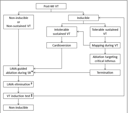
Figure 1. Flow chart for ventricular tachycardia (VT) ablation in post–myocardial infarction (post-MI) patients. *In case with no local
abnor-mal ventricular activity (LAVA) during sinus rhythm (SR), ablation was guided by pace mapping. †LAVA-guided ablation was limited in case LAVA located near coronary arteries and phrenic nerve on the epicardium or persisted after extensive ablation. ‡In case of intolerable VTs with cardioversion >2 times during the procedure, VT inducibility was not tested.
respectively. When a 3D-EAM system was used (CARTO3; Biosense Webster, or NavX; St Jude Medical), mapping was performed during SR to create a voltage map and annotate LAVA. For hemodynami-cally tolerated VT, activation mapping was performed during VT.
For image integration, structural substrate was derived from MDCT or MRI. Images were processed using dedicated software (MUSIC; Liryc-Université de Bordeaux/Inria-Sophia Antipolis, France) to render patient-specific meshes of the cardiac chambers, epicardium, coronary sinus, and coronary arteries and left phrenic nerve in case of epicardial approach.16 The structural substrate was
segmented on imaging as areas of late gadolinium enhancement on MRI and/or areas of wall thinning <5 mm on MDCT.9 The imaging
model was registered to the mapping geometry using landmark-based registration and field scaling when using NavX system or landmark-based registration and automatic surface registration when using CARTO system.
VT Inducibility and Ablation
A flow chart illustrating procedural management is provided in Figure 1. VT inducibility was assessed at the beginning of the pro-cedure (600 and 400 ms drive trains, ≤3 extrastimuli decremented to 200 ms from the right ventricular apex). If hemodynamically stable VT was induced, conventional activation and entrainment was per-formed.17 For noninducible or poorly tolerated VT, mapping was
un-dertaken to identify LAVA during SR. LAVA was defined as sharp high-frequency ventricular potentials, distinct from the far-field ven-tricular electrogram occurring anytime during or after the far-field ventricular electrogram in SR.7 Radiofrequency current was
deliv-ered with an irrigated catheter (25–50 W endocardial and 25–35 W
Table 1. Patient Characteristics
Baseline clinical characteristics
Age, y 64±11 Male sex 135 (95) Hypertension 91 (64) Diabetes mellitus 25 (18) Dyslipidemia 121 (85) Smoking 76 (53) Multivessel disease 61 (43) Previous CABG 31 (22) Previous PCI 75 (53) ICD 116 (82)
Left ventricular ejection fraction, % 33±10 Amiodarone 94 (66) β-Blocker 134 (94) Ventricular arrhythmia storm 69 (49) Redo procedure 31 (22) Procedural techniques
Transseptal approach 124 (87) Retrograde aortic approach 48 (34) Epicardial access 52 (37) Multipolar catheter 73 (51)
3D-EAM 99 (70)
Real-time image integration 54 (38) Integration of MDCT data* 50 (93) Integration of MRI data* 21 (39) Baseline VT inducibility
Inducible VT 106 (75) Clinical VT induced 86 (61) Nonclinical VT induced 67 (47) Mean VT cycle length, ms 406±103 Shortest VT cycle length, ms 370±107 DCC for intolerable VT/VF 29 (20) Mapping characteristics
Number of endocardial mapping points† 370 (228–747) Low-voltage endocardial area, cm2† 68 (32–97)
Dense scar endocardial area, cm2† 26 (12–54)
Presence of LAVA on endocardium 125 (88) Number of epicardial mapping points† 432 (328–758) Low-voltage epicardial area, cm2† 71 (41–85)
Dense scar epicardial area, cm2† 33 (16–45)
Presence of LAVA on epicardium‡ 39 (75) Ablation and procedural end points
Total RF time 32 (20–50) (Continued )
Epicardial ablation 29 (20) Epicardial RF time§ 5 (2–13) Procedure time 250±82 Inducibility tested after ablation 112 (79) Noninducibility of VT‖ 93 (83) Complete LAVA elimination¶ 79 (60) Procedural complications 9 (6) Follow-up Follow-up duration, d 850 (439–1707) VT recurrence 53 (37) All-cause death 23 (16) Cardiovascular death 15 (11) Sudden death attributed to arrhythmia
recurrence 5 (4)
Follow-up duration until VT recurrence, d 119 (55–327) Follow-up duration until death, d 869 (78–1594)
3D-EAM indicates 3-dimensional electroanatomical mapping; CABG, coronary artery bypass graft; DCC, direct current cardioversion; ICD, implantable cardioverter-defibrillator; LAVA, local abnormal ventricular activity; MDCT, multidetector computed tomography; MRI, magnetic resonance imaging; PCI, percutaneous coronary intervention; RF, radiofrequency; VF, ventricular fibrillation; and VT, ventricular tachycardia.
*Among patients with image integration. †Applies to patients with 3D-EAM. ‡Among patients with epicardial access. §Among patients with epicardial ablation ‖Among patients with inducibility testing. ¶Among patients with LAVA.
Table 1. Continued
epicardial). After VT termination, further ablation was performed in SR to eliminate LAVA. The procedural end point was complete LAVA elimination and noninducibility of any VT.
Follow-Up
Patients were followed up with sequential implantable cardio-verter-defibrillator (ICD) interrogations (or Holter monitoring in patients without ICD) at 1, 3, 6, and 12 months for the first year and subsequently every 6 months. The primary end point was VT recurrence. Qualifying arrhythmias included any VT de-tected by ICDs, 12-lead ECGs, Holter monitors, or rhythm strips, regardless of morphology or rate. Death during follow-up was categorized as either cardiovascular or noncardiovascular. Among cardiovascular deaths, sudden deaths attributed to ventricular ar-rhythmia were recorded.
Statistical Analysis
Shapiro–Wilk and D’Agostino tests were used to assess whether quantitative data conformed to the normal distribution. Log transfor-mation was applied in case of non-normal distribution. Continuous variables are expressed as mean±SD (normally distribution) and median (interquartile range; non-normal distribution). Categorical variables are expressed as fraction (%). Continuous variables were compared using parametric (unpaired Student t test) or nonparametric tests (Mann–Whitney) depending on data normality. Categorical vari-ables were compared using χ2 tests. Relationships between variables
were assessed using Pearson or Spearman correlation coefficients (R). Factors associated with the 2 procedural end points (LAVA elimina-tion and VT noninducibility) were analyzed using univariable logistic regression. Univariable Cox proportional hazards regression analysis was used to identify predictors of VT recurrence, with candidate vari-ables including all patients’ baseline characteristics, procedural tech-niques, procedural findings, and procedural end points. Bonferroni correction was applied to account for multiple testing. Proportional hazards assumptions were verified by plotting Schoenfeld residuals
supplemented by testing for nonzero slopes. All candidate variables associated with a P value <0.05 on univariable analysis were consid-ered in an automated hierarchical forward multivariable Cox regres-sion model, except for mapping characteristics, because these were only available in case 3D-EAM was used. Assuming a VT recurrence rate of 40%,7 this study was populated to include a maximum of 5
variables in multivariate analyses. For each variable showing predic-tive of VT recurrence on multivariable analysis, VT-free survival rate was plotted against time since ablation in patients with and without the variable, according to the Kaplan–Meier method. All statistical tests were 2 tailed. A P value <0.05 was considered to indicate sta-tistical significance. Analyses were performed using NCSS 8 (NCSS Statistical Software, Kaysville, UT).
Results
Population
During the study period, 140 patients met the inclusion
crite-ria. Fifteen patients were excluded (8 patients for intracardiac
thrombus, 2 patients for insufficient delay post myocardial
infarction [VT spontaneously resolved in both], and 5 patients
declined consent). Therefore, the study population consisted
of 125 patients (age: 64±11 years; 7 women; 100 [80%] ICD)
who underwent 142 VT ablation procedures. Baseline
charac-teristics are summarized in Table 1.
Mapping and Ablation Procedure
Transseptal, retrograde aortic, and epicardial approaches
were performed in 124 (87%), 48 (34%), and 52 cases (37%),
respectively. Criteria for epicardial access were met in 64
cases (45%), in whom pericardial access was not possible
in 12 cases (3 cases because of pericardial adhesion; 7 cases
Figure 2. High-density mapping and real-time image integration. A case of post–myocardial infarction ventricular tachycardia (post-MI
VT; 60 y; man) is shown. A, Voltage map demonstrates large scar on anterior left ventricular (LV) wall with local abnormal ventricular
activity (LAVA) as recorded by a multipolar catheter. B, Image integration from multidetector computed tomography provides anatomic
details including the course of coronary arteries (red) and coronary sinus (CS) branches (blue). A decapolar catheter (green) is placed in distal CS to monitor image registration. Anatomic substrate demonstrated as areas of moderate and severe wall thinning (WT; light and dark brown surfaces). An anatomic isthmus of moderate WT is visible (green arrows), hosting most LAVAs, many of which show normal voltage (>1.5 mV). LAD indicates left anterior descending artery; and LCx, left circumflex artery.
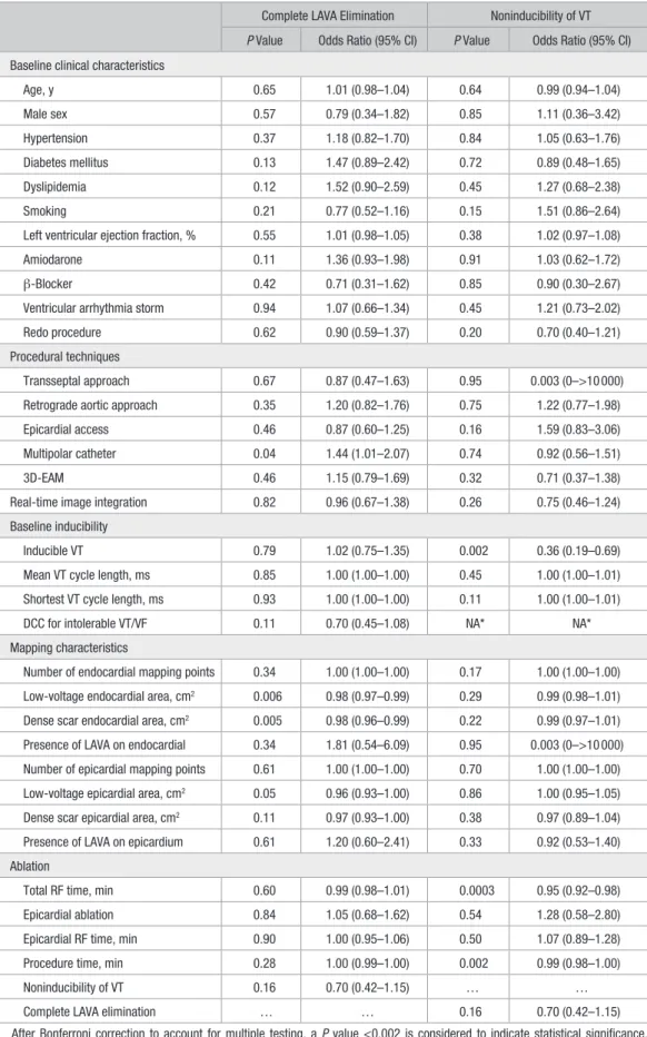
Table 2. Predictors of Procedural End Points
Complete LAVA Elimination Noninducibility of VT P Value Odds Ratio (95% CI) P Value Odds Ratio (95% CI) Baseline clinical characteristics
Age, y 0.65 1.01 (0.98–1.04) 0.64 0.99 (0.94–1.04) Male sex 0.57 0.79 (0.34–1.82) 0.85 1.11 (0.36–3.42) Hypertension 0.37 1.18 (0.82–1.70) 0.84 1.05 (0.63–1.76) Diabetes mellitus 0.13 1.47 (0.89–2.42) 0.72 0.89 (0.48–1.65) Dyslipidemia 0.12 1.52 (0.90–2.59) 0.45 1.27 (0.68–2.38) Smoking 0.21 0.77 (0.52–1.16) 0.15 1.51 (0.86–2.64) Left ventricular ejection fraction, % 0.55 1.01 (0.98–1.05) 0.38 1.02 (0.97–1.08) Amiodarone 0.11 1.36 (0.93–1.98) 0.91 1.03 (0.62–1.72) β-Blocker 0.42 0.71 (0.31–1.62) 0.85 0.90 (0.30–2.67) Ventricular arrhythmia storm 0.94 1.07 (0.66–1.34) 0.45 1.21 (0.73–2.02) Redo procedure 0.62 0.90 (0.59–1.37) 0.20 0.70 (0.40–1.21) Procedural techniques
Transseptal approach 0.67 0.87 (0.47–1.63) 0.95 0.003 (0–>10 000) Retrograde aortic approach 0.35 1.20 (0.82–1.76) 0.75 1.22 (0.77–1.98) Epicardial access 0.46 0.87 (0.60–1.25) 0.16 1.59 (0.83–3.06) Multipolar catheter 0.04 1.44 (1.01–2.07) 0.74 0.92 (0.56–1.51) 3D-EAM 0.46 1.15 (0.79–1.69) 0.32 0.71 (0.37–1.38) Real-time image integration 0.82 0.96 (0.67–1.38) 0.26 0.75 (0.46–1.24) Baseline inducibility
Inducible VT 0.79 1.02 (0.75–1.35) 0.002 0.36 (0.19–0.69) Mean VT cycle length, ms 0.85 1.00 (1.00–1.00) 0.45 1.00 (1.00–1.01) Shortest VT cycle length, ms 0.93 1.00 (1.00–1.00) 0.11 1.00 (1.00–1.01) DCC for intolerable VT/VF 0.11 0.70 (0.45–1.08) NA* NA* Mapping characteristics
Number of endocardial mapping points 0.34 1.00 (1.00–1.00) 0.17 1.00 (1.00–1.00) Low-voltage endocardial area, cm2 0.006 0.98 (0.97–0.99) 0.29 0.99 (0.98–1.01)
Dense scar endocardial area, cm2 0.005 0.98 (0.96–0.99) 0.22 0.99 (0.97–1.01)
Presence of LAVA on endocardial 0.34 1.81 (0.54–6.09) 0.95 0.003 (0–>10 000) Number of epicardial mapping points 0.61 1.00 (1.00–1.00) 0.70 1.00 (1.00–1.00) Low-voltage epicardial area, cm2 0.05 0.96 (0.93–1.00) 0.86 1.00 (0.95–1.05)
Dense scar epicardial area, cm2 0.11 0.97 (0.93–1.00) 0.38 0.97 (0.89–1.04)
Presence of LAVA on epicardium 0.61 1.20 (0.60–2.41) 0.33 0.92 (0.53–1.40) Ablation
Total RF time, min 0.60 0.99 (0.98–1.01) 0.0003 0.95 (0.92–0.98) Epicardial ablation 0.84 1.05 (0.68–1.62) 0.54 1.28 (0.58–2.80) Epicardial RF time, min 0.90 1.00 (0.95–1.06) 0.50 1.07 (0.89–1.28) Procedure time, min 0.28 1.00 (0.99–1.00) 0.002 0.99 (0.98–1.00) Noninducibility of VT 0.16 0.70 (0.42–1.15) … … Complete LAVA elimination … … 0.16 0.70 (0.42–1.15) After Bonferroni correction to account for multiple testing, a P value <0.002 is considered to indicate statistical significance. 3D-EAM indicates 3-dimensional electroanatomical mapping; CI, confidence interval; DCC, direct current cardioversion; LAVA, local abnormal ventricular activity; RF, radiofrequency; VF, ventricular fibrillation; and VT, ventricular tachycardia.
*Not assessable because inducibility was not tested again when the arrhythmia induced at baseline was poorly tolerated.
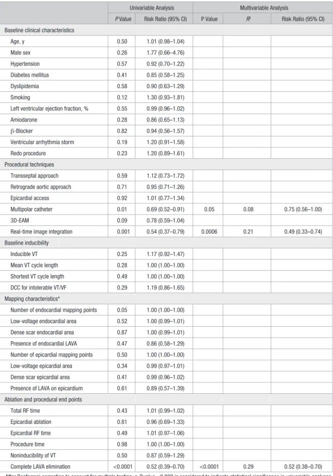
Table 3. Predictors of VT Recurrence
Univariable Analysis Multivariable Analysis
P Value Risk Ratio (95% CI) P Value R2 Risk Ratio (95% CI)
Baseline clinical characteristics
Age, y 0.50 1.01 (0.98–1.04) Male sex 0.26 1.77 (0.66–4.76) Hypertension 0.57 0.92 (0.70–1.22) Diabetes mellitus 0.41 0.85 (0.58–1.25) Dyslipidemia 0.58 0.90 (0.63–1.29) Smoking 0.12 1.30 (0.93–1.81) Left ventricular ejection fraction, % 0.55 0.99 (0.96–1.02) Amiodarone 0.28 0.86 (0.65–1.13) β-Blocker 0.82 0.94 (0.56–1.57) Ventricular arrhythmia storm 0.19 1.20 (0.91–1.58) Redo procedure 0.23 1.20 (0.89–1.61) Procedural techniques
Transseptal approach 0.59 1.12 (0.73–1.72) Retrograde aortic approach 0.71 0.95 (0.71–1.26) Epicardial access 0.92 1.01 (0.77–1.34)
Multipolar catheter 0.01 0.69 (0.52–0.91) 0.05 0.08 0.75 (0.56–1.00) 3D-EAM 0.09 0.78 (0.59–1.04)
Real-time image integration 0.001 0.54 (0.37–0.79) 0.0006 0.21 0.49 (0.33–0.74) Baseline inducibility
Inducible VT 0.25 1.17 (0.92–1.47) Mean VT cycle length 0.28 1.00 (1.00–1.00) Shortest VT cycle length 0.49 1.00 (1.00–1.00) DCC for intolerable VT/VF 0.29 1.19 (0.86–1.65) Mapping characteristics*
Number of endocardial mapping points 0.05 1.00 (1.00–1.00) Low-voltage endocardial area 0.52 1.00 (0.99–1.01) Dense scar endocardial area 0.87 1.00 (0.99–1.01) Presence of endocardial LAVA 0.47 0.86 (0.58–1.29) Number of epicardial mapping points 0.50 1.00 (1.00–1.00) Low-voltage epicardial area 0.34 0.99 (0.97–1.01) Dense scar epicardial area 0.41 0.99 (0.96–1.02) Presence of LAVA on epicardium 0.61 0.89 (0.57–1.39) Ablation and procedural end points
Total RF time 0.43 1.01 (0.99–1.02) Epicardial ablation 0.81 0.96 (0.69–1.33) Epicardial RF time 0.49 1.01 (0.97–1.06) Procedure time 0.98 1.00 (1.00–1.00) Noninducibility of VT 0.50 0.87 (0.59–1.29)
Complete LAVA elimination <0.0001 0.52 (0.39–0.70) <0.0001 0.29 0.52 (0.38–0.70) After Bonferroni correction to account for multiple testing, a P value <0.002 is considered to indicate statistical significance in univariable analyses. The criterion for entry in the multivariable model was P<0.05. 3D-EAM indicates 3-dimensional electroanatomical mapping; CI, confidence interval; DCC, direct current cardioversion; LAVA, local abnormal ventricular activity; RF, radiofrequency; VF, ventricular fibrillation; and VT, ventricular tachycardia.
*Mapping characteristics were not considered as candidates for multivariable analysis because these were only present in patients with 3D-EAM.
because of previous cardiac surgery; 1 case because of a risk
of bleeding by dual-antiplatelet therapy; and 1 case because
of pericardial bleeding). A multipolar mapping catheter was
used in 73 cases (51%). 3D-EAM was performed in 99 cases
(70%) and associated with real-time integration of anatomy
and scar from imaging in 54 cases (38%). Real-time image
integration consisted of MDCT data in 50 cases (93%), MRI
data in 21 cases (39%), and fused MDCT and MRI data in 17
cases (31%). A representative example of real-time
integra-tion and high-density mapping with a multipolar catheter is
illustrated in Figure 2.
Procedural findings are summarized in Table 1. VT was
inducible at baseline in 106 cases (75%). LAVAs were found
on the endocardium in 125 cases (88%) and on the epicardium
in 39 cases (75% of patients with epicardial access). A total of
216 VTs were induced, 55(25%) of which were mapped and
terminated by radiofrequency application, and 161(75%) of
which were unmappable because of hemodynamic intolerance,
conversion to another VT during mapping, or spontaneous
termination. After a total radiofrequency time of 32 minutes
(interquartile range, 20–50), complete LAVA elimination was
achieved in 79 cases (60% of patients with LAVA). At the end
of the procedure, VT inducibility was not tested in 30 out of
142 patients (21%) because of the induction of poorly
toler-ated VTs requiring cardioversion >2 times during the
proce-dure. Of the remaining 112 cases who underwent programmed
stimulation, the end point of VT noninducibility was achieved
in 93 out of 112 patients (83%). Eight patients had
pericar-dial bleeding (5 related to epicarpericar-dial approach). One patient
required surgery, whereas the others resolved spontaneously.
Permanent atrioventricular block occurred in 1 patient. There
were no strokes, phrenic palsies, coronary injuries, or
proce-dure-related deaths.
Follow-Up
Follow-up characteristics are summarized in Table 1. One
hun-dred out of 125 patients (80%) had an ICD implanted before
VT ablation. A further 3 patients had an ICD implanted after
VT ablation. After a median follow-up of 850 days
(interquar-tile range, 439–1707), VT recurred in 53 out of 146 patients
(36%), in which VTs were detected by the ICD (requiring
antitachycardia pacing or shock) in 48 patients and recorded
by the ambulance monitor or 12-lead ECG in the remaining 5
patients. Death from all causes occurred in 20 out of patients
125(16%), death from cardiac causes in 14 out 125 patients
(11%), and sudden death attributed to electrical storm in 5 out
of 125 patients (4%), despite ICD therapy.
Factors Associated With Procedural End Points
Results from univariable analysis for the prediction of
com-plete LAVA elimination and noninducibility of VT are shown
in Table 2. The ability to achieve complete LAVA elimination
or VT noninducibility was not related to any baseline
character-istics. Among procedural characteristics, low-voltage zone and
dense scar zone tended to be associated with failure to achieve
LAVA elimination, although the association did not reach
signif-icance after Bonferroni correction to account for multiple testing
(P=0.006 and P=0.005, respectively). Baseline inducibility was
associated with a lower rate of noninducibility at the end of the
procedure (P=0.002). Total radiofrequency and procedure time
were also associated with lower rates of noninducibility at the
end of the procedure (P=0.0003 and P=0.002, respectively).
Factors Associated With Clinical Outcome
Results from univariable and multivariable analyses for the
prediction of VT recurrence are summarized in Table 3. On
multivariable analysis, 3 characteristics were associated with
the outcome: (1) the ability to achieve complete LAVA
elimi-nation (R
2=0.29; P<0.0001; risk ratio 0.52 [0.38–0.70]), (2)
the use of real-time image integration (R
2=0.21; P=0.0006;
risk ratio 0.49 [0.33–0.74]), and (3) the use of multipolar
cath-eters (R
2=0.08; P=0.05; risk ratio 0.75 [0.56–1.00]). Kaplan–
Meier graphs illustrating the impact of these characteristics on
VT-free survival are shown in Figures 3–5.
Procedural data in patients with versus without
multipo-lar catheters and image integration are compared in Table 4.
Patients with multipolar catheters showed higher rates of
transseptal and epicardial approaches (P=0.01 and P<0.0001,
respectively), higher numbers of endocardial mapping points
(P<0.0001), and higher rate of endocardial LAVA (96%
ver-sus 80%; P=0.002). Patients with real-time image integration
showed higher numbers of endocardial LAVA sites (43 [27–
64] versus 24 [21–34]; P=0.03).
Follow-up at 1 year was available for 23 patients in whom
both multielectrode mapping and image integration had been
used. In this population, LAVA elimination had been achieved
in 18 out of 23 patients (78%), and 20 out of 23 patients (87%)
were free from recurrence at 1-year follow-up.
Discussion
This study is to our knowledge the first to analyze the
incre-mental effect of recent technological innovations on the
Figure 3. Ventricular tachycardia (VT)–free survival in procedures
with and without complete local abnormal ventricular activity (LAVA) elimination.
efficacy of post-MI VT ablation. Using constant procedural
end points in a cohort of post-MI VT patients, we
demon-strate that complete LAVA elimination, scar integration
from imaging, and high-density mapping with multipolar
catheters are independent predictors of postablation,
VT-free survival.
Impact of LAVA Elimination
Complete LAVA elimination was associated with a 2-fold
reduction in risk of VT recurrence in our cohort. VT
nonin-ducibility on the contrary did not influence outcome. These
findings provide further evidence that LAVA elimination of
VT substrate is an efficient procedural end point.
7,18The low
predictive value of VT noninducibility is also consistent with
many previous studies.
19,20Impact of Real-Time Image Integration
Real-time image integration was shown to be feasible with
processing times compatible with routine clinical practice.
Previous studies have reported good accuracy in
identify-ing VT substrate, usidentify-ing MDCT, MRI,
9or positron emission
tomography.
21Voltage mapping may fail to accurately
delin-eate the extent of diseased myocardium because of limitations
such as catheter contact issues, reduced sensitivity to far-field
signal in nontransmural or midwall scar,
22and epicardial fat
interposition.
23Additionally, the density and
comprehensive-ness of voltage mapping is highly dependent on operator
deci-sions and time constraints, whereas imaging methods provide
whole-heart assessment of the structural substrate with
sub-millimetric spatial resolution in a few heartbeats. Therefore,
integrating the structural substrate as defined from
prepro-cedural imaging might improve the accuracy of mapping in
identifying ablation targets.
In this study, the integration of anatomy and scar from
imaging was associated with a 2-fold reduction in VT
recur-rence. The superior outcome in patients with image
inte-gration might be explained by the ability of imaging to
comprehensively describe the structural substrate of VT,
thereby enabling a more focused mapping on critical areas,
at the same time ensuring that no abnormal myocardium is
left unexplored. Interestingly, we identified a higher number
of LAVA sites in patients with real-time image integration
despite a similar number of mapping points. This
obser-vation confirms that mapping is more efficiently focused
toward critical areas when guided by imaging data. Of note,
however, the identification of more LAVA did not translate
into higher rates of complete LAVA elimination. Therefore,
the impact of image integration on outcome seems to be
mediated by a more comprehensive treatment of VT
sub-strate; however, the issue of unreachable targets remains
(midwall circuits and coronary interposition).
24In this study,
VT-free survival rates in the population without image
inte-gration were comparable to those reported in the VTACH
study (Ventricular Tachycardia Ablation in Coronary Heart
Disease),
13whereas the outcome in patients treated with
image integration was more favorable. However, this
lat-ter outcome was similar to a previous study by Deneke et
al
14that did not use image integration (77% VT-free
sur-vival rate during a median follow-up of 16 months). This
is likely because of differences in patient characteristics
between studies because VTs with suspected epicardial
origin, noninducible clinical VTs, and ongoing VTs
dur-ing the procedure were excluded in the study by Deneke et
al.
14Moreover, in this study, poorly tolerated VTs requiring
cardioversion >2 times during the procedure were observed
in 21% of cases, which suggests a severe condition in the
included population.
Figure 4. Ventricular tachycardia (VT)–free survival in procedures
with and without real-time image integration.
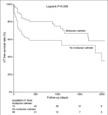
Figure 5. Ventricular tachycardia (VT)–free survival in procedures
with and without multipolar catheter.
Impact of Multipolar Catheters
High-density mapping with multipolar catheters has been
reported to enhance accurate depiction of slow conducting
areas during SR and improves understanding of VT
mecha-nisms.
7,25,26However, the impact of this technology on ablation
outcome has not been defined. In this study, the use of
mul-tipolar catheters was associated with a lower VT recurrence
rate in patients with post-MI VT. Consistent with a recent
report from our group
27, mapping with multipolar catheters
was associated with a higher mapping density that translated
into a higher rate of endocardial LAVA identification. The
rate of complete LAVA elimination tended to be higher when
using multipolar catheters. This might be explained by a more
mechanistic understanding of slow conduction areas during
SR and therefore by a more efficient targeting of the entry site
to interconnected channels.
28Overall, the impact of multipolar
catheters on patient outcome seems to be mediated by a more
comprehensive treatment of VT substrate.
Study Limitations
The objective of this study was to evaluate the impact of
tech-nological advances on efficacy of VT ablation. Therefore, a
Table 4. Procedural Characteristics in Patients With Vs Without Image Integration and Multipolar Catheters
With RII (N=54) Without RII (N=88) P value
With Multipolar Catheters (N=73) Without Multipolar Catheters (N=69) P Value Techniques Transseptal approach 49 (91) 75 (85) 0.20 69 (95) 55 (80) 0.01 Retrograde aortic approach 13 (24) 35 (40) 0.07 27 (37) 21 (30) 0.35 Epicardial access 17 (31) 35 (40) 0.32 38 (52) 14 (20) <0.0001 Multipolar catheter 31 (57) 42 (48) 0.27 NA NA … 3D-EAM 54 (100) 45 (51) <0.0001 51 (70) 48 (70) 0.97 Real-time image integration NA NA … 31 (42) 23 (33) 0.27 Baseline VT inducibility
Inducible VT 37 (69) 69 (78) 0.19 58 (79) 48 (70) 0.18 Mean VT cycle length, ms 398±113 410±96 0.57 388±103 431±97 0.03 Shortest VT cycle length, ms 368±119 372±100 0.87 360±107 385±106 0.24 DCC for untolerable VT/VF 11 (20) 18 (20) 0.45 17 (23) 12 (17) 0.17 Mapping characteristics*
Number of endocardial mapping points 452 (284–855) 268 (198–567) 0.26 658 (378–1062) 246 (165–354) <0.0001 Low-voltage endocardial area, cm2 54 (25–96) 75 (45–105) 0.49 70 (39–96) 66 (17–98) 0.54
Presence of LAVA on endocardium 48 (89) 77 (88) 0.38 70 (96) 55 (80) 0.002 Number of LAVA sites on endocardium 43 (27–64) 24 (21–34) 0.03 38 (22–66) 32 (21–55) 0.16 Number of epicardial mapping points 432 (362–755) 453 (328–952) 0.83 433 (366–1092) 381 (262–576) 0.15 Low-voltage epicardial area, cm2† 58 (39–87) 79 (53–85) 0.90 72 (41–87) 75 (63–83) 0.83
Presence of LAVA on epicardium† 15 (88) 24 (69) 0.13 30 (41) 9 (13) 0.29 Number of LAVA sites on epicardium† 19 (11–22) 36 (14–46) 0.31 19 (11–46) 20 (14–29) 0.38 Ablation and procedural end points
Total RF time 31 (23–55) 33 (17–49) 0.20 34 (23–53) 31 (16–48) 0.21 Epicardial ablation 11 (20) 18 (20) 0.99 20 (27) 9 (13) 0.03 Procedure time 278±68 233±86 0.003 271±71 226±88 0.002 Inducibility tested after ablation 46 (85) 66 (75) 0.15 55 (75) 57 (83) 0.22 Noninducibility of VT‡ 36 (78) 57 (86) 0.27 45 (82) 48 (84) 0.95 Complete LAVA elimination§ 30 (59) 49 (61) 0.82 49 (68) 30 (51) 0.05 Procedural complications 3 (6) 6 (7) 0.77 6 (8) 3 (4) 0.37 3D-EAM indicates 3-dimensional electroanatomical mapping; DCC, direct current cardioversion; LAVA, local abnormal ventricular activity; RF, radiofrequency; RII, real-time image integration; and VT, ventricular tachycardia.
*Applies to patients with 3D-EAM. †Among patients with epicardial access. ‡Among patients with inducibility testing. §Among patients with LAVA.
retrospective nature was mandatory. We acknowledge that the
use of different techniques during the course of the study likely
led to a systematic underestimation of the impact of baseline
characteristics. The impact of image integration and multipolar
catheters on patient outcome after post-MI VT ablation should
be confirmed in a randomized-controlled fashion. The second
limitation of this study is related to its sample size, which
par-ticularly prevented us from analyzing predictors of mortality.
Indeed, only 4 patients experienced sudden deaths attributed
to arrhythmia recurrence in the studied population. Third, one
of the inherent limitations of the nonrandomized study design
is a potential confounding effect of accumulating experience
for one technique influencing outcome of subsequently
intro-duced techniques. Although this effect is negated to a degree
by the fact that procedures were performed by specialists in VT
ablation with >10 years of experience, it remains a potential
confounding factor. Another potential bias would be an
evolu-tion in the indicaevolu-tion to perform VT ablaevolu-tion during the course
of the study. However, this indication remained unchanged,
and particularly, the baseline characteristics (age, sex,
previ-ous percutaneprevi-ous coronary intervention, previprevi-ous coronary
artery bypass graft, ICD, left ventricular ejection fraction, VT
storm, and number of past procedures and drugs) did not
dif-fer between patients with and without image integration and
between patients with and without multipolar catheters.
Conclusions
We demonstrate that the ability to achieve complete LAVA
elimination, the integration of scar data from preprocedural
imaging, and the use of multipolar catheters to perform
high-density mapping enhances VT-free survival after
post-MI VT ablation. Our results further confirm that complete
LAVA elimination is a procedural end point of high
predic-tive value.
Sources of Funding
The research leading to these results has received funding from the Leducq Foundation under grant agreement 09 CVD 03, and l’Agence Nationale de la Recherche (ANR) under Grant Agreements Equipex MUSIC ANR-11-EQPX-0030, MIGAT ANR-13-PRTS-0014-01, and IHU LIRYC ANR-10-IAHU-04.
Disclosures
None.
References
1. Stevenson WG, Wilber DJ, Natale A, Jackman WM, Marchlinski FE, Talbert T, Gonzalez MD, Worley SJ, Daoud EG, Hwang C, Schuger C, Bump TE, Jazayeri M, Tomassoni GF, Kopelman HA, Soejima K, Nakagawa H; Multicenter Thermocool VT Ablation Trial Investigators. Irrigated radiofrequency catheter ablation guided by electroanatomic mapping for recurrent ventricular tachycardia after myocardial infarction: the multicenter thermocool ventricular tachy-cardia ablation trial. Circulation. 2008;118:2773–2782. doi: 10.1161/ CIRCULATIONAHA.108.788604.
2. Reddy VY, Reynolds MR, Neuzil P, Richardson AW, Taborsky M, Jongnarangsin K, Kralovec S, Sediva L, Ruskin JN, Josephson ME. Prophylactic catheter ablation for the prevention of defibrillator therapy. N Engl J Med. 2007;357:2657–2665. doi: 10.1056/NEJMoa065457. 3. Bunch TJ, Weiss JP, Crandall BG, Day JD, May HT, Bair TL, Osborn JS,
Mallender C, Fischer A, Brunner KJ, Mahapatra S. Patients treated with catheter ablation for ventricular tachycardia after an ICD shock have lower
long-term rates of death and heart failure hospitalization than do patients treated with medical management only. Heart Rhythm. 2014;11:533–540. doi: 10.1016/j.hrthm.2013.12.014.
4. Marchlinski FE, Callans DJ, Gottlieb CD, Zado E. Linear ablation lesions for control of unmappable ventricular tachycardia in patients with ischemic and nonischemic cardiomyopathy. Circulation. 2000;101:1288–1296. 5. Arenal A, Glez-Torrecilla E, Ortiz M, Villacastín J, Fdez-Portales J, Sousa
E, del Castillo S, Perez de Isla L, Jimenez J, Almendral J. Ablation of electrograms with an isolated, delayed component as treatment of unmap-pable monomorphic ventricular tachycardias in patients with structural heart disease. J Am Coll Cardiol. 2003;41:81–92.
6. Soejima K, Stevenson WG, Maisel WH, Sapp JL, Epstein LM. Electrically unexcitable scar mapping based on pacing threshold for identification of the reentry circuit isthmus: feasibility for guiding ventricular tachycardia ablation. Circulation. 2002;106:1678–1683.
7. Jaïs P, Maury P, Khairy P, Sacher F, Nault I, Komatsu Y, Hocini M, Forclaz A, Jadidi AS, Weerasooryia R, Shah A, Derval N, Cochet H, Knecht S, Miyazaki S, Linton N, Rivard L, Wright M, Wilton SB, Scherr D, Pascale P, Roten L, Pederson M, Bordachar P, Laurent F, Kim SJ, Ritter P, Clementy J, Haïssaguerre M. Elimination of local abnormal ventricular activities: a new end point for substrate modification in patients with scar-related ventricular tachycardia. Circulation. 2012;125:2184–2196. doi: 10.1161/CIRCULATIONAHA.111.043216.
8. Shpun S, Gepstein L, Hayam G, Ben-Haim SA. Guidance of radiofre-quency endocardial ablation with real-time three-dimensional magnetic navigation system. Circulation. 1997;96:2016–2021.
9. Yamashita S, Sacher F, Mahida S, Berte B, Lim HS, Komatsu Y, Amraoui S, Denis A, Derval N, Laurent F, Sermesant M, Montaudon M, Hocini M, Haïssaguerre M, Jaïs P, Cochet H. Image integration to guide catheter ab-lation in scar-related ventricular tachycardia. J Cardiovasc Electrophysiol. 2016;27:699–708. doi: 10.1111/jce.12963.
10. Stevenson WG, Friedman PL, Kocovic D, Sager PT, Saxon LA, Pavri B. Radiofrequency catheter ablation of ventricular tachycardia after myocar-dial infarction. Circulation. 1998;98:308–314.
11. Della Bella P, Riva S, Fassini G, Giraldi F, Berti M, Klersy C, Trevisi N. Incidence and significance of pleomorphism in patients with postmyocar-dial infarction ventricular tachycardia. Acute and long-term outcome of radiofrequency catheter ablation. Eur Heart J. 2004;25:1127–1138. doi: 10.1016/j.ehj.2004.01.021.
12. Calkins H, Epstein A, Packer D, Arria AM, Hummel J, Gilligan DM, Trusso J, Carlson M, Luceri R, Kopelman H, Wilber D, Wharton JM, Stevenson W. Catheter ablation of ventricular tachycardia in patients with structural heart disease using cooled radiofrequency energy: results of a prospective multicenter study. Cooled RF Multi Center Investigators Group. J Am Coll Cardiol. 2000;35:1905–1914.
13. Kuck KH, Schaumann A, Eckardt L, Willems S, Ventura R, Delacrétaz E, Pitschner HF, Kautzner J, Schumacher B, Hansen PS; VTACH study group. Catheter ablation of stable ventricular tachycardia before defibrilla-tor implantation in patients with coronary heart disease (VTACH): a multi-centre randomised controlled trial. Lancet. 2010;375:31–40. doi: 10.1016/ S0140-6736(09)61755-4.
14. Deneke T, Lawo T, Grewe PH, Calcum B, Rausse R, Bösche L, Shin DI, Zarse M, Horlitz M, Mügge A, Lemke B. Usefulness of a limited linear ablation of post-myocardial infarction ventricular tachycardia using a standardized approach based on sinus rhythm mapping. Am J Cardiol. 2010;105:1235–1239. doi: 10.1016/j.amjcard.2009.12.038.
15. Lim HS, Sacher F, Cochet H, Berte B, Yamashita S, Mahida S, Zellerhoff S, Komatsu Y, Denis A, Derval N, Hocini M, Haïssaguerre M, Jaïs P. Safety and prevention of complications during percutaneous epicardial access for the ablation of cardiac arrhythmias. Heart Rhythm. 2014;11:1658–1665. doi: 10.1016/j.hrthm.2014.05.041.
16. Yamashita S, Sacher F, Mahida S, Berte B, Lim HS, Komatsu Y, Amraoui S, Denis A, Derval N, Laurent F, Montaudon M, Hocini M, Haïssaguerre M, Jaïs P, Cochet H. Role of high-resolution image integration to visual-ize left phrenic nerve and coronary arteries during epicardial ventricular tachycardia ablation. Circ Arrhythm Electrophysiol. 2015;8:371–380. doi: 10.1161/CIRCEP.114.002420.
17. Stevenson WG, Khan H, Sager P, Saxon LA, Middlekauff HR, Natterson PD, Wiener I. Identification of reentry circuit sites during catheter map-ping and radiofrequency ablation of ventricular tachycardia late after myo-cardial infarction. Circulation. 1993;88(4 pt 1):1647–1670.
18. Silberbauer J, Oloriz T, Maccabelli G, Tsiachris D, Baratto F, Vergara P, Mizuno H, Bisceglia C, Marzi A, Sora N, Guarracini F, Radinovic A, Cireddu M, Sala S, Gulletta S, Paglino G, Mazzone P, Trevisi N, Della Bella P. Noninducibility and late potential abolition: a novel combined
prognostic procedural end point for catheter ablation of postinfarction ventricular tachycardia. Circ Arrhythm Electrophysiol. 2014;7:424–435. doi: 10.1161/CIRCEP.113.001239.
19. Cooper MJ, Hunt LJ, Richards DA, Denniss AR, Uther JB, Ross DL. Effect of repetition of extrastimuli on sensitivity and reproducibility of mode of induction of ventricular tachycardia by programmed stimulation. J Am Coll Cardiol. 1988;11:1260–1267.
20. Daubert JP, Zareba W, Hall WJ, Schuger C, Corsello A, Leon AR, Andrews ML, McNitt S, Huang DT, Moss AJ; MADIT II Study Investigators. Predictive value of ventricular arrhythmia inducibility for subsequent ventricular tachycardia or ventricular fibrillation in Multicenter Automatic Defibrillator Implantation Trial (MADIT) II patients. J Am Coll Cardiol. 2006;47:98–107. doi: 10.1016/j.jacc.2005.08.049.
21. Kettering K, Weig HJ, Reimold M, Schwegler AC, Busch M, Laszlo R, Gawaz M, Schreieck J. Catheter ablation of ventricular tachycardias in pa-tients with ischemic cardiomyopathy: validation of voltage mapping criteria for substrate modification by myocardial viability assessment using FDG PET. Clin Res Cardiol. 2010;99:753–760. doi: 10.1007/s00392-010-0182-2. 22. Wijnmaalen AP, van der Geest RJ, van Huls van Taxis CF, Siebelink HM,
Kroft LJ, Bax JJ, Reiber JH, Schalij MJ, Zeppenfeld K. Head-to-head com-parison of contrast-enhanced magnetic resonance imaging and electroana-tomical voltage mapping to assess post-infarct scar characteristics in patients with ventricular tachycardias: real-time image integration and reversed regis-tration. Eur Heart J. 2011;32:104–114. doi: 10.1093/eurheartj/ehq345. 23. Desjardins B, Morady F, Bogun F. Effect of epicardial fat on
electroana-tomical mapping and epicardial catheter ablation. J Am Coll Cardiol. 2010;56:1320–1327. doi: 10.1016/j.jacc.2010.04.054.
24. Fernández-Armenta J, Berruezo A, Andreu D, Camara O, Silva E, Serra L, Barbarito V, Carotenutto L, Evertz R, Ortiz-Pérez JT, De Caralt TM, Perea RJ, Sitges M, Mont L, Frangi A, Brugada J. Three-dimensional architecture of scar and conducting channels based on high resolution ce-CMR: insights for ventricular tachycardia ablation. Circ Arrhythm Electrophysiol. 2013;6:528–537. doi: 10.1161/CIRCEP.113.000264. 25. Tung R, Nakahara S, Maccabelli G, Buch E, Wiener I, Boyle NG,
Carbucicchio C, Bella PD, Shivkumar K. Ultra high-density multipolar mapping with double ventricular access: a novel technique for ablation of ventricular tachycardia. J Cardiovasc Electrophysiol. 2011;22:49–56. doi: 10.1111/j.1540-8167.2010.01859.x.
26. Nayyar S, Wilson L, Ganesan AN, Sullivan T, Kuklik P, Chapman D, Brooks AG, Mahajan R, Baumert M, Young GD, Sanders P, Roberts-Thomson KC. High-density mapping of ventricular scar: a comparison of ventricular tachycardia (VT) supporting channels with channels that do not support VT. Circ Arrhythm Electrophysiol. 2014;7:90–98. doi: 10.1161/CIRCEP.113.000882.
27. Berte B, Relan J, Sacher F, Pillois X, Appetiti A, Yamashita S, Mahida S, Casassus F, Hooks D, Sellal JM, Amraoui S, Denis A, Derval N, Cochet H, Hocini M, Haïssaguerre M, Weerasooriya R, Jaïs P. Impact of electrode type on mapping of scar-related VT [published online ahead of print July 22, 2015]. J Cardiovasc Electrophysiol. doi: 10.1111/jce.12761. 28. Tung R, Mathuria NS, Nagel R, Mandapati R, Buch EF, Bradfield JS,
Vaseghi M, Boyle NG, Shivkumar K. Impact of local ablation on inter-connected channels within ventricular scar: mechanistic implications for substrate modification. Circ Arrhythm Electrophysiol. 2013;6:1131–1138. doi: 10.1161/CIRCEP.113.000867.