HAL Id: hal-02901599
https://hal.archives-ouvertes.fr/hal-02901599
Submitted on 17 Jul 2020
HAL is a multi-disciplinary open access
archive for the deposit and dissemination of
sci-entific research documents, whether they are
pub-lished or not. The documents may come from
teaching and research institutions in France or
abroad, or from public or private research centers.
L’archive ouverte pluridisciplinaire HAL, est
destinée au dépôt et à la diffusion de documents
scientifiques de niveau recherche, publiés ou non,
émanant des établissements d’enseignement et de
recherche français ou étrangers, des laboratoires
publics ou privés.
On the design of a pelvic phantom for magnetic
resonance and ultrasound image fusion
Fabrice Vidal, Oumaima El Mansouri, Denis Kouamé, Adrian Basarab
To cite this version:
Fabrice Vidal, Oumaima El Mansouri, Denis Kouamé, Adrian Basarab. On the design of a pelvic
phan-tom for magnetic resonance and ultrasound image fusion. IEEE International Ultrasonics Symposium
(IUS 2019), Oct 2019, Glasgow, United Kingdom. pp.2400-2403, �10.1109/ULTSYM.2019.8926218�.
�hal-02901599�
Official URL
https://doi.org/10.1109/ULTSYM.2019.8926218
Any correspondence concerning this service should be sent
to the repository administrator: tech-oatao@listes-diff.inp-toulouse.fr
This is an author’s version published in:
http://oatao.univ-toulouse.fr/26247
Open Archive Toulouse Archive Ouverte
OATAO is an open access repository that collects the work of Toulouse
researchers and makes it freely available over the web where possible
To cite this version: Vidal, Fabien and El Mansouri, Oumaima
and Kouamé, Denis and Basarab, Adrian On the design of a pelvic
phantom for magnetic resonance and ultrasound image fusion.
(2019) In: IEEE International Ultrasonics Symposium (IUS 2019),
6 October 2019 - 9 October 2019 (Glasgow, United Kingdom).
ON THE DESIGN OF A PELVIC PHANTOM FOR MAGENTIC
RESONONANCE AND UTLTRASOUND IMAGE FUSION
Fabien Vidal, Oumaima El Mansouri, Denis Kouam´e, Adrian Basarab
University of Toulouse, IRIT, CNRS UMR 5505, Universit´e Paul Sabatier, INP-ENSEEIHT, Toulouse, France {vidal, el-mansouri, kouame, basarab}@irit.fr
ABSTRACT
The purpose of this paper is to introduce a customized multi-modality phantom designed to facilitate the proof-of-concept of MRI/ultrasound fusion approaches. Phantom experiments are often required before in vivo validation, giving access to more challenging data than numerical simulations. Neverthe-less, manufactured phantoms are expensive and usually lack of flexibility. In contrast, the proposed model was inexpensive and accurately designed to overcome multimodal registration issues.
1. INTRODUCTION
Ultrasound (US) and Magnetic Resonance Imaging (MRI) are both routinely used in clinical practice, particularly in the field of gyne-cology. Due to the differences in their own imaging process, they present specific strengths and limits. Indeed, US images provide real time high-resolution images with enhanced anatomic landmarks but suffer from low signal to noise ratio and reduced field of view. Conversely, MRI offers a wide field of view with a good signal to noise ratio while its lower spatial resolution hinders the recogni-tion of millimetric anatomic details. Hence, informarecogni-tion triggered by both modalities are often required to accurately identify and plan the treatment of numerous conditions.
Endometriosis is a common and benign disease which affects women in their reproductive age. It is defined histologically by the presence of endometrial glands and/or stroma outside the uterus, while endometrial tissue is only located in the uterus in disease-free women. Among clinical presentations, deeply infiltrating en-dometriosis (DIE) is characterized by fibrous/muscular plaques in-filtrating the serosa and the muscular layer of pelvic organs. DIE with bowel involvement is a good illustration of the need for com-plementary properties of MRI and US. Indeed, surgical removal of DIE lesions may require segmental resection when the disease in-filtrates the deep layers of the bowel wall [1]. It is thus of tremen-dous importance that preoperative imaging workup accurately pre-dicts the extent of surgery to avoid unnecessary radical procedures. While MRI is associated with high diagnostic performances, with pooled sensitivity and specificity of 92% and 96% for rectosigmoid localization [2], it is less accurate in the evaluation of the depth of in-filtration [3]. As a consequence, the choice in the surgical approach (conservative or radical) cannot thus be exclusively based on MRI findings. In contrast, transrectal and transvaginal US better assess disease extent than MRI and are thus used to accurately schedule surgery [4].
Such observations greatly support the need for MR/US image fusion. Specifically, the combination of information arising from both MRI and US into a single image may improve preoperative
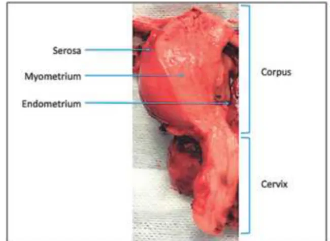
Fig. 1. Uterus specimen demonstrating successive layers of the wall.
mapping and surgical plan. However, to date, most studies have only considered MRI/US fusion from the perspective of image reg-istration [5]. Recently we have developed an MRI/US fusion al-gorithm validated on simulated data [6]. While in vivo validation is still difficult because of the challenging multimodal registration task, an intermediate step consists in testing fusion algorithms on phantoms. A multimodality MRI/US phantom must fulfill imaging and safety constraints of both modalities. Additionally, it has to sim-ulate tissue contrast. Several manufactured MRI/US phantoms are available. However, their expensiveness and lack of flexibility con-siderably hinder a wide use [7]. Herein, we propose the design of a customized multimodality phantom that facilitates the proof-of-concept of MRI/US fusion approaches.
2. MATERIAL AND METHODS 2.1. Phantom design
Phantom pre-requisites were: to fulfill constraints of both MRI and US imaging systems; to mimic uterine tissue and endometriotic cysts (endometrioma); to highlight easy-to-spot anatomical landmarks to facilitate MRI/US registration. As highlighted in Fig. 1, from the innermost to the outermost, uterine layers are the endometrium, my-ometrium and serosa. The mymy-ometrium is the thicker layer and mostly consists of smooth muscle fibers. To mimic the uterine wall, we thus used a piece of beefsteak of size 17× 10 × 1.5cm. Beyond its muscular component, beefsteak contains several greasy bays that
Fig. 2. Representation obtained from MRI 3D reconstruction of the customized phantom.
may be useful to facilitate the imaging registration process. Ovarian endometriosis, also called endometrioma, is an ovarian cyst poured with hemorrhagic fluid. To mimic endometrioma, we used a spheroid inclusion of cryogel. Cryogel was obtained from a mixture of polyvinyl acid (PVA, Sigma-Aldrich, USA), deionized water and silica powder. The following proportion of ingredients was used: 10% of PVA, 89% of water and 1% of silica. Briefly, PVA was introduced into hot water (90 to 100C) gradually at intensive magnetic stirring (500 to 700 rpm). Silica powder was added af-ter complete PVA dissolution (approximately 1 hour). The mixture was stored at room temperature for one hour then transferred into a spheroidal plastic mold measuring 4.3× 3 × 1.5cm. It was subse-quently submitted to freeze-thaw cycles until solidification. At least two cycles were necessary to obtain the adequate consistency.
Finally, we stuck the inclusion on the top of the beefsteak us-ing cyanoacrylate glue (see Fig. 2). After image acquisition, the phantom was stored at−20◦C.
2.2. Imaging techniques
MRI acquisitions were performed using a 3T clinical imaging sys-tem (Philips Achieva dStream, Inserm/UPS UMR 1214, ToNIC Technical plateform, Toulouse, France). Axial fat-suppressed T1-weighted sequences (multishot mode; 4 mm slice thickness; voxel matrix 4× 1 × 4 mm) and axial, sagittal and coronal T2-weighted sequences (multishot mode; 2 mm slice thickness; voxel matrix 0
.8× 2 × 2 mm) were acquired.
For US image acquisition, the phantom was immersed in a bucket full of water. US examination was performed using a Volu-son S10 system (General Electrics, USA). All images were acquired with a 10-MHz linear array transducer. The first series of US im-ages were performed right after MRI acquisitions. Further US acquisitions were performed at 3 and 8 months following phantom production.
3. RESULTS 3.1. Cost and manufacturing time
The proposed phantom costs approximately 10 dollars. It was easy to make using basic laboratory devices (glass laboratory bottle, heated
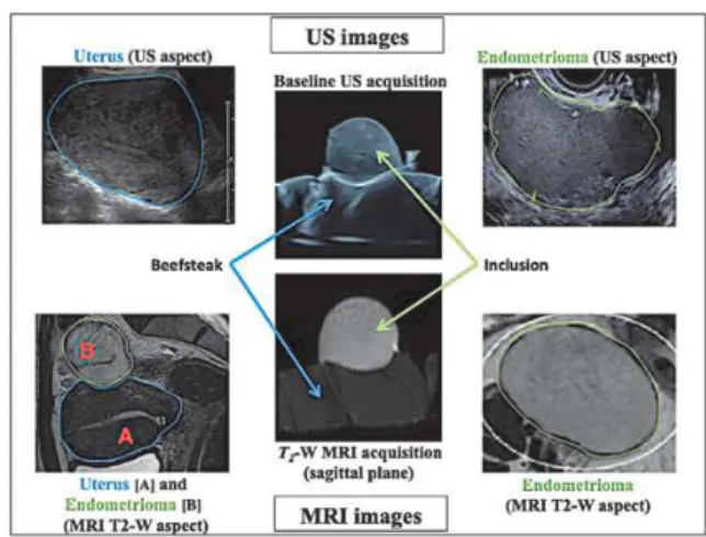
Fig. 3. US and MRI image acquired using the proposed customized phantom, showed by comparison with uterus and endometrioma im-ages acquired in vivo.
magnetic stirrer, electronic weighing scale). Its production lasted three days, considering the need for two 12 hours cycles of freeze-thaw.
3.2. Imaging results
The muscular part of the phantom demonstrated echogenicity and MRI signal similar to myometrium (see Fig. 3). Qualitatively the in-clusion showed at baseline US features close to those of endometri-omas: unilocular cyst with a homogenous low-level echogenicity content. MRI features in T1-weighted sequence were similar to en-dometrioma as well, displaying high signal intensity with no signal loss in fat-suppressed T1 sequence. In T2-weighted sequence, the in-clusion revealed a less typical aspect since signal intensity remained high while endometriomas typically appear hypointense.
3.3. Example of image fusion
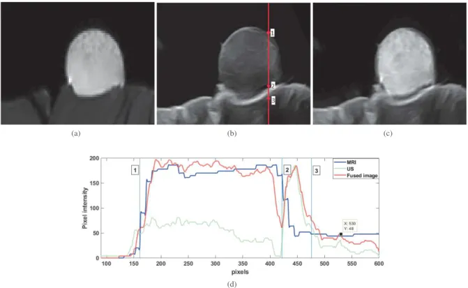
Fig. 4 shows an example of MR-US image using the algorithm pro-posed in [6]. It thus highlights the ability of the propro-posed phantom to facilitate the experimental validation of such fusion algorithms. In particular, the registration task needed before image fusion was easily conducted using a standard affine model-based registration al-gorithm, while it would require sophisticated techniques within in vivodata. In the example provided in Fig. 4, one can appreciate the result given by the fusion algorithm, that offers a good compromise between the good contrast offered by MRI and the good spatial res-olution enabled by US. More precisely, the contrast to noise ratio computed between two regions extracted from muscular and respec-tivly cryogel parts of the phantom was equal to 49.65 dB on the MRI, 19.65dB on the US image and 39.63dB on the fused image. The plot shown in Fig. 4 gives an insight about the spatial resolu-tion of the three images. While the cyanoacrylate glue can be clearly distinguishable on US and fused images, it is not on the MRI. 3.4. Impact of storage on US features
As mentioned above, the phantom was stored at−20◦C for several months. Noteworthy, US features evolved throughout the storage, particularly for the cryogel component (see Fig. 5). Indeed, the
(a) (b) (c)
(d)
Fig. 4. Example of image fusion using data acquired on the proposed phantom: (a) MR image, (b) US image, (c) fused MR and US image, (d) normalized profiles corresponding to the vertical in (b) extracted from MR, US and fused images.
Fig. 5. Evolution in time of the phantom showing, on the first row, good conservation at 3 months and considerable degradation 9 months after the fabrication. The second row plots the histograms of two blocks of pixels extracted from the cryogel regions at 0 and 3 months.
inclusion dried despite precautions for optimal preservation. At 3 months, it appeared less homogeneous while overall echogenicity remained stable. The more heterogeneous aspect at 3 months com-pared to baseline appearance is confirmed by the two histograms plotted in the second row of Fig. 5. Finally, at 9 months, the phan-tom was almost anechogenic, except its hyperechogenic wall. This experiment demonstrates one of the main limitations of the proposed phantom, which can be however mitigated by its low cost and easy-to-make advantages.
4. CONCLUSION
In conclusion, we propose in this article an inexpensive and easy to make multimodality phantom suitable for MRI/US image fusion val-idation. It provided radiological characteristics close to uterus and endometriotic cysts and may thus be particularly relevant in the field of gynecology. Its main limitation was the progressive deterioration of the cryogel component, which advocates a use restricted to a close period following production.
5. REFERENCES
[1] O. Donnez and H. Roman, “Choosing the right surgical technique for deep endometriosis: shaving, disc excision, or bowel resection?” Fertil-ity and SterilFertil-ity, vol. 108, no. 6, pp. 931 – 942, 2017. [Online]. Available: http://www.sciencedirect.com/science/article/pii/S0015028217319106 [2] V. Nisenblat, L. Prentice, P. Bossuyt, C. Farquhar, M. L. Hull, and
N. Johnson, “Combination of the noninvasive tests for the diagnosis of endometriosis,” Cochrane Database of Systematic Reviews, no. 7, 2016. [3] A. Kim, P. Fernandez, B. Martin, L. Palazzo, L. Ribeiro-Parenti, F. Walker, M. Bucau, H. Collinot, D. Luton, and M. Koskas, “Magnetic resonance imaging compared with rectal endoscopic sonography for the prediction of infiltration depth in colorec-tal endometriosis,” Journal of Minimally Invasive Gynecology, vol. 24, no. 7, pp. 1218 – 1226, 2017. [Online]. Available: http://www.sciencedirect.com/science/article/pii/S1553465017304454 [4] V. Nisenblat, P. Bossuyt, C. Farquhar, N. Johnson, and M. L. Hull,
“Imaging modalities for the noninvasive diagnosis of endometriosis,” Cochrane Database of Systematic Reviews, no. 2, 2016.
[5] K. Bolten, T. Fischer, Y. Y.-N. Bender, G. Diederichs, and A. Thomas, “Pilot study of mri/ultrasound fusion imaging in postpartum assess-ment of cesarean section scar,” Ultrasound in Obstetrics & Gynecology, vol. 50, no. 4, pp. 520–526, 2017.
[6] O. El Mansouri, A. Basarab, F. Vidal, D. Kouam, and J. Tourneret, “Fusion of magnetic resonance and ultrasound images: A preliminary study on simulated data,” in 2019 IEEE 16th International Symposium on Biomedical Imaging (ISBI 2019), April 2019, pp. 1733–1736. [7] W. R. Walter, C. J. Burke, M. Diallo, and R. S. Adler, “Use of a simple,
inexpensive dual-modality phantom as a learning tool for magnetic res-onance imagingultrasound fusion techniques,” Journal of Ultrasound in Medicine, vol. 37, no. 8, pp. 2083–2089, 2018.