HAL Id: inserm-00357465
https://www.hal.inserm.fr/inserm-00357465
Submitted on 26 Oct 2010
HAL is a multi-disciplinary open access
archive for the deposit and dissemination of
sci-entific research documents, whether they are
pub-lished or not. The documents may come from
teaching and research institutions in France or
abroad, or from public or private research centers.
L’archive ouverte pluridisciplinaire HAL, est
destinée au dépôt et à la diffusion de documents
scientifiques de niveau recherche, publiés ou non,
émanant des établissements d’enseignement et de
recherche français ou étrangers, des laboratoires
publics ou privés.
intermediary factor 1beta and heterochromatin protein
1 leads to a switch from DNA hyper- to
hypomethylation and H3K9 to H3K27 trimethylation on
the MEST promoter correlating with gene reactivation.
Raphaël Riclet, Mariam Chendeb, Jean-Luc Vonesch, Dirk Koczan,
Hans-Juergen Thiesen, Régine Losson, Florence Cammas
To cite this version:
Raphaël Riclet, Mariam Chendeb, Jean-Luc Vonesch, Dirk Koczan, Hans-Juergen Thiesen, et al..
Disruption of the interaction between transcriptional intermediary factor 1beta and heterochromatin
protein 1 leads to a switch from DNA hyper- to hypomethylation and H3K9 to H3K27 trimethylation
on the MEST promoter correlating with gene reactivation.: TIF1beta–HP1 Association Functions in
MEST Repression. Molecular Biology of the Cell, American Society for Cell Biology, 2009, 20 (1),
pp.296-305. �10.1091/mbc.E08-05-0510�. �inserm-00357465�
Molecular Biology of the Cell Vol. 20, 296 –305, January 1, 2009
Disruption of the Interaction between Transcriptional
Intermediary Factor 1
 and Heterochromatin Protein 1
Leads to a Switch from DNA Hyper- to Hypomethylation
and H3K9 to H3K27 Trimethylation on the MEST Promoter
Correlating with Gene Reactivation
Raphae¨l Riclet,* Mariam Chendeb,* Jean-Luc Vonesch,* Dirk Koczan,
†Hans-Juergen Thiesen,
†Re´gine Losson,* and Florence Cammas*
*Institut de Ge´ne´tique et de Biologie Mole´culaire et Cellulaire, Centre National de la Recherche Scientifique/
Institut National de la Sante´ et de la Recherche Me´dicale/Universite´ Louis Pasteur/Colle`ge de France, 67404
Illkirch-Cedex, France; and
†Institute for Immunology, Medical Faculty, 18057 Rostock, Germany
Submitted May 22, 2008; Revised September 26, 2008; Accepted October 3, 2008 Monitoring Editor: Wendy Bickmore
Here, we identified the imprinted mesoderm-specific transcript (MEST) gene as an endogenous TIF1 primary target gene and demonstrated that transcriptional intermediary factor (TIF) 1, through its interaction with heterochromatin protein (HP) 1, is essential in establishing and maintaining a local heterochromatin-like structure on MEST promoter region characterized by H3K9 trimethylation and hypoacetylation, H4K20 trimethylation, DNA hypermethylation, and enrich-ment in HP1 that correlates with preferential association to foci of pericentromeric heterochromatin and transcriptional repression. On disruption of the interaction between TIF1 and HP1, TIF1 is released from the promoter region, and there is a switch from DNA hypermethylation and histone H3K9 trimethylation to DNA hypomethylation and histone H3K27 trimethylation correlating with rapid reactivation of MEST expression. Interestingly, we provide evidence that the imprinted MEST allele DNA methylation is insensitive to TIF1 loss of function, whereas the nonimprinted allele is regulated through a distinct TIF1–DNA methylation mechanism.
INTRODUCTION
Cell functions result from the interpretation of genetic and epigenetic information established by a large cohort of com-plexes acting at the chromatin level (reviewed in Mellor, 2006). Gene silencing, in particular, plays essential roles during development and cell differentiation that require progressive extinction of pluripotent genes and specific cell lineage genes (reviewed in Rajasekhar and Begemann, 2007). Several histone modifications, believed to establish a “his-tone code,” are thought to be essential in this silencing program; these modifications include H3K9 trimethylation (3meH3K9) and H4K20 trimethylation (3meH4K20), two modifications well known to be associated with heterochro-matin structures (Schotta et al., 2004; Grewal and Jia, 2007); H3K27 trimethylation (3meH3K27), a mark assumed to be associated with facultative heterochromatin and to be par-ticularly enriched in polycomb response elements (Bracken et al., 2006). Moreover, recent studies indicate that different combinations of these and other histone marks can lead to different outputs that are difficult to anticipate considering the complexity of this combinatorial code (reviewed in Hirose,
2007; Reik, 2007). Another key epigenetic modification is DNA methylation that has well-characterized functions in genomic imprinting, X chromosome inactivation, silencing of tumor suppressor genes, and repression of viral elements (reviewed in Latham et al., 2008). It has been predicted that ⬃6% of CpG islands display tissue- and or developmental stage-specific DNA methylation pattern (Song et al., 2005; Weber et al., 2007). Although DNA methylation correlates with gene repression, the functional relevance of this level of regulation remains largely to be established in vivo.
Among complexes known to be involved in establishing gene silencing are those containing the transcriptional core-pressor, TIF1 (Transcriptional Intermediary factor 1), that plays essential roles during early embryonic development and terminal cell differentiation (Cammas et al., 2000; 2004). TIF1 (Le Douarin et al., 1996) (also known as KAP-1, Friedman et al., 1996; or TRIM28) is the universal corepressor for the Kru¨ppel-associated box domain contain-ing zinc-fcontain-inger proteins (KRAB-ZFP) family of transcription factors that constitutes the largest family of repressors in mammals (Friedman et al., 1996; Kim et al., 1996; Moosmann et al., 1996; Abrink et al., 2001). TIF1 is an intrinsic compo-nent of two chromatin remodeling and histone deacetylase complexes, N-CoR1 and NuRD (Underhill et al., 2000; Schultz et al., 2001) and directly interacts with the histone methyltransferase SETDB1, which specifically methylates H3K9 preferentially within euchromatin (Schultz et al., 2002). TIF1 also interacts with members of the heterochro-matin protein (HP) 1 family through a specific pentapeptide
This article was published online ahead of print in MBC in Press (http://www.molbiolcell.org/cgi/doi/10.1091/mbc.E08 – 05– 0510) on October 15, 2008.
Address correspondence to: Re´gine Losson (losson@igbmc.u-stras-bg.fr).
PxVxL called HP1box (Le Douarin et al., 1996; Nielsen et al., 1999; Ryan et al., 1999; Thiru et al., 2004; Cammas et al., 2007). HP1 is a structurally and functionally highly conserved protein with family members found in eukaryotic organisms ranging from Schizosaccharomyces pombe to humans (Eissenberg et al., 1990; Wang et al., 2000). These proteins participate in chro-matin packaging and have a well-established function in heterochromatin-mediated silencing (reviewed in Grewal and Jia, 2007). More recent data suggest that HP1 functions are much more diversified than initially assumed. These functions include gene specific silencing (Cryderman et al., 2005; Smallwood et al., 2007), gene activation (Cryderman et al., 2005), and transcriptional elongation (Vakoc et al., 2005). Mice and humans have three different HP1 proteins (HP1␣, , and ␥) that are associated, although not exclu-sively, with pericentromeric heterochromatin (Nielsen et al., 1999). It is currently speculated that HP1 serves as a bridg-ing protein, connectbridg-ing histones to nonhistone chromosomal proteins through specific recognition of di- and trimethyl-ated H3K9 by the HP1 chromodomain and association with diverse proteins through their HP1box and HP1 chro-moshadow domain (Lomberk et al., 2006). We and others have demonstrated previously that the interaction between TIF1 and HP1 is required for 1) TIF1 transcriptional re-pression activity, which also requires histone deacetylase activity (Nielsen et al., 1999; Ryan et al., 1999); 2) relocation of TIF1 from euchromatin to heterochromatin that accompa-nies the retinoic acid (RA)-induced primitive endodermal (PrE) differentiation of mouse embryonal carcinoma F9 cells (Cammas et al., 2002) and for 3) progression through F9 cell differentiation into RA plus cAMP-induced parietal endodermal differentiation (Cammas et al., 2004). Up to now, the molecular mechanisms underlying TIF1 functions as a corepressor of the KRAB-ZFP have been assessed in reporter systems (Ayyanathan et al., 2003; Sripathy et al., 2006). In these studies, the authors demonstrated that the artificial recruitment of TIF1 to the promoter region of a reporter gene induces stable silencing through the establishment of a heterochromatin-like structure characterized by trimethyla-tion of the H3K9, DNA methylatrimethyla-tion, and HP1 recruitment that is maintained for several generations.
In the present study, we identified mesoderm-specific transcript (MEST) as an endogenous TIF1 primary target gene and characterized the influence of the interaction be-tween TIF1 and HP1 on its expression, its chromatin struc-ture, and its nuclear positioning.
MATERIALS AND METHODS
Details on individual plasmid constructs, which were all verified by sequenc-ing, are available upon request.
Antibodies
Antibodies used in this study were as follows: mouse anti-TIF1 monoclonal antibody (mAb), 1Tb3, raised against recombinant Escherichia coli expressed mouse TIF1 (123-834) (Nielsen et al., 1999); rabbit anti-TIF1 polyclonal antibody (pAb), PF64, raised against TIF1 (amino acids 141–155; Cammas et
al., 2002); mouse anti-FLAG mAb 2FLB11; anti-HP1␣ mAb 2HP-2G9; and anti-HP1 mAb 1MOD-1A9; anti-HP1␥ mAb, 2 Mod-1G6 (Nielsen et al., 1999). The antibodies specific for the different histone modifications were purchased from Millipore (Billerica, MA).
Cell Culture
Wild-type (WT) and mutant F9 cells were grown as monolayers in DMEM (Invitrogen, France) supplemented with 10% fetal calf serum as previously described (Boylan and Gudas, 1991). Cells were counted with a particle counter (Coulter Z2).
Immunoprecipitation and Western Blot Analysis
Isolation of whole cell extracts from F9 cells and Western blot detection were performed as described previously (Chiba et al., 1997; Nielsen et al., 1999).
Reverse Transcription-Polymerase Chain Reaction (RT-PCR)
RNA extraction was performed with TRIzol reagent (Invitrogen). MEST cDNA was amplified with AHY249 (5⬘-GAAATTCAGAAGACGCTGGG-3⬘) and AHN102 (5⬘-CTCCAAAAACTCTGGATACG-3⬘); PEG3 cDNA with BBJ400 (5⬘-CCTGATCAATGGGTTCCTTG-3⬘) and BBJ401 (5⬘-CTTCTG-GAAGCCGACATTATG-3); PEG10 cDNA with BBJ402 (5⬘-CGAGTGTACT-TATTGGTCCC-3⬘) and BBJ403 (5⬘-TGACTGTCATCTGGCATTCC-3⬘);
COPG2 cDNA with BBH298 (5⬘-CTTGCTGTCTCCAACATG-3⬘) and BBH299 (5⬘-ATTTCGCAAGCAGCTCTC-3⬘); MEG3 cDNA with BBZ369 (5⬘-GACT-TCACGCACAACACG-3⬘) and BBZ370 (5⬘-ACAAGGGCGCTTCCAATC-3⬘);
IGF2R cDNA with BAM406 (5⬘-CAAAGGGAAGAGCTATGATG-3⬘) and BAM407 (5⬘-ATCTTCACTTTCATCACACG-3⬘); and HPRT cDNA with QG197 (5⬘-GTAATGATCAGTCAACGGGGGAC-3⬘) and QG198 (5⬘-CCAG-CAAGCTTGCAACCTTAACCA-3⬘).
Chromatin Immunoprecipitation (ChIP) Assay
ChIP assays were performed according to the Millipore protocol with some minor modifications. Cells were cross-linked with 1% formaldehyde for 10 min at 37°C resuspended in lysis buffer (0.1% SDS, 50 mM HEPES, pH 7.9, 140 mM NaCl, 1 mM EDTA, 1% Triton X-100, and 0.1% Na-deoxycholate) at a final concentration of 12.5⫻ 106cells/500l, incubated on ice for 10 min, and
sonicated to average fragment size of 200 –500 base pairs. The clarified solu-bilized chromatin was diluted fivefold in ChIP dilution buffer (16.7 mM Tris-HCl, pH 8.1, 1.2 mM EDTA, 167 mM NaCl, 0.01% SDS, and 1.1% Triton X-100). Immunoprecipitation was performed with 8l of mAb (TIF1 and HP1), 10l of pAb (TIF1), or 3 l of pAb (histone modifications). The beads were washed sequentially once with low salt buffer (20 mM Tris-HCl, pH 8.1, 2 mM EDTA, 150 mM NaCl, 0.1% SDS, and 1% Triton X-100), high salt buffer (20 mM Tris-HCl, pH 8.1, 2 mM EDTA, 500 mM NaCl, 0.1% SDS, and 1% Triton X-100), LiCl buffer (10 mM Tris-HCl, pH 8.1, 1 mM EDTA, 0.25 M LiCl, 1% NP40, and 1% deoxycholate) and twice with TE buffer (10 mM Tris-HCl, pH 8.0, and 1 mM EDTA). Immunocomplexes were eluted twice with 250l of elution buffer (1% SDS and 0.1 M NaHCO3) for 15 min at RT. Eluates and
input chromatin were heated at 65°C overnight in the presence of 0.2 M NaCl. ChIP DNA were quantified by real-time PCR using the QuantiTect SYBR Green Kit (QIAGEN, Hilden, Germany), and the final results for each sample were normalized to the inputs. PCR reactions were performed in triplicate in a LightCycler (Roche Diagnostics, Mannheim, Germany) with 3l of ChIP DNA. Primer sequences for the MEST promoter were as follows: forward, 5⬘-CAGCAGCTTCTGGCATGTGG-3⬘ and reverse, 5⬘-AACCCCAGAT-TCTAGTGAAG-3⬘; for the region 5⬘ 10 kb upstream of the MEST promoter: forward, 5⬘-TGGTGGCAGATGACTGTTAG-3⬘ and reverse, 5⬘-GAAGAAT-AGGCAATGCAGTG-3⬘; for the region 5⬘ 4 kb upstream of the MEST pro-moter: forward, 5⬘-ATCTGCAGTTTTGCCTCAGG-3⬘ and reverse, 5⬘-AT-GAAGGCACACAGAGATGC-3⬘; for the region 3⬘ 5 kb downstream of the
MEST promoter: forward, 5⬘-TTTCCTGAGACGCATCGTCC-3⬘ and reverse, ATAGACTGGCTCATCACCAC-3⬘; for the HPRT promoter: forward, 5⬘-TTATCTGGGAATCCTCTGGG-3⬘ and reverse, 5⬘-AAAGGCAGTTCCG-GAACTCT-3⬘; and for the major satellites: forward, 5⬘-GACGACTTGAAAAAT-GACGAAATC-3⬘ and reverse, 5⬘-CATATTCCAGGTCCTCAGTGTGC-3⬘.
DNA Fluorescence in Situ Hybridization (FISH)
Wild-type and mutant F9 cells were grown on gelatin-coated coverslips for 72h washed for 5 min in 1⫻ PBS, fixed in 2% paraformaldehyde 10 min at room temperature (RT). Coverslips were treated with 0.1 M Tris-Cl, pH 7.2, for 10 min at RT and washed in 1⫻ phosphate-buffered saline (PBS) for 5 min. Cells were permeabilized for 10 min at RT with 1⫻ PBS, 0.1% Triton X-100, and 0.1% saponin, and then they were incubated in 20% glycerol, 1⫻ PBS solution for 20 min. Coverslips were immersed three times in liquid nitrogen and allowed to thaw at RT, washed 5 min in 1⫻ PBS, and treated with 100 g/ml DNase-free RNase A in 1⫻ PBS for 1 h at 37°C. Coverslips were washed in 1⫻ PBS for 5 min and then in 1⫻ PBS, 0.1% Triton X-100, and 0.1% saponin for 30 min at RT. Coverslips were then washed in 1⫻ PBS, dehy-drated by an ethanol series (80, 90, and 100%) for 3 min each, and air-dried. Seven microliters of hybridization cocktail containing 100 ng of dCTP-Cy3 (GE Healthcare, Little Chalfont, Buckinghamshire, United Kingdom)-labeled probe, 4g of mouse Cot-1 DNA, 1 g of sheared salmon-sperm DNA in 50% formamide, 2⫻ SSC, and 10% dextran sulfate was heated at 76°C for 10 min and added to each coverslip. Coverslips were mounted on slides, and DNA-probe and cellular DNA were denatured simultaneously on a hot block at 75°C for 3 min and hybridized in a humid atmosphere at 37°C for 24 h. On the following day, coverslips were washed once in 50% formamide, 2⫻ SSC, pH 7.5, for 20 min, three times in 0.1⫻ SSC 5 min each at 37°C, once in 0.1% Tween 20, 4⫻ SSC for 5 min at 45°C, and finally in 1⫻ PBS for 5 min at RT, stained for DNA with Hoechst 33258 at 5g/ml and mounted in PBS 5% propyl gallate, 80% glycerol. Images acquisition was performed using a Leica
TCS-SP5 confocal scanning microscope (Leica Microsystems, Mannheim, Ger-many) that was equipped with an HCX PLAPO 63⫻ 1.4 oil immersion objective lens. Images of 20 –30 serial optical sections, spaced by 0.25m, were acquired per cell nucleus. Approximately 250 nuclei were analyzed per cell type by using an “in-house” software (TIMT) that calculates the minimal distance though the three dimensions between heterochromatin and the FISH signal. To this end, the distribution of the heterochromatin and euchromatin domains (images recorded at 480 nm) as well as the position of the two FISH signals (images recorded at 570 nm) within the nucleus were first delimited and marked on the projection of all recorded sections by using Photoshop software (Adobe Systems, Mountain View, CA). The distance between the FISH signal and the nearest heterochromatin domain was then searched and measured through the whole image stacks and normalized to the surface area of the nucleus (Hoechst staining). The MEST and HPRT probes were bacterial artificial chromosome (BAC) RP24-211G11 and BAC RP24-335G16, respec-tively.
Bisulfite Conversion
Five micrograms of genomic DNA was digested with EcoRI for 1 h at 37°C, purified, and denatured with 0.3 M NaOH for 10 min at 42°C. DNA was converted by adding a solution containing 0.55 mg of hydroquinine (Sigma Chemie, Deisenhofen, Germany) and 0.202 g of sodium hydrogen sulfite (Aldrich, Germany) at pH 5.05 for 15 h at 54°C. Converted DNA was purified with nucleospin columns (Macherey-Nagel, Du¨ren, Germany) and denatured 10 min at RT with 0.3 M NaOH. DNA fragments were ethanol precipitated in presence of glycoblue (Invitrogen) and cloned in blunt-ended pBluescript.
RESULTS
Identification of MEST as a TIF1 Primary Target Gene To identify TIF1 endogenous target genes, we compared the transcriptome of WT F9 cells with that of the previously described TIF1HP1box/⫺ F9 cells expressing a mutated
TIF1 protein (TIF1HP1box) unable to interact with HP1
(Cammas et al., 2004). The MEST gene displayed one of the highest -fold induction in mutant cells compared with WT cells (data not shown). To validate this microarray analysis, we performed quantitative (q)RT-PCR analysis with MEST-specific primers on RNA prepared from TIF1⫹/⫺ and
TIF1HP1box/⫺cells (Figure 1A). MEST expression is
unde-tectable in TIF1⫹/⫺cells, whereas it is highly expressed in
TIF1HP1box/⫺cells, confirming the microarray analysis and
demonstrating that MEST repression requires the interaction between TIF1 and HP1 within F9 EC cells (Figure 1A). MEST is an imprinted gene also known as paternally ex-pressed gene 1 (PEG1) (Kaneko-Ishino et al., 1995). It was therefore important to investigate whether TIF1 could be a specific regulator of imprinted genes. To this end, we ana-lyzed the expression of several imprinted genes (Murphy and Jirtle, 2003) by using our preliminary microarrays anal-ysis. On the 50 imprinted genes spotted on the microarray, 19 (38%) were found to be up-regulated and none down-regulated in TIF1HP1box/⫺compared with TIF1⫹/⫺cells
(with a criteria of a⬎2-fold change with a p value; p ⬍ 0.05), whereas on the whole 25,785 genes spotted on the microar-ray, 3580 (14%) and 31 (0.1%) were up- and down-regulated, respectively, in TIF1HP1box/⫺ compared with TIF1⫹/⫺
cells by using the criteria described above (data not shown). The expression of four imprinted genes that was predicted by our preliminary microarray analysis to be either not modified (PEG3 and PEG10) or induced (IGF2R and MEG3)
Figure 1. MEST is a TIF1 primary target gene. (A) Expression of MEST (PEG1), PEG3, PEG10, MEG3, IGF2R, and COPG2 was assessed in TIF1⫹/⫺and TIF1HP1box/⫺F9 cells by RT-PCR analysis. HPRT was used as a control house-keeping gene. (B) Schematic representation of the MEST gene with the black boxes representing the exons. The arrow labeled⫹ 1 represents the transcription start site described in Lefebvre et al. (1997). The different regions amplified for the ChIP analysis are shown. (C and D) RA-inducible expression of MEST is independent of TIF1–HP1 interaction. TIF1⫹/⫺(C) and TIF1HP1box/⫺(D) cells were treated for 96 h with either ethanol (no) or 1M all-trans RA (RA).
MEST expression was measured by qRT-PCR and normalized with HPRT expression. (E) ChIP assay with two anti-TIF1 antibodies (p, PF64 pAb; m, 1TB3 mAb) was performed in WT EC F9 cells. The three positions analyzed on the MEST gene are shown in B, and the HPRT promoter region was also analyzed. Results are the average of at least three independent experiments. *p⬍ 0.05 and **p ⬍ 0.005. R. Riclet et al.
Molecular Biology of the Cell 298
in TIF1HP1box/⫺compared with TIF1⫹/⫺cells was
vali-dated by qRT-PCR (Figure 1A). These results indicate that TIF1 regulates either directly or indirectly the expression of a large number of genes, including a significant proportion of imprinted genes. We also measured the expression of COPG2 whose 3⬘-untranslated region (UTR) overlaps MEST 3⬘-UTR on the mouse chromosome 6 (Lee et al., 2000). This gene is expressed at a slightly, although not statistically significant, higher level in TIF1HP1box/⫺ compared with
TIF1⫹/⫺ cells, implicating that TIF1-HP1 is specifically
involved in the regulation of MEST gene expression within this chromosomal locus (Figure 1A). To assess the regulation of MEST gene expression during differentiation, TIF1⫹/⫺
and TIF1HP1box/⫺cells were treated for 4 d with 1M RA
to induce PrE. qRT-PCR analysis shows that MEST expres-sion is equivalently induced in both genetic backgrounds and remains 104 lower in TIF1⫹/⫺ cells than in
TIF1HP1box/⫺cells (Figure 1, C and D). These results
indi-cate that MEST expression is inducible during PrE differen-tiation through a TIF1–HP1 interaction-independent mech-anism. Therefore, MEST gene expression regulation was only analyzed in noninduced conditions.
To establish whether MEST is a direct TIF1 target gene, we performed ChIP in WT F9 cells with two independent TIF1 antibodies, the monoclonal 1TB3 (Nielsen et al., 1999; Figure 1E, m) and the polyclonal PF64 (Cammas et al., 2002; Figure 1E, p) followed by real-time PCR. Results are repre-sented as (signal with specific antibodies⫺ signal with no antibody) normalized with the initial materials (input). TIF1 is significantly enriched on the MEST promoter region compared with regions 3.9 kb upstream and 6.3 kb down-stream of this MEST promoter region as well as to the promoter region of the constitutively active HPRT gene
(10.5; 17.4- and 12.5-fold, respectively, with the mAb; Figure 1E). These data strongly suggest that, in EC F9 cells, MEST is a primary target of TIF1 that maintains repression of this gene via a mechanism requiring its interaction with HP1.
The Interaction between TIF1 and HP1 Is Essential for Stable Recruitment of Both TIF1 and HP1 to MEST Promoter
To investigate the recruitment of HP1 to an endogenous TIF1 target gene, we performed ChIP with antibodies spe-cific of the three HP1 isotypes, HP1␣, HP1, and HP1␥, in TIF1⫹/⫺F9 cells. All three HP1 isotypes are significantly
enriched on the MEST promoter region, whereas they are not significantly enriched in the 5⬘ upstream or 3⬘ down-stream positions (Figure 2, A–C, Het). None of the HP1 isotypes are significantly recruited to the promoter of the HPRT gene, whereas all of them are, as expected, recruited to the constitutive heterochromatic major satellites, confirm-ing that the signals detected on MEST promoter correspond to specific recruitment of the three HP1 isotypes (Figure 2, A–C). Furthermore, to determine whether the interaction between TIF1 and HP1 is required for HP1 recruitment to the MEST promoter, the same ChIPs were performed in TIF1HP1box/⫺cells. As illustrated on Figure 2, A–C (Mut),
no HP1 is significantly detected at any position of the MEST gene after ChIP with HP1␣, HP1, or HP1␥ antibodies within TIF1HP1box/⫺ cells. In contrast, all three HP1
iso-types are still clearly detectable on the major satellites at a level equivalent to that observed in TIF1⫹/⫺cells (Figure 2,
A–C). This demonstrates that the interaction between TIF1 and HP1 is essential for specific HP1 recruitment to the MEST promoter, whereas it is not involved in the
recruit-Figure 2. The interaction between TIF1 and HP1 is essential for TIF1 and HP1 recruitment to MEST promoter. ChIP assays were
performed on TIF1⫹/⫺cells (black bars; Het) and TIF1HP1box/⫺cells (white bars; Mut) with antibodies directed against HP1␣ (A), HP1
(B), HP1␥ (C), and TIF1 (D). PCR was performed with primers specific to the three MEST gene positions described in Figure 1B, the HPRT promoter, and the major satellites. *p⬍ 0.05, **p ⬍ 0.005, and ***p ⬍ 0.0005.
ment of these proteins to the constitutive heterochromatic major satellites.
To determine whether the interaction between TIF1 and HP1 is also involved in the recruitment of TIF1 to the MEST promoter, ChIPs with the two TIF1 Abs, was per-formed in TIF1⫹/⫺and TIF1HP1box/⫺cells. Surprisingly,
TIF1HP1box recruitment to MEST promoter is drastically
decreased compared with the recruitment of TIF1 in TIF1⫹/⫺cells, although it remains significantly above the
signal detected on the HPRT promoter and on the major satellites sequences (Figure 2D; data not shown). This strongly suggests that the interaction between TIF1 and HP1 is not only essential for the recruitment of HP1 to the MEST promoter, but also for efficient TIF1 recruitment and/or stabilization to this region. Altogether, these data indicate that TIF1 and HP1 are recruited to MEST promoter through a mechanism that requires their mutual interaction to maintain gene repression within F9 EC cells.
TIF1, through Its Interaction with HP1, Maintains a Heterochromatin-like Structure on the MEST Promoter MEST is an imprinted gene that possesses a CpG island in its promoter region (Lefebvre et al., 1997; Figure 1B). It was therefore important to assess the DNA methylation status of this MEST CpG island in TIF1⫹/⫺and TIF1HP1box/⫺cells.
To this end, we performed bisulfite sequencing and ana-lyzed the methylation status of the MEST gene from position ⫺455 base pairs to ⫹269 base pairs comprising 56 CpG dinucleotides. This region of the MEST gene is fully meth-ylated on both alleles in TIF1⫹/⫺ cells (Figure 3A). A
notable exception to this full methylation pattern is the first position upstream to the transcription start site (at⫺33 base pairs) that is methylated in only 60% of MEST alleles (Figure 3A). In contrast to TIF1⫹/⫺ cells, only ⬃50% of MEST
alleles are methylated within the CpG island in TIF1HP1box/⫺ cells (Figure 3A). This loss of MEST gene
methylation is not a consequence of a general demethylation process within TIF1HP1box/⫺ cells, because digest of
TIF1⫹/⫺and TIF1HP1box/⫺genomic DNA by the
methy-lation-sensitive enzyme Hpa2 shows the same pattern on agarose gel (data not shown).
To establish the profile of histone modifications induced by TIF1–HP1 recruitment to MEST promoter, we per-formed ChIP with antibodies specific of different histone modifications in TIF1⫹/⫺cells. This analysis reveals that
MEST promoter is highly enriched in 3meH3K9 and 3meH4K20, two modifications well known to be associated with heterochromatin structures and gene silencing (Fig-ure 3B). Interestingly, 3meH3K9 and to a lesser extent 3meH4K20 specifically spreads up to 4 kb upstream from the promoter region (Figure 3B). These two marks are absent of the promoter region of the actively transcribed HPRT gene and highly enriched in the heterochromatic major sat-ellite sequences demonstrating the specificity of the ChIP (Figure 3B). Furthermore, acetylation of H3K9 (AcH3K9), a mark associated with gene expression, is not significantly enriched on MEST promoter region of TIF1⫹/⫺ cells,
whereas it is highly enriched on the promoter region of the constitutively transcribed HPRT (Figure 3B). As expected, this mark is also absent at the silent heterochromatic major satellites. Last, we analyzed the enrichment in 3meH3K27. 3meH3K27 is not significantly enriched within neither the MEST promoter, or the HPRT promoter or the heterochro-matic major satellite sequences (Figure 3B). It is noteworthy that AcH3K9 and 3meH3K27 are slightly, although signifi-cantly, enriched 4 and 10 kb upstream the MEST promoter region within TIF1⫹/⫺ cells (Figure 3B). To demonstrate
the specific role of TIF1 interaction with HP1 in the main-tenance of this chromatin structure, the same ChIP experi-ments were performed in TIF1HP1box/⫺cells. As expected,
considering the loss of MEST repression in this cell line, 3meH3K9 and 3meH4K20 are lost from the MEST gene in TIF1HP1box/⫺ cells, with a concomitant enrichment of
AcH3K9. More surprisingly, 3meH3K27 becomes highly en-riched in the MEST promoter region and at 4 kb upstream this region (11.9- and 6-fold, respectively, compared with the signal in TIF1⫹/⫺cells). This phenomenon is specific of the
MEST promoter and its 4 kb upstream region since none of the histone modifications presented above are significantly altered at 10 kb upstream the MEST promoter region nor within the HPRT promoter nor the major satellite sequences in TIF1HP1box/⫺compared with TIF1⫹/⫺cells. To find out
whether the 3meH3K27 mark that occurs specifically in TIF1HP1box/⫺ cells displays any preferential association
with the methylated or unmethylated MEST allele, we per-formed bisulfite sequencing after a 3meH3K27 ChIP in TIF1HP1box/⫺cells. As illustrated on Figure 3C, 3meH3K27
is exclusively associated with the unmethylated MEST allele.
MEST Preferentially Associates with Pericentromeric Heterochromatin in a TIF1–HP1 Interaction-dependent Manner
It has been suggested that nuclear organization and in par-ticular the localization of specific genomic loci close to het-erochromatin has important implications for gene silencing (Brown et al., 1999). We therefore assessed the localization of the MEST gene within TIF1⫹/⫺and TIF1HP1box/⫺cells by
FISH and confocal microscopy. The distance between the spots corresponding to the hybridization of the MEST gene to the nearest heterochromatin domain defined as bright spots of Hoechst staining was determined throughout the three dimensions of the nucleus using an in-house (TIMT) software. The distance was normalized by the surface of the nucleus. In both cell lines, three types of nuclei were ob-served: 1) both MEST alleles associated with heterochroma-tin (2HC), 2) both MEST alleles excluded from heterochro-matin (2EU), and 3) one MEST allele within heterochroheterochro-matin and one excluded from this compartment (1HC ⫹ 1EU) (Figure 3D). Interestingly, the proportion of the (1EU ⫹ 1HC) and (2EU) populations are significantly different within TIF1⫹/⫺and TIF1HP1box/⫺cells lines according to
the statistical chi-square test (2⬍ 0.01), whereas the (2HC)
populations are equivalently represented in both cell lines (7.1 and 10.2% in TIF1⫹/⫺and TIF1HP1box/⫺cells,
respec-tively). The (1EU ⫹ 1HC) population represents 46.5% of TIF1⫹/⫺ cells, whereas it is found in only 24%
of TIF1HP1box/⫺ cells (Figure 3D). Furthermore 43.3% of
TIF1⫹/⫺ cells are (2EU) compared with 68.9% for
TIF1HP1box/⫺cells (Figure 3D). These changes are unlikely
to result from a global difference of heterochromatin distri-bution between the two cell lines because no obvious differ-ence is observed by Hoechst staining. Finally the average distance between the “EU” spots and the nearest heterochro-matin domain is equivalent in both genetic backgrounds (0.952 and 0.895m in TIF1⫹/⫺and TIF1HP1box/⫺cells,
respectively) (Figure 3D). Furthermore, we analyzed the subnuclear localization of the chromosome X-associated gene, HPRT, and we found that this gene is euchromatic in 84.4 and 85.9% of TIF1⫹/⫺and TIF1HP1box/⫺nuclei,
re-spectively (data not shown). These data strongly suggest that the association of TIF1 with HP1 facilitates the anchor-age of the MEST locus within pericentromeric heterochro-matin.
R. Riclet et al.
Molecular Biology of the Cell 300
TIF1 Is Permanently Required on MEST Promoter to Maintain Transcriptional Repression
To address the kinetic of loss of TIF1-mediated MEST repression upon disruption of TIF1–HP1 interaction, we established a cell line allowing hydroxytamoxifen (OHT)-inducible disruption of the interaction between these two
proteins. This cell line (TIF1L2/HP1box/Cre-ERT2) carries 1)
one floxed TIF1 allele (TIF1L2), 2) the other TIF1 allele
with the HP1box motif mutated by homologous recombina-tion (TIF1HP1box), and 3) an expression vector driving
sta-ble expression of the OHT-inducista-ble CreERT2recombinase
(Indra et al., 1999). These cells were treated for different
Figure 3. TIF1, through its interaction with HP1, maintains a heterochromatin-like structure on the MEST promoter. (A) DNA methylation
of the MEST CpG island from position⫺455 base pairs to position 269 base pairs was analyzed in TIF1⫹/⫺(black square) and TIF1HP1box/⫺
F9 cell lines (open triangles) by bisulfite sequencing. (B) The histone modification status of MEST promoter and of its surrounding regions, of the HPRT promoter and of the major satellite sequences were analyzed by ChIP in nontreated TIF1⫹/⫺ (black bars; Het) and
TIF1HP1box/⫺F9 cell lines (white bars; Mut). As annotated trimethylation of H3K9 (3meH3K9), H4K20 (3meH4K20), H3K27 (3meH3K27) and acetylation of H3K9 (AcH3K9) were assessed. (C) The DNA methylation status of the MEST promoter region associated with 3meH3K27 was assessed by bisulfite sequencing after ChIP with an anti-3meH3K27 pAb in TIF1HP1box/⫺cells. The open circles represent the unmethylated
CpG and the black circles the methylated CpG. (D) DNA FISH analysis was performed using a MEST gene specific probe in TIF1⫹/⫺and
TIF1HP1box/⫺F9 cell lines (red dots). Pericentromeric heterochromatin was labeled by Hoechst staining and occurs as bright blue spots. Representative cells with two spots within heterochromatin (A and B), one spot within euchromatin and one spot within heterochromatin (C and D) or two spots within euchromatin (E and F) are shown. The percentages represent the analysis of⬎250 cells for each genotype. *p ⬍ 0.05, **p⬍ 0.005, and ***p ⬍ 0.0005.
times with OHT and collected for genotyping and analysis of MEST expression. Excision of the floxed TIF1L2allele
was detectable as early as 12 h of OHT treatment and 48 h of OHT treatment were sufficient to detect MEST expres-sion, which was further increased by 122-fold until 9 d of OHT treatment (Figure 4, A and B). It is noteworthy, that, although MEST expression increases with time after OHT treatment, it only reaches 10% of the level obtained in TIF1HP1box/⫺cells (data not shown). These results indicate
that MEST repression is rapidly lost upon disruption of the interaction between TIF1 and HP1 and strongly suggest that the interaction between these two proteins is perma-nently required to maintain MEST repression.
TIF1 Is Sufficient to Establish the Heterochromatin-like Structure on MEST Gene
As described above, TIF1, through its interaction with HP1, is essential to maintain MEST repression. A key question that remained to be answered was whether TIF1 is also involved for the establishment of MEST repression. To ad-dress this question, we used the previously established cell line TIF1HP1box/⫺/rTA-f.TIF1 allowing inducible
expres-sion of FLAG-TIF1 (f.TIF1) by doxycycline (Dox) treat-ment within TIF1HP1box/⫺ cells (Cammas et al., 2004).
f.TIF1 expression was verified by Western using the anti-FLAG mAb in TIF1HP1box/⫺/rTA-f.TIF1 cells. As
ex-pected, f.TIF1 is expressed only in presence of Dox; how-ever, this does not lead to any significant increase in the total level of TIF1 in these cells, strongly suggesting that f.TIF1 level is very low compared with endogenous TIF1 (Figure 5A). f.TIF1 expression was also verified by immunofluo-rescence and found to be homogenous in each Dox-treated TIF1HP1box/⫺/rTA-f.TIF1 cell (data not shown). f.TIF1
recruitment to MEST promoter was then assessed by ChIP with the anti-FLAG antibody. As illustrated on Figure 5B, f.TIF1 is efficiently recruited to MEST promoter only in presence of Dox.
MEST expression was quantified in presence or absence of Dox treatment. Dox induces a twofold decrease of MEST expression compared with nontreated cells (Figure 5C), demonstrating that f.TIF1 is able to partially restore MEST repression. This twofold decrease in MEST expression was maintained even after 15 passages, strongly suggesting that these cells are refractory to further repression (data not shown), most likely due in part to the low level of f.TIF1 expression. It was then essential to determine whether f.TIF1 could reestablish the heterochromatin-like structure observed in TIF1⫹/⫺cells. To this end, we analyzed
spe-cific histone modifications in TIF1HP1box/⫺/rTA-f.TIF1
cells in presence or absence of Dox treatment. ChIP experi-ments show that 3meH4K20 and 3meH3K9 are increased by 3.6- and 3.1-fold, respectively, in the promoter region of Dox-treated cells compared with nontreated cells, whereas 3meH3K27 and AcH3K9 are both decreased by 1.7-fold in these same conditions (Figure 5D). Although the amplitude of these changes is relatively limited, they demonstrate that f.TIF1 is able to reestablish to some extent the histone modification profile observed in TIF1⫹/⫺cells. As
men-tioned above, the level of f.TIF1 is extremely low compared with that of the endogenous protein and might be a limiting factor for detection of DNA methylation by bisulfite se-quencing. We therefore analyzed DNA methylation of the ⫺176- to ⫺57-bp region upstream the MEST transcription start site after immunoprecipitation of Dox-treated TIF1HP1box/⫺/rTA-f.TIF1 chromatin with the anti-FLAG
mAb. As control, total DNA before ChIP (input) was also analyzed. As shown on Figure 5E, ⬃100% of the MEST alleles associated with f.TIF1 are methylated, whereas only ⬃50% are methylated in the input. This demonstrates that f.TIF1 is able to establish DNA methylation at the MEST promoter; however, most likely because of the low level of f.TIF1 expression, this rescue is undetectable on total TIF1HP1box/⫺/rTA-f.TIF1 cell DNA.
MEST Proximal Promoter Region Is Highly Methylated in Liver
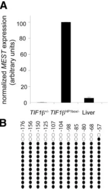
It has been suggested that several imprinted loci become aberrantly methylated on both alleles in F9 cells (Yeivin et al., 1996). To decipher whether the full methylation of MEST observed in our F9 model corresponds to any physiological condition, we tested MEST expression in different adult mouse tissues. As shown previously, MEST expression is very low in all tissues and in particular in liver in which MEST expression is 19-fold lower than in TIF1HP1box/⫺F9
cells (Figure 6A; data not shown; Lui et al., 2008). We ana-lyzed the methylation status of the MEST promoter region in adult liver. As illustrated on Figure 6B, bisulfite sequencing analysis shows that the MEST promoter region between ⫺107 and ⫺57 bp is methylated on 90% of the alleles. These results strongly suggest that, as in F9 EC cells, MEST prox-imal promoter region is methylated on both alleles.
DISCUSSION
In this study, we identified the MEST gene as an endoge-nous TIF1 primary target gene in embryonal carcinoma F9 cells. We demonstrate that the interaction between TIF1 and HP1 proteins is critical for MEST repression, being essential for mutual recruitment and/or stabilization of both proteins on the promoter region. We further show that TIF1–HP1 interaction is critical for the maintenance and the establishment of a local heterochromatin-like structure char-acterized by H3K9 trimethylation and hypoacetylation, H4K20 trimethylation, DNA hypermethylation, and
enrich-Figure 4. TIF1-HP1 interaction is permanently required to
main-tain MEST repression. TIF1L2/HP1box/CreERT2cells were treated
for the indicated times with 10⫺7M OHT and collected for RNA and DNA preparations. (A) Cells were genotyped at each time point by PCR as described previously (Cammas et al., 2004). (B) MEST ex-pression was measured by qRT-PCR at each time and normalized by HPRT expression.
R. Riclet et al.
Molecular Biology of the Cell 302
ment in HP1. This structure correlates with a preferential association of the MEST gene to foci of pericentromeric heterochromatin and MEST repression.
Interestingly, MEST is an imprinted gene, and three of our results strongly suggest that TIF1 regulates only one of the two MEST alleles: 1) although both alleles are fully methyl-ated in TIF1⫹/⫺EC F9 cells, only 50% of the MEST
frag-ments sequenced are methylated in TIF1HP1box/⫺, strongly
suggesting that only one allele is demethylated upon dis-ruption of the interaction between TIF1 and HP1; 2) only one of the two alleles seems to be significantly associated with pericentromeric heterochromatic foci, a localization that requires the interaction between TIF1 and HP1; and 3) upon disruption of the interaction between TIF1 and HP1, there is a switch from high levels of 3meH3K9 and 3meH4K20 and low level of 3meH3K27 to low levels of 3meH3K9 and 3meH4K20 and high level of 3meH3K27 that is exclusively associated with the demethylated MEST allele. These results indicate that the two MEST alleles are clearly silenced by two different mechanisms that both rely on DNA methylation, but of which, only one involves the formation of a heterochromatin-like structure and is dependent upon the interaction between TIF1 and HP1. Because 50% of MEST methylation is refractory to TIF1 loss of function, it is very likely that TIF1 does not regulate the allele of maternal origin that is imprinted but rather the paternal nonimprinted allele. This strongly suggests that the molec-ular mechanisms of gene repression described in this study apply to both imprinted and nonimprinted genes, a conclu-sion in line with the large number of imprinted and nonim-printed genes up-regulated upon loss of interaction between
TIF1 and HP1. Very interestingly, we show in liver that the MEST proximal promoter region between ⫺107 and ⫺57 base pairs is methylated on 90% of the alleles, which corre-lates with a very low level of MEST expression. This result is in striking contrast with the observation made on the MEST distal promoter region (⫺1001 to ⫺792 bp) that is methyl-ated only on 50% of MEST alleles (Lui et al., 2008). The different methylation status between these two MEST pro-moter regions strongly suggests that there is a boundary element between –792 and –107 base pairs that inhibits DNA methylation spreading on the nonimprinted allele but not on the imprinted alleles that is methylated, even on the MEST distal promoter. Together, these results highlight the exis-tence of different mechanisms of DNA methylation estab-lishment and/or maintenance. The marks that allow the preferential association of TIF1 with the nonimprinted al-lele and the establishment and/or maintenance of DNA methylation on specific gene area remain to be elucidated, but most likely they involve a complex combinatorial pres-ence of histone modifications on each MEST allele. This is the first demonstration that the nonimprinted allele of an imprinted gene is regulated by DNA methylation in vivo.
Concerning the molecular mechanism underlying TIF1 functions as a corepressor, it is interesting to note that, although 3meH3K9 and to a lesser extent 3meH4K20 extend around 4kb upstream the MEST transcription start site, HP1 binding is limited to the proximal MEST promoter region, indicating that, in contrast to constitutive heterochromatin, HP1 is not spreading to surrounding regions and that 3meH3K9 and 3meH4K20 on their own are not sufficient for HP1 recruitment. This conclusion is in agreement with the
Figure 5. Flag-TIF1 expression partially rescues MEST repression. (A) Western blot analysis of f.TIF1 expression was performed on
TIF1HP1box/⫺/rTA-f.TIF1 cells treated for the indicated times with Dox by using an anti-FLAG antibody. Total TIF1 expression was also assessed with an anti-TIF1 antibody. Protein loading was controlled with an anti-actin antibody. (B) ChIP using an anti-FLAG antibody was performed on TIF1HP1box/⫺/rTA-f.TIF1 cells in absence or presence of Dox. (C) MEST expression was assessed by qRT-PCR in
TIF1HP1box/⫺/rTA-f.TIF1 cells treated or not with Dox and in TIF1⫹/⫺cells. MEST expression was normalized with HPRT. (D) ChIP with
antibodies of the specified histone modifications were performed on TIF1HP1box/⫺/rTA-f.TIF1 cells in presence or absence of Dox. (E) DNA
methylation was assessed by bisulfite sequencing in TIF1HP1box/⫺/rTA-f.TIF1 after ChIP with an anti-FLAG mAb. The open lozenges represent
finding that in heterochromatin, HP1 association to chroma-tin requires both H3K9 trimethylation and direct association with Suv39H1 (Stewart et al., 2005). It is very likely that, here, HP1 chromatin association necessitates both H3K9 trimethylation and direct binding to TIF1. Furthermore, we find that MEST repression is rapidly lost upon disruption of the interaction between TIF1 and HP1 strongly suggesting that this repression requires permanent loading of TIF1 on MEST promoter region. This is in contrast with studies using integrated artificial TIF1 target genes, in which the authors concluded that a single pulse of TIF1 expression allowed transgene silencing for several passages (Sripathy et al., 2006). These data clearly demonstrates that HP1 loading on MEST promoter for repression can rapidly be displaced for gene activation. The loss of HP1 on MEST promoter is accompanied by loss of 3meH3K9, 3meH4K20, DNA meth-ylation and a gain of 3meH3K27, demonstrating that all these chromatin modifications are very dynamic and inter-dependent as previously suggested in other systems (Freitag et al., 2004; Smallwood et al., 2007; Wu et al., 2008). This conclusion is also in line with studies indicating that HP1 distribution within the nucleus is highly dynamic (Cheutin et al., 2003) and that DNA methylation is not always as stably established as assumed previously (Kangaspeska et al., 2008; Me´tivier et al., 2008). It is also intriguing that in most studies 3meH3K27 is associated with gene silencing, whereas in the present study, 3meH3K27 is associated with gene activation in TIF1HP1box/⫺ cells. This association of
3meH3K27 with gene activation has, however, been ob-served in some cases and often correlates with loss of DNA methylation, suggesting that 3meH3K27 could be a “default mark” when DNA methylation is lost on genes normally regulated by the formation of a heterochromatin-like
struc-ture (Peters et al., 2003; Mathieu et al., 2005; McGarvey et al., 2006; Papp and Mu¨ller, 2006).
In conclusion, we have demonstrated that the expression of the imprinted MEST gene expression is tightly regulated by a complex interplay of epigenetic modifications and in particular by two distinct DNA methylation-dependent mechanisms of which, only one is dependent upon TIF1.
ACKNOWLEDGMENTS
We thank Prof. A.J.L. Clark and Dr. I. Bogdarina, Saint Bartholomew’s Hos-pital, London, United Kingdom, for helpful technical advice for bisulfite sequencing; S. Dumanoir for helpful technical advice for FISH analysis; and B. Jost, D. Dembele, and C. Bole-Feysot for help in the microarray analysis. We thank M. Cervin˜o for technical assistance; M. Beglin, J.-L., M. Koch, Y. Lutz, P. Kessler, and D. Hentsch for confocal laser scanning microscopy; S. Vicaire and D. Stephan for DNA sequencing; M. Oulad-Abdelghani for antibodies supply; and B. Heller for medium supply. This work was supported by the Centre National de la Recherche Scientifique, the Institut National de la Sante´ et de la Recherche Me´dicale, the Agence Nationale de la Recherche (ANR06-BLAN-0377), the Association pour la Recherche sur le Cancer, and the Colle`ge de France. R. R. was supported by a fellowship from the Ministe`rede la Recherche et de la Technologie.
REFERENCES
Abrink, M., Ortiz, J. A., Mark, M., Sanchez, C., Looman, C., Hellman, L., Chambon, P., and Losson, R. (2001). Conserved interaction between distinct Kruppel-associated box domains and the transcriptional intermediary factor 1 . Proc. Natl. Acad. Sci. USA 98, 1422–1426.
Ayyanathan, K., Lechner, M. S., Bell, P., Maul, G. G., Schultz, D. C., Yamada, Y., Tanaka, K., Torigoe, K., and Rauscher, F. J., 3rd (2003). Regulated recruit-ment of HP1 to a euchromatic gene induces mitotically heritable, epigenetic gene silencing: a mammalian cell culture model of gene variegation. Genes Dev. 17, 1855–1869.
Boylan, J. F., and Gudas, L. J. (1991). Overexpression of the cellular retinoic acid binding protein-I (CRABP-I) results in a reduction in differentiation-specific gene expression in F9 teratocarcinoma cells. J. Cell Biol. 112, 965–979. Bracken, A. P., Dietrich, N., Pasini, D., Hansen, K. H., and Helin, K. (2006). Genome-wide mapping of Polycomb target genes unravels their roles in cell fate transitions. Genes Dev. 20, 1123–1136.
Brown, K. E., Baxter, J., Graf, D., Merkenschlager, M., and Fisher, A. G. (1999). Dynamic repositioning of genes in the nucleus of lymphocytes preparing for cell division. Mol. Cell 3, 207–217.
Cammas, F., Mark, M., Dolle´, P., Dierich, A., Chambon, P., and Losson, R. (2000). Mice lacking the transcriptional corepressor TIF1 are defective in early postimplantation development. Development 127, 2955–2963. Cammas, F., Oulad-Abdelghani, M., Vonesch, J.-L., Chambon, P., and Losson, R. (2002). Cell differentiation induces TIF1association with centromeric het-erochromatin through HP1 interaction. J. Cell Sci. 115, 3439 –3448. Cammas, F., Herzog, M., Lerouge, T., Chambon, P., and Losson, R. (2004). Association of the transcriptional corepressor TIF1 with Heterochromatin Protein 1 (HP1): an essential role for progression through differentiation. Genes Dev. 18, 2147–2160.
Cammas, F., Janoshazi, A., Lerouge, T., and Losson, R. (2007). HP1 Dynamic and selective interactions of the transcriptional corepressor TIF1 beta with the heterochromatin protein HP1 isotypes during cell differentiation. Differenti-ation 7, 627– 637.
Cheutin, T., McNairn, A. J., Jenuwein, T., Gilbert, D. M., Singh, P. B., and Misteli, T. (2003). Maintenance of stable heterochromatin domains by dy-namic HP1 binding. Science 299, 721–725.
Chiba, H., Clifford, J., Metzger, D., and Chambon, P. (1997). Specific and redundant functions of retinoid X receptor/retinoic acid receptor het-erodimers in differentiation, proliferation, and apoptosis of F9 embryonal carcinoma cells. Cell Biol. 139, 735–747.
Cryderman, D. E., Grade, S. K., Li, Y., Fanti, L., Pimpinelli, S., and Wallrath, L. L. (2005). Role of Drosophila HP1 in euchromatic gene expression. Dev. Dyn.
232, 767–774.
Eissenberg, J. C., James, T. C., Foster-Hartnett, D. M., Hartnett, T., Ngan, V., and Elgin, S. C. (1990). Mutation in a heterochromatin-specific chromosomal protein is associated with suppression of position-effect variegation in
Dro-sophila melanogaster. Proc. Natl. Acad. Sci. USA 87, 9923–9927. Figure 6. MEST proximal promoter region is hypermethylated in
liver. (A) qRT-PCR analysis of MEST expression in TIF1⫹/⫺and
TIF1HP1box/⫺F9 cells and adult liver. Results are normalized with HPRT expression and shown as arbitrary units. (B) MEST proximal promoter region is highly methylated in adult liver. Methylation of the MEST promoter region between⫺107 and ⫺57 base pairs was analyzed by bisulfite sequencing on genomic DNA prepared from three independent adult animal livers. The illustration of a repre-sentative experiment on an adult liver is shown. The open circles represent the unmethylated CpG, and the closed circles the meth-ylated CpG.
R. Riclet et al.
Molecular Biology of the Cell 304
Freitag, M., Hickey, P. C., Khlafallah, T. K., Read, N. D., and Selker, E. U. (2004). HP1 is essential for DNA methylation in Neurospora. Mol. Cell 13, 427– 434.
Friedman, J. R., Fredericks, W. J., Jensen, D. E., Speicher, D. W., Huang, X. P., Neilson, E. G., and Rauscher, F. J., 3rd (1996). KAP-1, a novel corepressor for the highly conserved KRAB repression domain, Genes Dev. 10, 2067–2078. Grewal, S. I., and Jia, S. (2007). Heterochromatin revisited. Nat. Rev. Genet. 8, 35– 46.
Hirose, S. (2007). Crucial roles for chromatin dynamics in cellular memory. J. Biochem. 14, 615– 619.
Indra, A. K., Warot, X., Brocard, J., Bornert, J. M., Xiao, J. H., Chambon, P., and Metzger, D. (1999). Temporally-controlled site-specific mutagenesis in the basal layer of the epidermis: comparison of the recombinase activity of the tamoxifen-inducible Cre-ER(T) and Cre-ER(T2) recombinases. Nucleic Acids Res. 27, 4324 – 4327.
Kaneko-Ishino, T., Kuroiwa, Y., Miyoshi, N., Kohda, T., Suzuki, R., Yokoyama, M., Viville, S., Barton, S. C., Ishino, F., and Surani, M. A. (1995). Peg1/MEST imprinted gene on chromosome 6 identified by cDNA subtrac-tion hybridizasubtrac-tion. Nat. Genet. 11, 52–59.
Kangaspeska, S., Stride, B., Me´tivier, R., Polycarpou-Schwarz, M., Ibberson, D., Carmouche, R. P., Benes, V., Gannon, F., and Reid, G. (2008). Transient cyclical methylation of promoter DNA. Nature 452, 112–115.
Kim, S. S., Chen, Y. M., O’Leary, E., Witzgall, R., Vidal, M., and Bonventre, J. V. (1996). A novel member of the RING finger family, KRIP-1, associates with the KRAB-A transcriptional repressor domain of zinc finger proteins, Proc. Natl. Acad. Sci. USA 93, 15299 –15304.
Latham, T., Gilbert, N., and Ramsahoye, B. (2008). DNA methylation in mouse embryonic stem cells and development. Cell Tissue Res. 331, 31–55. Le Douarin, B., Nielsen, A. L., Garnier, J.-M., Ichinose, H., Jeanmougin, F., Losson, R., and Chambon, P. (1996). A possible involvement of TIF1␣ and TIF1 in the epigenetic control of transcription by nuclear receptors. EMBO J.
15, 6701– 6715.
Lee, Y. J., Park, C. W., Hahn, Y., Park, J., Lee, J., Yun, J. H., Hyun, B., and Chung, J. H. (2000). Mit1/Lb9 and Copg2, new members of mouse imprinted genes closely linked to Peg1/MEST(1). FEBS Lett. 472, 230 –234.
Lefebvre, L., Viville, S., Barton, S. C., Ishino, F., and Surani, M. A. (1997). Genomic structure and parent-of-origin-specific methylation of Peg1. Hum. Mol. Genet. 6, 1907–1915.
Lomberk, G., Wallrath, L., and Urrutia, R. (2006). The heterochromatin pro-tein 1 family. Genome Biol. 7, 228
Lui, J. C., Finkielstain, G. P., Barnes, K. M., and Baron, J. (2008). An imprinted gene network that controls mammalian somatic growth is down-regulated during postnatal growth deceleration in multiple organs. Am. J. Physiol. Regul. Integr. Comp. Physiol. 295, R189 –R196.
Mathieu, O., Probst, A. V., and Paszkowski, J. (2005). Distinct regulation of histone H3 methylation at lysines 27 and 9 by CpG methylation in Arabidopsis. EMBO J. 24, 2783–2791.
McGarvey, K. M., Fahrner, J. A., Greene, E., Martens, J., Jenuwein, T., and Baylin, S. B. (2006). Silenced tumor suppressor genes reactivated by DNA demethylation do not return to a fully euchromatic chromatin state. Cancer Res. 66, 3541–3549.
Mellor, J. (2006). Dynamic nucleosomes and gene transcription. Trends Genet.
22, 320 –329.
Me´tivier, R. et al. (2008). Cyclical DNA methylation of a transcriptionally active promoter. Nature 452, 45–50.
Moosmann, P., Georgiev, O., Le Douarin, B., Bourquin, J. P., and Schaffner, W. (1996). Transcriptional repression by RING finger protein TIF1 that interacts with the KRAB repressor domain of KOX1. Nucleic Acids Res. 24, 4859 – 4867. Murphy, S. K., and Jirtle, R. L. (2003). Imprinting evolution and the price of silence. Bioessays 25, 577–588.
Nielsen, A. L., Ortiz, J. A., You, J., Oulad-Abdelghani, M., Khechumian, R., Gansmuller, A., Chambon, P., and Losson, R. (1999). Interaction with mem-bers of the heterochromatin protein 1 (HP1) family and histone deacetylation are differentially involved in transcriptional silencing by members of the TIF1 family. EMBO J. 18, 6385– 6399.
Papp, B., and Mu¨ller, J. (2006). Histone trimethylation and the maintenance of transcriptional ON and OFF states by trxG and PcG proteins. Genes Dev. 20, 2041–2054.
Peters, A. H. et al. (2003). Partitioning and plasticity of repressive histone methylation states in mammalian chromatin. Mol. Cell 12, 1577–1589. Rajasekhar, V. K., and Begemann, M. (2007). Concise review: roles of poly-comb group proteins in development and disease: a stem cell perspective. Stem Cells 25, 2498 –2510.
Reik, W. (2007). Stability and flexibility of epigenetic gene regulation in mammalian development. Nature 447, 425– 432.
Ryan, R. F., Schultz, D. C., Ayyanathan, K., Singh, P. B., Friedman, J. R., Fredericks, W. J., and Rauscher, F. J. 3rd. (1999). KAP-1 corepressor protein interacts and colocalizes with heterochromatic and euchromatic HP1 proteins: a potential role for Kru¨ppel-associated box-zinc finger proteins in heterochro-matin-mediated gene silencing. Mol. Cell. Biol. 19, 4366 – 4378.
Schotta, G., Lachner, M., Sarma, K., Ebert, A., Sengupta, R., Reuter, G., Reinberg, D., and Jenuwein, T. (2004). A silencing pathway to induce H3–K9 and H4 –K20 trimethylation at constitutive heterochromatin. Genes Dev. 18, 1251–1262.
Schultz, D. C., Friedman, J. R., and Rauscher, F. J. 3rd. (2001). Targeting histone deacetylase complexes via KRAB-zinc finger proteins: the PHD and bromodomains of KAP-1 form a cooperative unit that recruits a novel isoform of the Mi-2␣ subunit of NuRD. Genes Dev. 15, 428–443.
Schultz, D. C., Ayyanathan, K., Negorev, D., Maul, G. G., and Rauscher, F. J., 3rd (2002). SETDB 1, a novel KAP-1-associated histone H3, lysine 9-specific methyltransferase that contributes to HP1-mediated silencing of euchromatic genes by KRAB zinc-finger proteins. Genes Dev. 16, 919 –932.
Smallwood, A., Esteve, P. O., Pradhan, S., and Carey, M. (2007). Functional cooperation between HP1 and DNMT1 mediates gene silencing. Genes Dev.
21, 1169 –1178.
Song, F., Smith, J. F., Kimura, M. T., Morrow, A. D., Matsuyama, T., Nagase, H., and Held, W. A. (2005). Association of tissue-specific differentially meth-ylated regions (TDMs) with differential gene expression. Proc. Natl. Acad. Sci. USA 102, 3336 –3341.
Sripathy, S. P., Stevens, J., and Schultz, D. C. (2006). The KAP1 corepressor functions to coordinate the assembly of de novo HP1-demarcated microen-vironments of heterochromatin required for KRAB zinc finger protein-medi-ated transcriptional repression. Mol. Cell. Biol. 26, 8623– 8638.
Stewart, M. D., Li, J., and Wong, J. (2005). Relationship between histone H3 lysine 9 methylation, transcription repression, and heterochromatin protein 1 recruitment. Mol. Cell. Biol. 25, 2525–2538.
Thiru, A., Nietlispach, D., Mott, H. R., Okuwaki, M., Lyon, D., Nielsen, P. R., Hirshberg, M., Verreault, A., Murzina, N. V., and Laue, E. D. (2004). Structural basis of HP1/PXVXL motif peptide interactions and HP1 localisation to heterochromatin. EMBO J. 23, 489 – 499.
Underhill, C., Qutob, M. S., Yee, S.-P., and Torchia, J. (2000). A novel nuclear receptor corepressor complex, N-CoR, contains components of the mamma-lian SWI/SNF complex and the corepressor KAP-1. J. Biol. Chem. 275, 40463– 40470.
Vakoc, C. R., Mandat, S. A., Olenchock, B. A., and Blobel, G. A. (2005). Histone H3 lysine 9 methylation and HP1gamma are associated with transcription elongation through mammalian chromatin. Mol. Cell 19, 381–391. Wang, G., Ma, A., Chow, C. M., Horsley, D., Brown, N. R., Cowell, I. G., and Singh, P. B. (2000). Conservation of heterochromatin protein 1 function. Mol. Cell. Biol. 20, 6970 – 6983.
Weber, M., Hellmann, I., Stadler, M. B., Ramos, L., Pa¨a¨bo, S., Rebhan, M., and Schu¨beler, D. (2007). Distribution, silencing potential and evolutionary impact of promoter DNA methylation in the human genome. Nat. Genet. 39, 457– 466.
Wu, L. P. et al. (2008). HDAC inhibitor depsipeptide activates silenced genes through decreasing both CpG and H3K9 methylation on the promoter. Mol. Cell. Biol. 28, 3219 –3235.
Yeivin, A., Levine, A., and Razin, A. (1996). DNA methylation patterns in tumors derived from F9 cells resemble methylation at the blastula stage. FEBS Lett. 395, 11–16.