HAL Id: hal-01393194
https://hal.univ-reunion.fr/hal-01393194
Submitted on 7 Nov 2016
HAL is a multi-disciplinary open access
archive for the deposit and dissemination of sci-entific research documents, whether they are pub-lished or not. The documents may come from teaching and research institutions in France or abroad, or from public or private research centers.
L’archive ouverte pluridisciplinaire HAL, est destinée au dépôt et à la diffusion de documents scientifiques de niveau recherche, publiés ou non, émanant des établissements d’enseignement et de recherche français ou étrangers, des laboratoires publics ou privés.
Leptospirosis Is Not Associated with Increased
Expression of Granulocyte Cell Activation Markers
Loïc Raffray, Claude Giry, David Vandroux, Barbara Kuli, Andry
Randrianjohany, Anne-Marie Pequin, Frédéric Renou, Marie-Christine
Jaffar-Bandjee, Philippe Gasque
To cite this version:
Loïc Raffray, Claude Giry, David Vandroux, Barbara Kuli, Andry Randrianjohany, et al.. Major Neutrophilia Observed in Acute Phase of Human Leptospirosis Is Not Associated with Increased Expression of Granulocyte Cell Activation Markers. PLoS ONE, Public Library of Science, 2016, 11, pp.165716. �10.1371/journal.pone.0165716.t001�. �hal-01393194�
Major Neutrophilia Observed in Acute Phase
of Human Leptospirosis Is Not Associated
with Increased Expression of Granulocyte
Cell Activation Markers
Loic Raffray1,2,3*, Claude Giry1,4, David Vandroux5, Barbara Kuli6,
Andry Randrianjohany7, Anne-Marie Pequin8, Fre´de´ric Renou3, Marie-Christine
Jaffar-Bandjee1,4, Philippe Gasque1,2
1 Universite´ de La Re´union, CNRS 9192, INSERM U1187, IRD 249, CHU de La Re´union, Unite´ Mixte Processus Infectieux en Milieu Insulaire Tropical (PIMIT), Plateforme Technologique CYROI, Sainte-Clotilde, La Re´union, France, 2 Laboratoire d’immunologie clinique et expe´rimentale ZOI (LICE OI), CHU La Re´union site Fe´lix Guyon, St Denis, La Re´union, France, 3 Internal Medicine Unit, CHU La Re´union site Fe´lix Guyon, St Denis, La Re´union, France, 4 Microbiology/Virology Laboratory, CHU La Re´union site Fe´lix Guyon, St Denis, La Re´union, France, 5 Intensive Care Unit, CHU La Re´union site Fe´lix Guyon, St Denis, La Re´union, France, 6 Infectious Diseases Unit, CHU La Re´union site Fe´lix Guyon, St Denis, La Re´union, France, 7 Internal Medicine Unit, GHER Hospital, St Benoit, La Re´union, France, 8 Hematology laboratory, CHU La Re´union site Fe´lix Guyon, St Denis, La Re´union, France
*loic.raffray@chu-reunion.fr
Abstract
It has long been known that pathogenic Leptospira can mobilize the immune system but the specific contribution of neutrophils to control the infectious challenge remains to be clari-fied. We herein analyzed the phenotype of circulating neutrophils of patients with leptospi-rosis and healthy controls for the expression of toll-like receptor (TLR) type 2 (TLR2, to sense the leptospiral LPS) and several activation markers: interleukin 8 chemokine recep-tor CD182 (CXCR2), CD11b of the integrin/opsonin complement receprecep-tor type 3 (CR3) and CD15 (ligand of the selectin). The plasmatic level of the main CD182 ligand, interleukin 8 (CXCL8), was measured by ELISA. Hospitalized leptospirosis cases showed marked neu-trophilia, particularly in the most severe cases. Interestingly, TLR2 was significantly increased in leptospirosis but identical levels of CD182 and CD11b were detected when compared to controls. CD15 was significantly decreased on neutrophils in leptospirosis but returned to normal within 1 month. Basal levels of IL-8 were measured in control subjects and were not increased in leptospirosis cases at the initial stage of the disease. In conclu-sion, we observed that neutrophils failed to regulate the expression of several of the recep-tors involved in cell activation and recruitment. This study further emphasizes the paradigm that neutrophils may be impaired in their overall capacity to thwart bacterial infection in lep-tospirosis patients.
a11111
OPEN ACCESS
Citation: Raffray L, Giry C, Vandroux D, Kuli B,
Randrianjohany A, Pequin A-M, et al. (2016) Major Neutrophilia Observed in Acute Phase of Human Leptospirosis Is Not Associated with Increased Expression of Granulocyte Cell Activation Markers. PLoS ONE 11(11): e0165716. doi:10.1371/journal. pone.0165716
Editor: Yung-Fu Chang, Cornell University, UNITED
STATES
Received: August 22, 2016 Accepted: October 17, 2016 Published: November 1, 2016
Copyright:© 2016 Raffray et al. This is an open access article distributed under the terms of the Creative Commons Attribution License, which permits unrestricted use, distribution, and reproduction in any medium, provided the original author and source are credited.
Data Availability Statement: All FCS files
(cytometry data) are available from the FlowRepository database (accession URL: https://flowrepository.org/id/RvFriKtWmvLiM exq8aLzze9zXb9fJ1kWAtVbFtUMQbM pX1isPFWZxcjVOYmneg83).
Funding: This study was supported financially by
“Contrat de Projets E´tat-Re´gion/Fonds Europe´en de De´veloppement Re´gional” (CPER/FEDER), grant number 32648 coordinated by PG.
Introduction
Leptospirosis is a worldwide infectious disease caused by Leptospira species, with a recent esti-mate of 1 million cases per year [1]. This spirochaetal zoonosis is potentially life-threatening with mortality rates ranging from 5% to 15% [1,2]. The course of the disease includes a broad spectrum of manifestations, encompassing asymptomatic or influenza-like illness to multi-organ failure with icteric hepatitis, acute renal failure and intra-alveolar hemorrhage notably [2]. Although detrimental complement evasion is increasingly documented [3], there are rela-tively few data regarding the role of innate immune phagocytic cells such as polymorphonu-clear neutrophils (PMN).
During the first two weeks of leptospirosis, PMN count is usually characterized by a moder-ate increase and is correlmoder-ated to disease severity [4–6]. This is also a non-specific feature of acute sepsis and bacteremia in general. PMN may act against Leptospira using soluble factors like antimicrobial peptides or oxidative stress [7–9]. The production of reactive oxygen species (ROS) seems to be more elevated in leptospirosis patients compared to controls, and is corre-lated to levels of markers of tissue injury, although the source of ROS and the contribution of PMN was not evaluated [10]. Recent findings suggest that the production of neutrophil’s extra-cellular traps (NETosis) could be a reliable mechanism of defense to prevent bacterial dissemi-nation [11]. Beside these weapons, pathogenic leptospires are able to evade the immune response of PMN. A study has shown that the pathogenic strains are adherent to the granulo-cyte cell surfaces but are barely phagocytized by PMN [12]. In addition, Leptospira is able to exploit pyruvate to rescue H2O2killing in vitro, and avoiding H2O2killing [13]. A clearer
understanding of the role of PMN during leptospirosis warrants further studies.
After the initial phase of bacteremia, Leptospira invades several organs, including liver, kid-neys and lungs. Recruitment of PMN in inflamed tissues is a complex and coordinated
sequence of events mediated by multiple soluble and cellular factors such as chemokines, selec-tins and integrins [14,15]. A tight regulation of PMN migration is mandatory to permit patho-gen elimination in the inflamed tissues. While few studies have indicated that endothelial cells can be activated in leptospirosis [16–18], little is known about the capacity of PMN to be acti-vated and mobilized from the circulation to adhere to vessels and to infiltrate target organs.
Canonically, during sepsis and septic shock mediated by Gram-negative bacteria, one major chemokine implicated in PMN chemotaxis is interleukin 8 (CXCL8), and its receptor, CD182 (CXCR2), is critical for the recruitment of PMNs [19,20]. During infection, CXCL8 is up-regu-lated at the site of inflammation, allowing the immune system to direct PMN in inflamed tis-sues via CD182 [21]. However, in severe sepsis, in humans and animal models of septic shock, it has been shown that the expression of CD182 was decreased, possibly altering the recruit-ment of PMN [22,23]. This down-regulation of CD182 may be explained by its internalization in the presence of high levels of circulating CXCL8, but it can also be a consequence of TLR2 stimulation as demonstrated in several reports [24,25]. Strikingly, TLR2 is the obligate receptor of LPS from Leptospira [26]. To date, the levels of the cellular molecules (markers) implicated in PMN response to LPS (TLR2) and cell activation (CD11b, CD15 and CD182) to mediate a robust innate immune response have not been studied in the setting of acute leptospirosis in humans. This was the main objective of this study.
Material and Methods
Cohort study and ethics
Our study was conducted in a medical center of the University Hospital of Reunion Island: 13 healthy subjects and 15 patients with confirmed leptospirosis (by PCR or serology) were Competing Interests: The authors have declared
enrolled. Patients were hospitalized in the intensive care unit or conventional medical units. They were treated according to the standards of care. Clinical and laboratory data were recorded until the point of discharge or death.
Definitions of the disease hallmarks:
• Severe leptospirosis was defined as a disease associated with severe organ injury and corre-sponded to patients fulfilling at least one of the following criteria: either jaundice
(bilirubin > 50 μmol/L), aspartate aminotransferase increase (> 3 fold the upper normal limit), acute renal failure with creatinine clearance inferior to 30 ml/min (MDRD) or require-ment of hemodialysis, mechanical ventilation or oxygen requirerequire-ment, hypotension requiring fluid resuscitation.
• Leptospirosis stages: patients were enrolled during the first days after onset of symptoms and were defined as acute phase or M0 (month 0). Biological and immunological evaluations were also performed 1 month later after discharge of the patient and defined as convalescent phase or M1 (month 1).
Healthy controls (workers at the hospital) were matched with patients for age and sex. This study was conducted according to the principles expressed in the Declaration of Helsinki and was approved by the local human ethic committee of “CHU de La Réunion” (number R15018). All patients provided written informed consent for the collection of samples and subsequent analyses were performed anonymously.
Real-time quantitative PCR analyses for diagnosis and quantification of
leptospirosis
Biological specimens for diagnosis of leptospirosis were sampled at admittance of patients. Lep-tospires in plasma or urine were detected by quantitative real-time PCR (qPCR) using the Light cycler LC480 system (Roche), and TaqMan1 Universal PCR Mastermix with primers and probe specific for 23S rRNA gene of Leptospira as detailed before [27]. For quantification of bacterial burden in plasma by PCR, serial dilutions of genomic DNA extracts from L.
inter-rogans serogroup Icterohaemorragiae serovar Copenhageni were performed. These dilutions
corresponded to concentrations from 4 x 10⁶to 4 bacteria/ml and the number of bacteria per ml in plasma samples were inferred from the cycle threshold (Ct) values of PCR according to the log-transformed standard curve, as detailed in previous reports [28].
Serological testing for leptospirosis
For patients with a strong suspicion for acute leptospirosis but negative by PCR in blood or urine samples, we have performed serological testing. If the detection of anti-leptospiral IgM antibodies by ELISA (SERION) was positive (>50 U/mL), the testing was completed with a micro-agglutination test (MAT) performed by the national reference centre of Pasteur Institute in Paris. Patients were considered positive above a MAT titer of 1/400. The MAT was also used to identify the serovars in several patients after the convalescent phase.
Flow cytometry analyses
Peripheral blood was collected in EDTA vacutainer tubes from 13 healthy controls and 15 patients with acute leptospirosis confirmed by PCR or serology. Within 6 hours after sampling on tubes containing ethylenediaminetetraacetic acid (EDTA), 100 μL of whole blood was mixed with 5 μL monoclonal antibodies against different cell surface markers. The following antibodies were used: isotypic controls coupled with FITC (fluorescein isothyocyanate,
Biolegend 400108) and PE (phycoerythrin, Biolegend 400212), anti-CD16-ECD (Beckman Coulter A33098), anti-CD182-FITC (Biolegend 320704), anti-CD11b-FITC (Beckman Coulter IM 0503), anti-CD15-PE (Biolegend 323006) and anti-TLR2-PE (Biolegend 309708). Tubes were vortexed after adding each antibody. After 30 minutes of incubation at room temperature in the dark, red blood cells were lysed with Beckman ImmunoPrep™ Reagent System (Beckman Coulter ref 7546999) using TQ-Prep™ Workstation. Cells surface markers data were acquired by flow cytometry (Beckman Coulter, Navios™ Cytometer and Navios™ acquisition software, version 1.2) and analyzed with Kaluza1 Analysis Software version 1.3 (Beckman Coulter™). PMN were gated according to FSC and SSC characteristics, and then CD16+ cells were consid-ered for further analysis. The mean fluorescence intensity (MFI) values are evaluated after adjusting cytometer to obtain MFI of isotypic controls to 10−1.
ELISA
Interleukin 8 (CXCL8) chemokine levels were measured in plasma from healthy subjects and leptospirosis patients. Plasma were conserved at -80°C and tested by ELISA using the kit from eBioscience™ (Human ELISA Ready-Set-Go ref. 88-8086-88).
Statistics
Data are expressed as medians and interquartile ranges for quantitative variables; and as num-bers and percentages for qualitative variables. Owing to non-Gaussian distribution, statistical significance of difference between groups was determined by non-parametric Mann-Whitney U-test for continuous variables and by Khi-2 square test for qualitative variables. For paired data the Wilcoxon non-parametrical test was used. The Spearman test was used to analyze cor-relations among variables. P-values below 0.05 were considered statistically significant. Statis-tics were performed with GraphPad Prism™.
Results
Population study
Patients were included in the study prospectively between February and June 2015, during the rainy season in Réunion Island. Among the 15 hospitalized patients, 14 were male and the median age was 46.8 years (IQR = interquartile range: 28.2–61.4), seeTable 1. The source of contamination was recreational activities for 9 patients: bathing in rivers (n = 5), gardening (n = 3) or hunting (n = 1). The 6 patients exposed to professional risks were farmers (n = 4) or green space workers (n = 2). Seven patients had at least one additional risk with residential exposure to rats in the neighborhood, or living in a rural area. All patients had influenza-like illnesses or isolated fever as a first sign of illness. The median time frame to hospitalization was 5 days (ranges 1–7). Patients presented with markers of classical organ damages: jaundice (n = 6), acute renal failure (n = 8), and rhabdomyolysis with creatinine kinase >1000 UI/L (n = 9). Patients presented also thrombocytopenia (<100.109/L) (n = 10). The values of biologi-cal data recorded at admittance were significantly different from the healthy subject controls (Table 1), and systematically associated to the more severe cases of leptospirosis (n = 10) com-pared to milder forms (n = 5). Among the 10 severe forms, 9 patients were hospitalized in intensive care unit as they required dialysis (n = 6), mechanical ventilation (n = 3) and/or vaso-pressor drugs support (n = 3). Despite receiving antibiotics, 1 patient died because of septic shock complicated by multiorgan failure within 48 hours after admittance. All other patients received antibiotic course, mainly amoxicillin, and had a favorable outcome after median stay in hospital of 6 days (ranges 2–25).
The diagnostic was established by plasma real-time PCR for 12 patients, urine PCR analysis for 2 patients, and serological testing for 1 patient (IgM+) for whom PCR were negative. One patient was positive by PCR in blood and urine samples. Of note, the bacterial burden estab-lished from PCR data was more elevated in the severe group: P = 0.04 (Fig 1). For 9 conva-lescent individuals, a complementary analysis was performed 1 month after hospital discharge (M1 patients) in order to identify the Leptospira species implicated. The MAT screening at M1 indicated infection consecutive to L. Icterohaemorragiae in 5 cases, while it was impossible to conclude in 4 cases, as a consequence of test positivity (titer>1:400) associated to multiple ser-ovars: L. Icterohaemorragiae, L. canicola, and one for L. ballum.
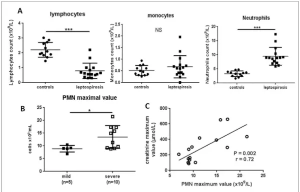
Unique neutrophilia during the course of leptospirosis in patients
The quantification of circulating immune cells at admittance (M0) showed marked lymphope-nia, no significant change in monocytes count, while there was a significant increase in PMN cells to 8.9 x10⁹/L on average (7.4–9.8) in patients with leptospirosis,Fig 2A. Notably, there was no difference between severe and mild forms regarding PMN counts determined at admit-tance (Table 1). We further analyzed PMN count and possible links with clinical hallmarks. There were no correlations between PMN counts in leptospirosis patients with either age, dura-tion of hospital stay, or number of damaged organs. Along the same lines, PMN count was not linked to the levels of the markers associated with organ injury such as AST, bilirubin, platelets, creatinine or creatinine phosphokinase levels. Moreover, PMN count was not statistically cor-related to plasma bacterial load at admittance for 12 of leptospirosis patients.
Table 1. Characteristics of leptospirosis patients group at admittance and comparison to controls. Characteristic (units) Controls Leptospirosis cases Mild
leptospirosis Severe leptospirosis P value controls vs. lepto mild vs. severe Number of individuals 13 15 5 10 Ratio M/F 12/1 14/1 5/0 9/1 NS NS Age (years) 34.1 (25.8– 48.6) 46.8 (28.2–61.4) 46 (24.6–46.8) 52.5 (28.6–61.9) NS NS Neutrophils (109/L) 3.4 (2.6–3.8) 8.9 (7.4–9.8) 9 (8.9–9.2) 8.7 (7.3–11.6) <0.0001 NS Lymphocytes (109/L) 2.0(1.9–2.7) 0.62 (0.53–0.92) 0.69 (0.56–0.84) 0.59 (0.42–0.91) <0.0001 NS Monocytes (109/L) 0.48(0.35– 0.64) 0.61 (0.39–0.74) 0.65 (0.61–0.76) 0.52 (0.38–0.71) NS NS Platelets (109/L) 236 (226–260) 50 (34–103) 101 (95–194) 37.5 (30–49) 0.0001 0.02 Creatinine (μmol/L) 84 (81–91) 151 (99–341) 103 (96–112) 282 (115–399) 0.001 NS Total bilirubin (μmol/L) 11 (9–12) 41 (25–89) 16 (14–23) 53 (41–126) 0.0002 0.005
AST (IU/L) 26 (18–31) 70 (39–146) 39 (36–56) 97 (66–198) 0.0003 0.049
CPK (IU/L) 128 (106–216) 1273 (845–2378) 714 (324–1080) 1960 (1264–4911) 0.0013 NS CRP (mg/L) 0.5 (0.4–2) 194 (187–215) (n = 9) 194 (151–215) 203 (192–256)
(n = 4)
0.0001 NS
Plasma bacterial load*(bact./ mL) NA 262 (67–1560) (n = 12) 52 (24–163) (n = 4) 1002 (92–2689) (n = 8) NA 0.04
Data are expressed as medians (interquartile ranges). Statistics between 2 groups are performed with nonparametric unpaired tests (Mann-Whitney U-test) for quantitative variables, and with Khi-2 square test for categorical data. P value inferior to 0.05 was considered significant.
*Plasma bacterial load is inferred from plasma PCR values according to the log-transformed standard curve. (AST = aspartate aminotransferase; CPK = creatinine phosphokinase; CRP = C-reactive protein; NS = not significant)
In contrast, bacterial load was found to be positively correlated to the length of hospital stay (P = 0.03, r value at 0.8 with Spearman test) or the level of bilirubin (P = 0.03 and r = 0.64) and negatively correlated to the platelet counts (P = 0.02, r = 0.36).
Given that the duration of the disease prior to hospitalization was different from one patient to another, we also performed analyses with the maximal values of PMN counts during the dis-ease course. The maximal values of PMN were at a median of 9.7 x10⁹/L (9–15.4), and these values were observed in median at the second day after admittance (IQR 1–4). This corre-sponded to the 7thday (6–9) from the onset of symptoms (fever and myalgia).
The severe forms had a greater increase in PMN counts (P = 0.04) when considering the maximal value of PMN during hospitalization (Fig 2B). Moreover, the maximal value of PMN count presented a significant correlation with the maximal value of creatinine: r = 0.72, P = 0.002 (Fig 2C).
Fig 1. Bacterial burden is higher in severe leptospirosis. Plasma bacterial load is established from the plasma PCR values (n = 12) and using the log-transformed standard curve as described [27]. Horizontal bars indicate the median. Comparison with non-parametric Mann-Whitney test.*indicates P-value inferior to 0.05.
Expression of TLR2 and markers of cell activation on PMN in
leptospirosis
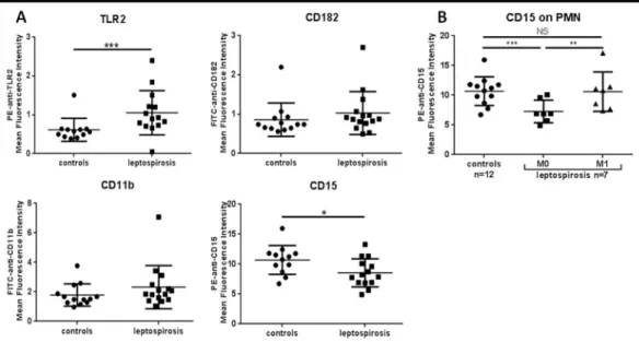
Next, we investigated the expression of selected PMN cell surface molecules implicated in LPS sensing, and cell activation (chemotaxis, adhesion and phagocytosis) for patients in the acute phase of the disease (M0) (Fig 3A).
Focusing on TLR2 expression levels on PMN, we observed a significant increase in the lep-tospirosis group (P = 0.001). The MFI values were 0.92 (0.75–1.21) versus 0.57 (0.48–0.63) in healthy controls.
In contrast, we found no differences between controls and leptospirosis cases at M0 regard-ing CD182 and CD11b surface levels on PMN identified by the CD16 counterstainregard-ing. Con-cerning CD15, the expression in leptospirosis group was decreased compared to controls.
The changes in the expression levels of CD182, CD11b, CD15 and TLR2 were not correlated to any of the clinical and biological markers of disease and tissue injuries. The levels of expres-sion of these receptors and PMN counts were further obtained from convalescent patients eval-uated at 1 month after discharge (M1). The median value of PMN decreased at M1: 4.7 x10⁹ cells/L (4.1–6.1) compared to 8.9 x10⁹cells/L (7.5–10.3) at M0 (P = 0.04). The levels for CD182 and CD11b between M0 and M1 remained identical. The level of CD15 expression on PMN at 1 month was significantly higher compared to the acute phase of leptospirosis: P = 0.02 with non-parametrical Wilcoxon test for paired data,Fig 3B. Of note, the MFI values at M1 post infection were not significantly different from the healthy controls and indicating a return to basal levels of expression for all PMN markers analyzed in our study.
Fig 2. Neutrophil levels at admittance and during the disease course of leptospirosis patients. (A) Counts of immune cells for healthy controls (circles) and leptospirosis patients (squares) at admittance (M0). (B) The maximal value of PMN count reached during hospital stay was compared between mild and severe forms. For A and B, the largest horizontal bars indicate the median value, upper and lower bars the interquartile ranges. Comparisons with non-parametric Mann-Whitney test.*,***indicate P-value inferior to 0.05 and 0.0001 respectively. (C) Among the patients with leptospirosis, the maximal value of PMN was correlated to the maximal value of creatinine with Spearman test.
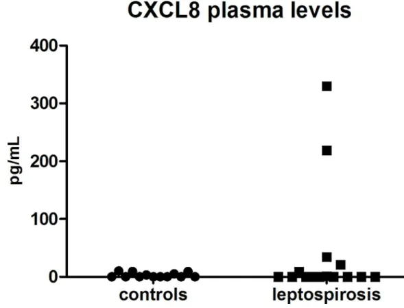
Finally, we investigated the levels of CXCL8 in plasma of the 15 patients and 13 healthy con-trols. The chemokine was barely detectable (<10 pg/mL), except for four of the severe leptospi-rosis cases with values of 21, 34, 219 and 330 pg/mL (Fig 4). Three of these patients had septic shock with multiorgan failure and one of them died.
Discussion
Innate immune cells such as PMN play a critical role to control bacterial infection. In this study we showed that the expression of several cell surface PMN molecules, implicated in che-motaxis and recruitment to inflamed tissues, were either not upregulated when compared to healthy subject controls, or decreased. Moreover, CXCL8, a major chemokine for leukocyte recruitment was barely elevated in leptospirosis. Counterintuitively, only four patients with severe forms of the disease had elevated levels of CXCL8.
To our knowledge, this is the first study assessing the capacity of circulating PMN to be acti-vated during the acute phase of leptospirosis in humans. With the limits of partial exploration of the complex process implicated in the chemotaxis, adhesion and phagocytosis of PMN, our results indicate unique behavior between leptospirosis patients and those with classical Gram-negative multiple organ infection, as discussed below.
The studied population presented mild and severe forms of leptospirosis. Cases were con-firmed by PCR or IgM serological assay. Although the number of individuals included in the study is limited, this number is sufficient to evaluate significant variations in cell counts and cell immunophenotyping.
The observed neutrophilia during the acute phase of leptospirosis was in agreement with previous reports. Moderate increase of PMN counts have been reported in patients with
Fig 3. Expression of cell surface molecules implicated in PMN recruitment and activation during
leptospirosis. (A) Expression levels of receptors and ligands on circulating PMN at admittance, assessed by flow cytometry. The mean fluorescence intensity (MFI) values were evaluated while isotypic negative controls were set with an MFI of 10−1. One value is missing for CD15. (B) Evolution of CD15 expression among 7 leptospirosis
patients and comparison between admittance (M0) and at 1-month (M1) post infection. Largest horizontal bars indicate the median value, upper and lower bars the interquartile ranges. Comparison of the patient group and healthy donor group with non-parametric Mann-Whitney test, and comparison within patients between M0 and M1 with non-parametric Wilcoxon paired test.*,**,***indicate P-value inferior to 0.05, 0.001 and 0.0001
respectively.
Leptospira and particularly in the most severe forms [4–6]. Neutrophilia has also been found in other Gram-negative bacterial infection and to control the infectious challenge and to limit tis-sue damage [29,30].
Interestingly, we found that this neutrophilia in blood sampled at admittance (M0 stage) was not correlated to the level of major organ injuries either at the clinical or biological levels. In contrast, a significant association was observed between the maximal PMN count and the maximum creatinine level during the disease course of leptospirosis.
Interestingly, the levels of CXCL8 was low in our leptospirosis cohort and contrasting to data published for classical Gram-negative severe sepsis cases [19]. CXCL8 was increased only in 4 patients and in the context of the more severe forms of the disease. Literature data have also indicated low levels of CXCL8 in human leptospirosis [31] but several other reports dem-onstrated increased levels and correlation to severity and mortality [32–34]. How can we possi-bly reconcile such discrepancies? The differences may be a consequence of different infecting serovars and this information is unfortunately lacking in many of the published studies. Another explanation may be that CXCL8 levels may be fluctuating during the disease course. Papa et al. recently demonstrated that CXCL8 peaked at 6–10 days post-infection [35]. In our study we sampled CXCL8 levels at 5 days post-infection in median. It would be interesting to study whether other chemokines are differentially expressed in leptospirosis. Experiments along these lines are now highly warranted.
Fig 4. Circulating CXCL8 (interleukin 8) levels are not significantly elevated in leptospirosis cases. CXCL8 levels were assessed by ELISA of plasma from healthy control individuals (circles, n = 13) and leptospirosis cases (squares, n = 15). For leptospirosis cases, assessment was performed with sampling at hospital admittance corresponding to acute phase (M0), with PCR positive patients. Comparison between two groups with non-parametric Mann-Whitney U-test showed no significant difference.
TLR2 and TLR4 regulate important PMN functions such as release of chemokines, produc-tion of reactive oxygen species (ROS), and activaproduc-tion of major proinflammatory signaling path-ways [36]. The expression levels of cell surface TLR2 and TLR4 on PMN is usually
up-regulated upon stimulation by bacterial products, LPS or lipopeptides, as well as during sepsis [36,37]. TLR expression levels on PMN during infection by Leptospira have never been reported. It has been demonstrated that LPS of Leptospira could signal through murine TLR4 and TLR2 but that human TLR2 is the major receptor for leptospiral LPS [26,38]. Noticeably, it has been shown that a specific polymorphism in human TLR2 gene was associated with an increased risk of leptospirosis and influencing its severity [39]. Hence, we focused on TLR2 expression and showed that it was significantly up-regulated during leptospirosis. It has been found that TLR2 is upregulated on endothelial cells in lung samples of fatal leptospirosis [40]. TLR2 stimulation by LPS should induce CXCL8 synthesis and secretion at least in vitro [36] and yet this was not detected in our patients. This would indicate that the innate immune response of circulating PMN in the presence of LPS from Leptospira is tightly regulated.
With regards to the activation status of PMN, this work indicates no major changes in the expression of CD182, CD11b while CD15 expression was decreased. The number of receptors studied is limited and other key effectors molecules should also be considered and studied. The membrane-bound receptors studied are nevertheless representative of the main steps of the PMN adhesion cascade to an activated endothelium [15,21]. It remains to be ascertained whether the levels of LFA-1 (integrin CD11a/CD18) or PSGL1, the P-selectin glycoprotein ligand 1 are affected on PMN during the course of leptospirosis. CD15 is a glycoprotein that binds to the major P-, E- and L-selectins indispensable for the rolling step of PMN[41]. Data from other studies regarding CD15 expression in the setting of sepsis are scarce but clearly indicated a basal or increased CD15 expression [42,43]. Thus, the decrease of CD15 level in leptospirosis cases might reflect a poor capacity of circulating PMN to be recruited to the tis-sues infected by Leptospira. CD11b (alpha subunit of CR3) is crucial for the slow rolling, adher-ence and transendothelial migration towards inflamed tissues as well as phagocytosis of complement opsonized-bacteria [15]. During the coordinated response to canonical bacterial sepsis, PMN activation is accompanied by CD11b up-regulation [44–46]. Moreover, infection by the spirochete Borrelia burgdorferi demonstrated an activation state of PMN with an increase of CR3 expression [47]. In our study we did not observe such increase. This unique observation adds to the plausible impaired innate immune function of PMN in leptospirosis.
CD182 is a receptor for several important CXCL chemokines, including CXCL8, and it induces activation of PMN during the rolling step next to an activated endothelium. As men-tioned previously, sepsis is usually associated with CD182 down-regulation [22,23,45], a mech-anism thought to participate to immune paralysis during sepsis and septic shock. This down-regulation may be induced either by high amounts of CXCL8, the most potent ligand of CXCR2 or by TLR2 stimulation as demonstrated by Alves-Filho et al [24]. This feed-back loop would allow PMN to respond to high CXCL8 gradient released by the vasculature and in order to invade inflamed tissues [21]. Of note, CXCL8 and CD182 are not the only ligand/receptor to engage PMN chemotaxis and lower expression of CD182 is not inevitably associated to chemo-taxis impairment as demonstrated by Sabroe et al [25]. As a note of caution, we have studied the phenotype of circulating PMN at a given time of the disease process (acute phase of the dis-ease-M0- and in convalescent phase–M1) and longitudinal studies are now warranted. Inter-estingly, the levels of PMN cell markers returned to control basal levels at M1 post infection. The activation status of PMN can change greatly between bloodstream and inflamed tissues [48] and despite the difficulties in performing such studies in humans, experiments along these lines are warranted.
Conclusion
Our study provides unique information regarding the phenotype and possible functional impairments of PMN during the process of Leptospira infection. In sharp contrast to the para-digm reported in Gram-negative sepsis, CD182 expression remained at basal levels, CD11b expression did not increase and CD15 level was down regulated. Taken together our novel observations question the ability of PMN to be mobilized into inflamed target-tissues during leptospirosis; PMN migration and phagocytic functions may also be impaired. Further studies should be performed to address the expression of other receptors essential for PMN’s mobiliza-tion as well as performing funcmobiliza-tional phagocytic assays in the presence of Leptospira.
Acknowledgments
The authors thank the practitioners implicated in the patients’ care as well as the members of the microbiology laboratory of St Denis, La Réunion. We also thank the French national refer-ence centre of leptospirosis, Pasteur Institute in Paris, for performing MAT testing.
Author Contributions
Conceptualization: LR PG MCJB. Data curation: LR PG. Formal analysis: LR PG. Funding acquisition: PG. Investigation: LR CG. Methodology: LR PG MCJB AMP. Resources: LR DV BK FR AR. Software: LR CG AMP. Validation: LR PG CG AMP.Writing – original draft: LR PG MCJB. Writing – review & editing: LR CG MCJB PG.
References
1. Costa F, Hagan JE, Calcagno J, Kane M, Torgerson P, Martinez-Silveira MS, et al. Global Morbidity and Mortality of Leptospirosis: A Systematic Review. PLoS Negl Trop Dis. 2015; 9: e0003898. doi:10. 1371/journal.pntd.0003898PMID:26379143
2. Levett PN. Leptospirosis. Clin Microbiol Rev. 2001; 14: 296–326. doi:10.1128/CMR.14.2.296–326. 2001PMID:11292640
3. Meri T, Murgia R, Stefanel P, Meri S, Cinco M. Regulation of complement activation at the C3-level by serum resistant leptospires. Microb Pathog. 2005; 39: 139–147. doi:10.1016/j.micpath.2005.07.003
PMID:16169184
4. Craig SB, Collet TA, Wynwood SJ, Smythe LD, Weier SL, McKay DB. Neutrophil counts in leptospiro-sis patients infected with different serovars. Trop Biomed. 2013; 30: 579–583. PMID:24522125
5. De Silva NL, Niloofa M, Fernando N, Karunanayake L, Rodrigo C, De Silva HJ, et al. Changes in full blood count parameters in leptospirosis: a prospective study. Int Arch Med. 2014; 7: 31. doi:10.1186/ 1755-7682-7-31PMID:25018781
6. Craig SB, Graham GC, Burns M-A, Dohnt MF, Smythe LD, McKay DB. Haematological and clinical-chemistry markers in patients presenting with leptospirosis: a comparison of the findings from
uncomplicated cases with those seen in the severe disease. Ann Trop Med Parasitol. 2009; 103: 333– 341. doi:10.1179/136485909X435058PMID:19508751
7. Scocchi M, Romeo D, Cinco M. Antimicrobial activity of two bactenecins against spirochetes. Infect Immun. 1993; 61: 3081–3083. PMID:8514417
8. Murgia R, Garcia R, Cinco M. Leptospires are killed in vitro by both oxygen-dependent and -indepen-dent reactions. Infect Immun. 2002; 70: 7172–7175. doi:10.1128/IAI.70.12.7172-7175.2002PMID:
12438405
9. Cinco M, Perticarari S, Presani G, Dobrina A, Liut F. Biological activity of a peptidoglycan extracted from Leptospira interrogans: in vitro studies. J Gen Microbiol. 1993; 139: 2959–2964. doi:10.1099/ 00221287-139-12-2959PMID:8126423
10. Arau´jo AM, Reis EAG, Athanazio DA, Ribeiro GS, Hagan JE, Araujo GC, et al. Oxidative stress mark-ers correlate with renal dysfunction and thrombocytopenia in severe leptospirosis. Am J Trop Med Hyg. 2014; 90: 719–723. doi:10.4269/ajtmh.13-0667PMID:24493675
11. Scharrig E, Carestia A, Ferrer MF, Ce´dola M, Pretre G, Drut R, et al. Neutrophil Extracellular Traps are Involved in the Innate Immune Response to Infection with Leptospira. PLoS Negl Trop Dis. 2015; 9: e0003927. doi:10.1371/journal.pntd.0003927PMID:26161745
12. Wang B, Sullivan J, Sullivan GW, Mandell GL. Interaction of leptospires with human polymorphonu-clear neutrophils. Infect Immun. 1984; 44: 459–464. PMID:6715045
13. Troxell B, Zhang J-J, Bourret TJ, Zeng MY, Blum J, Gherardini F, et al. Pyruvate protects pathogenic spirochetes from H2O2 killing. PLoS ONE. 2014; 9: e84625. doi:10.1371/journal.pone.0084625
PMID:24392147
14. Medzhitov R. Inflammation 2010: new adventures of an old flame. Cell. 2010; 140: 771–776. doi:10. 1016/j.cell.2010.03.006PMID:20303867
15. Ley K, Laudanna C, Cybulsky MI, Nourshargh S. Getting to the site of inflammation: the leukocyte adhesion cascade updated. Nat Rev Immunol. 2007; 7: 678–689. doi:10.1038/nri2156PMID:
17717539
16. Dobrina A, Nardon E, Vecile E, Cinco M, Patriarca P. Leptospira icterohemorrhagiae and leptospire peptidolgycans induce endothelial cell adhesiveness for polymorphonuclear leukocytes. Infect Immun. 1995; 63: 2995–2999. PMID:7542637
17. Goeijenbier M, Gasem MH, Meijers JCM, Hartskeerl RA, Ahmed A, Goris MGA, et al. Markers of endo-thelial cell activation and immune activation are increased in patients with severe leptospirosis and associated with disease severity. J Infect. 2015; 71: 437–446. doi:10.1016/j.jinf.2015.05.016PMID:
26048204
18. Atzingen MV, Go´mez RM, Schattner M, Pretre G, Gonc¸ales AP, de Morais ZM, et al. Lp95, a novel lep-tospiral protein that binds extracellular matrix components and activates e-selectin on endothelial cells. J Infect. 2009; 59: 264–276. doi:10.1016/j.jinf.2009.07.010PMID:19665803
19. Jean-Baptiste E. Cellular mechanisms in sepsis. J Intensive Care Med. 2007; 22: 63–72. doi:10.1177/ 0885066606297123PMID:17456726
20. Remick DG. Interleukin-8. Crit Care Med. 2005; 33: S466–467. PMID:16340423
21. Phillipson M, Kubes P. The neutrophil in vascular inflammation. Nat Med. 2011; 17: 1381–1390. doi:
10.1038/nm.2514PMID:22064428
22. Cummings CJ, Martin TR, Frevert CW, Quan JM, Wong VA, Mongovin SM, et al. Expression and func-tion of the chemokine receptors CXCR1 and CXCR2 in sepsis. J Immunol. 1999; 162: 2341–2346. PMID:9973513
23. Stadtmann A, Zarbock A. CXCR2: From Bench to Bedside. Front Immunol. 2012; 3: 263. doi:10.3389/ fimmu.2012.00263PMID:22936934
24. Alves-Filho JC, Freitas A, Souto FO, Spiller F, Paula-Neto H, Silva JS, et al. Regulation of chemokine receptor by Toll-like receptor 2 is critical to neutrophil migration and resistance to polymicrobial sepsis. Proc Natl Acad Sci USA. 2009; 106: 4018–4023. doi:10.1073/pnas.0900196106PMID:19234125
25. Sabroe I, Jones EC, Whyte MKB, Dower SK. Regulation of human neutrophil chemokine receptor expression and function by activation of Toll-like receptors 2 and 4. Immunology. 2005; 115: 90–98. doi:10.1111/j.1365-2567.2005.02133.xPMID:15819701
26. Werts C, Tapping RI, Mathison JC, Chuang TH, Kravchenko V, Saint Girons I, et al. Leptospiral lipo-polysaccharide activates cells through a TLR2-dependent mechanism. Nat Immunol. 2001; 2: 346– 352. doi:10.1038/86354PMID:11276206
27. Raffray L, Giry C, Thirapathi Y, Binois F, Moiton M-P, Lagrange-Xelot M, et al. High leptospiremia is associated with low gamma-delta T cell counts. Microbes Infect. 2015; 17: 451–455. doi:10.1016/j. micinf.2015.04.001PMID:25899947
28. Levett PN, Morey RE, Galloway RL, Turner DE, Steigerwalt AG, Mayer LW. Detection of pathogenic leptospires by real-time quantitative PCR. J Med Microbiol. 2005; 54: 45–49. doi:10.1099/jmm.0. 45860-0PMID:15591254
29. Alves-Filho JC, de Freitas A, Spiller F, Souto FO, Cunha FQ. The role of neutrophils in severe sepsis. Shock. 2008; 30 Suppl 1: 3–9. doi:10.1097/SHK.0b013e3181818466PMID:18704017
30. Mulvey MA, Schilling JD, Martinez JJ, Hultgren SJ. Bad bugs and beleaguered bladders: interplay between uropathogenic Escherichia coli and innate host defenses. Proc Natl Acad Sci USA. 2000; 97: 8829–8835. PMID:10922042
31. Kyriakidis I, Samara P, Papa A. Serum TNF-α, sTNFR1, IL-6, IL-8 and IL-10 levels in Weil’s syndrome. Cytokine. 2011; 54: 117–120. doi:10.1016/j.cyto.2011.01.014PMID:21316985
32. Rizvi M, Azam M, Ajmal MR, Shukla I, Malik A. Prevalence of leptospira in acute hepatitis syndrome and assessment of IL-8 and TNF-alpha level in leptospiral hepatitis. Ann Trop Med Parasitol. 2011; 105: 499–506. doi:10.1179/1364859411Y.0000000041PMID:22185944
33. Reis EAG, Hagan JE, Ribeiro GS, Teixeira-Carvalho A, Martins-Filho OA, Montgomery RR, et al. Cyto-kine response signatures in disease progression and development of severe clinical outcomes for lep-tospirosis. PLoS Negl Trop Dis. 2013; 7: e2457. doi:10.1371/journal.pntd.0002457PMID:24069500
34. Wagenaar JFP, Gasem MH, Goris MGA, Leeflang M, Hartskeerl RA, van der Poll T, et al. Soluble ST2 levels are associated with bleeding in patients with severe Leptospirosis. PLoS Negl Trop Dis. 2009; 3: e453. doi:10.1371/journal.pntd.0000453PMID:19488407
35. Papa A, Kotrotsiou T. Cytokines in human leptospirosis. Trans R Soc Trop Med Hyg. 2015; 109: 749– 754. doi:10.1093/trstmh/trv095PMID:26626338
36. Sabroe I, Dower SK, Whyte MKB. The role of Toll-like receptors in the regulation of neutrophil migra-tion, activamigra-tion, and apoptosis. Clin Infect Dis. 2005; 41 Suppl 7: S421–426. doi:10.1086/431992
PMID:16237641
37. Ha¨rter L, Mica L, Stocker R, Trentz O, Keel M. Increased expression of toll-like receptor-2 and -4 on leukocytes from patients with sepsis. Shock. 2004; 22: 403–409. PMID:15489631
38. Goris MGA, Wagenaar JFP, Hartskeerl RA, van Gorp ECM, Schuller S, Monahan AM, et al. Potent innate immune response to pathogenic leptospira in human whole blood. PLoS ONE. 2011; 6: e18279. doi:10.1371/journal.pone.0018279PMID:21483834
39. Ce´dola M, Chiani Y, Pretre G, Alberdi L, Vanasco B, Go´mez RM. Association of Toll-like receptor 2 Arg753Gln and Toll-like receptor 1 Ile602Ser single-nucleotide polymorphisms with leptospirosis in an Argentine population. Acta Trop. 2015; 146: 73–80. doi:10.1016/j.actatropica.2015.03.007PMID:
25784560
40. Del Carlo Bernardi F, Ctenas B, da Silva LFF, Nicodemo AC, Saldiva PHN, Dolhnikoff M, et al. Immune receptors and adhesion molecules in human pulmonary leptospirosis. Hum Pathol. 2012; 43: 1601– 1610. doi:10.1016/j.humpath.2011.11.017PMID:22436623
41. Foxall C, Watson SR, Dowbenko D, Fennie C, Lasky LA, Kiso M, et al. The three members of the selectin receptor family recognize a common carbohydrate epitope, the sialyl Lewis(x) oligosaccha-ride. J Cell Biol. 1992; 117: 895–902. PMID:1374413
42. Weinschenk NP, Farina A, Bianchi DW. Premature infants respond to early-onset and late-onset sep-sis with leukocyte activation. J Pediatr. 2000; 137: 345–350. doi:10.1067/mpd.2000.107846PMID:
10969258
43. Januszkiewicz A, Lore´ K, Esse´n P, Andersson B, McNurlan MA, Garlick PJ, et al. Response of in vivo protein synthesis in T lymphocytes and leucocytes to an endotoxin challenge in healthy volunteers. Clin Exp Immunol. 2002; 130: 263–270. doi:10.1046/j.1365-2249.2002.01983.xPMID:12390314
44. Sabroe I, Jones EC, Usher LR, Whyte MKB, Dower SK. Toll-like receptor (TLR)2 and TLR4 in human peripheral blood granulocytes: a critical role for monocytes in leukocyte lipopolysaccharide responses. J Immunol. 2002; 168: 4701–4710. PMID:11971020
45. Chishti AD, Shenton BK, Kirby JA, Baudouin SV. Neutrophil chemotaxis and receptor expression in clinical septic shock. Intensive Care Med. 2004; 30: 605–611. doi:10.1007/s00134-004-2175-yPMID:
14991094
46. Russwurm S, Vickers J, Meier-Hellmann A, Spangenberg P, Bredle D, Reinhart K, et al. Platelet and leukocyte activation correlate with the severity of septic organ dysfunction. Shock. 2002; 17: 263–268. PMID:11954824
47. Cinco M, Panfili E, Presani G, Perticarari S. Interaction with Borrelia burgdorferi causes increased expression of the CR3 integrin and increased binding affinity to fibronectin via CR3. J Mol Microbiol Biotechnol. 2000; 2: 575–579. PMID:11075934
48. Fortunati E, Kazemier KM, Grutters JC, Koenderman L, Van den Bosch van JMM. Human neutrophils switch to an activated phenotype after homing to the lung irrespective of inflammatory disease. Clin Exp Immunol. 2009; 155: 559–566. doi:10.1111/j.1365-2249.2008.03791.xPMID:19077082

![Fig 1. Bacterial burden is higher in severe leptospirosis. Plasma bacterial load is established from the plasma PCR values (n = 12) and using the log-transformed standard curve as described [27]](https://thumb-eu.123doks.com/thumbv2/123doknet/13786596.440109/7.918.209.856.111.728/bacterial-leptospirosis-plasma-bacterial-established-transformed-standard-described.webp)