HAL Id: hal-02110834
https://hal-amu.archives-ouvertes.fr/hal-02110834
Submitted on 25 Apr 2019HAL is a multi-disciplinary open access archive for the deposit and dissemination of sci-entific research documents, whether they are pub-lished or not. The documents may come from teaching and research institutions in France or abroad, or from public or private research centers.
L’archive ouverte pluridisciplinaire HAL, est destinée au dépôt et à la diffusion de documents scientifiques de niveau recherche, publiés ou non, émanant des établissements d’enseignement et de recherche français ou étrangers, des laboratoires publics ou privés.
Antimicrobial Agents With Bimodal Activity
Marine Blanchet, Diane Borselli, Anne Rodallec, Franck Peiretti, Nicolas
Vidal, Jean-Michel Bolla, Carole Digiorgio, Kelly Morrison, William Wuest,
Jean Michel Brunel
To cite this version:
Marine Blanchet, Diane Borselli, Anne Rodallec, Franck Peiretti, Nicolas Vidal, et al.. Claramines: A New Class Of Broad-Spectrum Antimicrobial Agents With Bimodal Activity. ChemMedChem, Wiley-VCH Verlag, 2018, 13 (10), pp.1018-1027. �10.1002/cmdc.201800073�. �hal-02110834�
Claramines : A new class of broad-spectrum antimicrobial agents with bimodal activity Marine Blanchet[a], Diane Borselli[b], Anne Rodallec[a], Franck Peiretti[c], Nicolas Vidal[d], Jean-Michel Bolla[b], Carole Digiorgio[e], Kelly R. Morrison[f], William M. Wuest[f] and Jean
Michel Brunel[a]* [a] Dr. M. Blanchet, Dr. A. Rodallec, Dr. J. M. Brunel
Centre de Recherche en Cancérologie de Marseille (CRCM), CNRS, UMR 7258, Institut Paoli Calmette, Aix-Marseille Université
UM 105, Inserm, U1068, Faculté de pharmacie, 13385 Marseille, France. E-mail : bruneljm@yahoo.fr
[b] Dr. D. Borselli, Dr. J. M. Bolla
Aix-Marseille Université, IRBA, TMCD2 UMR-MD1 Faculté de Médecine, 13385 Marseille, France.
[c] Dr. Fr. Peiretti
Aix Marseille Université, INSERM 1263, INRA1260, C2VN, 13385 Marseille, France. [d] N. Vidal
YELEN, 10 bd tempête, 13820 Ensues la redonne, France. [e] Dr. C. Digiorgio
Aix Marseille Université, CNRS, IRD, IMBE UMR 7263, Laboratoire de Mutagénèse Environnementale, 13385 Marseille, France.
[f] K. R. Morrison, Pr. W. M. Wuest
Emory University, Department of Chemistry, 1515 Dickey Dr. Atlanta GA 30322, USA. Supporting information for this article is given via a link at the end of the document.
Abstract:
The emergence of multidrug resistant bacteria/pathogens has highlighted the need for the development of new antibiotics. In this manuscript, we report herein our results dealing with the broad spectrum of antibacterial activity against both sensitive and resistant Gram-negative and Gram-positive bacterial strains of an easily prepared water soluble polyaminosterol compound namely claramine A1. We will also demonstrate its peculiar mechanism of action (different from all the well-known classes of antibiotics) towards Gram-negative and Gram-positive bacteria. Finally, due to its low cytotoxicity, this class of molecules could constitute an effective response to combat the emergence of multidrug resistant bacteria and nosocomial diseases.Over the last 75 years, the development of antibiotics has contributed greatly to the increase in life expectancy. Unfortunately, as a consequence of both their overuse and natural defense mechanisms, multidrug resistance has emerged and has seriously limited the efficacy of antibiotic therapy.[1-3] Since 1928, thousands of compounds which target essential pathways have been isolated from natural sources. Nevertheless, few classes have been identified since 1987, and when coupled with the over-prescribing of existing treatments, have led to a drastic increase in resistant bacteria. Antibiotic resistance is an issue of great concern that has attracted the attention of health agencies, media, and global leaders. More specifically, the WHO, European Commission, and CDC have all run public health awareness campaigns to bring greater attention to a specific subset of bacteria, named the ESKAPE pathogens, comprised of Enterococcus faecium, Staphylococcus aureus, Klebsiella pneumoniae, Acinetobacter baumannii, Pseudomonas aeruginosa, and Enterobacter spp., which constitute the main cause of nosocomial infections throughout the world. In this context, there is an urgent medical need for new classes of antimicrobial agents that both target resistant ESKAPE pathogens and be refractory to all bacterial resistance mechanisms. These new antibiotics should present the following features: (i) a mechanism of action ideally avoiding, but more likely prolonging, the rise of antibiotic-resistant strains, (ii) specific targeting of pathogens without affecting the commensal microbiota and (iii) overcoming or limiting natural forms of resistance such as the formation of biofilms and efflux mechanisms. Bacterial membrane structure constitutes an appealing target for the design of new potent antimicrobial agents since it is generally conserved among most species of both Gram-negative and Gram-positive bacteria. This is evident in Nature as bacteria have developed antimicrobial peptides and other small molecules like the polymyxins which target this structural element.[4] Resistance to membrane active compounds requires either an increase in efflux pumps or a major change in membrane structure that in turn influences the permeability barrier, increasing susceptibility to hydrophobic antibiotics.
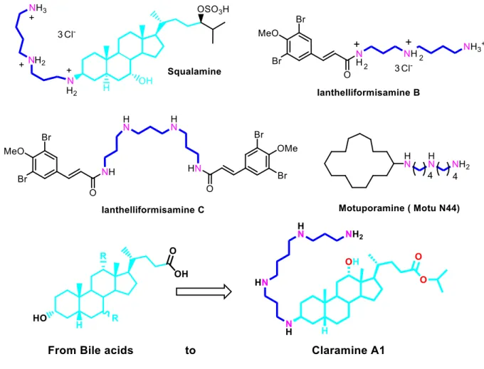
Polyamino natural products such as squalamine[5,6], ianthelliformisamines (B and C)[7], and motuporamines[8] are known to target bacterial cell membranes with modest activity (Figure 1).
Figure 1. Strategy involved for claramine A1 design
Thus, we and others were able to demonstrate the importance of the amphiphilicity of the molecules as well as the crucial nature of the polyamino group linked to a well-chosen lipophilic molecule to encounter interesting antimicrobial activities.[9-12]
Results and Discussion
Based on our continued interest in squalamine and polyaminosterol derivatives[13-14] we posited
that the bile acids sterol core could serve as a platform for the design of potent antimicrobial compounds. Based on all these considerations, we postulated that we could couple these two structural features to design a new class of polyaminosterol compounds. This hypothesis was born out in the first derivative disclosed below which we have named claramine A1. Herein,
we present the synthesis of novel antimicrobial derivative claramine A1 demonstrating its broad-spectrum activity as well as its antibiotic-enhancement properties, and elucidation of its unusual mechanism of action against both Gram-positive and Gram-negative bacteria by implementing chemical tools to investigate changes in membrane depolarization and permeabilization.
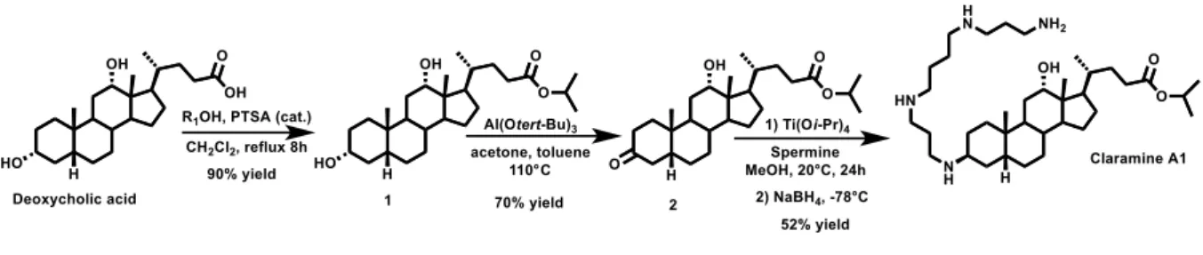
In order to further investigate the interesting biological activity[5] we were interested in designing a concise and economical synthesis of squalamine derivatives. Toward this end, polyaminosterol parent compound claramine A1 (Figure S1) was prepared in a three-step synthesis (33% overall yield) from deoxycholic acid using an efficient stereoselective titanium-mediated reductive amination that controlled the −stereochemistry at the C-3 position (Figure 2).
Figure 2. Claramine A1 synthetic pathway
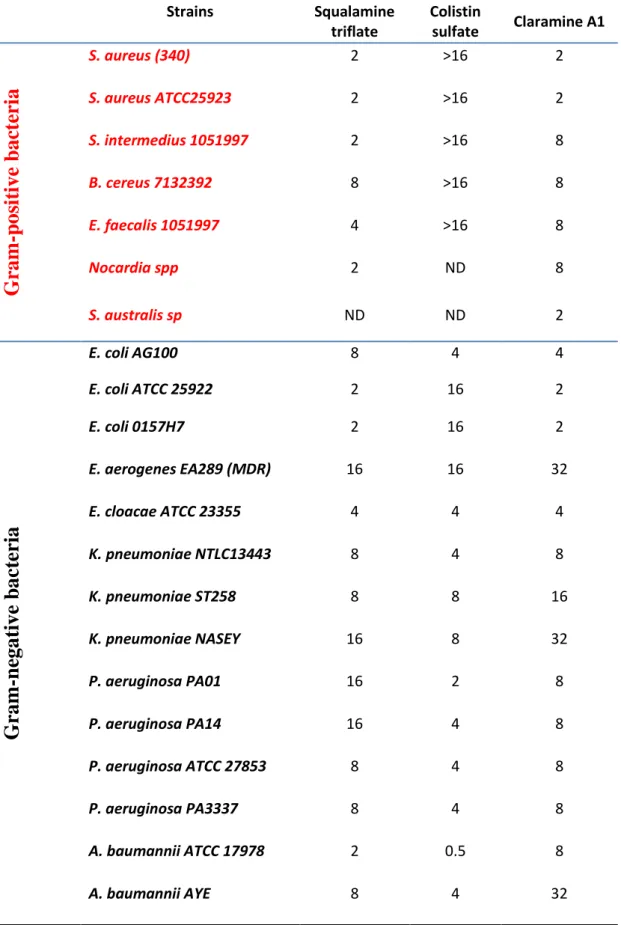
Claramine A1 was found to be a potent antimicrobial with activity against a wide panel of both Gram-positive and Gram-negative bacteria including Multi-Drug Resistant (MDR) pathogens (Table 1). Evaluation of the activities of claramine A1 and controls (squalamine and colistin) against reference strains was repeated and reproducible, and accurate MICs were obtained. For Gram-negative E. coli ATCC 25922 and P. aeruginosa ATCC 27853, MICs were 2, 2, 16 µg/mL and 8, 8, 4 µg/mL for squalamine, claramine A1, colistin, respectively. For Gram-positive S. aureus ATCC 25923, MICs were 2, 2, >16 µg/mL for squalamine, claramine A1 and colistin, respectively. In general, claramine A1 demonstrates antimicrobial activities against a large panel of both Gram-positive and negative bacterial strains with MIC values varying from 2 to 32 µg/mL (Table 1).
Strains Squalamine triflate Colistin sulfate Claramine A1
Gra
m
-p
o
si
ti
v
e bac
te
ri
a
S. aureus (340) 2 >16 2 S. aureus ATCC25923 2 >16 2 S. intermedius 1051997 2 >16 8 B. cereus 7132392 8 >16 8 E. faecalis 1051997 4 >16 8 Nocardia spp 2 ND 8 S. australis sp ND ND 2Gra
m
-ne
g
a
ti
v
e b
a
ct
eri
a
E. coli AG100 8 4 4 E. coli ATCC 25922 2 16 2 E. coli 0157H7 2 16 2 E. aerogenes EA289 (MDR) 16 16 32 E. cloacae ATCC 23355 4 4 4 K. pneumoniae NTLC13443 8 4 8 K. pneumoniae ST258 8 8 16 K. pneumoniae NASEY 16 8 32 P. aeruginosa PA01 16 2 8 P. aeruginosa PA14 16 4 8 P. aeruginosa ATCC 27853 8 4 8 P. aeruginosa PA3337 8 4 8 A. baumannii ATCC 17978 2 0.5 8 A. baumannii AYE 8 4 32Claramine A1 exhibited complete killing of the P. aeruginosa reference strain in the same time period (3 hours) than colistin (Figure 3B). However, contrary to colistin which is inactive against Gram-positive bacteria, claramine A1 showed a rapid direct bactericidal effect against the S. aureus reference strain which was favorable when compared to tobramycin, reflected by a nearly 2.5 log drop in the counts of this strain by 0.75 h, with complete killing achieved in 2 hours (Figure 3A).
Figure 3. Time kill studies for claramine A1 against (A) P. aeruginosa ATCC27853 and (B)
S. aureus ATCC25923
The first study dealing with the mechanism of action of squalamine against Gram-negative bacteria suggested that squalamine amino groups (positively-charged) strongly interact with the phosphate groups (negatively-charged) in the lipopolysaccharide (LPS) structure disrupting the bacterial membrane integrity.[11] Such a mechanism had been previously described for
polymyxin antibiotic colistin, which can be antagonized by increased concentrations of Ca2+ and Mg2+ divalent cations, which further inhibits the binding of this polycationic antibiotic to
LPS.[15,16] This result indicates that the interaction with the negatively charged phosphate groups in the LPS structure is mandatory for both agents to be active. Although required, the cationic charge is not sufficient to cause either the permeabilization of bacterial membranes or the bacteria killing.[17,18] On the other hand, Dynamic Light Scattering (DLS) was performed to determine whether the compound aggregates demonstrating that samples concentrations (i.e.,
500 µM, 50 µM, 16 µM and 2 µM) were too low to meet DLS quality criteria, suggesting the absence of colloidal aggregates at all tested concentrations (Figure S2).
Thus, we attempted to elucidate more precisely the mechanism of action of claramine A1 by investigating its potent permeabilizing, depolarizing, and disrupting behavior of the outer and/or inner membranes of S. aureus ATCC 25923 and E. aerogenes EA289 bacteria, as well as its ability to potentially act as an efflux pump inhibitor (Figure 4A).
Figure 4. General points investigated for claramine A1 mechanism of action study against both
Gram-positive and Gram-negative bacteria
Squalamine, polymyxin B, and cetyltrimethylammonium bromide (CTAB) were chosen for these studies as positive controls whereas claramine A19 (lacking the polyamine) and spermine (Sp, lacking the sterol) were selected as inactive compounds to compare their different behaviors (Figure 4B).
In a first approach, a bioluminescence method was developed to determine the behavior of claramine A1 on the intracellular pool of bacterial ATP. The detection of the external concentration of ATP was then used as a reporter reflecting the permeabilizing effect of claramine A1 along with providing a dose-response curve (Figure 5A). Claramine A1 dramatically disrupted the S. aureus membrane after two minutes as observed by intracellular ATP release kinetics, which was similar to the positive control squalamine. (Figure 5B).
Figure 5. Dose dependent ATP release for claramine A1 (A) treated S. aureus ATCC 25923
(B) and E. aerogenes EA289 (C) strains
Conversely, no significant effect was found by using spermine as a polyamine negative control during the test time. By performing the same experiment against Gram-negative E. aerogenes EA289 a lower ATP efflux was observed for both Claramine A1 and squalamine with only a 20 and 10% ATP efflux release compared to CTAB positive control, respectively, hinting at a different mechanism of action (Figure 5C). These results suggest that LPS damage induced by claramine A1 is clearly greater and faster than that caused by squalamine. Furthermore, this has recently been demonstrated in a study on the interaction of squalamine and colistin with the bacterial lipid bilayer and the consequences of such interactions on the electrical properties of these membranes.[19] Accordingly, the activity of claramine A1 against Gram-negative bacteria may be simulated by the carpet model previously proposed for describing cationic peptide
antibiotics, which led to a large disruption of the bacterial membrane due to a detergent-like effect of micelle formation.[20] However, this model is not valid for Gram-positive bacteria since they are devoided of LPS.
To clarify this alternate mechanism, claramine A1 was investigated for its ability to alter the outer membrane integrity of E. aerogenes EA289 via a nitrocefin colorimetric assay wherein a color change from yellow to red occurs when the chromogenic ß-lactam is efficiently hydrolyzed by periplasmic ß-lactamases. As presented in Figure 6, even at low concentration (1.79 µM), claramine A1 increased the rate of nitrocefin hydrolysis compared to the A19-treated or unA19-treated control. Otherwise, a similar behavior is observed as seen with the positive controls polymyxin-B (PMB) and squalamine which also provided an increase in the nitrocefin hydrolysis rate. All these data suggest that claramine A1 can permeabilize the outer membrane or disrupt the integrity of Gram-negative bacteria.
Figure 6. Study of outer membrane permeabilization of E. aerogenes EA289 by evaluation of
nitrocefin hydrolysis rate (A-C) and claramine A1 dose dependent effect on nitrocefin hydrolysis rate (D).
On the other hand, efflux systems function via an energy-dependent mechanism (active transport) to pump out unwanted substances such as toxins, antibiotics, or dyes through specific efflux pumps. To determine if claramine A1 could act as a disruptor of the transmembrane potential, we used the membrane potential sensitive probe DiSC3(5) which concentrates at the
When a compound impairs the membrane potential, the dye is released in the liquid environment leading to a fluorescence increase. Thus, a strong depolarization of the membrane after 15 minutes of incubation was observed against Gram-positive S. aureus strain both for squalamine and Claramine A1 (Figure 7B) whereas EA289 treatment resulted in a lower but significant depolarization of the membrane (Figure 7C) suggesting a potent disruption of the proton gradient which may affect efflux pumps from the RND family such as AcrAB-TolC.
Figure 7. A-D) Membrane perturbation study of E. aerogenes EA289 and S. aureus ATCC
25923 by DiSC3(5) fluorescence evaluation. E-F) Study of effect on efflux performance of E.
aerogenes EA289 by evaluation of glucose-triggered 1,2'-diNA efflux
Therefore, claramine A1 was tested for its ability to inhibit efflux pumps that are proteinaceous transporters localized in the cytoplasmic membrane of bacteria and are active transporters, meaning that they require a source of chemical energy to perform their function. (Figure 7E). Some efflux systems are drug-specific, whereas others may accommodate multiple drugs, and
thus contribute to bacterial multidrug resistance (MDR). Some are primary active transporters utilizing adenosine triphosphate hydrolysis as a source of energy, whereas others are secondary active transporters in which transport is coupled to an electrochemical potential difference created by pumping hydrogen or sodium ions from or to the outside of the cell. In this context, the transport measurement of a known transport substrate, can be used to directly monitor the function of efflux pumps. As already mentioned Gram-negative EA289 is a drug resistant bacterium overexpressing the AcrAB-TolC pump, which belongs to the RND efflux pumps and uses the proton gradient across the inner membrane as an energy source. By loading EA289 bacteria with the 1,2'-diNA dye which is well known to be a substrate of the AcrAB-TolC efflux pump and deenergization by adding CCCP (Figure 7E, step 2), we rendered the bacteria fluorescent after incubation. Bacteria were then incubated with claramine A1 (Figure 7E, step 3) at different concentrations before addition of glucose as an energy source (Figure 7E, step 4).
In this case, the active transport of more than 80% of the dye is rapidly observed in non-treated bacteria (Figure 7F, green line). When claramine A1 is added to the bacteria, a significant dose-dependent inhibition is observed resulting in more than 90% retention of the dye at a concentration up to 25 µM (Figure 7F, grey line). These results suggest that claramine A1 may act as an inhibitor of the AcrAB-TolC efflux pump. It is noteworthy than squalamine presents a similar behavior but at a higher concentration (of up to 100 µM) demonstrating the better efficiency of claramine A1.
An alternative strategy to safeguard the efficiency of old existing drugs is using antibiotic adjuvants, which can enhance the activity of current drugs and minimize or even directly block, resistance.[21-23] This concept of antibiotic adjuvants comes from the successful use of two antibiotic combinations such as gentamicin/ampicillin for the treatment of enterococcal infections.[24,25] Synergistic interactions are achieved through a variety of mechanisms,
including enhancement of uptake or suppression of efflux. The result is that efficacy is greater than the sum of the individual agents (synergy) with faster kill times and limiting the emergence of resistant organisms. Thus, we decided to use claramine A1 as a potent adjuvant in combination with various antibiotics based on our recent success using polyamine motuporamine derivatives.[6] We investigated whether this polyaminosterol derivative could restore the potency of the antibiotic doxycycline at doses below its MIC. In our hands the MIC of doxycycline against P. aeruginosa PAO1 and E. aerogenes EA289 was 16 and 40 µg/mL, respectively, so we investigated the use of doxycycline at a lower concentration (2 µg/mL, corresponding to its pharmacokinetic properties in humans[5]) in the presence of claramine A1. We speculated that the polyamine agents would disrupt bacterial membrane integrity and enhance antibiotic delivery to the bacteria and thus increase doxycycline potency. Thus, even at this low doxycycline concentration, claramine A1 restored doxycycline activity against E. aerogenes EA289 and P. aeruginosa PAO1 by using 0.5 and 2 µg/mL, respectively. Unfortunately, under the same experimental conditions no synergy was observed for MDR K. pneumoniae Nasey and A. baumannii AYE. Nevertheless, chloramphenicol activity can also be restored at 2 µg/mL against P. aeruginosa PAO1 by using 4 µg/mL of claramine A1 (Table 2).
Strains P. aeruginosa PAO1 E. aerogenes EA289 K. pneumoniae NASEY A. baumannii AYE
Antibiotica DOX CHL DOX CHL DOX DOX
A1b 2 4 0.5 60c 45c 30c
a antibiotic used: DOX: doxycycline, CHL: chloramphenicol. b Concentration of claramine A1 used (µg/mL). c
Values equivalent to the MIC of claramine A1 against these bacterial strains (cf Table 1)
Table 2. Concentration of claramine A1 used to restore antibiotics activity against various
bacterial strains
The cytotoxic activity of claramine A1 was also investigated. Cell viability was assessed by evaluating two different physiological mechanisms: the activity of mitochondrial dehydrogenases which cleave the tetrazolium salt WST-1 to formazan, and the ability of
lysosomal membranes to concentrate Neutral Red. Results showed that claramine A1 exerted a weak cytotoxic activity with an IC50 around 70 µM. No differences could be observed in IC50
regarding the cell line or the vital dye, suggesting that the cytotoxic activities did not result from a specific mechanism but from a global impact on several intracellular targets (Table 3).
IC50 (µM) as assessed by WST1 vital dye IC50 (µM) as assessed by neutral red vital dye
CHO HEPG2 MDCK 3T3 Hacat CHO HEPG2 MDCK 3T3 Hacat
70.1+2.6 81.3+2.6 74.3+11.2 77.4+ 3.8 86.4+3.5 75.3+2.4 79.8+3.4 79.4+6.5 82.6 + 0.7 83.1+5.3.2 IC50 Inhibitory Concentration 50% (µM)
Table 3. Cytotoxic activities of claramine A1
Conversely, claramine A1 was unable to induce micronuclei in CHO-K1 cells with or without S9 metabolizing mixture. They indicated that claramine A1 was deprived from direct clastogenic/aneugenic activity and that it was not transformed into clastogenic/aneugenic metabolites by P450 cytochromes (Table S1).
Because of its structural resemblance to steroidal detergents, claramine A1 was assayed for hemolytic activity against human erythrocytes. This activity occurs at higher concentrations than observed for nonselective membrane disruptive amphipathic molecules such as CTAB (Figure S3). Thus, 35% haemolysis was observed for claramine A1 used at a 200 µM concentration. It is also noteworthy that claramine A1 exerts antibiotic activity at concentrations below which erythrocyte disruption occurs.
Recently, the use of insects has become a method of choice for measuring the virulence of microbial pathogens due to the numerous similarities existing between the immune system of insects and mammals.[26, 27] Insects are also inexpensive to purchase, house, and led to results in 24–48 h. All these reasons rationalize the fact that insects can be used to evaluate the in vivo activity of antimicrobial drugs.[28, 29] Thus, Galleria mellonella larvae were used as an animal model to determine their tolerance to claramine A1 as well as the in vivo activity of claramine A1 against a S. aureus infection. In a first approach, the results indicate no toxicity up to a
concentration of 50 µg/mL however larvae inoculate with a dose of 100 µg/mL showed a 90% reduction in viability after 24 h. On the other hand, larvae inoculated with claramine A1 (3.125, 6.25, 12.5, 25 µg/mL) did not show signs of cuticular darkening, an unambiguous sign of acute toxicity (Figure 8A).
Figure 8. A) Larval survival 24h after administration with water or claramine A1 (3.125, 6.25,
12.5, 25, 50, 100, 1000 µg/mL). B) Survival of larvae infected with S. aureus ATCC25923 following administration with claramine A1 solution (6.25 µg/mL) 4h after infection.
In order to ascertain the in vivo activity of claramine A1, larvae were infected with S. aureus ATCC25923 (20 µL of a solution containing 107 bacterial cells) and subsequently administered with or without claramine A1 (6.25 µg/mL) 4h post infection. Those treated larvae showed 80% survival at 24h whereas only 10% of the untreated ones survived at the same time point (Figure 8B). Nevertheless, caution must be exercised in larvae use as the introduction of foreign material into the insect haemoceol can provoke a non-specific immune response.[30, 31]
Conclusion
More broadly, we demonstrated that claramine A1, the first member of a new class of potent antimicrobial agents, possesses an unusual multifaceted mechanism of action against both Gram-positive and Gram-negative bacteria. Claramine A1 exerts its bactericidal effect in a rapid fashion via membrane depolarization against Gram-positive bacteria thereby disrupting
its integrity. In contrast, further studies indicate that changes in the transmembrane electrical potential in Gram-negative E. aerogenes EA289 are correlated with the permeabilization of the cell membranes by claramine A1 leading (or concomitantly facilitating) an altered proton homeostasis. Finally, claramine A1 which can disrupt the proton gradient can be considered as a new member of the class of efflux pump inhibitors. These bimodal activities, when coupled with its synergizing abilities, highlights the enormous potential of this class of compounds to fight MDR bacteria. Studies are now underway to improve the activity and cytotoxicity of claramine derivatives to provide a potent human therapeutic.
Experimental Section
All solvents were purified according to reported procedures, and reagents were used as commercially available. Methanol, ethyl acetate, dichloromethane, ammonia and petroleum ether (35-60 °C) were purchased from SDS and used without further purification. Column chromatography was performed on SDS silica gel (70-230 mesh). 1H NMR and 13C NMR spectra were recorded in CDCl3 on a Bruker AC 250 spectrometer working at 250 MHz and 63
MHz, respectively (the usual abbreviations are used: s: singlet, d: doublet, t: triplet, q: quadruplet, m: multiplet). Tetramethylsilane was used as internal standard. All chemical shifts are given in ppm.
Synthesis of isopropyl deoxycholate 1
In a 250 mL two necked round flask was introduced in 70 mL of isopropanol and 30 mL of dichloromethane 10 g of deoxycholic acid (0.0255 mol) and 2.2 g of para-toluene sulfonic acid (0.013 mol). The mixture was heated at reflux under vigorous stirring for 8 hours. The solvents were subsequently removed and 100 mL of dichloromethane was added. The organic phase was washed 3 times with 50mL of NHCO3 (10%) solution. The aqueous phases were extracted twice
concentrated in vacuo to afford the expected product as a white solid in 90% yield. NMR 1H (250 MHz, CDCl3) : δ (ppm) = 4.94 (m, 1H), 3.92 (m, 1H), 3.53 (m, 1H), 2.31-2.09 (m, 2H), 1.78-0.85 (m, 38H), 0.61 (s, 3H). NMR 13C (63 MHz, CDCl 3) : δ (ppm) = 173.68, 72.86, 71.37, 67.17, 47.99, 47.05, 46.32, 41.95, 36.23, 35.86, 35.17, 35.11, 33.97, 33.38, 31.61, 30.80, 30.20, 28.49, 27.42, 27.05, 26.01, 23.58, 22.99, 21.70, 17.06, 12.53.
Synthesis of 3-oxo isopropyl deoxycholate 2
In a 250 mL two necked round flask was introduced in 100 mL of toluene and 50 mL of acetone 11 g of isopropyl deoxycholate 1 (0.025 mol). 2 equivalents of aluminum tert-butoxide (12.3 g, 0.050 mol) were subsequently added and stirring was maintained under reflux for 12 hours. 50 mL of a 2N H2SO4 solution was added and the mixture was stirred for an additional 1 hour.
The organic phase was washed 3 times of a 2N H2SO4 solution and 50 mL of water. The
combined organic phases were dried over Na2SO4, filtered, and concentrated in vacuo to afford
a crude product which was purified by flash chromatography on silica gel (ethylacetate/ petroleum ether (1/1)). The expected 3-oxo isopropyl deoxycholate 2 was successfully obtained as a white solid in 70% yield. NMR 1H (250 MHz, CDCl3) : δ (ppm) = 5.00 (m, 1H), 4.04 (m,
1H), 2.46-2.12 (m, 4H), 2.06-0.96 (m, 35H), 0.71 (s, 3H). NMR 13C (63 MHz, CDCl3) : δ (ppm)
= 212.74, 173.57, 72.98, 67.34, 48.22, 47.59, 46.70, 44.30, 42.33, 37.04, 36.92, 35.87, 35.05, 34.45, 34.11, 31.76, 31.00, 29.06, 27.40, 26.64, 25.55, 23.58, 22.38, 21.81, 17.42, 12.75.
Synthesis of 3-spermino isopropyl deoxycholate (claramine A1)
In a 100 mL two necked round flask was introduced 100 mL of methanol. A mixture of the ketone 2 (3.5 g, 8 10-3mol), titanium(IV) isopropoxide (7.15 mL, 2.4 10-2 mol), and spermine (3.2g, 1.6 10-2 mol) was stirred under argon at room temperature for 24 hours. After cooling the flask at -20°C, sodium borohydride (0.9 g, 2.4 10-2 mol) was then added and the resulting
mixture was stirred for additional 12 hours. The reaction was then quenched by adding water (4 mL). Stirring was continued at room temperature for 1 hour then the reaction mixture was filtered over a pad of Celite which was subsequently rinsed with NH4OH and methanol. The
mixture was concentrated in vacuo to afford the expected crude compound which was purified by flash chromatography on silica gel using CH2Cl2/MeOH/NH4OH (32%) 7:3:1 as eluent. The
expected claramine A1 was obtained as a viscous yellow oil in 52% yield. NMR 1H (250 MHz, CD3OD) : δ (ppm) = 4.96 (m, 1H), 3.94 (m, 1H), 2.88-2.46 (m, 13H), 2.39-2.18 (m, 2H), 2.04-1.12 (m, 40H), 1.01-0.95 (m, 7H), 0.71 (s, 3H). NMR 13C (63 MHz, CD3OD) : δ (ppm) = 175.66, 74.15, 68.95, 59.06, 50.74, 50.70, 49.30, 48.97, 48.42, 48.27, 47.77, 45.83, 44.08, 40.78, 37.58, 37.14, 36.82, 35.97, 35.92, 34.94, 34.59, 33.56, 32.72, 32.45, 30.52, 29.98, 28.84, 28.65, 28.40, 28.29, 27.64, 25.04, 24.10, 22.27, 17.71, 13.37. MS (ESI+): m/z 619.5519 ([M+H]+)
Synthesis of claramine A1 hydrochloride salt
Claramine A1 is dissolved in a minimum of anhydrous methanol and a anhydrous HCl solution in diethylether (2M, 8 equivalents) was slowly added under vigorous. The formed precipitate was filtrated, washed with anhydrous diethylether and dried under vacuum. The claramine A1 hydrochloride salt is obtained in a quantitative yield as a white solid stable to air and moisture.
Determination of minimal inhibitory concentrations
Antimicrobial activity of the compounds was studied by determination of minimal inhibitory concentrations (MIC) according to the NCCLS guidelines M7-A2 using the microbroth dilution methods. The bacteria strains were grown on trypticase soy agar (Becton Dickinson) at 37 °C for 24h. Inocula were prepared in TCE (tryptone 0.1%, NaCl 8%, wt/vol) by ajusting the turbidity at 623 nm to obtain 1-3 105 CFU/mL.
Antimicrobial activities of the compounds were determined by using a broth microdilution method performed in sterile 96-well microplates. All compounds were solubilized in water at a concentration of 10mM and were transferred to each microplate well in order to obtain a two-fold serial dilution in 100 µL of broth and 100 µL of inoculum containing 2-6 105 CFU of each bacterium were added to each well. Some wells were reserved for positive controls and inoculum viability. After 24 h incubation, MIC was defined for each agent from duplicate observations as the lowest concentration of compound allowing no visible growth.
Time-kill assays
Time-kill assays were conducted with concentrations corresponding to the MIC value of claramine A1, colistin and tobramycin against reference strains of P. aeruginosa ATCC27853 and S. aureus ATCC25923. Claramine A1, colistin or tobramycin was added to a bacterial suspension of approximately 5 × 10⁵ CFU/mL of each bacteria. 2 mL of the tested suspension were sampled at 0, 0.5, 1, 2, 3, and 4 h for viable cell counting that was conducted by spiral plating on Trypticase Soy Agar medium (bioMérieux, Craponne, France) followed by incubation at 37°C for 24 h.
Membrane depolarization assays
Bacteria were grown in MH broth for 24 h at 37°C and centrifuged at 10000 rpm at 20°C. The supernatant was discarded, and the bacteria were washed twice with buffered sucrose solution (250 mM) and magnesium sulfate solution (5 mM). The fluorescent dye 3,3′-diethylthiacarbocyanine iodide was added to a final concentration of 3 µM, and it could penetrate bacterial membranes during 1 h of incubation at 37°C. Bacteria were then washed to remove the unbound dye before adding compound A1 at different concentrations Compound was then added at different concentrations. Fluorescence measurements were performed using
a Jobin Yvon Fluoromax 3 spectrofluorometer with slit widths of 5/5 nm. The relative corrected fluorescence (RCF) was recorded at time intervals of 0, 3, 5, 7, 9, 11, 13, 15, 17, 19, and 21 min. The maximum RCF was that recorded with a pure solution of the fluorescent dye in buffer (3 µM).
Nitrocefin hydrolysis assay
Outer membrane permeabilization was measured using nitrocefin as a chromogenic substrate of periplasmic -lactamase. Ten milliliters of MH broth were inoculated with 0.1 mL of an overnight culture of EA289 bacteria and grown at 37°C until the OD600 nm reached 0.5. The remaining steps were performed at room temperature. Cells were recovered by centrifugation (4000 rpm for 20 min) and washed once in 20 mM potassium phosphate buffer (pH 7.2) containing MgCl2 (1 mM). After a second centrifugation, the pellet was re-suspended and
adjusted to a OD600 nm to 0.5. Then, 50 µL of either Polymyxin B (positive control) or the A1 compound were added to 100 µL of the cell suspension to obtain a final concentration varying from 0.98 µM to 500 µM. Fifty microliters of nitrocefin were then added to obtain a final concentration of 50 µg/mL. Nitrocefin hydrolysis was monitored spectrophotometrically by measuring the increase in absorbance at 490 nm. Assays were performed in 96-well plates using a M200 Pro Tecan spectrophotometer.
Glucose-triggered 1,2'-diNA efflux assays
Bacteria were grown until the stationary phase was reached, collected by centrifugation and re-suspended at OD600 nm= 0.25 in PPB supplemented with Carbonyl cyanide m-chlorophenyl
hydrazone (CCCP, 5 μM), and incubated overnight with 1,2'-Dinaphthylamine (1,2'-diNA, 32 μM) at 37°C. Before addition of the desired compounds at a 100 μM concentration, the cells were washed with potassium phosphate buffer (PPB). Glucose (50 mM) was added after 300 s
to initiate bacterial energization. Membrane-incorporated 1,2'-diNA was followed by monitoring the fluorescence (λex= 370 nm ; λem= 420 nm). Cell suspension was added at 100
μL/well and the fluorescence read every 30 s at 37°C. An Infinite M200Pro reader (Tecan) was used. Assays were performed in Greiner Bio-One 96 well plates, ref 675076 (half area, black with solid bottom).
Measurement of ATP efflux
Squalamine solutions were prepared in doubly distilled water at different concentrations. A suspension of growing S. aureus or E. aerogenes (EA289) to be studied in MH broth was prepared and incubated at 37°C. 90 µL of this suspension was added to 10 µL of squalamine solution and vortexed for 1 second. Luciferin-luciferase reagent (Yelen, France; 50 µL) was immediately added to the precedent mix and luminescent signal quantified with an Infinite M200 microplate reader (Tecan) for five seconds. ATP concentration was quantified by internal sample addition. A similar procedure was done for spermine (100 µg/mL) and for A1 (4 times the MIC).
Cytotoxicity assays
The cytotoxic activities of compounds were assessed on 5 cell lines provided from ATCC-LGC Standards Sarl (Molsheim, France): Chinese hamster Ovary cells (CHO-K1), Hepatocellular carcinoma cells (HepG2), Canine kidney cells (MDCK), primary mouse embryonic fibroblasts (3T3), and immortalized human keratinocytes (Hacat). Cells were maintained in Mc Coy’s 5A (CHO) or DMEM (HEPG2, MDCK, 3T3, Hacat) media supplemented with 10% bovine calf serum, 2 mM glutamine, and 100 U.mL-1/10 µg.mL-1 penicillin/streptomycin mixtures. For the cytotoxicity experiments, they were seeded in 96-well plates and incubated at 37°C in humidified atmosphere containing 5% CO2 overnight, then concentrations of compounds were
incorporated in triplicate cultures. After a 24-hours incubation period at 37°C, cells were submitted to three successive washes in phosphate buffer saline (PBS) and cell viability was evaluated by two different vital dyes:
- A first set of cell cultures was incubated in PBS containing 10% WST-1 for 30 min at 37°C, 5% CO2. Cell viability was evaluated by the assessment of WST-1 absorbance at 450 nm
in a microplate spectrophotometer.
- A second set of cell cultures was placed into Neutral Red medium (50 μg/ml Neutral Red in complete medium) and incubated for 3 hours at 37°C, 5% CO2. Then the Neutral Red
medium was removed and the distaining solution (50% ethanol, 1% acetic acid, 49% distilled water; 50 µL per well) was added into the wells. The plates were shaken for 15-20 min at room temperature in the dark. Cell viability was evaluated by the assessment of absorbance at 540 nm in a microplate spectrophotometer.
Results were expressed as percentages of cell viability about the control (culture medium-only), which corresponded to 100% cell viability. Dose-response curves were calculated by non-linear regression analysis with TableCurve V2 software. The Inhibitory Concentration 50% (IC50) was
defined as the concentration of compounds that induced a 50% decrease of viable cells.
In vitro micronucleus assay
The micronucleus assay was performed on a Chinese Hamster Ovary cell line CHO-K1 (ATCC, USA). This cell line was chosen for its rapid cell cycle (doubling time of 24 hours) and its genetic stability. It has been validated and accepted for the MNvit test by the OECD. Cells were maintained in McCoy's 5A medium supplemented with 10% fetal calf serum, 1 mM glutamine and 100-U/mL-10 µg.mL-1 of a mixture of penicillin-streptomycin. They were incubated at 37°C in 5% CO2.The CHO-K1 cells, suspended in Mac Coys'5A medium, were transferred into Labteck wells at a concentration of 100,000 cells/ml, and incubated for 24 hours at 37°C in
CO2 (5%). When the test was performed without metabolic activation, the compounds were added into cell cultures at concentrations previously defined. A negative control containing culture medium, and a positive control containing 0.6 µg/ml of mitomycin C were added. When the assay was performed in the presence of metabolic activation, S9 mix metabolizing mixture was added to cell cultures at a concentration of 10%. The S9 fraction was made with a centrifuged supernatant (9000 x g) of a liver homogenate prepared from male Sprague-Dawley rats treated with a single injection of Aroclor 1254 (500 mg kg-1 body weight). The metabolizing mixture (S9 mix) contained 10 % S9, 5 mM G6P, 4 mM NADP, 33 mM KCl and
8 mM MgCl2.Then the compounds were added to the cell cultures at concentrations previously
defined. A negative control containing culture medium, and a positive control containing 5 µg/ml of benzo-a-pyrene were added. After 3 hours of incubation at 37 °C in CO2 (5%), the culture medium was removed, the cells were rinsed with phosphate buffered saline (PBS), and then returned to culture in McCoy's 5A medium containing 3 µg/ml of cytochalasin B. After a 21-hour incubation period at 37°C, cells were rinsed with phosphate buffered saline (PBS), fixed with methanol and stained with 10% Giemsa for 20 minutes. The analysis of results was performed under a microscope at x1000 magnification. The antiproliferative activity of test substances was estimated by counting the number of binucleated cells relative to the number of mononucleated cells on a total of 500 cells for each dose (250 cells counted per well). The proliferation index (Cytokinesis Blocked Proliferative Index CBPI) was calculated using the following formula:
CBPI =2 ∗ BI + MONO 500
BI : number of binucleated cells MONO : number of mononucleated cells
The cytostasis index (CI%), i.e. the percentage of cell replication inhibition, was calculated using the following formula:
CI% : 100- {100x(CBPItest material-1)/(CBPIsolvent control-1)}
After this step, only the doses inducing a decrease of less than 55+5% of CI% as compared to the negative control were considered for counting micronuclei. The rates of micronuclei were evaluated for the presence of independent nuclear core entities in 1000 binucleated cells per well, which corresponds to 2000 cells examined by test substance dose. Micronuclei were identified as small nuclei well differentiated from cell nucleus, stained in the same manner and having a diameter less than one third of that of the cell nucleus. Micronuclei rates obtained for different doses of test substances were compared to the negative control by a χ2 test. The assay was considered positive if:
- A dose-response relationship was obtained between the rate of micronuclei and the doses tested,
- At least one of these doses induced a statistically significant increase (P < 0.05) in the number of micronucleated cells as compared to the negative control.
Inoculation of Galleria mellonella larvae
Ten larvae of G. mellonella were stored in the dark at 15°C. Larvae of the same age and weighing around 0.2 g were inoculated with 20 µL of water containing 5 107 S. aureus cells through the last pro-leg using a Myjector U100 insulin syringe (Terumo Europe, Leuven, Belgium).
In vivo toxicity assay
Larvae were injected with 20 µL of claramine A1 solution (31.25, 62.5, 125, 250, 500 or 1000 µM) or water. Larvae were incubated at 30°C for 24 h prior to quantifying survival.
Larvae were injected with 20 µL of claramine A1 solution (62.5 µM) 4 h post-inoculation with S. aureus. The control consisted of larvae inoculated with S. aureus treated or not with water (20µL). Larvae were incubated at 30°C and survival was assessed at 24 h.
Acknowledgements
M.B. acknowledges Virbac company for a PhD grant.
Keywords:
Antibiotics – Steroids- Polyamine derivative – Antibacterial agents – Bile acidReferences
[1] P. Fernandes Nat. Biotechnol. 2006, 24, 1497-1503.
[2] J. Clardy, M. A. Fischbach, C. T. Walsh Nat. Biotechnol. 2006, 24, 1541-1550. [3] S. E. Rossiter, M. H. Fletcher, W. M. Wuest N Chem. Rev. 2017, 17, 12415-12474. [4] S. Biswas, J. M. Brunel, J. C. Dubus, M. Reynaud-Gaubert, J. M. Rolain Expert Rev.
Anti Infect. Ther. 2012, 10, 917-934.
[5] K. Alhanout, J. M. Rolain, J. M. Brunel Curr. Med. Chem. 2010, 17, 3909-3917.
[6] J. M. Brunel, C. Salmi, C. Loncle, N. Vidal, Y. Letourneux, Y. Current Cancer Drug Targets 2005, 5, 267-272.
[7] C. Pieri, D. Borselli, C. Di Giorgio, M. De Méo, J. M. Bolla, N. Vidal, S. Combes, J. M. Brunel, J. Med. Chem. 2014, 57, 4263-4272.
[8] D. Borselli, M. Blanchet, J. M., Bolla, A. Muth, K. Skruber, O. Phanstiel, J. M. Brunel ChemBioChem 2017, 18, 276-283.
[9] J. M. Bolla, J. M. Brunel Future Med. Chem. 2014, 6, 1849-1851.
[10] M. Blanchet, D. Borselli, J. M. Brunel Future Med. Chem. 2016, 8, 963-973.
[11] C. Salmi, C. Loncle, N. Vidal, Y. Letourneux, J. Fantini, M. Maresca, N. Taïeb, J. M. Pagès, J. M. Brunel Plos One 2008, 3, e2765.
[12] K. Alhanout, S. Malesinki, N. Vidal, V. Peyrot, J. M. Rolain, J. M. Brunel J. Antimicrob. Chemother. 2010, 65, 1688-1693.
[13] L. Djouhri, N. Vidal, J. M. Rolain, J. M. Brunel J. Med. Chem. 2011, 54, 7417-7421. [14] C. Loncle, C. Salmi, Y. Letourneux, J. M. Brunel Tetrahedron 2007, 63, 12968-12974.
[15] A. P. Zavascki, L. Z. Goldani, J. Li, R. L. Nation J. Antimicrob. Chemother. 2007, 60, 1206-1215.
[16] J. Li, R. L. Nation, R. W. Milne, J. D. Turnidge, K. Coulthard Int. J. Antimicrob. Agents
2005, 25, 11-25.
[17] T. Katsu, H. Nakagawa, K. Yasuda Antimicrob. Agents Chemother. 2002, 46, 1073-1079. [18] P. B. Savage, C. Li, U. Taotafa, B. Ding, Q. Guan FEMS Microbiol. Lett. 2002, 217,
1-7.
[19] E. Di Pasquale, C. Salmi-Smail, J. M. Brunel, P. Sanchez, J. Fantini, M. Maresca Chem. Phys. Lipids 2010, 163, 131-140.
[20] H. Jenssen, P. Hamill, R. E. Hancock Clin. Microbiol. Rev. 2006, 19, 491-511.
[21] P. Bernal, C. Molina-Santiago, A. Daddoua, M. A. Llamas Microb. Biotechnol. 2013, 6, 445-449.
[22] E. E. Gill, O. L. Franco, R. E. Hancock Chem. Biol. Drug Des. 2015, 85, 56-78. [23] L. Kalan, G. D. Wright Expert. Rev. Mol. Med. 2011, 13, e5.
[24] G. L. Drusano, W. Hope, A. MacGowan, A. Louie Antimicrob. Agents Chemother. 2015, 60, 1194-1201.
[25] R. J. Worthington, C. Melander Trends Biotechnol. 2013, 31, 177-184.
[26] M. Brennan, D. Y. Thomas, M. Whiteway, K. Kavanagh FEMS Immunol. Med. Microbiol. 2002, 34, 153-157.
[27] G. Jander, L. Rahme, F. Ausbel J. Bacteriol. 2000, 182, 3843-3845.
[28] H. Hamamoto, K. Kurokawa, C. Kaito, K. Kamura, I. Manitra Razanajatovo, H. Kusuhara, T. Santa, K. Sekimizu Antimicrob. Agents Chemother. 2004, 48, 774-779. [29] M. S. Lionakis, D. P. Kontoyiannis Med. Mycol. 2005, 43, 111-114.
[30] P. Mowlds, C. Coates, J. Renwick, K. Kavanagh, K. Microb. Infect. 2010, 12, 146-153. [31] J. Kelly, K. Kavanagh J. Med. Microbiol. 2011, 60, 189-196.
Table of contents
Claramines : A new class of broad-spectrum antimicrobial agents with bimodal activity
Marine Blanchet, Dr Diane Borselli, Anne Rodallec, Nicolas Vidal, Dr Jean-Michel Bolla, Dr Carole Digiorgio, Dr Kelly R. Morrison, Dr William M. Wuest and Dr Jean Michel Brunel
The synthesis of water soluble polyaminosterol compound namely claramine A1 and its broad spectrum of antibacterial activity against both sensitive and resistant bacterial strains were reported. The peculiar mechanisms of action towards negative and Gram-positive bacteria was fully investigated suggesting that this class of molecules could constitute one of the most appropriate response against the questionable emergence of multidrug resistant bacteria