HAL Id: hal-01537909
https://hal.inria.fr/hal-01537909
Submitted on 13 Jun 2017
HAL is a multi-disciplinary open access
archive for the deposit and dissemination of
sci-entific research documents, whether they are
pub-lished or not. The documents may come from
teaching and research institutions in France or
abroad, or from public or private research centers.
L’archive ouverte pluridisciplinaire HAL, est
destinée au dépôt et à la diffusion de documents
scientifiques de niveau recherche, publiés ou non,
émanant des établissements d’enseignement et de
recherche français ou étrangers, des laboratoires
publics ou privés.
Battogtokh, Aly Baumgartner, Brad Binder, Siobhan Braybrook, Cynthia
Chang, Viktoirya Coneva, Thomas Dewitt, et al.
To cite this version:
Alexander Bucksch, Acheampong Atta-Boateng, Akomian Azihou, Dorjsuren Battogtokh, Aly
Baum-gartner, et al..
Morphological Plant Modeling: Unleashing Geometric and Topological
Poten-tial within the Plant Sciences.
Frontiers in Plant Science, Frontiers, 2017, 8 (900), pp.16.
�10.3389/fpls.2017.00900�. �hal-01537909�
doi: 10.3389/fpls.2017.00900
Edited by: Katrin Kahlen, Hochschule Geisenheim University, Germany Reviewed by: Evelyne Costes, Institut National de la Recherche Agronomique (INRA), France Leo Marcelis, Wageningen University and Research, Netherlands *Correspondence: Alexander Bucksch bucksch@uga.edu Daniel H. Chitwood dhchitwood@gmail.com Specialty section: This article was submitted to Plant Biophysics and Modeling, a section of the journal Frontiers in Plant Science Received: 05 October 2016 Accepted: 12 May 2017 Published: 09 June 2017 Citation: Bucksch A, Atta-Boateng A, Azihou AF, Battogtokh D, Baumgartner A, Binder BM, Braybrook SA, Chang C, Coneva V, DeWitt TJ, Fletcher AG, Gehan MA, Diaz -Martinez DH, Hong L, Iyer -Pascuzzi AS, Klein LL, Leiboff S, Li M, Lynch JP, Maizel A, Maloof JN, Markelz RJC, Martinez CC, Miller LA, Mio W, Palubicki W, Poorter H, Pradal C, Price CA, Puttonen E, Reese JB, Rellán-Álvarez R, Spalding EP, Sparks EE, Topp CN, Williams JH and Chitwood DH (2017) Morphological Plant Modeling: Unleashing Geometric and Topological Potential within the Plant Sciences. Front. Plant Sci. 8:900. doi: 10.3389/fpls.2017.00900
Morphological Plant Modeling:
Unleashing Geometric and
Topological Potential within the Plant
Sciences
Alexander Bucksch
1,2,3*, Acheampong Atta-Boateng
4, Akomian F. Azihou
5,
Dorjsuren Battogtokh
6, Aly Baumgartner
7, Brad M. Binder
8, Siobhan A. Braybrook
9,
Cynthia Chang
10, Viktoirya Coneva
11, Thomas J. DeWitt
12, Alexander G. Fletcher
13,
Malia A. Gehan
11, Diego Hernan Diaz-Martinez
14, Lilan Hong
15, Anjali S. Iyer-Pascuzzi
16,
Laura L. Klein
17, Samuel Leiboff
18, Mao Li
14, Jonathan P. Lynch
19, Alexis Maizel
20,
Julin N. Maloof
21, R. J. Cody Markelz
21, Ciera C. Martinez
22, Laura A. Miller
23,
Washington Mio
14, Wojtek Palubicki
9, Hendrik Poorter
24, Christophe Pradal
25,
Charles A. Price
26, Eetu Puttonen
27,28, John B. Reese
29, Rubén Rellán-Álvarez
30,
Edgar P. Spalding
31, Erin E. Sparks
32, Christopher N. Topp
11, Joseph H. Williams
29and
Daniel H. Chitwood
11*
1Department of Plant Biology, University of Georgia, Athens, GA, United States,2Warnell School of Forestry and Natural
Resources, University of Georgia, Athens, GA, United States,3Institute of Bioinformatics, University of Georgia, Athens, GA,
United States,4School of Forestry and Environmental Studies, Yale University, New Haven, CT, United States,5Laboratory
of Applied Ecology, Faculty of Agronomic Sciences, University of Abomey-Calavi, Cotonou, Benin,6Department of Biological
Sciences, Virginia Polytechnic Institute and State University, Blacksburg, VA, United States,7Department of Geosciences,
Baylor University, Waco, TX, United States,8Department of Biochemistry and Cellular and Molecular Biology, University of
Tennessee, Knoxville, Knoxville, TN, United States,9The Sainsbury Laboratory, University of Cambridge, Cambridge, United
Kingdom,10Division of Biology, University of Washington, Bothell, WA, United States,11Donald Danforth Plant Science
Center, St. Louis, MO, United States,12Department of Wildlife and Fisheries Sciences–Department of Plant Pathology and
Microbiology, Texas A&M University, College Station, TX, United States,13School of Mathematics and Statistics and Bateson
Centre, University of Sheffield, Sheffield, United Kingdom,14Department of Mathematics, Florida State University,
Tallahassee, FL, United States,15Weill Institute for Cell and Molecular Biology and Section of Plant Biology, School of
Integrative Plant Sciences, Cornell University, Ithaca, NY, United States,16Department of Botany and Plant Pathology,
Purdue University, West Lafayette, IN, United States,17Department of Biology, Saint Louis University, St. Louis, MO, United
States,18School of Integrative Plant Science, Cornell University, Ithaca, NY, United States,19Department of Plant Science,
The Pennsylvania State University, University Park, PA, United States,20Center for Organismal Studies, Heidelberg
University, Heidelberg, Germany,21Department of Plant Biology, University of California, Davis, Davis, CA, United States, 22Department of Molecular and Cell Biology, University of California, Berkeley, Berkeley, CA, United States,23Program in
Bioinformatics and Computational Biology, The University of North Carolina, Chapel Hill, NC, United States,24Plant Sciences
(IBG-2), Forschungszentrum Jülich GmbH, Jülich, Germany,25CIRAD, UMR AGAP, INRIA, VirtualPlants, Montpellier, France, 26National Institute for Mathematical and Biological Synthesis, University of Tennessee, Knoxville, Knoxville, TN, United
States,27Department of Remote Sensing and Photogrammetry, Finnish Geospatial Research Institute, National Land Survey
of Finland, Masala, Finland,28Centre of Excellence in Laser Scanning Research, National Land Survey of Finland, Masala,
Finland,29Department of Ecology and Evolutionary Biology, University of Tennessee, Knoxville, Knoxville, TN, United States, 30Unidad de Genómica Avanzada, Laboratorio Nacional de Genómica para la Biodiversidad, Center for Research and
Advanced Studies of the National Polytechnic Institute (CINVESTAV), Irapuato, Mexico,31Department of Botany, University
of Wisconsin–Madison, Madison, WI, United States,32Department of Plant and Soil Sciences and Delaware Biotechnology
Institute, University of Delaware, Newark, DE, United States
The geometries and topologies of leaves, flowers, roots, shoots, and their arrangements
have fascinated plant biologists and mathematicians alike. As such, plant morphology
is inherently mathematical in that it describes plant form and architecture with
geometrical and topological techniques. Gaining an understanding of how to modify
plant morphology, through molecular biology and breeding, aided by a mathematical
perspective, is critical to improving agriculture, and the monitoring of ecosystems
is vital to modeling a future with fewer natural resources. In this white paper, we begin
with an overview in quantifying the form of plants and mathematical models of patterning
in plants. We then explore the fundamental challenges that remain unanswered
concerning plant morphology, from the barriers preventing the prediction of phenotype
from genotype to modeling the movement of leaves in air streams. We end with a
discussion concerning the education of plant morphology synthesizing biological and
mathematical approaches and ways to facilitate research advances through outreach,
cross-disciplinary training, and open science. Unleashing the potential of geometric and
topological approaches in the plant sciences promises to transform our understanding
of both plants and mathematics.
Keywords: plant biology, plant science, morphology, mathematics, topology, modeling
INTRODUCTION
Morphology from the Perspective of
Plant Biology
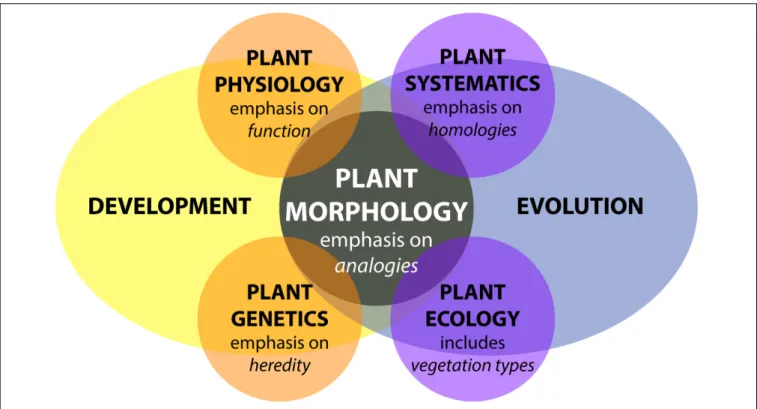
The study of plant morphology interfaces with all biological
disciplines (Figure 1). Plant morphology can be descriptive
and categorical, as in systematics, which focuses on biological
homologies to discern groups of organisms (
Mayr, 1981
;
Wiens,
2000
). In plant ecology, the morphology of communities defines
vegetation types and biomes, including their relationship to the
environment. In turn, plant morphologies are mutually informed
by other fields of study, such as plant physiology, the study of the
functions of plants, plant genetics, the description of inheritance,
and molecular biology, the underlying gene regulation (
Kaplan,
2001
).
Plant morphology is more than an attribute affecting plant
organization, it is also dynamic. Developmentally, morphology
reveals itself over the lifetime of a plant through varying
rates of cell division, cell expansion, and anisotropic growth
(
Esau, 1960
;
Steeves and Sussex, 1989
;
Niklas, 1994
). Response
to changes in environmental conditions further modulate
the abovementioned parameters. Development is genetically
programmed and driven by biochemical processes that are
responsible for physical forces that change the observed
patterning and growth of organs (
Green, 1999
;
Peaucelle et al.,
2011
;
Braybrook and Jönsson, 2016
). In addition, external
physical forces affect plant development, such as heterogeneous
soil densities altering root growth or flows of air, water, or
gravity modulating the bending of branches and leaves (
Moulia
and Fournier, 2009
). Inherited modifications of development
over generations results in the evolution of plant morphology
(
Niklas, 1997
). Development and evolution set the constraints
for how the morphology of a plant arises, regardless of whether
in a systematic, ecological, physiological, or genetic context
(Figure 1).
Plant Morphology from the Perspective
of Mathematics
In 1790, Johann Wolfgang von Goethe pioneered a perspective
that transformed the way mathematicians think about plant
morphology: the idea that the essence of plant morphology
is an underlying repetitive process of transformation (
Goethe,
1790
;
Friedman and Diggle, 2011
). The modern challenge
that Goethe’s paradigm presents is to quantitatively describe
transformations resulting from differences in the underlying
genetic, developmental, and environmental cues. From a
mathematical perspective, the challenge is how to define shape
descriptors to compare plant morphology with topological
and geometrical techniques and how to integrate these shape
descriptors into simulations of plant development.
Mathematics to Describe Plant Shape and
Morphology
Several areas of mathematics can be used to extract quantitative
measures of plant shape and morphology. One intuitive
representation of the plant form relies on the use of skeletal
descriptors that reduce the branching morphology of plants to
a set of intersecting lines or curve segments, constituting a
mathematical graph. These skeleton-based mathematical graphs
can be derived from manual measurement (
Godin et al.,
1999
;
Watanabe et al., 2005
) or imaging data (
Bucksch et al.,
2010
;
Aiteanu and Klein, 2014
). Such skeletal descriptions
can be used to derive quantitative measurements of lengths,
diameters, and angles in tree crowns (
Bucksch and Fleck, 2011
;
Raumonen et al., 2013
;
Seidel et al., 2015
) and roots, at a
single time point (
Fitter, 1987
;
Danjon et al., 1999
;
Lobet
et al., 2011
;
Galkovskyi et al., 2012
) or over time to capture
growth dynamics (
Symonova et al., 2015
). Having a skeletal
description in place allows the definition of orders, in a biological
and mathematical sense, to enable morphological analysis from
a topological perspective (Figure 2A). Topological analyses
can be used to compare shape characteristics independently
of events that transform plant shape geometrically, providing
a framework by which plant morphology can be modeled.
The relationships between orders, such as degree of
self-similarity (
Prusinkiewicz, 2004
) or self-nestedness (
Godin and
Ferraro, 2010
) are used to quantitatively summarize patterns
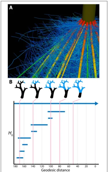
of plant morphology. Persistent homology (Figure 2B), an
extension of Morse theory (
Milnor, 1963
), transforms a given
plant shape gradually to define self-similarity (
MacPherson and
Schweinhart, 2012
) and morphological properties (
Edelsbrunner
and Harer, 2010
;
Li et al., 2017
) on the basis of topological
event statistics. In the example in Figure 2B, topological
FIGURE 1 | Plant morphology from the perspective of biology. Adapted fromKaplan (2001). Plant morphology interfaces with all disciplines of plant biology—plant physiology, plant genetics, plant systematics, and plant ecology—influenced by both developmental and evolutionary forces.
events are represented by the geodesic distance at which
branches are “born” and “die” along the length of the
structure.
In the 1980s, David Kendall defined an elegant statistical
framework to compare shapes (
Kendall, 1984
). His idea was
to compare the outline of shapes in a transformation-invariant
fashion. This concept infused rapidly as morphometrics into
biology (
Bookstein, 1997
) and is increasingly carried out using
machine vision techniques (
Wilf et al., 2016
). Kendall’s idea
inspired the development of methods such as elliptical Fourier
descriptors (
Kuhl and Giardina, 1982
) and new trends employing
the Laplace Beltrami operator (
Reuter et al., 2009
), both relying
on the spectral decompositions of shapes (
Chitwood et al.,
2012
;
Laga et al., 2014
;
Rellán-Álvarez et al., 2015
). Beyond
the organ level, such morphometric descriptors were used to
analyze cellular expansion rates of rapidly deforming primordia
into mature organ morphologies (
Rolland-Lagan et al., 2003
;
Remmler and Rolland-Lagan, 2012
;
Das Gupta and Nath,
2015
).
From a geometric perspective, developmental processes
construct surfaces in a three-dimensional space. Yet, the
embedding of developing plant morphologies into a
three-dimensional space imposes constraints on plant forms.
Awareness of such constraints has led to new interpretations
of plant morphology (
Prusinkiewicz and de Reuille, 2010
;
Bucksch et al., 2014b
) that might provide avenues to
explain symmetry and asymmetry in plant organs (e.g.,
Martinez et al., 2016
) or the occurrence of plasticity as a
morphological response to environmental changes (e.g.,
Royer et al., 2009
;
Palacio-López et al., 2015
;
Chitwood et al.,
2016
).
Mathematics to Simulate Plant Morphology
Computer simulations use principles from graph theory, such as
graph rewriting, to model plant morphology over developmental
time by successively augmenting a graph with vertices and edges
as plant development unfolds. These rules unravel the differences
between observed plant morphologies across plant species
(
Kurth, 1994
;
Prusinkiewicz et al., 2001
;
Barthélémy and Caraglio,
2007
) and are capable of modeling fractal descriptions that reflect
the repetitive and modular appearance of branching structures
(
Horn, 1971
;
Hallé, 1971, 1986
). Recent developments in
functional-structural modeling abstract the genetic mechanisms
driving the developmental program of tree crown morphology
into a computational framework (
Runions et al., 2007
;
Palubicki
et al., 2009
;
Palubicki, 2013
). Similarly, functional-structural
modeling techniques are utilized in root biology to simulate the
efficiency of nutrient and water uptake following developmental
programs (
Nielsen et al., 1994
;
Dunbabin et al., 2013
).
Alan Turing, a pioneering figure in 20th-century science, had a
longstanding interest in phyllotactic patterns. Turing’s approach
to the problem was twofold: first, a detailed geometrical analysis
of the patterns (
Turing, 1992
), and second, an application of
his theory of morphogenesis through local activation and
long-range inhibition (
Turing, 1952
), which defined the first
reaction-diffusion system for morphological modeling. Combining
physical experiments with computer simulations,
Douady and
Coudert (1996)
subsequently modeled a diffusible chemical signal
FIGURE 2 | Plant morphology from the perspective of mathematics. (A) The topological complexity of plants requires a mathematical framework to describe and simulate plant morphology. Shown is the top of a maize crown root 42 days after planting. Color represents root diameter, revealing topology and different orders of root architecture. Image provided by JPL (Pennsylvania State University). (B) Persistent homology deforms a given plant morphology using functions to define self-similarity in a structure. In this example, a geodesic distance function is traversed to the ground level of a tree (that is, the shortest curved distance of each voxel to the base of the tree), as visualized in blue in successive images. The branching structure, as defined across scales of the geodesic distance function is recorded as an H0
(zero-order homology) barcode, which in persistent homology refers to connected components. As the branching structure is traversed by the function, connected components are “born” and “die” as terminal branches emerge and fuse together. Each of these components is indicated as a bar in the H0barcode, and the correspondence of the barcode to different points in
the function is indicated by vertical lines, in pink. Images provided by ML (Danforth Plant Science Center).
produced by a developing primordium that would inhibit the
initiation of nearby primordia, successfully recapitulating known
phyllotactic patterns in the shoot apical meristem (
Bernasconi,
1994
;
Meinhardt, 2004
;
Jönsson et al., 2005
;
Nikolaev et al., 2007
;
Hohm et al., 2010
;
Fujita et al., 2011
), the number of floral organs
(
Kitazawa and Fujimoto, 2015
), the regular spacing of root hairs
(
Meinhardt and Gierer, 1974
), and the establishment of specific
vascular patterns (
Meinhardt, 1976
).
EMERGING QUESTIONS AND BARRIERS
IN THE MATHEMATICAL ANALYSIS OF
PLANT MORPHOLOGY
A true synthesis of plant morphology, which comprehensively
models observed biological phenomena and incorporates a
mathematical perspective, remains elusive. In this section, we
highlight current focuses in the study of plant morphology,
including the technical limits of acquiring morphological data,
phenotype prediction, responses of plants to the environment,
models across biological scales, and the integration of complex
phenomena, such as fluid dynamics, into plant morphological
models.
Technological Limits to Acquiring Plant
Morphological Data
There are several technological limits to acquiring plant
morphological data that must be overcome to move this field
forward. One such limitation is the acquisition of quantitative
plant images. Many acquisition systems do not provide
morphological data with measurable units. Approaches that rely
on the reflection of waves from the plant surface can provide
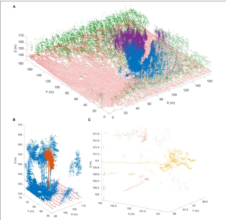
quantitative measurements for morphological analyses. Time of
flight scanners, such as terrestrial laser scanning, overcome
unit-less measurement systems by recording the round-trip time of
hundreds of thousands of laser beams sent at different angles
from the scanner to the first plant surface within the line of sight
(
Vosselman and Maas, 2010
) (Figure 3). Leveraging the speed of
light allows calculation of the distance between a point on the
plant surface and the laser scanner.
Laser scanning and the complementary, yet unitless, approach
of stereovision both produce surface samples or point clouds as
output. However, both approaches face algorithmic challenges
encountered when plant parts occlude each other, since both
rely on the reflection of waves from the plant surface (
Bucksch,
2014
). Radar provides another non-invasive technique to study
individual tree and forest structures over wide areas. Radar
pulses can either penetrate or reflect from foliage, depending
on the selected wavelength (
Kaasalainen et al., 2015
). Most
radar applications occur in forestry and are being operated from
satellites or airplanes. Although more compact and agile systems
are being developed for precision forestry above- and
below-ground (
Feng et al., 2016
), their resolution is too low to acquire
the detail in morphology needed to apply hierarchy or similarity
oriented mathematical analysis strategies.
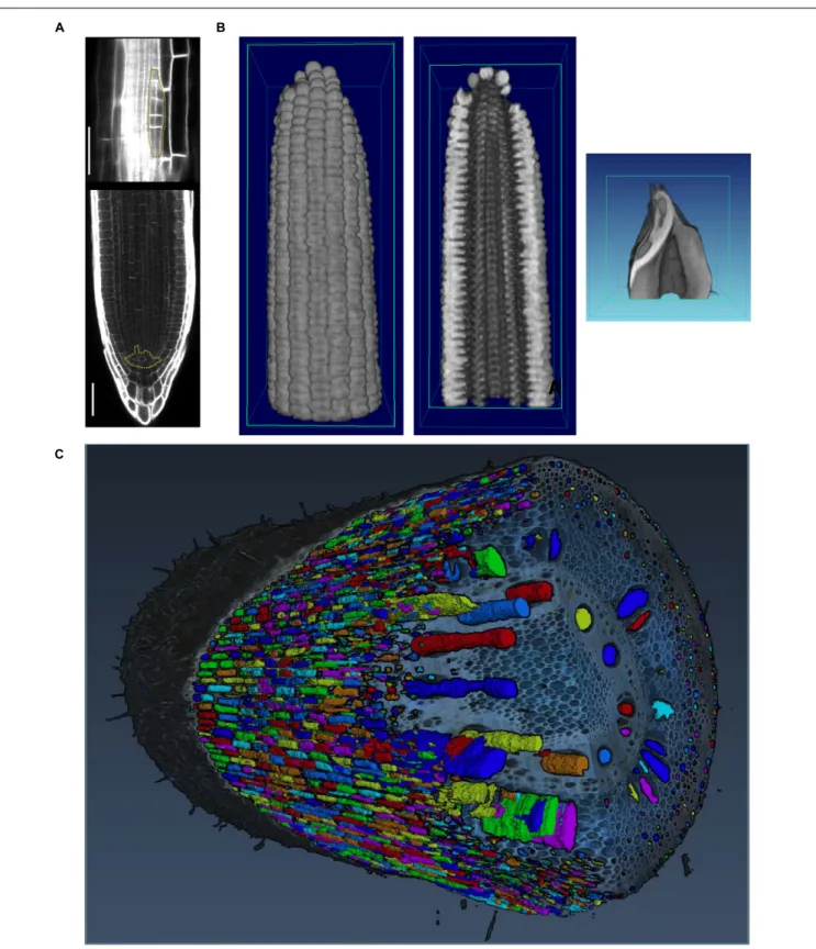
Image acquisition that resolves occlusions by penetrating plant
tissue is possible with X-ray (
Kumi et al., 2015
) and magnetic
resonance imaging (MRI;
van Dusschoten et al., 2016
). While
both technologies resolve occlusions and can even penetrate
soil, their limitation is the requirement of a closed imaging
FIGURE 3 | Terrestrial laser scanning creates a point cloud reconstruction of a Finnish forest. (A) Structure of a boreal forest site in Finland as seen with airborne (ALS) and terrestrial (TLS) laser scanning point clouds. The red (ground) and green (above-ground) points are obtained from National Land Survey of Finland national ALS point clouds that cover hundreds of thousands of square kilometers with about 1 point per square meter resolution. The blue and magenta point clouds are results of two individual TLS measurements and have over 20 million points each within an area of about 500 m2. TLS point density varies with range but can be
thousands of points per square meter up to tens of meters away from the scanner position. (B) An excerpt from a single TLS point cloud (blue). The TLS point cloud is so dense that individual tree point clouds (orange) and parts from them (yellow) can be selected for detailed analysis. (C) A detail from a single TLS point cloud. Individual branches (yellow) 20 m above ground can be inspected from the point cloud with centimeter level resolution to estimate their length and thickness. Images provided by EP (Finnish Geospatial Research Institute in the National Land Survey of Finland). ALS data was obtained from the National Land Survey of Finland Topographic Database, 08/2012 (National Land Survey of Finland open data license, version 1.0).
volume. Thus, although useful for a wide array of purposes, MRI
and X-ray are potentially destructive if applied to mature plant
organs such as roots in the field or tree crowns that are larger
than the imaging volume (
Fiorani et al., 2012
). Interior plant
anatomy can be imaged destructively using confocal microscopy
and laser ablation (Figure 4) or nano- or micro-CT tomography
techniques, that are limited to small pot volumes, to investigate
the first days of plant growth.
FIGURE 4 | Imaging techniques to capture plant morphology. (A) Confocal sections of an Arabidopsis root. The upper panel shows a new lateral root primordium at an early stage of development (highlighted in yellow). At regular intervals new roots branch from the primary root. The lower panel shows the primary root meristem and the stem cell niche (highlighted in yellow) from which all cells derive. Scale bars: 100µm. Images provided by AM (Heidelberg University). (B) Computational tomographic (CT) x-ray sections through a reconstructed maize ear (left and middle) and kernel (right). Images provided by CT (Donald Danforth Plant Science Center). (C) Laser ablation tomography (LAT) image of a nodal root from a mature, field-grown maize plant, with color segmentation showing definition of cortical cells, aerenchyma lacunae, and metaxylem vessels. Image provided by JPL (Pennsylvania State University).
The Genetic Basis of Plant Morphology
One of the outstanding challenges in plant biology is to link the
inheritance and activity of genes with observed phenotypes. This
is particularly challenging for the study of plant morphology,
as both the genetic landscape and morphospaces are complex:
modeling each of these phenomena alone is difficult, let alone
trying to model morphology as a result of genetic phenomena
(
Benfey and Mitchell-Olds, 2008
;
Lynch and Brown, 2012
;
Chitwood and Topp, 2015
). Although classic examples exist
in which plant morphology is radically altered by the effects
of a few genes (
Doebley, 2004
;
Clark et al., 2006
;
Kimura
et al., 2008
), many morphological traits have a polygenic basis
(
Langlade et al., 2005
;
Tian et al., 2011
;
Chitwood et al.,
2013
).
Quantitative trait locus (QTL) analyses can identify the
polygenic basis for morphological traits that span scales from
the cellular to the whole organ level. At the cellular level, root
cortex cell number (
Ron et al., 2013
), the cellular basis of carpel
size (
Frary et al., 2000
), and epidermal cell area and number
(
Tisné et al., 2008
) have been analyzed. The genetic basis of
cellular morphology ultimately affects organ morphology, and
quantitative genetic bases for fruit shape (
Paran and van der
Knaap, 2007
;
Monforte et al., 2014
), root morphology (
Zhu et al.,
2005
;
Clark et al., 2011
;
Topp et al., 2013
;
Zurek et al., 2015
),
shoot apical meristem shape (
Leiboff et al., 2015
;
Thompson et al.,
2015
), leaf shape (
Langlade et al., 2005
;
Ku et al., 2010
;
Tian et al.,
2011
;
Chitwood et al., 2014a,b
;
Zhang et al., 2014
;
Truong et al.,
2015
), and tree branching (
Kenis and Keulemans, 2007
;
Segura
et al., 2009
) have been described.
Natural variation in cell, tissue, or organ morphology
ultimately impacts plant physiology, and vice versa. For example,
formation of root cortical aerenchyma was linked to better plant
growth under conditions of suboptimal availability of water and
nutrients (
Zhu et al., 2010
;
Postma and Lynch, 2011
;
Lynch,
2013
), possibly because aerenchyma reduces the metabolic costs
of soil exploration. Maize genotypes with greater root cortical
cell size or reduced root cortical cell file number reach greater
depths to increase water capture under drought conditions,
possibly because those cellular traits reduce metabolic costs of
root growth and maintenance (
Chimungu et al., 2015
). The
control of root angle that results in greater water capture in
rice as water tables recede was linked to the control of auxin
distribution (
Uga et al., 2013
). Similarly, in shoots, natural
variation can be exploited to find genetic loci that control shoot
morphology, e.g., leaf erectness (
Ku et al., 2010
;
Feng et al.,
2011
).
High-throughput phenotyping techniques are increasingly
used to reveal the genetic basis of natural variation (
Tester
and Langridge, 2010
). In doing so, phenotyping techniques
complement classic approaches of reverse genetics and often lead
to novel insights, even in a well-studied species like
Arabidopsis
thaliana. Phenotyping techniques have revealed a genetic basis
for dynamic processes such as root growth (
Slovak et al., 2014
)
and traits that determine plant height (
Yang et al., 2014
).
Similarly, high-resolution sampling of root gravitropism has
led to an unprecedented understanding of the dynamics of the
genetic basis of plasticity (
Miller et al., 2007
;
Brooks et al., 2010
;
Spalding and Miller, 2013
).
The Environmental Basis of Plant
Morphology
Phenotypic plasticity is defined as the ability of one genotype
to produce different phenotypes based on environmental
differences (
Bradshaw, 1965
;
DeWitt and Scheiner, 2004
) and
adds to the phenotypic complexity created by genetics and
development. Trait variation in response to the environment
has been analyzed classically using ‘reaction norms,’ where the
phenotypic value of a certain trait is plotted for two different
environments (
Woltereck, 1909
). If the trait is not plastic, the
slope of the line connecting the points will be zero; if the reaction
norm varies across the environment the trait is plastic and the
slope of the reaction norm line will be a measure of the plasticity.
As most of the responses of plants to their environment are
non-linear, more insight into phenotypic plasticity can be obtained
by analyzing dose-response curves or dose-response surfaces
(
Mitscherlich, 1909
;
Poorter et al., 2010
).
Seminal work by
Clausen et al. (1941)
demonstrated using
several clonal species in a series of reciprocal transplants that,
although heredity exerts the most measureable effects on plant
morphology, environment is also a major source of phenotypic
variability. Research continues to explore the range of phenotypic
variation expressed by a given genotype in the context of different
environments, which has important implications for many fields,
including conservation, evolution, and agriculture (
Nicotra et al.,
2010
;
DeWitt, 2016
). Many studies examine phenotypes across
latitudinal or altitudinal gradients, or other environmental clines,
to characterize the range of possible variation and its relationship
to the process of local adaptation (
Cordell et al., 1998
;
Díaz et al.,
2016
).
Below-ground,
plants
encounter
diverse
sources
of
environmental variability, including water availability, soil
chemistry, and physical properties like soil hardness and
movement. These factors vary between individual plants (
Razak
et al., 2013
) and within an individual root system, where plants
respond at spatio-temporal levels to very different granularity
(
Drew, 1975
;
Robbins and Dinneny, 2015
). Plasticity at a
micro-environmental scale has been linked to developmental and
molecular mechanisms (
Bao et al., 2014
). The scientific challenge
here is to integrate these effects at a whole root system level and
use different scales of information to understand the optimal
acquisition in resource limited conditions (
Rellán-Álvarez et al.,
2016
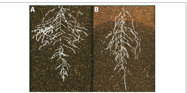
) (Figure 5).
Integrating Models from Different Levels
of Organization
Since it is extremely difficult to examine complex interdependent
processes
occurring
at
multiple
spatio-temporal
scales,
mathematical modeling can be used as a complementary
tool with which to disentangle component processes and
investigate how their coupling may lead to emergent patterns
at a systems level (
Hamant et al., 2008
;
Band and King, 2012
;
FIGURE 5 | The environmental basis of plant morphology. Root system architecture of Arabidopsis Col-0 plants expressing ProUBQ10:LUC2o growing in (A) control and (B) water-deficient conditions using the GLO-Roots system (Rellán-Álvarez et al., 2015). Images provided by RR-Á (Laboratorio Nacional de Genómica para la Biodiversidad, CINVESTAV) are a composite of a video originally published (Rellán-Álvarez et al., 2015).
multiscale model should generate well-constrained predictions
despite significant parameter uncertainty (
Gutenkunst et al.,
2007; Hofhuis et al., 2016
). It is desirable that a multiscale model
has certain modularity in its design such that individual modules
are responsible for modeling specific spatial aspects of the system
(
Baldazzi et al., 2012
). Imaging techniques can validate multiscale
models (e.g.,
Willis et al., 2016
) such that simulations can reliably
guide experimental studies.
To illustrate the challenges of multi-scale modeling,
we highlight an example that encompasses molecular and
cellular scales. At the molecular scale, models can treat some
biomolecules as diffusive, but others, such as membrane-bound
receptors, can be spatially restricted (
Battogtokh and Tyson,
2016
). Separately, at the cellular scale, mathematical models
describe dynamics of cell networks where the mechanical
pressures exerted on the cell walls are important factors for cell
growth and division (
Jensen and Fozard, 2015
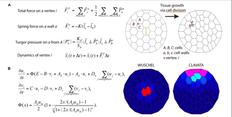
) (Figure 6A).
In models describing plant development in a two-dimensional
cross-section geometry, cells are often modeled as polygons
defined by walls between neighboring cells. The spatial position
of a vertex, where the cell walls of three neighboring cells
coalesce, is a convenient variable for mathematical modeling of
the dynamics of cellular networks (
Prusinkiewicz and Runions,
2012
). A multiscale model can then be assembled by combining
the molecular and cellular models. Mutations and deletions of the
genes encoding the biomolecules can be modeled by changing
parameters. By inspecting the effects of such modifications
on the dynamics of the cellular networks, the relationship
between genotypes and phenotypes can be predicted. For
example,
Fujita et al. (2011)
model integrates the dynamics of
cell growth and division with the spatio-temporal dynamics of
the proteins involved in stem cell regulation and simulates shoot
apical meristem development in wild type and mutant plants
(Figure 6B).
Modeling the Impact of Morphology on
Plant Function
Quantitative measures of plant morphology are critical to
understand function.
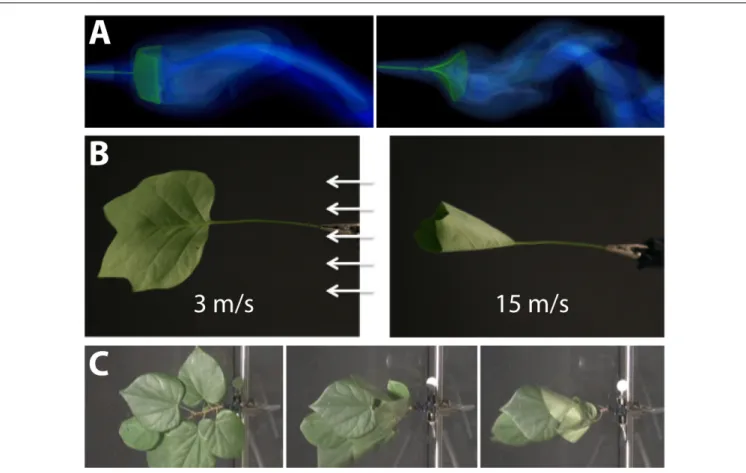
Vogel (1989)
was the first to provide
quantitative data that showed how shape changes in leaves reduce
drag or friction in air or water flows. He found that single
broad leaves reconfigure at high flow velocities into cone shapes
to reduce flutter and drag (Figures 7A,B). More recent work
discovered that the cone shape is significantly more stable than
other reconfigurations such as U-shapes (
Miller et al., 2012
).
Subsequent experimental studies on broad leaves, compound
leaves, and flowers also support rapid repositioning in response
to strong currents as a general mechanism to reduce drag
(
Niklas, 1992
;
Ennos, 1997
;
Etnier and Vogel, 2000
;
Vogel, 2006
)
(Figure 7C). It is a combination of morphology and anatomy,
and the resultant material properties, which lead to these optimal
geometric re-configurations of shape.
From a functional perspective, it is highly plausible that leaf
shape and surface-material properties alter the boundary layer of
a fluid/gas over the leaf surface or enhance passive movement that
can potentially augment gas and heat exchange. For example, it
has been proposed that the broad leaves of some trees flutter for
the purpose of convective and evaporative heat transfer (
Thom,
1968
;
Grant, 1983
). Any movement of the leaf relative to the
FIGURE 6 | Integration of tissue growth and reaction-diffusion models. (A) Vertex model of cellular layers (Prusinkiewicz and Lindenmayer, 1990). K, Ela, and El0are
the spring constant, current length, and rest length for wall a. KPis a constant and SAis the size of cell A.1t is time step. Shown is a simulation of cell network
growth. (B) Reaction diffusion model of the shoot apical meristem for WUSCHEL and CLAVATA interactions (Fujita et al., 2011). u = WUS, v = CLV, i = cell index,8 is a sigmoid function. E, B, AS, Ad, C, D, um, Du, Dvare positive constants. Shown are the distributions of WUS and CLV levels within a dynamic cell network. Images
provided by DB (Virginia Tech).
movement of the air or water may decrease the boundary layer
and increase gas exchange, evaporation, and heat dissipation
(
Roden and Pearcy, 1993
). Each of these parameters may be
altered by the plant to improve the overall function of the leaf
(
Vogel, 2012
).
The growth of the plant continuously modifies plant topology
and geometry, which in turn changes the balance between
organ demand and production. At the organismal scale, the
3D spatial distribution of plant organs is the main interface
between the plant and its environment. For example, the 3D
arrangement of branches impacts light interception and provides
the support for different forms of fluxes (water, sugars) and
signals (mechanical constraints, hormones) that control plant
functioning and growth (
Godin and Sinoquet, 2005
).
MILESTONES IN EDUCATION AND
OUTREACH TO ACCELERATE THE
INFUSION OF MATH INTO THE PLANT
SCIENCES
Mathematics and plant biology need to interact more closely to
accelerate scientific progress. Opportunities to interact possibly
involve cross-disciplinary training, workshops, meetings, and
funding opportunities. In this section, we outline perspectives
for enhancing the crossover between mathematics and plant
biology.
Education
Mathematics has been likened to “biology’s next microscope,”
because of the insights into an otherwise invisible world
it has to offer. Conversely, biology has been described as
“mathematics’ next physics,” stimulating novel mathematical
approaches because of the hitherto unrealized phenomena that
biology studies (
Cohen, 2004
). The scale of the needed interplay
between mathematics and plant biology is enormous and may
lead to new science disciplines at the interface of both: ranging
from the cellular, tissue, organismal, and community levels to
the global; touching upon genetic, transcriptional, proteomic,
metabolite, and morphological data; studying the dynamic
interactions of plants with the environment or the evolution
of new forms over geologic time; and spanning quantification,
statistics, and mechanistic mathematical models.
Research is becoming increasingly interdisciplinary, and
undergraduate, graduate, and post-graduate groups are actively
trying to bridge the gap between mathematics and biology
skillsets. While many graduate programs have specialization
tracks under the umbrella of mathematics or biology-specific
programs, increasingly departments are forming specially
designed
graduate
groups
for
mathematical/quantitative
biology
1,2to strengthen the interface between both disciplines.
1BioQuant at University of Heidelberg, http://www.bioquant.uni-heidelberg.de(retrieved February 28, 2017)
2Quantitative Biosciences at Georgia Tech in Atlanta, http://qbios.gatech.edu
FIGURE 7 | Modeling the interaction between plant morphology and fluid dynamics. (A) 3D immersed boundary simulations of flow past a flexible rectangular sheet (left) and disk with a cut from the center to edge (right). Both structures are attached to a flexible petiole, and the flow is from left to right. The contours show the magnitude of vorticity (the rotation in the air). The circular disk reconfigures into a cone shape, similar to many broad leaves. (B) Reconfiguration of tulip poplar leaves in 3 m/s (left) and 15 m/s flow (right). The leaves typically flutter at lower wind speeds and reconfigure into stable cones at high wind speeds. (C) A cluster of redbud leaves in wind moving from right to left. The wind speed is increased from 3 m/s (left) to 6 m/s (middle) and 12 m/s (right). Note that the entire cluster reconfigures into a cone shape. This is different from the case of tulip poplars and maples where each leaf individually reconfigures into a conic shape. Images provided by LM (University of North Carolina, Chapel Hill, NC, United States).
This will necessitate team-teaching across disciplines to train the
next generation of mathematical/computational plant scientists.
Public Outreach: Citizen Science and the
Maker Movement
Citizen science, which is a method to make the general public
aware of scientific problems and employ their help in solving
them
3, is an ideal platform to initiate a synthesis between
plant biology and mathematics because of the relatively
low cost and accessibility of each field. Arguably, using
citizen science to collect plant morphological diversity has
already been achieved, but has yet to be fully realized. In
total, it is estimated that the herbaria of the world possess
greater than 207 million voucher specimens
4, representing
the diverse lineages of land plants collected over their
respective biogeographies over a timespan of centuries.
3For example, see the White Paper on Citizen Science for Europe, http://www.
socientize.eu/sites/default/files/white-paper_0.pdf (retrieved May 29, 2016)
4List of herbaria, https://en.wikipedia.org/wiki/List_of_herbaria (retrieved May 29,
2016)
Digital documentation of the millions of vouchers held by
the world’s botanic gardens is actively underway, allowing
for researchers and citizens alike to access and study for
themselves the wealth of plant diversity across the globe
and centuries (
Smith et al., 2003
;
Corney et al., 2012
;
Ryan,
2013
).
The developmental changes in plants responding to
environmental variability and microclimatic changes over
the course of a growing season can be analyzed by studying
phenology. Citizen science projects such as the USA National
Phenology Network
5or Earthwatch
6and associated programs
such as My Tree Tracker
7document populations and individual
plants over seasons and years, providing a distributed,
decentralized network of scientific measurements to study
the effects of climate change on plants.
5https://www.usanpn.org/# (retrieved May 29, 2016)
6http://earthwatch.org/scientific-research/special-initiatives/urban-resiliency
(retrieved May 29, 2016)
7http://www.mytreetracker.org/cwis438/websites/MyTreeTracker/About.php?
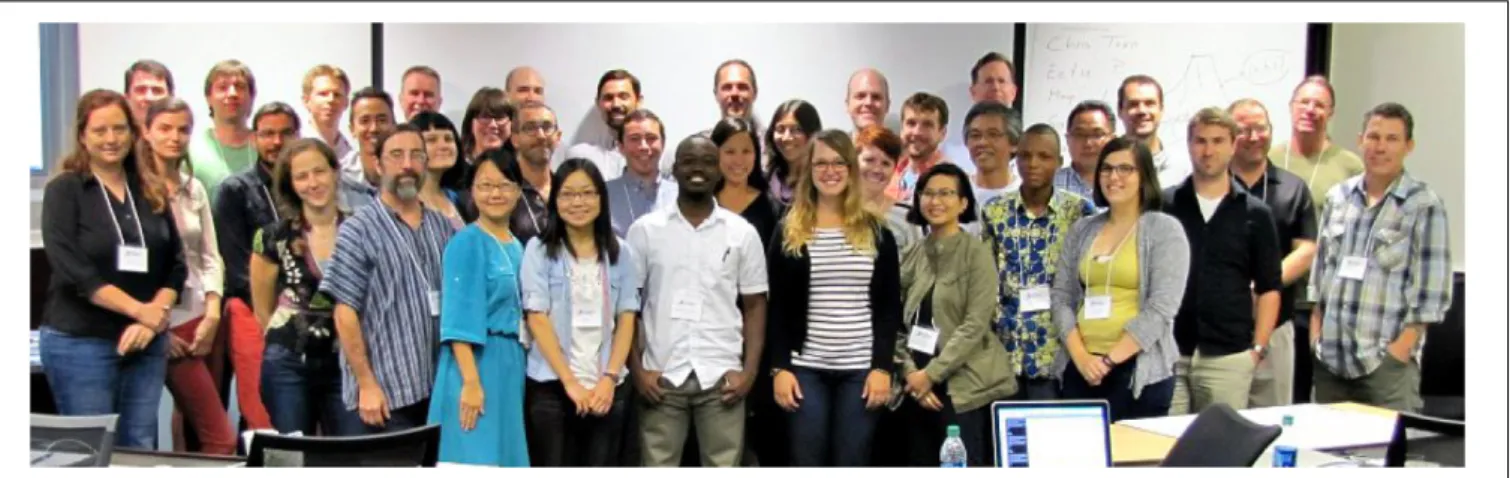
FIGURE 8 | Milestones to accelerate the infusion of math into the plant sciences. Group photo of the authors from the National Institute for Mathematical and Biological Synthesis (NIMBioS) meeting on plant morphological models (University of Tennessee, Knoxville, September 2–4, 2015) that inspired this manuscript. Workshops such as these, bringing mathematicians and plant biologists together, will be necessary to create a new synthesis of plant morphology.
Citizen science is also enabled by low-cost, specialized
equipment. Whether programming a camera to automatically
take pictures at specific times or automating a watering
schedule for a garden, the maker movement—a
do-it-yourself cultural phenomenon that intersects with hacker
culture—focuses
on
building
custom,
programmable
hardware, whether via electronics, robotics, 3D-printing,
or time-honored skills such as metal- and woodworking.
The focus on programming is especially relevant for
integrating mathematical approaches with plant science
experiments.
The
low-cost
of
single-board
computers
like Raspberry Pi, HummingBoard, or CubieBoard is a
promising example of how to engage citizen scientists into
the scientific process and enable technology solutions to specific
questions.
Workshops and Funding Opportunities
Simply bringing mathematicians and plant biologists together
to interact, to learn about new tools, approaches, and
opportunities in each discipline is a major opportunity for
further integration of these two disciplines and strengthen
new disciplines at the interface of both. This white paper itself
is a testament to the power of bringing mathematicians and
biologists together, resulting from a National Institute for
Mathematical and Biological Synthesis (NIMBioS) workshop
titled “Morphological Plant Modeling: Unleashing Geometric
and Topologic Potential within the Plant Sciences,” held
at the University of Tennessee, Knoxville, September 2–
4, 2015
8(Figure 8). Other mathematical institutes such
as the Mathematical Biology Institute (MBI) at Ohio
State University
9, the Statistical and Applied Mathematical
Sciences Institute (SAMSI) in Research Triangle Park
10,
the Institute for Mathematics and Its Applications at
8http://www.nimbios.org/workshops/WS_plantmorph (retrieved May 29, 2016) 9https://mbi.osu.edu/ (retrieved May 29, 2016)
10http://www.samsi.info/ (retrieved May 29, 2016)
University
of
Minnesota
11,
and
the
Centre
for
Plant
Integrative Biology at the University of Nottingham
12have
also hosted workshops for mathematical and quantitative
biologists from the undergraduate student to the faculty
level.
There are efforts to unite biologists and mathematics
through initiatives brought forth from The National Science
Foundation,
including
Mathematical
Biology
Programs
13and the Joint DMS/NIGMS Initiative to Support Research at
the Interface of the Biological and Mathematical Sciences
14(DMS/NIGMS). Outside of the Mathematics and Life Sciences
Divisions, the Division of Physics houses a program on the
Physics of Living Systems. Societies such as The Society
for Mathematical Biology and the Society for Industrial
and Applied Mathematics (SIAM) Life Science Activity
Group
15are focused on the dissemination of research at the
intersection of math and biology, creating many opportunities
to present research and provide funding. We emphasize the
importance that funding opportunities have had and will
continue to have in the advancement of plant morphological
modeling.
Open Science
Ultimately, mathematicians, computational scientists, and plant
biology must unite at the level of jointly collecting data,
analyzing it, and doing science together. Open and timely
data sharing to benchmark code is a first step to unite these
disciplines along with building professional interfaces to bridge
11https://www.ima.umn.edu/ (retrieved May 29, 2016)
12https://www.cpib.ac.uk/outreach/cpib-summer-school/ (retrieved May 29,
2016)
13https://www.nsf.gov/funding/pgm_summ.jsp?pims_id=5690 (retrieved May 29,
2016)
14http://www.nsf.gov/funding/pgm_summ.jsp?pims_id=5300&org=DMS
(retrieved May 29, 2016)
between the disciplines (
Bucksch et al., 2017
;
Pradal et al.,
2017
).
A number of platforms provide open, public access to
datasets, figures, and code that can be shared, including
Dryad
16, Dataverse
17, and Figshare
18. Beyond the ability
to share data is the question of open data formats and
accessibility. For example, in remote sensing research it is
unfortunately common that proprietary data formats are
used, which prevents their use without specific software.
This severely limits the utility and community building
aspects of plant morphological research. Beyond datasets,
making code openly available, citable, and user-friendly is
a means to share methods to analyze data. Places to easily
share code include web-based version controlled platforms
like Bitbucket
19or Github
20and software repositories like
Sourceforge
21. Furthermore, numerous academic Journals (e.g.,
Nature Methods, Applications in Plant Sciences, and Plant
Methods) already accept publications that focus on methods
and software to accelerate new scientific discovery (
Pradal et al.,
2013
).
Meta-analysis datasets provide curated resources where
numerous published and unpublished datasets related to a
specific problem (or many problems) can be accessed by
researchers
22. The crucial element is that data is somehow
reflective of universal plant morphological features, bridging the
gap between programming languages and biology, as seen in the
Root System Markup Language (
Lobet et al., 2015
) and OpenAlea
(
Pradal et al., 2008, 2015
). Bisque is a versatile platform to store,
organize, and analyze image data, providing simultaneously open
access to data and analyses as well as the requisite computation
(
Kvilekval et al., 2010
). CyVerse
23(formerly iPlant) is a similar
platform, on which academic users get 100 GB storage for free
and can create analysis pipelines that can be shared and reused
(
Goff et al., 2011
). For example, DIRT
24is an automatic, high
throughput computing platform (
Bucksch et al., 2014a
;
Das
et al., 2015
) that the public can use hosted on CyVerse using
the Texas Advanced Computing Center
25(TACC) resources at
UT Austin that robustly extracts root traits from digital images.
The reproducibility of these complex computational experiments
can be improved using scientific workflows that capture and
automate the exact methodology followed by scientists (
Cohen-Boulakia et al., 2017
). We emphasize here the importance of
adopting open science policies at the individual investigator and
journal level to continue strengthening the interface between
plant and mathematically driven sciences.
16http://datadryad.org/ (retrieved May 29, 2016) 17http://dataverse.org/ (retrieved May 29, 2016) 18https://figshare.com/ (retrieved May 29, 2016) 19https://bitbucket.org/ (retrieved May 29, 2016) 20https://github.com/ (retrieved May 29, 2016) 21https://sourceforge.net/ (retrieved May 29, 2016)
22BAAD: a Biomass And Allometry Database for woody plants, https://github.
com/dfalster/baad (retrieved May 29, 2016)
23http://www.cyverse.org/ (retrieved August 20, 2016) 24http://dirt.iplantcollaborative.org/ (retrieved August 20, 2016) 25https://www.tacc.utexas.edu/ (retrieved August 20, 2016)
CONCLUSION: UNLEASHING
GEOMETRIC AND TOPOLOGICAL
POTENTIAL WITHIN THE PLANT
SCIENCES
Plant morphology is a mystery from a molecular and
quantification point of view. Hence, it fascinates both
mathematical and plant biology researchers alike. As such,
plant morphology holds the secret by which predetermined
variations of organizational patterns emerge as a result of
evolutionary, developmental, and environmental responses.
The persistent challenge at the intersection of plant
biology and mathematical sciences might be the integration
of measurements across different scales of the plant. We have to
meet this challenge to derive and validate mathematical models
that describe plants beyond the visual observable. Only then
we will be able to modify plant morphology through molecular
biology and breeding as means to develop needed agricultural
outputs and sustainable environments for everybody.
Cross-disciplinary training of scientists, citizen science, and
open science are inevitable first steps to develop the interface
between mathematical-driven and plant biology-driven sciences.
The result of these steps will be new disciplines, that will add to
the spectrum of researchers in plant biology. Hence, to unleash
the potential of geometric and topological approaches in the
plant sciences, we need an interface familiar with both plants
and mathematical approaches to meet the challenges posed by
a future with uncertain natural resources as a consequence of
climate change.
AUTHOR CONTRIBUTIONS
AB and DC conceived, wrote, and organized the manuscript. All
authors contributed to writing the manuscript.
FUNDING
This
work
was
assisted
through
participation
in
the
Morphological Plant Modeling: Unleashing geometric and
topological potential within the plant sciences Investigative
Workshop at the National Institute for Mathematical and
Biological Synthesis, sponsored by the National Science
Foundation through NSF Award #DBI-1300426, with additional
support from The University of Tennessee, Knoxville, Knoxville,
TN, United States.
ACKNOWLEDGMENTS
The authors are grateful to the National Institute for
Mathematical and Biological Synthesis (NIMBioS, University
of Tennessee, Knoxville) for hosting and funding the workshop
“Morphological Plant Modeling: Unleashing geometric and
topological potential within the plant sciences” that inspired this
manuscript. We thank the reviewers Evelyne Costes and Leo
Marcelis for creative and open discussions.
REFERENCES
Aiteanu, F., and Klein, R. (2014). Hybrid tree reconstruction from inhomogeneous point clouds.Visual Comput. 30, 763–771. doi: 10.1007/s00371-014-0977-7 Baldazzi, V., Bertin, N., De Jong, H., and Génard, M. (2012). Towards multiscale
plant models: integrating cellular networks.Trends Plant Sci. 17, 728–736. doi: 10.1016/j.tplants.2012.06.012
Band, L. R., Fozard, J. A., Godin, C., Jensen, O. E., Pridmore, T., Bennett, M. J., et al. (2012). Multiscale systems analysis of root growth and development: modeling beyond the network and cellular scales.Plant Cell 24, 3892–3906. doi: 10.1105/tpc.112.101550
Band, L. R., and King, J. R. (2012). Multiscale modelling of auxin transport in the plant-root elongation zone.J. Math. Biol. 65, 743–785. doi: 10.1007/s00285-011-0472-y
Bao, Y., Aggarwal, P., Robbins, N. E., Sturrock, C. J., Thompson, M. C., Tan, H. Q., et al. (2014). Plant roots use a patterning mechanism to position lateral root branches toward available water.Proc. Natl. Acad. Sci. U.S.A. 111, 9319–9324. doi: 10.1073/pnas.1400966111
Barthélémy, D., and Caraglio, Y. (2007). Plant architecture: a dynamic, multilevel and comprehensive approach to plant form, structure, and ontogeny.Ann. Bot. 99, 375–407. doi: 10.1093/aob/mcl260
Battogtokh, D., and Tyson, J. J. (2016). A bistable switch mechanism for stem cell domain nucleation in the shoot apical meristem.Front. Plant Sci. 7:674. doi: 10.3389/fpls.2016.00674
Benfey, P. N., and Mitchell-Olds, T. (2008). From genotype to phenotype: systems biology meets natural variation.Science 320, 495–497. doi: 10.1126/science. 1153716
Bernasconi, G. P. (1994). Reaction-diffusion model for phyllotaxis.Physica D 70, 90–99. doi: 10.1016/0167-2789(94)90058-2
Bookstein, F. L. (1997).Morphometric Tools for Landmark Data: Geometry and Biology. New York, NY: Cambridge University Press.
Bradshaw, A. D. (1965). Evolutionary significance of phenotypic plasticity in plants.Adv. Genet. 13, 115–155. doi: 10.1016/s0065-2660(08)60048-6 Braybrook, S. A., and Jönsson, H. (2016). Shifting foundations: the mechanical cell
wall and development.Curr. Opin. Plant Biol. 29, 115–120. doi: 10.1016/j.pbi. 2015.12.009
Brooks, T. L. D., Miller, N. D., and Spalding, E. P. (2010). Plasticity of Arabidopsis root gravitropism throughout a multidimensional condition space quantified by automated image analysis.Plant Physiol. 152, 206–216. doi: 10.1104/pp.109. 145292
Bucksch, A. (2014). A practical introduction to skeletons for the plant sciences. Appl. Plant Sci. 2:1400005. doi: 10.3732/apps.1400005
Bucksch, A., Burridge, J., York, L. M., Das, A., Nord, E., Weitz, J. S., et al. (2014a). Image-based high-throughput field phenotyping of crop roots.Plant Physiol. 166, 470–486. doi: 10.1104/pp.114.243519
Bucksch, A., Das, A., Schneider, H., Merchant, N., and Weitz, J. S. (2017). Overcoming the law of the hidden in cyberinfrastructures.Trends Plant Sci. 22, 117–123. doi: 10.1016/j.tplants.2016.11.014
Bucksch, A., and Fleck, S. (2011). Automated detection of branch dimensions in woody skeletons of fruit tree canopies.Photogramm. Eng. Remote Sens. 77, 229–240. doi: 10.14358/PERS.77.3.229
Bucksch, A., Lindenbergh, R., and Menenti, M. (2010). SkelTre.Vis. Comput. 26, 1283–1300. doi: 10.1007/s00371-010-0520-4
Bucksch, A., Turk, G., and Weitz, J. S. (2014b). The fiber walk: a model of tip-driven growth with lateral expansion.PLoS ONE 9:e85585. doi: 10.1371/journal.pone. 0085585
Chimungu, J. G., Maliro, M. F., Nalivata, P. C., Kanyama-Phiri, G., Brown, K. M., and Lynch, J. P. (2015). Utility of root cortical aerenchyma under water limited conditions in tropical maize (Zea mays L.). Field Crops Res. 171, 86–98. doi: 10.1016/j.fcr.2014.10.009
Chitwood, D. H., Headland, L. R., Ranjan, A., Martinez, C. C., Braybrook, S. A., Koenig, D. P., et al. (2012). Leaf asymmetry as a developmental constraint imposed by auxin-dependent phyllotactic patterning.Plant Cell 24, 2318–2327. doi: 10.1105/tpc.112.098798
Chitwood, D. H., Kumar, R., Headland, L. R., Ranjan, A., Covington, M. F., Ichihashi, Y., et al. (2013). A quantitative genetic basis for leaf morphology in a set of precisely defined tomato introgression lines.Plant Cell 25, 2465–2481. doi: 10.1105/tpc.113.112391
Chitwood, D. H., Ranjan, A., Kumar, R., Ichihashi, Y., Zumstein, K., Headland, L. R., et al. (2014a). Resolving distinct genetic regulators of tomato leaf shape within a heteroblastic and ontogenetic context.Plant Cell 26, 3616–3629. doi: 10.1105/tpc.114.130112
Chitwood, D. H., Ranjan, A., Martinez, C. C., Headland, L. R., Thiem, T., Kumar, R., et al. (2014b). A modern ampelography: a genetic basis for leaf shape and venation patterning in grape.Plant Physiol. 164, 259–272. doi: 10.1104/pp. 113.229708
Chitwood, D. H., Rundell, S. M., Li, D. Y., Woodford, Q. L., Tommy, T. Y., Lopez, J. R., et al. (2016). Climate and developmental plasticity: interannual variability in grapevine leaf morphology. Plant Physiol. 170, 1480–1491. doi: 10.1104/pp.15.01825
Chitwood, D. H., and Topp, C. N. (2015). Revealing plant cryptotypes: defining meaningful phenotypes among infinite traits.Curr. Opin. Plant Biol. 24, 54–60. doi: 10.1016/j.pbi.2015.01.009
Clark, R. M., Wagler, T. N., Quijada, P., and Doebley, J. (2006). A distant upstream enhancer at the maize domestication gene tb1 has pleiotropic effects on plant and inflorescent architecture.Nat. Genet. 38, 594–597. doi: 10.1038/ ng1784
Clark, R. T., MacCurdy, R. B., Jung, J. K., Shaff, J. E., McCouch, S. R., Aneshansley, D. J., et al. (2011). Three-dimensional root phenotyping with a novel imaging and software platform.Plant Physiol. 156, 455–465. doi: 10.1104/pp.110.169102 Clausen, J., Keck, D. D., and Hiesey, W. M. (1941). Regional differentiation in plant
species.Am. Nat. 75, 231–250. doi: 10.1086/280955
Cohen, J. E. (2004). Mathematics is biology’s next microscope, only better; biology is mathematics’ next physics, only better. PLoS Biol. 2:e439. doi: 10.1371/ journal.pbio.0020439
Cohen-Boulakia, S., Belhajjame, K., Collin, O., Chopard, J., Froidevaux, C., Gaignard, A., et al. (2017). Scientific workflows for computational reproducibility in the life sciences: status, challenges and opportunities. Fut. Gen. Comput. Syst. (in press). doi: 10.1016/j.future.2017.01.012
Cordell, S., Goldstein, G., Mueller-Dombois, D., Webb, D., and Vitousek, P. M. (1998). Physiological and morphological variation inMetrosideros polymorpha, a dominant Hawaiian tree species, along an altitudinal gradient: the role of phenotypic plasticity.Oecologia 113, 188–196. doi: 10.1007/s004420050367 Corney, D., Clark, J. Y., Tang, H. L., and Wilkin, P. (2012). Automatic extraction of
leaf characters from herbarium specimens.Taxon 61, 231–244.
Danjon, F., Bert, D., Godin, C., and Trichet, P. (1999). Structural root architecture of 5-year-oldPinus pinaster measured by 3D digitising and analysed with AMAPmod.Plant Soil 217, 49–63. doi: 10.1023/A:1004686119796
Das, A., Schneider, H., Burridge, J., Ascanio, A. K. M., Wojciechowski, T., Topp, C. N., et al. (2015). Digital imaging of root traits (DIRT): a high-throughput computing and collaboration platform for field-based root phenomics.Plant Methods 11:51. doi: 10.1186/s13007-015-0093-3
Das Gupta, M., and Nath, U. (2015). Divergence in patterns of leaf growth polarity is associated with the expression divergence of miR396.Plant Cell 27, 2785–2799. doi: 10.1105/tpc.15.00196
DeWitt, T. J. (2016). Expanding the phenotypic plasticity paradigm to broader views of trait space and ecological function. Curr. Zool. 62, 463–473. doi: 10.1093/cz/zow085
DeWitt, T. J., and Scheiner, S. M. (eds) (2004).Phenotypic Plasticity: Functional and Conceptual Approaches. Oxford: Oxford University Press.
Díaz, S., Kattge, J., Cornelissen, J. H., Wright, I. J., Lavorel, S., Dray, S., et al. (2016). The global spectrum of plant form and function.Nature 529, 167–171. doi: 10.1038/nature16489
Doebley, J. (2004). The genetics of maize evolution.Annu. Rev. Genet. 38, 37–59. doi: 10.1146/annurev.genet.38.072902.092425
Douady, S., and Coudert, Y. (1996). Phyllotaxis as a dynamical self organizing process part I: the spiral modes resulting from time-periodic iterations.J. Theor. Biol. 178, 255–273. doi: 10.1006/jtbi.1996.0024
Drew, M. C. (1975). Comparison of the effects of a localised supply of phosphate, nitrate, ammonium and potassium on the growth of the seminal root system, and the shoot, in barley.New Phytol. 75, 479–490. doi: 10.1111/j.1469-8137. 1975.tb01409.x
Dunbabin, V. M., Postma, J. A., Schnepf, A., Pagès, L., Javaux, M., Wu, L., et al. (2013). Modelling root–soil interactions using three–dimensional models of root growth, architecture and function.Plant Soil 372, 93–124. doi: 10.1007/ s11104-013-1769-y