The following is a reprint from Operat Orthop Traumatol 2003;15:3–19 and continues the
new series of articles at providing continuing education on operative techniques to the
European trauma community.
Late Missed Monteggia Lesions –
Reconstruction of the Humeroradial Joint
Lutz von Laer, Carol Hasler, Anna Kathrin Hell-Vocke
1Ab stract
Objective: Reconstruction of the humeroradial joint
re-sulting in immediate functional stability.
Indications: Missed traumatic dislocation of the radial
head. Congenital dislocation of the radial head.
Contraindications: Secondary dislocation of the radial
head in instances of dysplasia or aplasia of the capitel-lum. Relative: deformation of the radial head in adults.
Surgical Technique: Open reduction of the radial head
without reconstruction of the annular ligament. Oste-otomy of the shaft of the proximal ulna and installa-tion of an external fixator with open clamps. Three-di-mensional correction of the ulna under visual control of the humeroradial joint until the radial head is relocated in its anatomic position.
Results: Between 01/1998 and 05/2001, we performed
an osteotomy of the proximal ulna, external fixation, and open reduction of the radial head in 14 patients presenting with a late missed Monteggia lesion (Bado type I). The average age of the seven girls and seven boys at the time of reconstruction was 9 (5–15) years, the mean interval between index trauma and recon-struction amounted to 21 months (2 weeks to 7 years). Removal of the external fixator after an average of 12 (7–16) weeks. In twelve patients the reduction was maintained, and in two patients the radial head redis-located postoperatively. In one of these patients a closed reduction was successful, whereas in the other patient an open reduction was done and the external fixation modified. In both patients the joint position was maintained.
Preoperatively seven out of 14 patients showed a de-creased range of motion; it improved postoperatively in most. A clinical and radiologic follow-up averaging 14 (3–44) months was possible in 13 patients. No com-plications were recorded.
Key Words
Late missed Monteggia lesion · Ulnar osteotomy · External fixation
Eur J Trau ma 2005;31:597–607 DOI 10.1007/s00068-005-6605-5
Introductory Remarks
A traumatic anterior dislocation of the radial head (Ba-do type I [2], Table 1) cannot occur without a simultane-ous lesion of the ulna such as bowing of the ulna, the classic greenstick fracture, or a complete fracture [2, 8, 15, 19, 20, 25, 27, 28, 30]. Even when the dislocation has been missed, the fracture of the ulna consolidates, often under conservative care. The axial deviation is often minimal and corrects spontaneously during further growth. A relative lengthening of the radius when com-pared to the ulna occurs, particularly in children < 5 years of age, most probably due to the absent contact between radius and humerus.
The goal of the surgical reconstruction of the re-mote radial head dislocation is the correction of the ma-lalignment of the ulna by an osteotomy and, if
neces-1 Orthopädisch-traumatologische Abteilung,
Universitätskinder-spital beider Basel, Schweiz.
Reprint from:
Operat Orthop Traumatol 2003;15:3–19 DOI 10.1007/s00064-003-1059-6
sary, correction of the length discrepancy. With few exceptions [1, 11, 16, 18], most authors agree with such an approach [3, 4, 6, 9, 12, 21, 23, 24]. The discrepancy of opinion relates to the indication for a reconstruction of the annular ligament [5]. In none of the published series did such an intervention lead to good results. Our own experience based on 15 attempts at reconstruction for missed dislocations led to poor results in 50% [13]. We therefore reconsidered our approach and opted for an ulnar osteotomy. The choice of the extent and plane of this osteotomy entails two problems: 1. We do not know the original axial deviation that led to the dislocation. The axial deviation as seen in the original anteroposte-rior (AP) and lateral radiographs cannot tell us the di-rection nor the extent of the displacement. Besides, malrotation can neither be diagnosed clinically nor ra-diologically. 2. The estimation of the axial deviation months to years after the injury is even more difficult given the spontaneous, growth-induced correction.
Plates or rods are limited in their ability to deter-mine the direction of the osteotomy necessary for re-duction of the radial head; their use does not allow to obtain the direction optimal for reduction of the dislo-cation. Additional soft tissue procedures, even the often chosen but obsolete method according to Witt [31, 32] do not solve this problem. The method of Witt forces the radial head in its proper place and holds it there with a transarticular wire. As repeatedly shown, these ap-proaches lead to new problems such as wire breakage, infection, chronic epiphysiolysis, and growth distur-bances [7, 17].
Only the radial head once properly reduced can de-termine the final direction and extent of angulation of the ulnar osteotomy. These prerequisites are met by a system such as an external fixator that allows unlimited motion in all planes. Difficulties in respect of timing: the earlier the reconstruction is done, the better the results [10, 13, 14, 18, 21–24, 26, 29]. During early correction one does not need to deal with length discrepancy, the
head has kept its original shape, and the patients are usually completely symptom-free and have a full range of motion postoperatively. The prognosis of late post-traumatic radial head dislocations is unknown. There-fore, we are unable to predict the late outcome and the occurrence of secondary symptoms, even in symptom-free patients. This is additional reason to recommend early reconstruction.
Surgical Principles and Objective
Corrective osteotomy of the ulnar shaft and open reduc-tion of the radial head. Retenreduc-tion of the fragments with an external fixator. No soft tissue reconstruction. Im-mediate stable reduction of the humero-radial joint al-lowing early movements. Restoration of shape and function of the elbow joint.
Advantages
• Immediate mobilization of the reconstructed joint.
Disadvantages
• In the presence of a discrepancy in length between ra-dius and ulna, two interventions are necessary: first, a distraction osteotomy of the ulna to regain the proper length of the ulna and then, second, later an ulnar cor-rectional osteotomy and open reduction of the radial head.
Indications
• Remote, untreated traumatic radial head dislocation: – with functional disturbances and/or pain secondary
to instability,
– symptom-free patients in whom the dislocation has been diagnosed 1–2 years after the trauma.
Contraindications
• Radial head dislocation secondary to a dysplasia or aplasia of the capitellum.
• Deformed radial head with recognizable convex shape of the radial head in adults.
Patient Information
• No promise as to the function of the humeroradial joint.
• Possible loss of function. • Late outcome uncertain.
• Risk of nerve and/or vessel injury.
• Risk of infection, particularly after screw loosening and neglect of fixator care.
Table 1. Monteggia fractures – classification according to Bado [2]. Type I Anterior dislocation of radial head, anterior angulation of ulnar
shaft fracture
Type II Posterior dislocation of radial head, posterior angulation of ulnar shaft fracture
Type III Lateral dislocation of radial head, proximal metaphyseal fracture of ulna
Type IV Anterior dislocation of radial head, shaft fracture of ulna and radius
• Fixator does not necessitate limitation of daily activi-ties such as bathing.
• Screw loosening; may necessitate revision surgery. • Duration of external fixation 8–16 weeks, until bony
consolidation of osteotomy.
• Risk of delayed union or nonunion: cancellous bone grafting.
• Overgrowth in length of radius: two-stage surgery: dis-traction osteotomy of ulna and, later, corrective ulnar osteotomy. Decision often only possible during sur-gery. Discuss this point with parents.
• Show photos of patients with fixator (preferably dem-onstrating the function, Figure 1) to patient and/or parents.
• Discuss necessity for meticulous pin-site care.
• Discuss your previous experience with patient and/or parents.
Preoperative Work-up
• Radiographs in two planes of both forearms including neighboring joints.
• Photographic documentation of function and aspect of the involved limb.
• Availability of image intensifier.
Surgical Instruments and Implants
• External fixator “Hoffman II Compact II®” (Stryker-Howmedica).
• Six Apexpins®, 3 mm in diameter (drilling, self-tapping; Stryker-Howmedica Europe, P.O. Box 1568, 1820 Montreux, Switzerland; Figure 2).
• 2-mm drill bit. • Chisel, 10 mm broad.
Anesthesia and Positioning
• Endotracheal or plexus anesthesia.
• Supine, arm table, tourniquet at upper arm.
a b
d e
c
Figures 1a to 1e. 5-year-old girl (patient No. 5), no plaster cast immobilization and early function permitted. Documentation of flexion (a), extension (b), pronation (c) and supination (d) as well as alignment of el-bow (e) 4 weeks postoperatively with the fixator in situ.
Surgical Technique Figures 3 to 13
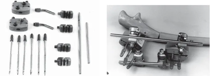
Figures 2a and 2b. Hoffman II Compact II® set.
a) Individual parts: two clamps; two bent connecting rods, six Apexpins®, 3 mm in diameter; two yellow-yellow rod-to-rod clamps; two yellow-gray rod-to-pin clamps; two rods.
b) Assembled external fixator (see also Figure 13).
a b
Proc. coronoideus
Figure 3. Installation of fixator: under image intensification stab inci-sion and insertion of the first Apexpin perpendicular to the longitudi-nal axis of the ulnar shaft and close to the posterior border in direction of the anterior surface of the ulna. The tip of the pin must exit extraar-ticularly just distal to the coronoid process.
Figure 4. A 4-hole clamp is used as a template and inserted over the Apexpin. Through another stab incision, the pin that should be most distally situated is inserted through the clamp into the ulna. Finally, the middle pin is introduced through the clamp. All pins must find pur-chase in the opposite cortex.
Figure 5. In patients > 10 years of age, a fourth Apexpin should be used. In this instance, the clamp has to be moved distally by one hole to al-low insertion of the fourth pin, and this is done to avoid its intraaar-ticular placement. Using the same principle, a second clamp is mount-ed freehand three fingerbreadths distal to the first clamp.
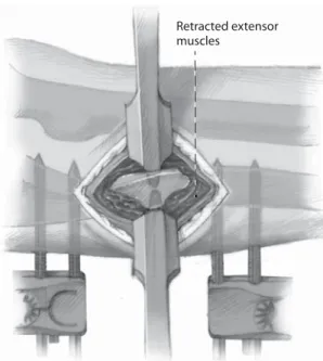
Retracted extensor muscles
Figure 6. Subperiosteal exposure of the ulna through a 3-cm skin inci-sion between both clamps and subperiosteal insertion of two small, blunt Hohmann retractors.
Figures 7a and 7b. Ulnar osteotomy. a) Drilling of three drill holes with a 2-mm drill bit under irrigation. b) Transverse osteotomy with a 10-mm chisel.
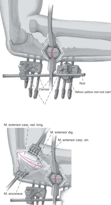
Rod Clamps
Yellow-yellow rod-rod clamp
Figure 8. Temporary fixation using two bent attachment rods that are inserted into the star-like hole of each clamp and secured with a hex screw. Now two yellow-yellow rod-rod clamps are slid over the attach-ment rods. The longitudinal rod is then passed through the clamps bridging both clamps. The rod-rod clamps are mounted on both rods with click mechanism and fastened with a hex screw.
Epicondylus radialis hum.
M. extensor carp. rad. long.
M. extensor dig.
M. extensor carp. uln.
M. anconeus Joint capsule
M. anconeus
Figures 9a to 9c. Reduction of the radial head.
a) Posteroradial longitudinal incision slightly anterior to the lateral epicondyle.
b) Division of fascia and deepening between anconeus and extensor carpi ulnaris posteriorly and extensor digitorum and extensor carpi radialis longus anteriorly.
c) After blunt retraction of the anconeus in a posterior direction, the humeroradial capsule joint is exposed. Longitudinal division of the capsule starting at its humeral origin and extension distally. Exposure of the dislocated radial head.
a
b
Caput radii Collum radii Lig. anulare radii
Epicondylus lateralis hum.
M. supinator Incisura ulnae rad.
Figure 10. Complete excision with a rongeur of the often scarred rem-nants of the annular ligament up to the articular facet of the ulna. The radial head must be completely freed of scar and fat tissue.
Figure 11. Loosening of the hex screw of the fixator to allow a posterior angulation at the ulnar osteotomy and a reduction of the radial head which had been dislocated anteriorly or anterolaterally. Tightening of the clamps with the ulna in a position of posterior angulation. With a blunt, small Hohmann retractor the radial head is kept in the reduced position.
Figures 12a to 12e. While holding the radial head in the reduced position with the Hohm-ann retractor, movements of pro- and supina-tion are performed and the fixator is tightened. After removal of the Hohmann retractor, the exactness of the reduction, particularly during pro- and supination (a to c) and in full flexion and extension (d, e), is checked under direct vi-sion. If during the extremes of these move-ments a tendency to dislocation is observed, the fixator is loosened, the head kept in a re-duced position with a blunt Hohmann retrac-tor, all movements are repeated up to their ex-tremes, and then the fixator is tightened again.
It is sometimes necessary to repeat this maneuver a few times. It may also be necessary to manually increase the angulation of the ulna. Once the reduction of the radial head during pro- and supination under direct vision proves to be satisfactory, the movements of flexion/exten-sion and pro- and supination are checked with the image intensifier in two planes and documented by radiographs.
a b c
Postoperative Management
• Immediately after surgery the patient can move his/ her arm as much as tolerated. Physiotherapy is not prescribed. First dressing change on day 1. The pa-tient can be discharged as soon as he/she and the par-ents are familiar with the pin-site care, usually on day 2 or 3.
• Fixator and wounds should be controlled weekly. First radiograph after 4 weeks and recording of the active range of motion. Depending on the findings, the next radiographic control is done 4–6 weeks later. After bony consolidation of the ulnar osteotomy, the exter-nal fixator is removed. Depending on the patient’s age, this is usually done under anesthesia, seldom only under sedation.
• Should the range of motion not improve considerably between first and second radiographic control, a care-ful physiotherapy under the guidance of an experi-enced physiotherapist should be instituted at the earli-est after 8 weeks.
Errors, Hazards, Complications
• Pin tract infection or pin loosening: depending on the time of occurrence:
– change of pin sites and, if necessary, of fixator, curet-tage of the pin tract; antibiotic therapy based on cul-ture and sensitivity studies;
– removal of the pin, curettage of pin tract; antibiotic therapy and, if indicated, immobilization in a plaster cast until the osteotomy has consolidated;
– improved pin-site care;
– pin loosening without infection (occurs only after several weeks): pin removal and, if necessary, immo-bilization in a plaster cast until bony consolidation of the ulnar osteotomy.
• Secondary displacement of the osteotomy and, thus, secondary dislocation of the radial head during phys-iotherapy: do not prescribe physiotherapy and allow patients to move freely. Physiotherapy, if needed, only Rod
Clamps
Bent connecting rods
Yellow-yellow rod-rod clamp
Additional rod
Yellow-gray rod-pin clamp
Figure 13. Final tightening of the clamps. An additional gray-gold rod is attached to the pins with pin-rod clamps for improved stability. Re-sorbable sub- and intracutaneous running suture for closure of the incisions over the ulnar osteotomy and over the humeroradial joint. Around the pin sites, gauze strips are placed. No external immobiliza-tion.
Table 2. Results. f: female; m: male.
Follow-up Age at Interval between Radial head Elbow flexion/extension Forearm pronation/supination periode surgery trauma and preoperative at follow-up preoperative at follow-up
(months) (years) reconstruction (°) (°) (°) (°)
1 f 14 8 6 months Reduceda 130-20-0 110-10-0 60-0-80 40-0-90 2 m 7 8,5 15 months Reduced 90-0-0 110-0-0 80-0-80 80-0-80 3 f 3 7 2 weeks Reduced 100-0-0 140-0-0 90-0-80 90-0-80 4 m 21 7 5 months Reduced 125-0-10 125-0-10 80-0-80 80-0-80 5 f 44 5 16 months Reduced 150-0-10 150-0-10 90-0-90 90-0-90 6 f 5 6 months Reduced 7 m 11 9 10 months Reduced 120-0-0 140-0-0 80-0-80 80-0-80 8 f 10 15 7 years Reduced 150-0-0 150-0-0 90-0-80 90-0-20 9 m 18 9,5 5 years Reduced 110-0-0 130-0-0 80-0-90 80-0-90 10 f 6 10 1 year Reduced 140-0-0 140-20-0 80-0-80 80-0-80 11 m 17 6 7 months Reduced 140-0-0 140-0-0 80-0-80 80-0-80 12 m 20 8 8 months Reduced 150-0-05 150-0-5 90-0-90 90-0-90 13 f 5 10 2 years Reducedb 130-0-10 140-0-10 80-0-90 80-0-90 14 m 7 15 3 years Reduced 140-25-0 140-10-0 60-0-40 80-0-20
after consolidation of the osteotomy. If physiotherapy has been performed and caused a loss of reduction, revision surgery is indicated depending on the find-ings.
• New dislocation such as after trauma in the presence of the fixator: repeat surgery (depending on the psy-chosocial situation).
Results
Between 01/1998 and 05/2001, we performed an open reduction of the radial head without ligamentoplasty, and a proximal ulnar osteotomy fixed with an external fixator in 14 patients presenting with a late missed Mon-teggia lesion (Bado type I). No postoperative
immobili-Figures 14a to 14d. a) 8-year-old girl (patient # 1): AP and lateral radiographs after open reduction and transarticular Kirschner wire fixation according to Witt [31, 32] done elsewhere.
b) AP and lateral radiographs showing anterior redislocation of the radial head.
c) Radiographs taken 2 and 3 months after open reduction of radial head, ulnar osteotomy, and ex-ternal fixation. Perfect reduction of radial head.
d) 1.5 years after removal of external fixator, the radial head lies in perfect position as seen in the AP and lateral radiographs. An anterior calcification of the capsule still seen in a) to c) has been com-pletely resorbed.
a b
c
zation was used. An ulnar lengthening was only required in one patient; in all others the reduction of the radial head posed no problems.
At the time of reconstruction, the average age of the seven girls and seven boys was 9 (5–15) years, and the average interval between index trauma and reconstruc-tion 21 months (2 weeks to 7 years). The external fixator was removed after an average of 12 (7–16) weeks. Clini-cal and radiologic follow-up of 13 children after an aver-age of 14 (3–44) months. During the clinical follow-up the axial alignment, elbow flexion and extension, and pro- and supination were recorded (Table 2). One girl (No. 6) could not be examined clinically. However, ra-diographs after 12 months done elsewhere were avail-able and showed a proper position of the radial head and a consolidation of the osteotomy. The examining colleague reported a full range of motion and symmet-rical axial alignment of the elbows in this patient.
Reduction of the radial head: the reduction was
maintained postoperatively in twelve patients. A loss of reduction occurred in two patients (No. 1 and 13). A closed reduction and a fixator adjustment were success-fully done in one patient (No. 13) after 2.5 weeks, the other patient required an open reduction after 2 weeks (Figure 14).
Elbow alignment: the axial alignment of the elbows
was clinically symmetric preoperatively as well as at the time of follow-up.
Range of motion: Seven out of 14 patients showed a
limited range of motion preoperatively, involving most-ly (five times) the flexion. The range returned to normal in four patients and was improved in one. Two patients had an extension lag preoperatively; it had improved at the time of follow-up. A preoperative limitation of fore-arm rotation deteriorated slightly in one patient, in an-other patient the pronation improved but was accompa-nied by an identical loss of supination. The range of motion decreased in four patients postoperatively (once in flexion, once in extension, once a 20° loss of prona-tion, and once a 60° loss of supination). No correlation could be found between loss of motion and age or inter-val between index trauma and reconstruction.
Symptoms: none of the patients complained about
pain either preoperatively or at the time of follow-up.
Aftercare: a plaster cast was never used, and the
pa-tients were allowed to move their elbow in all planes postoperatively. Physiotherapy was never prescribed. Already during the hospital stay patients and parents were instructed on the daily pin-site care. They were
asked to bathe the arm daily after wound healing (usu-ally after 14 days) or to take a shower.
Complications: no complication such as infection,
nerve or vessel lesion or pseudarthrosis was observed. All patients were satisfied with the cosmetic and func-tional result.
Experiences with the described method have not been published by other authors, with the exception of two case histories [6]. Corrective ulnar osteotomies fixed with plates and immobilized in a plaster cast and annular ligament reconstruction with and without transarticular Kirschner wire show an elevated incidence of complica-tions (plate breakage, limitation of range of motion, ra-dioulnar synostoses) and an incidence of redislocation of approximately 25% [10, 16, 18, 23, 24].
References
1. Abe M, Ishizu T, Morikawa J. Recurrent posterior dislocation of the head of the radius in posttraumatic cubitus varus. J Bone Joint Surg Br 1995;77:582–5.
2. Bado JL.The Monteggia lesion. Clin Orthop 1967;50:71–86. 3. Bouyala JM, Bollini G, Jacquemier M, et al. Le traitement des
luxa-tions anciennes da la tête radiale chez l’enfant par l’ostéotomie haute du cubitus. À propos des 15 cas. Rev Chir Orthop 1988;74:173–82.
4. Bouyala JM, Chrestian P, Ramaherison P. L’ostéotomie haute du cubitus dans le traitement de la luxation antérieure résiduelle après fracture de Monteggia. Chir Pediatr 1978;19:201–3. 5. De Boeck H. Radial neck osteolysis after annular ligament
recon-struction. A case report. Clin Orthop 1997;342:94–8. 6. Exner GU. Missed chronic anterior Monteggia lesion. Closed
reduction by gradual lengthening and angulation of the ulna. J Bone Joint Surg Br 2001;83:547–50.
7. Fischer M, Maroske D. Der Drahtbruch als Komplikation der trans-artikulären Spickung bei der kindlichen Radiushalsfraktur. Unfall-heilkunde 1976;79:277–9.
8. Hertel P. Radiusköpfchenfrakturen – einschließlich Monteggia-Verletzungen. Unfallmed Tagung Landesverb Gesetzl Berufs-genoss 1977;32:199.
9. Hertel P, Bernhard M, Moazami-Goudarzi Y. Die fehlverheilte kindliche Fraktur – der Monteggia-Schaden. Orthopäde 1991;20: 341–5.
10. Hirayama T, Takemitsu Y, Yagihara K, et al. Operation for chronic dislocation of the radial head in children. Reduction by osteoto-my of the ulna. J Bone Joint Surg Br 1987;69:639–42.
11. Hurst LC, Dubrow EN. Surgical treatment of symptomatic chronic radial head dislocation: a neglected Monteggia fracture. J Pediatr Orthop 1983;3:227–30.
12. Laer L von. Late missed Monteggia fractures. Osteosynthese Int 2000;8:1–4.
13. Laer L von, Pirwitz A, Hasler C. Late missed Monteggia fractures. Techn Orthop 2000;15:30–37.
14. Laer L von, Pirwitz A, Vocke AK. Posttraumatische Problemfälle am kindlichen Ellbogen. Orthopäde 1997;26:1030–6.
15. Letts M, Locht R, Wiens J. Monteggia fracture dislocation in chil-dren. J Bone Joint Surg Br 1985;67:724–7.
16. Lloyds-Roberts GC, Bucknill TM. Anterior dislocation of the radial head in children. J Bone Joint Surg Br 1977;59:402–7.
17. Luther R, Legal H. Ursachen von Fehlergebnissen in der Behand-lung kindlicher Radiusköpfchenfrakturen und -luxationen. Or-thop Prax 1973;9:450.
18. Oner FC, Diepstraten AF. Treatment of chronic post-traumatic dis-location of the radial head in children. J Bone Joint Surg Br 1993;75:577–81.
19. Peiro A, Andrei F, Fernandez-Esteve F. Acute Monteggia lesions in children. J Bone Joint Surg Am 1977;59:92–7.
20. Reckling FW. Unstable fracture-dislocations of the forearm (Mon-teggia and Galleazzi lesions). J Bone Joint Surg Am 1982;64: 57–63.
21. Schmitt E, Mittelmaier H, Katthagen BD. Operative Lang-zeitergebnisse bei veralteten Radiusköpfchenluxationen im Kindesalter. Akt Traumatol 1985;15:36–41.
22. Schulitz KP. Die operative Behandlung der veralteten Radiusköpf-chenluxationen im Kindesalter. Arch Orthop Unfallchir 1975;81: 225–37.
23. Seel MJ, Peterson HA. Management of chronic posttraumatic ra-dial head dislocation in children. J Pediatr Orthop 1999;19:306–12. 24. Stoll TM, Willis RB, Paterson DC. Treatment of the missed
Monteg-gia fracture in the child. J Bone Joint Surg Br 1992;74:436–40. 25. Trillat A, Marsan C, Lapeyre B. Classification et traitement des
fractures de Monteggia. Rev Chir Orthop 1969;55:639–57. 26. Verneret C, Langlais J, Pouliquen JC, Wieser R, Scheier HJ,
Gram-mont P, Chrestian JM, Ramaherison P, Bonyala JM, Jani L. Luxa-tions anciennes post-traumatiques de la tête radiale chez l’enfant. Rev Chir Orthop 1989;75:77–89.
27. Vinz H. Die Monteggia-Fraktur im Kindesalter. Beitr Orthop Trau-matol 1989;36:153–68.
28. Vinz H. Die isolierte Luxation des Radiusköpfchens im Kindes-alter. Beitr Orthop Traumatol 1989;36:169–76.
29. Wiesner R, Scheier HJG, Grammont P, Rigan HP. Veraltete Radi-usköpfchenluxationen bei Kindern nach Monteggia-Frakturen. Orthopäde 1981;10:307–10.
30. Wiley J, Pegington J, Horwich JP. Traumatic dislocation of the radius at the elbow. J Bone Joint Surg Br 1974;56:501–7. 31. Witt AN. Beitrag zur Fixation des Radiusköpfchens. Verh Dtsch
Orthop Ges 1958;90:394.
32. Witt AN. Die transartikuläre Fixation bei Frakturen und Luxa-tionen im Bereich des Humeroradialgelenkes. In: Maurer G, Hrsg. Chirurg im Fortschritt. Stuttgart: Enke, 1965.
Address for Correspondence
Dr. Carol Hasler
Orthopädisch-traumatologische Abteilung Universitätskinderspital beider Basel Postfach Römergasse 8 4005 Basel Switzerland Phone (+41/61) 685-5434, Fax -5006 e-mail: [email protected]