Proceedings
of
the
Nutrition
Society
The Summer Meeting of the Nutrition Society was held at the University of Surrey, Guildford on 30 June–2 July 2009
Conference on ‘Over- and undernutrition: challenges and approaches’
Postgraduate Symposium
Positive influence of nutritional alkalinity on bone health
E. Wynn
1*, M. A. Krieg
1, S. A. Lanham-New
2and P. Burckhardt
31University Hospital (CHUV), 1011 Lausanne, Switzerland
2Faculty of Health and Medical Sciences, University of Surrey, Guildford GU2 7XH, UK 3Clinic Bois-Cerf, 1003 Lausanne, Switzerland
There is growing evidence that consumption of a Western diet is a risk factor for osteoporosis through excess acid supply, while fruits and vegetables balance the excess acidity, mostly by providing K-rich bicarbonate-rich foods. Western diets consumed by adults generate approxi-mately 50–100 mEq acid/d; therefore, healthy adults consuming such a diet are at risk of chronic low-grade metabolic acidosis, which worsens with age as a result of declining kidney function. Bone buffers the excess acid by delivering cations and it is considered that with time an overstimulation of this process will lead to the dissolution of the bone mineral content and hence to reduced bone mass. Intakes of K, Mg and fruit and vegetables have been associated with a higher alkaline status and a subsequent beneficial effect on bone health. In healthy male volunteers an acid-forming diet increases urinary Ca excretion by 74 % and urinary C-terminal telopeptide of type I collagen (C-telopeptide) excretion by 19 % when compared with an alkali (base-forming) diet. Cross-sectional studies have shown that there is a correlation between the nutritional acid load and bone health measured by bone ultrasound or dual-energy X-ray absorptiometry. Few studies have been undertaken in very elderly women (> 75 years), whose osteoporosis risk is very pertinent. The EVAluation of Nutrients Intakes and Bone Ultra Sound Study has developed and validated (n 51) an FFQ for use in a very elderly Swiss population (mean age 80.4 (SD2.99) years), which has shown intakes of key nutrients (energy, fat, car-bohydrate, Ca, Mg, vitamin C, D and E) to be low in 401 subjects. A subsequent study to assess net endogenous acid production (NEAP) and bone ultrasound results in 256 women aged ‡ 75 years has shown that lower NEAP (P= 0.023) and higher K intake (P = 0.033) are correlated with higher bone ultrasound results. High acid load may be an important additional risk factor that may be particularly relevant in very elderly patients with an already-high fracture risk. The latter study adds to knowledge by confirming a positive link between dietary alkalinity and bone health indices in the very elderly. In a further study to complement these findings it has also been shown in a group of thirty young women that in Ca sufficiency an acid Ca-rich water has no effect on bone resorption, while an alkaline bicarbonate-rich water leads to a decrease in both serum parathyroid hormone and serum C-telopeptide. Further investigations need to be undertaken to study whether these positive effects on bone loss are maintained over long-term treatment. Mineral-water consumption could be an easy and inexpensive way of helping to prevent osteoporosis and could be of major interest for long-term prevention of bone loss.
Dietary alkalinity: Bone health: Acid–base balance: Elderly
Osteoporosis is a skeletal disorder characterised by low bone mass and microarchitectural deterioration of bone tissue, leading to bone fragility and an increased risk of
fracture. Optimum bone health is achieved through the contribution of numerous factors such as genetics, health and nutrition. It is common to consider bone health in
Abbreviations: BUA, broadband ultrasound attenuation; BMD, bone mineral density; C-telopeptide, C-terminal telopeptide of type I collagen; NEAP, net endogenous acid production.
*Corresponding author: Emma Wynn, present address BioAnalytical Science Department, Clinical Evaluation Group, Nestle´ Research Center, PO Box 44, CH-1000 Lausanne 26, Switzerland, email [email protected]
Proceedings
of
the
Nutrition
Society
terms of Ca intake only. Ca is indeed a critical component of the skeleton; however, bone health is much more com-plex. Whilst the optimum diet for osteoporosis prevention remains unknown, a variety of nutrients are important to the ageing skeleton including protein and micronutrients such as Ca, K and vitamin K(1,2). There is a growing belief that the Western diet may be a risk factor for osteoporosis through excess acid supply. Fruit and vegetable intake may balance the excess acidity by providing K(1,2). As the major determining factor for osteoporosis is an individual’s genetic makeup, which cannot be modified, it is important to identify modifiable risk factors that contribute to bone health and use them in prevention messages to the popu-lation. Nutrition is a modifiable factor that has the potential to be altered relatively easily and cheaply.
Osteoporosis Definition
In 1994 the WHO Working Group defined osteoporosis according to measurements of bone mineral density (BMD) using dual-energy X-ray absorptiometry. Thus, osteoporosis is defined as a bone density T-score of £ 2.5
SD below normal peak values for young adults(3). These
criteria were initially established for the assessment of osteoporosis in Caucasian women.
Epidemiology
Recent epidemiological data estimate that one in two women and one in five men aged > 50 years will suffer from an osteoporotic fracture(4). Thus, it is urgent for public health strategies to reduce the predicted increase in osteoporosis within the aging population by undertaking prevention campaigns. A public health strategy for optimising bone health throughout the life cycle is critically important.
Osteoporosis affects an estimated seventy-five million individuals in Europe, USA and Japan. In 2000 there were approximately 9· 106new osteoporotic fractures(5). Recent
epidemiological data from the Third National Health and Nutrition Examination Survey indicate that 13–18 % of white American women aged ‡ 50 years have osteoporosis of the hip(6). The lifetime risk in the UK for a hip, spine or forearm fracture at the age of 50 years has been estimated to be 53 % in women and 21 % in men(7). The projected rise in osteoporotic fracture worldwide clearly demonstrates that the future impact of osteoporosis will be enormous; the worldwide incidence of hip fracture in men is projected to increase by 310 % and 240 % in women by 2050(8).
In Switzerland between 2000 and 2020 osteoporotic hip, vertebral and forearm fracture are predicted to increase by 33 %, 27 % and 19 % respectively if present prevention and treatment patterns stay unchanged(9). In terms of duration of hospital stay, the annual cost of hospitalisations for osteoporotic fractures is higher than those for myo-cardial infarction, stroke and breast cancer(10). In 2000 62 535 hospitalisations for fractures were registered. The direct medical cost of hospitalisations of patients with osteoporosis and/or related fractures was 357· 106Swiss Francs (£15· 106), with hip fractures accounting for
approximately half these costs(11).
Acid–base balance
Disturbances of acid–base balance in the body are classi-fied as either acidosis, indicating an excess of H+ ions (leading to an acid environment; e.g. metabolic, latent or respiratory acidosis), or alkalosis (leading to a reduction in H+ ions and therefore a more alkaline environment). Such disturbances of acid–base balance are relevant to bone health. In vitro studies have shown that there is an induced Ca efflux from bone as a result of metabolic acidosis(12), which is the only form of acidosis relevant to bone. A moderate increase in alkaline equivalents (90 mmol potas-sium citrate/d) has been shown to reduce bone resorption, increase bone formation and improve Ca balance in post-menopausal women(13).
Measuring acid–base status
During the past few decades a number of researchers have used in vitro methods that are inappropriate to specifically determine the acid- or alkaline-producing potential of the diet(14). An in vitro measurement of acid:base has been made by using a calculation model(15,16). The net acid load is estimated from average intestinal absorption rates of ingested S-containing protein (cysteine and methionine) and additional minerals as well as an anthropometry-based estimate for organic acid excretion:
estimated net endogenous acid production (NEAP; mEq=d)= PRAL (mEq=d) + OAest(mEq=d)ð17Þ;
where PRAL is potential renal acid load and OAest is
estimated organic acids, i.e. estimated urinary organic anions, with these two components calculated as follows (we did not include Na and Cl in the PRAL equation be-cause they are usually balanced in the diet):
PRAL (mEq=d)= 0.49 · protein (g=d) + 0.037
· P (mg=d) - 0.021 · K (mg=d) - 0.026 · Mg (mg=d) - 0.013 · Ca (mg=d); OAest(mEq=d)= individual body surface area
· [0.007184 - height0.725(cm)- weight0.425(kg)]· 41=1.73:
Determination of acid:base of foods contributing to the overall diet consumed by individuals and population groups is a practical and useful method that can be used in assessing the role of the skeleton in acid–base home-ostasis(18). PRAL has been estimated for a variety of foods items using nutrient data expressed on a per 100 g food basis for protein (methionine, cysteine), P, Cl, K, Mg, Ca and Na (Table 1)(16).
Nutritional factors that influence acid–base balance Nutrition has long been known to strongly influence acid– base balance in human subjects(19). Intakes of K, Mg and fruit and vegetables have been associated with a more
Proceedings
of
the
Nutrition
Society
alkaline environment in the human body and therefore have a beneficial effect in relation to bone health(20).
Indeed, Western diets consumed by adults generate 50– 100 mEq acid/d(21). Thus, healthy adults consuming such a diet are at risk of chronic low-grade metabolic acidosis, which worsens with age as a result of a decline in kidney function(22).
Role of the skeleton
Maintenance of acid–base homeostasis is tightly regulated in the extracellular fluid at pH 7.4 °0.05(18). Almost every biological process in the human body is dependent on the acid–base balance, including bone metabolism. Bone con-tributes to acid–base homeostasis as it delivers cations such as Mg, K, Ca and Na, which can be associated with alkali salts such as citrate or carbonate. Over time, an over-stimulation of this process will lead to the dissolution of the bone mineral content and hence to a reduction in bone
mass(18,22). Thus, long-term nutritional acid load may be
harmful to bone health.
Protein
Protein is an important macronutrient contributing to BMD. Indeed, sufficient dietary intake appears to have a beneficial effect on bone healing. Supplementation with 20 g protein improves fracture healing and reduces bone loss in elderly subjects; the mechanism appearing to be at least partially through an increase in insulin-like growth factor 1, which stimulates proliferation and differentiation of osteoblasts(23). However, protein has an acidic effect on bone, which is thought to be undesirable. More precisely, two mechanisms come into play: excess protein induces an increase in renal Ca excretion; the metabolism of the S amino acids (methionine and cysteine) generates an acid load, which results in a reduction in blood and urinary
pH(22,24,25). However, it has been suggested that
high-protein diets are associated with an increased risk of fracture when Ca is low, but there is no overall association between total protein and fracture risk with high Ca intake(26). Furthermore, a study of 161 post-menopausal women has suggested that protein intake is positively associated with areal BMD, but benefit at the lumbar spine is offset by a negative impact of the protein S acid load(27). However, it has also been suggested that the negative association between meat intake and BMD may be a reflection of an inadequate consumption of fruit and vegetables rather than an excessive consumption of meat(28).
In vitro studies
In vitro studies have shown that metabolic acidosis induces a Ca efflux from bone(12). It has been demonstrated that the acidification of bone may not be linked only to osteoclastic activity; cultured osteoblasts show reduced collagen syn-thesis and mineralisation in an acidic environment(29,30). In both the animal and human model an acid environment is associated with a negative Ca balance and increased bone
loss(31–33). Any reduction in extracellular pH enhances
osteoclastic activity(34). Even a small change in pH, approximately (pH 7.1) but not exactly at the physiological level (pH 7.4), stimulates osteoclasts(35). On a long-term nutritional acidic load, pH is kept constant at the expense of bone, which delivers the buffering substances through bone resorption(36).
In vivo studies
In vivo studies in healthy subjects have shown that sup-plements of KHCO3, potassium citrate and even
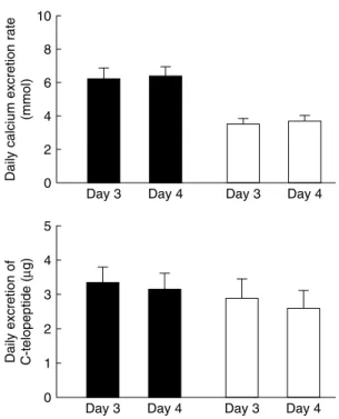
bicarbo-nate-rich mineral water decrease calciuria and bone resorption markers(13,37–39). In healthy male volunteers an acid-forming diet increases urinary Ca excretion by 74 % and urinary C-terminal telopeptide of type I collagen (C-telopeptide) excretion by 19 % when compared with an alkali (base-forming) diet(40) (Fig. 1). In cross-sectional studies assessment of the acid:base value has shown that there is a correlation between the nutritional acid load and bone health measured by bone ultrasound(41) or dual-energy X-ray absorptiometry(42). The Dietary Approaches to Stop Hypertension Trial, which examined the effects of two dietary patterns in 186 adults aged 23–76 years, has compared an experimental Ca-rich diet emphasising fruits, vegetables, whole grains, poultry, fish, nuts and low-fat dairy products with the typical American diet as the con-trol diet. The experimental diet was found to reduce serum osteocalcin by 8–11 % and C-telopeptide by 16–18 % (P< 0.001)(43). However, a recent randomised
placebo-controlled study has shown no effect of K supplementa-tion(44). It was found that potassium citrate supplementa-tion for 2 years does not decrease bone turnover or increase BMD at the spine or hip in post-menopausal healthy women. Moreover, in the group of women who received a supplement of 300 g fruit and vegetables no impact on bone turnover or BMD was observed(44). Fur-thermore, the findings of the EVAluation of Nutrients Intakes and Bone Ultra Sound Study (discussed later) have led to the hypothesis that very elderly subjects may be more sensitive to nutritional interventions. In a healthy population other factors such as weight and muscle mass have a substantial effect on bone(45,46).
Results from the EVAluation of Nutrients Intakes and Bone Ultra Sound Study
Development of a Swiss FFQ
Few studies have been undertaken in very elderly women (>75 years), who are at major risk of osteoporosis risk. As part of the EVAluation of Nutrients Intakes and Bone Table 1. Estimated potential renal acid load (PRAL) in a variety of
foods items (from Remer & Manz(16))
Food group PRAL (mEq/100 g)
Hard cheese 23.6
Meat and meat products 9.5
Bread 3.5
Milk and non-cheese products 1
Vegetables - 2.8
Proceedings
of
the
Nutrition
Society
Ultra Sound Study on osteoporosis in a Swiss very elderly population the influence of dietary intake and acid load on bone health indices was examined in a cohort of 401 ambulatory women (mean age 80.6 years) from the Lausanne area, an urban area of approximately 200 000 in-habitants. A detailed FFQ was developed, cross-validated against a further forty-four 4 d weighed records and the short- (1 month; n 15) and long-term (12 months; n 14) reproducibility examined(47). No significant difference was found between mean energy intakes by 4 d weighed records and the FFQ (6548 (SD 1469) kJ (1565 (SD351) kcal) and
6867 (SD2189) kJ (1641 (SD523) kcal) respectively). Mean
crude nutrient intakes were also found to be similar (paired t test), ranging from P= 0.13 for K to P = 0.48 for Mg. Similar results were found in the reproducibility studies.
Nutrient intake of the EVAluation of Nutrients Intakes and Bone Ultra Sound Study population
The validated FFQ was administered to the cohort of 401 subjects to assess dietary intake(48). The mean daily energy intake was found to be 6460 (SD1874) kJ (1544 (SD447.7)
kcal), with a protein intake of 65.2 (SD19.9) g, i.e. 1.03 g/
kg body weight per d. The mean daily intakes for energy, fat, carbohydrate, Ca, Mg, vitamin C, D and E were found
to be below their respective recommended nutrient intakes(49), while protein, P, K, Fe and vitamin B6intakes
were above their respective recommended nutrient in-takes(49) (Table 2). The NEAP for each subject was also calculated using the PRAL equation (Table 2).
Thus, the recommendation to optimise dietary intake is energy-dense foods rich in carbohydrate, Mg, Ca, vitamin D and E as well as regular sunshine exposure.
Association between net endogenous acid production estimates and bone ultrasound
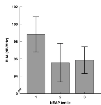
The association between NEAP estimates and bone ultra-sound results was assessed in the cohort of 401 subjects(50). No significant effects of the acid load were observed in the whole group, which led to a separate assessment of those women who had a history of fracture (n 256). Fig. 2 shows broadband ultrasound attenuation (BUA) for these 256 women stratified by tertiles of NEAP. Associations were found between higher BUA and both lower estimates of NEAP (r - 0.142, P <0.05) and higher estimates of Ca and K (r 0.151 and 0.134 respectively, P< 0.05), with the dif-ferences remaining significant after adjustment for age, BMI and osteoporosis treatment. The stepwise regression analysis included BUA, NEAP, BMI, osteoporosis treat-ment, mini nutritional assessment and age. It explains 7.6 % of the variation in BUA. BMI and osteoporosis treatment explained 6.3 % and NEAP 1.3 % of the variation (P< 0.05); it is known that £ 80 % of the variance is explained by genetic factors.
Thus, the findings suggest that lower estimates of NEAP (i.e. more-alkaline diets) are significantly associated with higher BUA measured by bone ultrasound in the subgroup of elderly women who had a history of fractures. High acid load may be an important additional risk factor that might Table 2. A comparison of daily dietary intake, as recorded by FFQ, and recommended nutrient intakes (RNI) for 401 very elderly Swiss
women (mean age 80.6 years)
Nutrient RNI(49) Recorded intake Mean SD % RNI Energy (kJ) 8657 6460 1874 74.6 Protein (g) 62.7 65.2 19.9 104 Fat (g) 69 63.9 21.3 92.6 Carbohydrate (g) > 259 164 54.4 63.3 Minerals Ca (mg) 1000 983 389 98.3 P (mg) 700 1164 392 166 Mg (mg) 300 288 93.1 95.9 K ( mg) 2000 2761 875 138 Fe (mg) 10 11.6 3.7 116 Vitamins Vitamin B6(mg) 1.2 1.25 0.44 104 Vitamin C (mg) 100 93.1 45.2 93.1 Vitamin D (mg) 10 2.49 1.45 24.9 Vitamin E (mg) 11 9.39 3.88 85.4 NEAP (mEq) - 3.22 - 42.4 to + 29.2*
NEAP, net endogenous acid production. *Range. 10 8 6 4 2 0
Daily calcium excretion rate
(mmol)
Day 3 Day 4 Day 3 Day 4
5 4 3 2 1 0 Daily excretion of C-telopeptide (
µ
g)
Day 3 Day 4 Day 3 Day 4
Fig. 1. Mean daily excretion rates of calcium and C-terminal telo-peptide of type I collagen (C-telotelo-peptide) for healthy male volun-teers (n 8; age 22–31 years) receiving an acid-forming diet (&) or a base-forming diet (K) with no calcium supplement on day 3 and 1 g calcium supplement on day 4 in a double-cross-over design study. To obtain optimal compliance both diets consisted of alternation of two daily menus, one for days 1 and 3, the other for days 2 and 4. Values are means with their standard errors represented by vertical bars. The effect of diet was significant for calcium (P= 0.0002) and C-telopeptide (P= 0.01). Calcium supplement had no significant effect. (From Buclinet al.(40); with kind permission of Springer Sci-ence and Business Media.)
Proceedings
of
the
Nutrition
Society
be particularly relevant in very elderly patients with an already-high fracture risk. The study adds to knowledge by confirming a positive link between dietary alkalinity and bone health indices in the very elderly.
The influence of drinking mineral water on bone health indices
The positive association between low estimates of dietary acid load and bone ultrasound in women >75 years of age with a history of fracture has led to the investigation of the effect of acid load in a younger population by assessing the effect of drinking mineral water on premenopausal women(51). The consumption of mineral water may be an easy and cheap way of influencing the acid–base balance.
A comparison was undertaken of the effect of an alka-line (base-forming) mineral water rich in bicarbonate (mg/l; Ca 547, bicarbonate 2172, sulphate 9; PRAL - 11.2 mEq/l) with that of an acid (acid-forming) mineral water rich in Ca only (mg/l; Ca 520, bicarbonate 291, sulphate 1160; PRAL + 9.2 mEq/l) on bone markers in young women with a normal Ca intake. Thirty female dietitians aged 26.3 (SD 7.3) years were randomised into
two groups, with each group following an identical weighed balanced diet (965 mg Ca/d) and drinking 1.5
litres of the assigned mineral water daily. Changes in blood and urine electrolytes, C-telopeptide, urinary pH and bicarbonate and serum parathyroid hormone were measured after 2 and 4 weeks. No difference was found between the groups at baseline and a similar increase in urinary Ca excretion was observed. Urinary pH and bicarbonate excretion were found to increase with the alkaline mineral water, but not with the acid mineral water. Parathyroid hormone (P= 0.022; Fig. 3) and serum C-telopeptide (P= 0.023; Fig. 4) were shown to decrease with the alkaline NEAP tertile 3 2 1 BUA (dB/MHz) 100 102 98 96 94 0
Fig. 2. Broadband ultrasound attenuation (BUA) in the 256 elderly women from the EVAluation of Nutrients Intakes and Bone Ultra Sound Study with fractures stratified by tertiles of net endogenous acid production (NEAP). Values are means and standard deviations represented by vertical bars for eighty-six, seventy-eight and ninety-two subjects for tertiles 1, 2 and 3 respectively. The mean NEAP values for tertiles 1, 2 and 3 were –15.4, –2.6 and 8.3 mEq/d respectively. The mean scores of BUA for the three groups were significantly different:P= 0.03 (one-way ANOVA with a post hoc test (Tukey test));P= 0.03 (F test for linearity). Comparison of the means for BUA by tertiles of NEAP (post hoc test (Tukey test)) showed trends: tertile 1v. tertile 2, P= 0.052; tertile 1 v. tertile 3, P= 0.07. (From Wynn et al.(50)
.) 4 2 0 Week 2 3 0 –2 –4 –6 –8 –10 PTH (ng/l)
Fig. 3. Mean changes in serum parathyroid hormone (PTH) in premenopausal women (age 26.3 (SD 7.3) years) after 2 and 4
weeks of consuming either an acid-forming mineral water (n 15;& & &) or a base-forming mineral water (n 15; ) and an identical
weighed balanced diet (965 mg Ca/d). Values are with their stan-dard errors represented by vertical bars. For the base-forming mineral water the decrease after 4 weeks was significant (P= 0.0043). The difference between the two mineral waters at 4 weeks was significant (P= 0.022). (Reprinted from Wynn et al.(51), with
permission from Elsevier.)
4 2 0 Week 0.00 –0.05 –0.10 C-telopeptides (µg/l)
Fig. 4. Mean changes in serum C-terminal telopeptide of type I collagen (C-telopeptides) in premenopausal women (age 26.3 (SD
7.3) years) after 2 and 4 weeks of consuming either an acid-forming mineral water (n 15;& & &) or a base-forming mineral water (n 15;
) and an identical weighed balanced diet (965 mg Ca/d). Values are with their standard errors represented by vertical bars. For the base-forming mineral water the decrease after 4 weeks was sig-nificant (P= 0.021). The difference between the two mineral waters at 4 weeks was significant (P= 0.023). (Reprinted from Wynn et al.(51), with permission from Elsevier.)
Proceedings
of
the
Nutrition
Society
mineral water but not with the acid mineral water. Thus, in Ca sufficiency an acid Ca-rich water has no effect on bone resorption, while an alkaline water rich in bicarbonate leads to a decrease in serum parathyroid hormone and serum-C-telopeptide(51). By careful choice of mineral water the population could possibly positively influence their bone health.
Concluding remarks
In the present paper the role of acid–base balance and bone health has been discussed. A dietary acid load appears to have a detrimental effect on bone health indices. Long-term studies in post-menopausal women, the group most affected by osteoporosis, would be of major interest to investigate the effect of an acidic diet and whether there is an influence on fracture risk.
Directions for future research work
Future work needs to be undertaken to assess whether certain fruits and vegetables have a more beneficial effect than others. Furthermore, some currently unidentified compounds found in a number of herbs, salad and vege-tables have been shown to play a beneficial role in rats(52) that is not mediated by their alkalinity but by their phar-macological activity; for example, certain phytonutrients, especially polyphenols, increase osteoblastic activity and decrease osteoclastic activity(53). A recent study has shown that onion consumption might have a beneficial effect on BMD in peri- and post-menopausal white women aged ‡ 50 years(54). Thus, increasing the amount of fruit and
vegetables in the Western population may be an additional prevention treatment for osteoporosis. However, increasing the daily fruit and vegetable consumption remains a diffi-cult challenge.
It should also be remembered that in man there is a mismatch between the genes and the modern diet that could explain the negative effects of a diet low in K salts on osteoporosis. Man’s pre-agricultural hunter–gatherer ancestors consumed wild animal-source foods and uncul-tivated plant-source foods that would have provided an NaCl-poor K-rich bicarbonate-precursor-rich diet. This K:NaCl has inverted with the dietary shift, as in the mod-ern human diet plant food has fallen and consumption of NaCl has increased(55). Human genes are not adapted to this modern diet as the shift to this contemporary diet has occurred too recently for the genome to adjust(56,57). It has been suggested that the dietary patterns responsible for low-grade metabolic acidosis, as well as low K intakes and high NaCl intakes, contribute to the pathogenesis of osteoporosis and other age-related disorders(58,59).
Acknowledgements
This study was funded by the Foundation for Research on Osteoporosis and Bone Diseases, Lausanne, Switzerland. The authors declare no conflict of interest. E. W. per-formed the studies and wrote the manuscript, S. A. L-N. provided permanent scientific supervision especially for
the nutritional aspects, M. A. K. provided permanent sci-entific supervision especially for the quantitative ultra-sound aspects and P. B. wrote the research protocols, directed and coordinated the studies and supervised the preparation of the manuscript.
References
1. Lanham-New SA (2008) Importance of calcium, vitamin D and vitamin K for osteoporosis prevention and treatment. Proc Nutr Soc 67, 163–176.
2. Macdonald HM, New SA, Fraser WD et al. (2005) Low dietary potassium intakes and high dietary estimates of net endogenous acid production are associated with low bone mineral density in premenopausal women and increased markers of bone resorption in postmenopausal women. Am J Clin Nutr 81, 923–933.
3. Kanis J (1994) Assessment of fracture risk and its application to screening for postmenopausal osteoporosis: synopsis of a WHO report. WHO Study Group. Osteoporos Int 4, 368–381. 4. Holroyd C, Cooper C & Dennison E (2008) Epidemiology of osteoporosis. Best Pract Res Clin Endocrinol Metab 22, 671–685.
5. Johnell O & Kanis J (2006) An estimate of the worldwide prevalence and disability associated with osteoporotic frac-tures. Osteoporos Int 17, 1726–1733.
6. Looker A, Orwoll E & Johnston C (1997) Updated data on proximal femur bone mineral levels of US adults from NHANES III. J Bone Miner Res 12, 1761–1768.
7. Van Staa TP, Dennison EM, Leufkens HG et al. (2001) Epidemiology of fractures in England and Wales. Bone 29, 517–522.
8. Gullberg B, Johnell O & Kanis J (1997) World-wide pro-jections for hip fracture. Osteoporos Int 7, 407–413. 9. Schwenkglenks M, Lippuner K, Ha¨uselmann H et al. (2005)
A model of osteoporosis impact in Switzerland 2000–2020. Osteoporos Int 16, 659–671.
10. Lippuner K, Von Overbeck J, Perrelet R et al. (1997) Inci-dence and direct medical costs of hospitalizations due to osteoporotic fractures in Switzerland. Osteoporos Int 7, 414– 425.
11. Lippuner K, Golder M & Greiner R (2005) Epidemiology and direct medical costs of osteoporotic fractures in men and women in Switzerland. Osteoporos Int 16, Suppl. 2, S8–S17. 12. Bushinsky D & Frick K (2000) The effects of acid on bone.
Curr Opin Nephrol Hypertens 9, 369–379.
13. Sellmeyer D, Schloetter M & Sebastian A (2002) Potassium citrate prevents increased urine calcium excretion and bone resorption induced by a high sodium chloride diet. J Clin Endocrinol Metab 87, 2008–2012.
14. Dwyer J, Folukes E, Evans M et al. (1985) Acid/alkaline ash diets: time for assessment and change. J Am Diet Assoc 85, 841–845.
15. Remer T & Manz F (1994) Estimation of the renal net acid excretion by adults consuming diets containing variable amounts of protein. Am J Clin Nutr 59, 1356–1361. 16. Remer T & Manz F (1995) Potential renal acid load of foods
and its influence on urine pH. J Am Diet Assoc 95, 791–797. 17. Frassetto L, Lanham-New S, Macdonald H et al. (2007) Standardizing terminology for estimating the diet-dependent net acid load to the metabolic system. J Nutr 137, 1491– 1492.
18. New S (2002) The role of the skeleton in acid–base home-ostasis. Proc Nutr Soc 61, 151–164.
Proceedings
of
the
Nutrition
Society
19. Remer T (2000) Influence of diet on acid-base balance. Semin Dial 13, 221–226.
20. New S, Robins S, Campbell M et al. (2000) Dietary influ-ences on bone mass and bone metabolism: further evidence of a positive link between fruit and vegetable consumption and bone health? Am J Clin Nutr 71, 142–151.
21. Vormann J & Remer T (2008) Dietary, metabolic, physio-logic, and disease-related aspects of acid-base balance: fore-word to the contributions of the Second International Acid-Base Symposium. J Nutr 138, 413S–414S.
22. Frassetto L, Curtis Morris R & Sebastian A (1996) Effect of age on blood acid-base composition in adult humans: role of age-related renal functional decline. Am J Physiol 271, F1114–F1122.
23. Bonjour JP, Schu¨rch MA, Chevalley T, Ammann P & Rizzoli R (1997) Protein intake, IGF-I and osteoporosis. Osteoporos Int 7, Suppl. 3, S36–S42.
24. Barzel U (1995) The skeleton as an ion exchange system: implication for the role of acid-base imbalance in the genesis of osteoporosis. J Bone Miner Res 10, 1431–1436.
25. Reid D & New S (1997) Nutritional influences on bone mass. Proc Nutr Soc 56, 977–987.
26. Dargent-Molina P, Sabia S, Touvier M et al. (2008) Proteins, dietary acid load, and calcium and risk of postmenopausal fractures in the E3N French Women Prospective Study. J Bone Miner Res 23, 1915–1922.
27. Thorpe M, Mojtahedi M, Chapman-Novakofski K et al. (2008) A positive association of lumbar spine bone mineral density with dietary protein is suppressed by negative asso-ciation with protein sulphur. J Nutr 138, 80–85.
28. Heaney R & Layman D (2008) Amount and type of protein influences bone health. Am J Clin Nutr 87, 1567S– 1570S.
29. Bushinsky D (1995) Stimulated osteoclastic and suppressed osteoblastic activity in metabolic but not respiratory acidosis. Am J Physiol 268, C80–C88.
30. Frick K & Bushinsky D (1998) Chronic metabolic acidosis reversibly inhibits extracellular matrix gene expression in mouse osteoblasts. Am J Physiol Renal Physiol 275, F840– F847.
31. Lemann J, Litzow J & Lennon E (1966) The effects of chronic acid loads in normal man: Further evidence for the participation of bone mineral in defense against chronic metabolic acidosis. J Clin Invest 45, 1608–1614.
32. Barzel U & Jowsey J (1969) The effects of chronic acid and alkali administration on bone turnover in adult rats. Clin Sci 36, 517–524.
33. Tucker K, Hannan M & Kiel D (2001) The acid-base hypothesis: diet and bone in the Framingham Osteoporosis Study. Eur J Nutr 40, 231–237.
34. Arnett T & Dempster D (1986) Effect of pH on bone resorption by rat osteoclasts in vitro. Endocrinology 119, 119–124.
35. Arnett T & Spowage M (1996) Modulation of the resorptive activity of rat osteoclasts by small changes in extracellular pH near the physiological range. Bone 18, 277–279. 36. Green J & Kleeman C (1991) Role of bone in regulation of
systemic acid-base balance. Kidney Int 39, 9–26.
37. Jehle S, Zanetti A, Muser J et al. (2006) Partial neutralization of the acidogenic western diet with potassium citrate increa-ses bone mass in postmenopausal women with osteopenia. J Am Soc Nephrol 17, 3213–3222.
38. Burckhardt P, Waldvogel S, Aeschlimann J et al. (2002) Bicarbonate in mineral water inhibits bone resorption. J Bone Miner Res 17, Suppl., S476.
39. Sebastian A, Harris S, Ottaway J et al. (1994) Improved mineral balance and skeletal metabolism in postmenopausal
women treated with potassium bicarbonate. New Engl J Med 330, 1776–1781.
40. Buclin T, Cosma M, Appenzeller M et al. (2001) Diet acids and alkalis influence calcium retention in bone. Osteoporos Int 12, 493–499.
41. Welch A, Bingham S, Reeve J et al. (2007) More acidic dietary acid-load is associated with reduced calcaneal broadband ultrasound attenuation in women but not in men: results from the EPIC-Norfolk cohort study. Am J Clin Nutr 85, 1134–1141.
42. New S, Macdonald H, Campbell M et al. (2004) Lower estimates of net endogenous noncarbonic acid production are positively associated with indexes of bone health in pre-menopausal and peripre-menopausal women. Am J Clin Nutr 79, 131–138.
43. Lin P-H, Gointy F, Appel L et al. (2003) The DASH diet and sodium reduction improve markers of bone turnover and calcium metabolism in adults. J Nutr 133, 3130–3136. 44. Macdonald H, Black A, Aucott L et al. (2008) Effect of
potassium citrate supplementation or increased fruit and vegetable intake on bone metabolism in healthy post-menopausal women: a randomized controlled trial. Am J Clin Nutr 88, 465–474.
45. Edelstein SL & Barrett-Connor E (1993) Relation between body size and bone mineral density in elderly men and women. Am J Epidemiol 138, 160–169.
46. Segal NA, Torner JC, Yang M et al. (2008) Muscle mass is more strongly related to hip bone mineral density than is quadriceps strength or lower activity level in adults over age 50 year. J Clin Densitom 11, 503–510.
47. Wynn Dumartheray E, Krieg M, Cornuz J et al. (2006) Validation and reproducibility of a semi-quantitative food frequency questionnaire for use in elderly Swiss women. J Hum Nutr Diet 19, 321–330.
48. Wynn Dumartheray E, Krieg M, Cornuz J et al. (2006) Energy and nutrient intake of Swiss women aged 75–87 years. J Hum Nutr Diet 19, 431–435.
49. Martin A (2001) Apports nutritionnels conseille´s pour la population franc¸aise (Recommended Nutrient Intakes for the French Population), 3rd ed. Paris: Tec and Doc Lavoisier.
50. Wynn E, Lanham-New S, Krieg M et al. (2008) Low esti-mates of dietary acid load are positively associated with bone ultrasound in women older than 75 years of age with a life-time fracture. J Nutr 138, 1349–1354.
51. Wynn E, Krieg M, Aeschlimann J et al. (2009) Alkaline mineral water lowers bone resorption even in calcium suffi-ciency. Bone 44, 120–124.
52. Muhlbauer R, Lozano A & Reinli A (2002) Onion and a mixture of vegetables, salads, and herbs affect bone resorp-tion in the rat by a mechanism independent of their base excess. J Bone Miner Res 17, 1230–1236.
53. Trzeciakiewicz A, Habauzit V & Horcajada M-N (2009) When nutrition interacts with osteoblast function: molecular mechanisms of polyphenols. Nutr Res Rev 22, 68–81.
54. Matheson E, Arch M & Carnemolla M (2009) The associa-tion between onion consumpassocia-tion and bone density in peri-menopausal and postperi-menopausal non-Hispanic white women 50 years and older. Menopause 16, 1–4.
55. Frassetto L, Morris C, Sellmeyer D et al. (2008) Adverse effects of sodium chloride on bone in the aging human population resulting from habitual consumption of typical American diets. J Nutr 138, 419S–422S.
56. Eaton S & Cordain L (1997) Evolutionary aspects of diet: old genes, new fuels. Nutritional changes since agriculture. World Rev Nutr Diet 81, 26–37.
Proceedings
of
the
Nutrition
Society
57. Eaton S, Konner M & Shostak M (1988) Stone agers in the fast lane: chronic degenerative disease in evolutionary per-spective. Am J Med 84, 739–749.
58. Frassetto L, Morris R, Sellmeyer D et al. (2001) Diet, evo-lution and aging – the pathophysiologic effects of the post-agricultural inversion of the potassium-to-sodium and base-to-chloride ratios in the human diet. Eur J Nutr 40, 200–213.
59. Frassetto L, Morris R & Sebastian A (2004) The natural dietary potassium intake of humans: the effect of diet-induced potassium-replete, chloride-sufficient, chronic low-grade metabolic alkalosis, or stone age diets for the 21st century. In Nutritional Aspects of Osteoporosis, 2nd ed., pp. 349–365 [P Burckhardt, B Dawson-Hughes and R Heaney, editors]. Amsterdam: Elsevier/Academic Press.