ABSTRACT: The peroxisome proliferator-activated receptors (PPAR) are ligand-activated transcription factors that belong to the nuclear hormone receptor family. Three isotypes (PPARα, PPARβ or δ, and PPARγ) with distinct tissue distributions and cellular functions have been found in vertebrates. All three PPAR isotypes are expressed in rodent and human skin. They were ini-tially investigated for a possible function in the establishment of the permeability barrier in skin because of their known function in lipid metabolism in other cell types. In vitro studies using spe-cific PPAR agonists and in vivo gene disruption approaches in mice indeed suggest an important contribution of PPARα in the formation of the epidermal barrier and in sebocyte differentiation. The PPARγ isotype plays a role in stimulating sebocyte develop-ment and lipogenesis, but does not appear to contribute to epi-dermal tissue differentiation. The third isotype, PPARβ, regulates the late stages of sebaceous cell differentiation, and is the most effective isotype in stimulating lipid production in these cells, both in rodents and in humans. In addition, PPARβ activation has pro-differentiating effects in kera-tinocytes under normal and inflammatory conditions. Finally, preliminary studies also point to a potential role of PPAR in hair follicle growth and in melanocyte differentiation. By their diverse biological effects on cell proliferation and differentiation in the skin, PPAR ago-nists or antagoago-nists may offer interesting oppotunities for the treatment of various skin disorders characterized by inflamma-tion, cell hyperproliferainflamma-tion, and aberrant differentiation.
Paper no. L9545 in Lipids 39, 1093–1099 (November 2004).
Peroxisome proliferator-activated receptors (PPAR) belong, together with the receptors for thyroid hormones, retinoids, steroid hormones, and vitamin D, to the nuclear hormone re-ceptor family. They require heterodimerization with the retinoid X receptors (RXR, NR2B) for binding to DNA as lig-and-activated transcription factors that regulate the expres-sion of target genes containing peroxisome proliferator re-sponse elements (PPRE) in their promoters. The PPAR sub-family consists of three isotypes, which are named PPARα (NR1C1), PPARβ or δ (NR1C2), and PPARγ (NR1C3). Each isoform is characterized by a distinct tissue distribution and
specific functions (1). PPARα has a main function in FA ca-tabolism in the liver, and regulates amino acid meca-tabolism, urea synthesis, and inflammatory responses (1,2). PPARγ plays a pivotal role in adipocyte differentiation and then in maintenance of the differentiated state, as well as in lipid stor-age. Furthermore, like PPARα, it has been implicated in the downregulation of multiple inflammatory processes (1,2). PPARβ is the most ubiquitously expressed isotype, but little is known about its functions, mainly because of the lack, until recently, of selective PPARβ agonists. Nevertheless, recent studies have suggested a role for PPARβ in embryonic devel-opment, colon tumorigenesis, skin wound healing, fat catabo-lism, and oligodendrocyte differentiation (3,4).
Specific roles for PPAR in vertebrate development have emerged from both in vitro and in vivo models, in particular during the differentiation of adipose tissue, brain, and pla-centa in mice (5). The importance of PPAR in lipid metabo-lism in various cell types has led to the investigation of PPAR expression and function during the differentiation of skin, which is a tissue with high rates of FA and cholesterol metab-olism largely devoted to the formation of the epidermal per-meability barrier. During skin development, several nuclear hormone receptors, including the estrogen, thyroid, androgen, and retinoid receptors, and their respective ligands have been implicated in the ontogeny of the epidermal barrier, hair folli-cle growth, and skin homeostasis (6,7).
In this review, we will summarize the PPAR functions re-cently identified in skin homeostasis, including epidermal barrier formation, hair follicle growth, sebocyte differentia-tion, and melanogenesis.
MAIN FUNCTIONS OF PPARα AND PPARβ IN THE HEALTHY AND INJURED EPIDERMIS
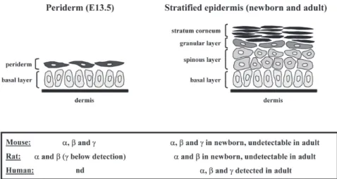
The epidermis is renewed continuously and its integrity is de-pendent on a tightly regulated balance between cell prolifera-tion, differentiaprolifera-tion, and apoptosis. During its maturaprolifera-tion, which happens mainly in the latest stages of fetal develop-ment, the epidermis evolves from a single layer of epithelial cells (periderm) to a fully stratified and differentiated epithe-lium (Fig. 1). This process involves the sequential expression of structural proteins (keratins, involucrin, loricrin, and filag-grin) and synthesis of specific lipids (sphingolipids, FFA, and cholesterol). The outermost layer of the epidermis, the stra-tum corneum, is the end product of keratinocyte differentia-tion and consists of a layer of cross-linked proteins and lipids,
*To whom correspondence should be addressed at Center for Integrative Genomics (CIG), University of Lausanne, Biology Building, CH-1015 Lau-sanne, Switzerland. E-mail: walter.wahli@unil.ch
Abbreviations: ADRP, adipose differentiation-related protein; E, embryonic day; FIAF, fasting-induced adipose factor; PPAR, peroxisome proliferator-activated receptor; PPRE, peroxisome proliferator response element; RXR, retinoid X receptor; TNF, tumor necrosis factor; TPA, tetradecanoylphorbol acetate.
Functions of Peroxisome Proliferator-Activated
Receptors (PPAR) in Skin Homeostasis
Nicolas Di-Poï, Liliane Michalik, Béatrice Desvergne, and Walter Wahli*
which functions as a barrier to transepidermal water loss and as a defense against physical damage, microbes, and xenobi-otics.
During mouse and rat embryonic development, all three PPAR isotypes, and predominantly PPARβ, have been de-tected in the interfollicular epidermis from embryonic day 13.5 (E13.5) onward (3,8). Interestingly, PPAR expression is associated with all major events of the fetal maturation of the epidermal barrier. After birth, PPAR gradually disappear from the interfollicular epidermis to become undetectable in the adult animals (Fig. 1) (3,9). In contrast, the three PPAR iso-types are highly expressed in the basal and suprabasal layers of human adult interfollicular epidermis (Fig. 1), with PPARβ being again the predominant subtype (10,11). Consistent with the expression pattern of PPAR in the developing rodent skin, several reports have concentrated on the involvement of PPAR in processes such as cell proliferation, differentiation, and permeability barrier development (see Table 1). A vari-ety of PPARα activators, including clofibrate, were shown to accelerate the morphologic and functional maturation of the epidermal permeability barrier in fetal rat skin both in vitro and in vivo (12–14). This is evidenced by decreased transepidermal water loss, increased epidermal stratification, and increased ex-pression of the two specific late keratinocyte differentiation markers, loricrin and filaggrin. PPARα ligands also inhibit epi-dermal proliferation and induce keratinocyte differentiation in adult mouse epidermis in vivo (15,16). Furthermore, these ac-tivators restore epidermal homeostasis in murine models of hyperproliferative epidermis (17). In contrast, the two PPARγ ligands, troglitazone and prostaglandin J2, did not affect the development of barrier function or epidermal morphology in fetal rat skin (12), and no specific function in skin maturation
has been attributed so far to this isotype (Table 1). In favor of a potential role of PPARβ in epidermal differentiation in ro-dents, the pan PPAR(α/β) activator linoleic acid (18) was shown to accelerate epidermal barrier development in fetal rat skin explants (12). In addition, an important role of PPARβ in mediating keratinocyte differentiation induced by inflammation was demonstrated in mouse primary ker-atinocytes (19). Importantly, the PPARβ-selective agonist GW1514 stimulated mouse epidermal differentiation without affecting cell proliferation in vivo, by inducing the expression of the late differentiation markers filaggrin and loricrin (20). Also, topical treatment of mice with GW1514 accelerates the restoration of permeability barrier functions after disruption by tape stripping, solvent, or detergent treatment (20), in sup-port of the imsup-portance of the pro-differentiating effect of PPARβ activation.
Novel information on the role of PPAR in epidermis homeo-stasis also came from PPAR mutant mouse models. Although normal skin architecture was initially reported in PPARα-knockout mice (5,21), these animals show delayed fetal skin de-velopment between E18.5 and birth, with defects in the forma-tion of the stratum corneum (22). Morphologic analysis of adult PPARα-null epidermis revealed a thinned stratum granulosum, with focal parakeratosis, indicative of impaired keratinocyte dif-ferentiation (15). Thus, consistent with its expression pattern, PPARα might be important for the maturation of the epidermis during late embryogenesis, but dispensable for normal renewal of the epidermis in the adult animals. Interestingly, PPARα also regulates the early inflammation phase during skin wound heal-ing, as the recruitment of immune cells to the wound site is im-paired in PPARα-null mice (3). PPARγ heterozygous mice, or PPARγ-null mice born after placental rescue, show no defect in
1094 N. DI-POÏ ET AL.
FIG. 1. Schematic representation of interfollicular epidermis development. During late fetal
development (between embryonic day E13.5 and the end of gestation) in mouse skin, the epi-dermis changes from a single basal undifferentiated layer (basal layer) covered by a transient superficial layer of cells (periderm), to a fully stratified and differentiated epithelium, with se-quential formation of suprabasal layers (spinous and granular layers). The end product of dif-ferentiation resides on the surface of the epidermis (stratum corneum) and provides the main permeability barrier of the skin. Expression patterns of PPARα (α), PPARβ (β), and PPARγ (γ) determined or not (nd) during mouse (3) and rat (8,9) epidermal development, and in human adult interfollicular epidermis (10,11), are summarized in the bottom panel.
epidermal maturation (3,23). In addition, PPARγ-null cells are able to participate in the formation of the epidermal tissue in PPARγ-null and wild-type chimeric mice, suggesting very little or no contribution of PPARγ in this process (24). Analysis of PPARβ-mutant skin reveals no defect in fetal and adult epider-mal architecture, or in the expression of keratinocyte differenti-ation markers (3). However, epidermal hyperplasia in response to tetradecanoylphorbol acetate (TPA) treatment was enhanced in PPARβ-mutant animals, emphasizing the role for PPARβ in the control of keratinocyte proliferation and differentiation (3,25). Similarly, the slightly increased keratinocyte prolifera-tion index in PPARβ heterozygous animals is also in favor of the existence of such a control (3). Consistent with these obser-vations, PPARβ expression is rapidly upregulated following challenges that stimulate keratinocyte proliferation, such as hair plucking or cutaneous injury, and skin wound healing is altered in PPARβ-mutant mice (3), largely due to a disrupted balance between proliferation and apoptosis (19,26), as well as to de-fects in kera-tinocyte adhesion and migration (3,27).
Important roles of PPARα and PPARβ in human kera-tinocyte differentiation were also reported (see Table 1). As already mentioned, PPARβ is the predominant isotype in these keratinocytes (10,11,28,29). Its expression remains high and unchanged during the differentiation of cultured kera-tinocytes, or during the stratification and keratinization of the epidermis in in vitro reconstructed skin (10,28), whereas it in-creases upon squamous differentiation in human tracheo-bronchial epithelial cells (29). PPARα and PPARγ are expressed at lower levels, but their expression increases upon keratinocyte
differentiation in similar models (10,28). In human ker-atinocytes, PPARβ- (L-165041 and GW1514) and PPARα (clofibrate and Wy-14,643)-selective agonists induce the expres-sion of a number of epidermal differentiation markers, including involucrin (10,20,30), whereas PPARγ ligands (BRL-49653 and prostaglandin J2) have no effect (10,31). The hypothesis that PPAR may also affect the metabolism of lipids in keratinocytes is supported by the observation that PPARα ligand Wy-14,643 increased both the synthesis of cer-amides and cholesterol de-rivatives in a human skin equivalent model (30). Also, the PPARβ selective agonist GW1514 increases TG accumulation and induces the adipose differentiation-related protein (ADRP) and fasting-induced adipose factor (FIAF) expres-sion in human keratinocytes, two proteins that have potential important roles in lipid metabolism (20).
IMPLICATION OF PPAR IN HYPERPROLIFERATIVE SKIN DISEASES
Based on their diverse biological effects on keratinocyte pro-liferation and differentiation, PPAR ligands may become in-teresting compounds for the treatment of various epidermal disorders characterized by inflammation, keratinocyte hyper-proliferation, and aberrant differentiation, such as psoriasis. In support of an involvement of PPAR in psoriatic epidermis, PPARβ expression was reported to be dramatically increased in the hyperproliferative lesional skin from psoriatic patients (11,28), probably as a response to pro-inflammatory signals in the lesions. It is indeed well established that PPARβ gene
PPAR AND SKIN HOMEOSTASIS 1095
TABLE 1
Effects of Peroxisome Proliferator-Activated Receptor Agonists in Various Cell Types of the Skina
Cell types Rodents Human
Keratinocytes α differentiation in vitro (WY; CLO; OA) differentiation (WY; CLO; OA) (15;30;31) (12;14;17) and in vivo (WY; CLO) (13;15) lipid accumulation (WY) (20;30)
proliferation in vivo (WY; CLO) (15;17) proliferation (WY; CLO) (10;31) β differentiation in vitro (LD; LA) (12;19) differentiation (LD; GW; TTA) (10;20)
and in vivo (GW) (20) lipid accumulation (GW) (20) No effect on proliferation in vivo (GW) (20) No effect on proliferation (LD) (10)
γ No significant effect on differentiation No effect on differentiation (BRL; PGJ2) (10;31)
in vitro (TRO; PGJ2) (12) proliferation (TRO; BRL) (10;33)
Hair follicles α nd survival of cultured hair follicles (CLO) (34)
and melanocytes proliferation and melanogenesis in melanocytes (WY) (42) β nd No effect on proliferation and melanogenesis
in melanocytes (BEZ) (42)
γ nd proliferation and melanogenesis in melanocytes (CIG) (42) Sebocytes α differentiation (WY) (36;37;39) No effect on differentiation (WY) (38)
No effect on proliferation (WY) (39)
β differentiation (LA; PGI2) (36;37;39) differentiation (LA) (38) proliferation (PGI2) (39)
γ differentiation (TRO; BRL) (36;37;39) No effect on differentiation (CIG) (38) No effect on proliferation (TRO) (39)
aThis table summarizes the stimulatory ( ) or inhibitory ( ) effects of treatment with PPARα (α), PPARβ (β), and PPARγ (γ) agonists on the
differ-entiation and proliferation in various cell types derived from rat and mouse (Rodents) as well as human (Human) skin. These results were obtained in in vitro studies, except in rodent keratinocytes where both in vitro and in vivo experiments were performed. Effects of PPAR ligands on the dif-ferentiation of hair follicles and melanocytes in rodents have not been determined (nd). PPAR agonists used in each study are indicated as follows: BEZ, bezafibrate; BRL, BRL-49653; CIG, ciglitazone; CLO, clofibrate; GW, GW1514; LA, linoleic acid; LD, L-165041; OA, oleic acid; PGI2, car-baprostacyclin; PGJ2, 15-deoxy-prostaglandin J2; TRO, troglitazone; TTA, tetradecylthoacetic acid; WY, Wy-14,643.
➚ ➚ ➚ ➚ ➚ ➚ ➚ ➚ ➚ ➚ ➚ ➚ ➚ ➚ ➚ ➚ ➚ ➚ ➚ ➚ ➚
expression is upregulated in mouse skin in response to inflammatory cytokines, such as tumor necrosis factor alpha (TNF-α) (19). In addition, putative PPARβ ligands, such as lipoxygenase products, are generated at high levels in psori-atic skin lesions, and may therefore activate the increased amount of PPARβ (11). The fact that PPARβ is probably nat-urally highly active in the psoriatic lesions may explain why the PPARβ agonist tetradecylthioacetic acid has no strong anti-psoriatic effect when applied topically (32). By contrast to PPARβ expression, no or little change in the cutaneous levels of PPARα and PPARγ was observed in lesional psoriatic skin (11,28), and the PPARα (clofibrate) and PPARγ (rosiglita-zone) agonists had no effect when applied on psoriasis plaques (32). Nevertheless, it is encouraging to note that treatment with the synthetic PPARγ agonist troglitazone was reported to normalize the histological characteristics of psoriatic skin in organ culture, and to reduce the epidermal hyperplasia of psoriasis in the severe combined immunodeficient mouse and human skin transplant model of psoriasis (33). Finally, two PPARα ligands (Wy-14,643 and clofibrate) were able to re-store epidermal homeostasis in subacute and chronic models
of hyperproliferative epidermis in hairless mice (17), even though these models do not perfectly mimic psoriatic or other human disorders. Obviously, further in vivo and clinical stud-ies are needed to better define the potentially beneficial roles of PPAR in this pathology.
ROLE OF PPARS IN HAIR FOLLICLE AND SEBOCYTE DIFFERENTIATION
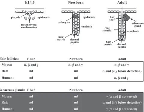
Skin epithelial progenitor cells give rise to the epidermis, as well as to the epithelial component of skin appendages, in-cluding hair follicles and their associated sebaceous glands. Hair follicle morphogenesis is governed by complex bidirec-tional interactions between epithelial keratinocytes and the underlying dermal cells of the mesenchymal condensations (Fig. 2). These interactions control a tight balance between keratinocyte proliferation and apoptosis. In rodents, all three PPAR isotypes are expressed in the differentiating hair folli-cles from the early embryonic developmental stages (Fig. 2). PPAR remain highly expressed in postnatal and adult hair fol-licles, whereas they disappear from the interfollicular
epider-1096 N. DI-POÏ ET AL.
FIG. 2. Schematic representation of murine hair follicle morphogenesis. Hair follicle
develop-ment occurs between embryonic day 14.5 (E14.5) and postnatal day 7 (P7) in mouse, and is governed by bidirectional epithelial–mesenchymal interactions between epithelial kera-tinocytes and dermal fibroblasts of underlying mesenchymal condensations. Following dermal induction, hair follicle precursors appear first as thickening of the uniform epithelial surface (placode, E14.5). Follicular epithelial cells will next proliferate, followed by downgrowth into the dermis, leading to the elongation of hair follicles (Newborn), and will finally differentiate into the typical structures of mature hair follicles, including root sheaths, hair matrix, and hair shaft (Adult) (46). In parallel, melanin starts to be produced in the precortex region, and the first sebocytes form the sebaceous gland (Newborn). Expression patterns of PPARα (α), PPARβ (β), and PPARγ (γ) determined or not (nd) in mouse (8), rat (10), and human (35) hair follicles at indicated stages, as well as in mature sebaceous glands (8,28,39), are given in the bottom panels.
mis after birth (3,8), as already mentioned (see Figs. 1, 2). A detailed analysis of the expression of PPAR in human hair fol-licles shows that they are specifically located in both epithe-lial and dermal hair follicle cells (34). In addition, the PPARα ligand clofibrate was reported to increase the survival of human hair follicles in vitro, within a narrow range of con-centrations (Table 1) (34). Although a possible function of the two other PPAR isotypes in hair follicle growth has not yet been examined, it is interesting to note that conditional abla-tion in the murine epidermis of RXRα, the most abundant heterodimeric partner of PPARs in keratinocytes, results in delayed hair follicle maturation and alopecia (35).
Chronologically, the latest differentiated cell type to ap-pear in the developing follicle is the oil-rich sebocytes. They arise from cells within the superficial hair follicle and will eventually form a gland located outside the hair follicle. Se-baceous lipogenesis, leading to the accumulation of lipid droplets and finally to sebum excretion, represents a major step in the differentiation process and differentiated state of sebaceous gland cells. Rodent and human sebocytes were shown to express the three PPAR, either in cell culture or in vivo(Fig. 2), the predominant isotype being PPARβ (9,24,36− 38). Consistently, and in addition to androgens that are well known to regulate the growth and maturation of sebocytes, PPAR agonists and retinoids were recently found to affect se-baceous gland differentiation (see Table 1). Activation of PPARγ and PPARα by respective selective agonists has no ef-fect on sebocyte growth (39), but stimulates lipid droplet ac-cumulation in cultured rat preputial sebocytes, as assessed histochemically using Oil Red O staining (36,37). This effect was not observed in epidermal cells (37) or in cultured human sebocytes (38), possibly due to the different selective PPAR agonists or to various ligand concentrations used in each study. In parallel, RXR selective ligands have prominent dif-ferentiative and weak proliferative effects on sebocytes (40). The RXRα selective rexinoid CD2809 also amplified the pro-differentiative effect of PPAR in preputial sebocytes, suggesting a cooperation between PPAR and RXR agonists in promoting differentiation of these cells (39). In both rat and human cul-tured sebocytes, the pan PPAR(α/β) activators carbaprostacy-clin and linoleic acid were more effective than PPARα or PPARγ agonists in stimulating sebocyte lipid droplet forma-tion, suggesting an important contribution of PPARβ in this process (37–39). Interestingly, PPARβ seems more important in the late stages of sebocyte differentiation (37,38), whereas it was involved in the early adipocyte differentiation (41), suggesting that it plays different roles in the differentiation program of each cell type.
It is noteworthy that, in spite of the relative importance of PPAR in lipid metabolism and their high expression in sebo-cytes, in neither PPARα nor PPARβ mutant mouse models has sebaceous gland function been closely examined so far. In contrast, inactivation of the PPARγ gene has underscored a crucial contribution of PPARγ in sebocyte differentiation, although it appears dispensable for epidermal differentiation.
Indeed, and as already discussed, chimeric mice for PPARγ-null and wild-type cells showed little or no contribution of mutant cells to the development of sebaceous glands, suggest-ing that PPARγ-null cells cannot develop into sebocytes (24). NEW PUTATIVE FUNCTION OF PPARS IN
MELANOCYTE DIFFERENTIATION
The pigment-producing cells of the skin are called melano-cytes and their activity is the major determinant of the color of the hair and skin. Melanocytes originate from the neural crest and migrate to the basal layer of the epidermis and the hair matrix during embryogenesis. Interestingly, all three PPAR were detected in cultured human melanocytes (42) and in melanoma cells (43). Consistent with the role of PPAR ag-onists in cellular proliferation and differentiation in kera-tinocytes, PPARα (Wy-14,643) and PPARγ (ciglitazone) ligands were shown to inhibit the proliferation and to stimulate the melanin synthesis of cultured melanocytes (Table 1), whereas bezafibrate, a preferential activator for PPARβ in Xenopus (44), had no effect on melanin content (42). In agreement with this study, several PPARγ agonists, including troglitazone and rosiglitazone, were previously demonstrated to inhibit cell growth in human malignant melanoma (43), and topical appli-cation of retinoic acid was shown to improve hyperpigmented skin lesions such as melasma (45). Because of their antipro-liferative and prodifferentiative effect on melanocytes, it is tempting to suggest that PPAR and RXR ligands may be ben-eficial in the treatment of melanomas. However, too little is known in this context for the moment, and further investiga-tion is needed.
DISCUSSION AND HYPOTHESIS
These studies suggest that PPARα may contribute to seba-ceous gland differentiation and epidermal permeability bar-rier formation by increasing both lipid metabolism and ex-pression of structural differentiation markers, whereas PPARγ plays a unique role in stimulating sebocyte function. Further-more, PPARβ was identified as the predominant isotype in the skin, and as a modulator of cell differentiation in both ker-atinocytes and sebocytes.
Because of their diverse biological activities in epidermal processes such as keratinocyte proliferation and differentia-tion, PPAR may represent a major research target for the un-derstanding and treatment of many skin diseases resulting in disturbance of normal tissue homeostasis and epidermal hy-perproliferation, such as benign epidermal tumors, papillo-mas, melanopapillo-mas, and psoriasis. In addition, due to the in-creasing number of studies implicating PPAR in the control of sebocyte differentiation, the development of PPAR antago-nists that can interfere selectively with sebum production may constitute an important element in the prevention of acne vul-garis, characterized by excess sebum production.
ACKNOWLEDGMENTS
This work was supported by the Swiss National Science Foundation (grants to Walter Wahli and to Béatrice Desvergne) and by the Etat de Vaud.
REFERENCES
1. Desvergne, B., and Wahli, W. (1999) Peroxisome Proliferator-Activated Receptors: Nuclear Control of Metabolism, Endocr. Rev. 20, 649–688.
2. Kersten, S., Desvergne, B., and Wahli, W. (2000) Roles of PPARs in Health and Disease, Nature 405, 421–424.
3. Michalik, L., Desvergne, B., Tan, N.S., Basu-Modak, S., Escher, P., Rieusset, J., Peters, J.M., Kaya, G., Gonzalez, F.J., Zakany, J., Metzger, D., Chambon, P., Duboule, D., and Wahli, W. (2001) Impaired Skin Wound Healing in Peroxisome Prolifera-tor-Activated Receptor (PPAR)α and PPARβ Mutant Mice, J. Cell Biol. 154, 799–814.
4. Michalik, L., Desvergne, B., and Wahli, W. (2003) Peroxisome Proliferator-Activated Receptors β/δ: Emerging Roles for a Pre-viously Neglected Third Family Member, Curr. Opin. Lipidol. 14, 129–135.
5. Michalik, L., Desvergne, B., Dreyer, C., Gavillet, M., Laurini, R.N., and Wahli, W. (2002) PPAR Expression and Function During Vertebrate Development, Int. J. Dev. Biol. 46, 105–114. 6. Alonso, L.C., and Rosenfield, R.L. (2003) Molecular Genetic and Endocrine Mechanisms of Hair Growth, Horm. Res. 60, 1–13.
7. Williams, M.L., Hanley, K., Elias, P.M., and Feingold, K.R. (1998) Ontogeny of the Epidermal Permeability Barrier, J. In-vestig. Dermatol. Symp. Proc. 3, 75–79.
8. Braissant, O., and Wahli, W. (1998) Differential Expression of Peroxisome Proliferator-Activated Receptor-α, -β, and -γ Dur-ing Rat Embryonic Development, Endocrinology 139, 2748–2754.
9. Braissant, O., Foufelle, F., Scotto, C., Dauca, M., and Wahli, W. (1996) Differential Expression of Peroxisome Proliferator-Acti-vated Receptors (PPARs): Tissue Distribution of PPAR-α, -β, and -γ in the Adult Rat, Endocrinology 137, 354–366.
10. Westergaard, M., Henningsen, J., Svendsen, M.L., Johansen, C., Jensen, U.B., Schroder, H.D., Kratchmarova, I., Berge, R.K., Iversen, L., Bolund, L., Kragballe, K., and Kristiansen, K. (2001) Modulation of Keratinocyte Gene Expression and Dif-ferentiation by PPAR-Selective Ligands and Tetradecyl-thioacetic Acid, J. Invest. Dermatol. 116, 702–712.
11. Westergaard, M., Henningsen, J., Johansen, C., Rasmussen, S., Svendsen, M.L., Jensen, U.B., Schroder, H.D., Staels, B., Iversen, L., Bolund, L., Kragballe, K., and Kristiansen, K. (2003) Expression and Localization of Peroxisome Proliferator-Activated Receptors and Nuclear Factor κB in Normal and Le-sional Psoriatic Skin, J. Invest. Dermatol. 121, 1104–1117. 12. Hanley, K., Jiang, Y., Crumrine, D., Bass, N.M., Appel, R.,
Elias, P.M., Williams, M.L., and Feingold, K.R. (1997) Activa-tors of the Nuclear Hormone RecepActiva-tors PPARα and FXR Ac-celerate the Development of the Fetal Epidermal Permeability Barrier, J. Clin. Invest. 100, 705–712.
13. Hanley, K., Komuves, L.G., Bass, N.M., He, S.S., Jiang, Y., Crumrine, D., Appel, R., Friedman, M., Bettencourt, J., Min, K., Elias, P.M., Williams, M.L., and Feingold, K.R. (1999) Fetal Epidermal Differentiation and Barrier Development in vivo Is Accelerated by Nuclear Hormone Receptor Activators, J. Invest. Dermatol. 113, 788–795.
14. Komuves, L.G., Hanley, K., Jiang, Y., Elias, P.M., Williams, M.L., and Feingold, K.R. (1998) Ligands and Activators of Nu-clear Hormone Receptors Regulate Epidermal Differentiation
During Fetal Rat Skin Development, J. Invest. Dermatol. 111, 429–433.
15. Komuves, L.G., Hanley, K., Lefebvre, A.M., Man, M.Q., Ng, D.C., Bikle, D.D., Williams, M.L., Elias, P.M., Auwerx, J., and Feingold, K.R. (2000) Stimulation of PPARα Promotes Epider-mal Keratinocyte Differentiation in vivo, J. Invest. Dermatol. 115, 353–360.
16. Hanley, K., Komuves, L.G., Ng, D.C., Schoonjans, K., He, S.S., Lau, P., Bikle, D.D., Williams, M.L., Elias, P.M., Auwerx, J., and Feingold, K.R. (2000) Farnesol Stimulates Differentiation in Epidermal Keratinocytes via PPARα, J. Biol. Chem. 275, 11484–11491.
17. Komuves, L.G., Hanley, K., Man, M.Q., Elias, P.M., Williams, M.L., and Feingold, K.R. (2000) Keratinocyte Differentiation in Hyperproliferative Epidermis: Topical Application of PPARα Activators Restores Tissue Homeostasis, J. Invest. Dermatol. 115, 361–367.
18. Yu, K., Bayona, W., Kallen, C.B., Harding, H.P., Ravera, C.P., McMahon, G., Brown, M., and Lazar, M.A. (1995) Differential Activation of Peroxisome Proliferator-Activated Receptors by Eicosanoids, J. Biol. Chem. 270, 23975–23983.
19. Tan, N.S., Michalik, L., Noy, N., Yasmin, R., Pacot, C., Heim, M., Fluhmann, B., Desvergne, B., and Wahli, W. (2001) Criti-cal Roles of PPAR β/δ in Keratinocyte Response to Inflamma-tion, Genes Dev. 15, 3263–3277.
20. Schmuth, M., Haqq, C.M., Cairns, W.J., Holder, J.C., Dorsam, S., Chang, S., Lau, P., Fowler, A.J., Chuang, G., Moser, A.H., et al. (2004) Peroxisome Proliferator-Activated Receptor (PPAR)-β/δ Stimulates Differentiation and Lipid Accumulation in Keratinocytes, J. Invest. Dermatol. 122, 971–983.
21. Lee, S.S., Pineau, T., Drago, J., Lee, E.J., Owens, J.W., Kroetz, D.L., Fernandez-Salguero, P.M., Westphal, H., and Gonzalez, F.J. (1995) Targeted Disruption of the α Isoform of the Peroxi-some Proliferator-Activated Receptor Gene in Mice Results in Abolishment of the Pleiotropic Effects of Peroxisome Prolifera-tors, Mol. Cell Biol. 15, 3012–3022.
22. Schmuth, M., Schoonjans, K., Yu, Q.C., Fluhr, J.W., Crumrine, D., Hachem, J.P., Lau, P., Auwerx, J., Elias, P.M., and Feingold, K.R. (2002) Role of Peroxisome Proliferator-Activated Recep-tor α in Epidermal Development in Utero, J. Invest. Dermatol. 119, 1298–1303.
23. Barak, Y., Nelson, M.C., Ong, E.S., Jones, Y.Z., Ruiz-Lozano, P., Chien, K.R., Koder, A., and Evans, R.M. (1999) PPAR γ Is Required for Placental, Cardiac, and Adipose Tissue Develop-ment, Mol. Cell 4, 585–595.
24. Rosen, E.D., Sarraf, P., Troy, A.E., Bradwin, G., Moore, K., Milstone, D.S., Spiegelman, B.M., and Mortensen, R.M. (1999) PPAR γ Is Required for the Differentiation of Adipose Tissue in vivo and in vitro, Mol. Cell 4, 611–617.
25. Peters, J.M., Lee, S.S., Li, W., Ward, J.M., Gavrilova, O., Everett, C., Reitman, M.L., Hudson, L.D., and Gonzalez, F.J. (2000) Growth, Adipose, Brain, and Skin Alterations Resulting from Targeted Disruption of the Mouse Peroxisome Prolifera-tor-Activated Receptor β(δ), Mol. Cell Biol. 20, 5119–5128. 26. Di Poï, N., Tan, N.S., Michalik, L., Wahli, W., and Desvergne,
B. (2002) Antiapoptotic Role of PPARβ in Keratinocytes via Transcriptional Control of the Akt1 Signaling Pathway, Mol. Cell 10, 721–733.
27. Di Poï, N., Michalik, L., Tan, N.S., Desvergne, B., and Wahli, W. (2003) The Anti-apoptotic Role of PPARβ Contributes to Efficient Skin Wound Healing, J. Steroid Biochem. Mol. Biol. 85, 257–265.
28. Rivier, M., Safonova, I., Lebrun, P., Griffiths, C.E., Ailhaud, G., and Michel, S. (1998) Differential Expression of Peroxisome Proliferator-Activated Receptor Subtypes During the Differenti-ation of Human Keratinocytes, J. Invest. Dermatol. 111, 1116–1121.
29. Matsuura, H., Adachi, H., Smart, R.C., Xu, X., Arata, J., and Jet-ten, A.M. (1999) Correlation Between Expression of Peroxi-some Proliferator-Activated Receptor β and Squamous Differ-entiation in Epidermal and Tracheobronchial Epithelial Cells, Mol. Cell Endocrinol. 147, 85–92.
30. Rivier, M., Castiel, I., Safonova, I., Ailhaud, G., and Michel, S. (2000) Peroxisome Proliferator-Activated Receptor-α Enhances Lipid Metabolism in a Skin Equivalent Model, J. Invest. Der-matol. 114, 681–687.
31. Hanley, K., Jiang, Y., He, S.S., Friedman, M., Elias, P.M., Bikle, D.D., Williams, M.L., and Feingold, K.R. (1998) Kera-tinocyte Differentiation Is Stimulated by Activators of the Nu-clear Hormone Receptor PPARα, J. Invest. Dermatol. 110, 368–375.
32. Kuenzli, S., and Saurat, J.H. (2003) Effect of Topical PPARβ/δ and PPARγ Agonists on Plaque Psoriasis. A Pilot Study, Der-matology 206, 252–256.
33. Ellis, C.N., Varani, J., Fisher, G.J., Zeigler, M.E., Pershadsingh, H.A., Benson, S.C., Chi, Y., and Kurtz, T.W. (2000) Troglita-zone Improves Psoriasis and Normalizes Models of Prolifera-tive Skin Disease: Ligands for Peroxisome Proliferator-Acti-vated Receptor-γ Inhibit Keratinocyte Proliferation, Arch. Der-matol. 136, 609–616.
34. Billoni, N., Buan, B., Gautier, B., Collin, C., Gaillard, O., Mahe, Y.F., and Bernard, B.A. (2000) Expression of Peroxisome Pro-liferator Activated Receptors (PPARs) in Human Hair Follicles and PPARα Involvement in Hair Growth, Acta Derm. Venereol. 80, 329–334.
35. Li, M., Chiba, H., Warot, X., Messaddeq, N., Gerard, C., Cham-bon, P., and Metzger, D. (2001) RXR-α Ablation in Skin Kera-tinocytes Results in Alopecia and Epidermal Alterations, Devel-opment 128, 675–688.
36. Rosenfield, R.L., Deplewski, D., Kentsis, A., and Ciletti, N. (1998) Mechanisms of Androgen Induction of Sebocyte Differ-entiation, Dermatology 196, 43–46.
37. Rosenfield, R.L., Kentsis, A., Deplewski, D., and Ciletti, N. (1999) Rat Preputial Sebocyte Differentiation Involves Peroxi-some Proliferator-Activated Receptors, J. Invest. Dermatol. 112, 226–232.
38. Chen, W., Yang, C.C., Sheu, H.M., Seltmann, H., and
Zouboulis, C.C. (2003) Expression of Peroxisome Proliferator-Activated Receptor and CCAAT/Enhancer Binding Protein Transcription Factors in Cultured Human Sebocytes, J. Invest. Dermatol. 121, 441–447.
39. Kim, M.J., Deplewski, D., Ciletti, N., Michel, S., Reichert, U., and Rosenfield, R.L. (2001) Limited Cooperation Between Per-oxisome Proliferator-Activated Receptors and Retinoid X Re-ceptor Agonists in Sebocyte Growth and Development, Mol. Genet. Metab. 74, 362–369.
40. Kim, M.J., Ciletti, N., Michel, S., Reichert, U., and Rosenfield, R.L. (2000) The Role of Specific Retinoid Receptors in Sebo-cyte Growth and Differentiation in Culture, J. Invest. Dermatol. 114, 349–353.
41. Grimaldi, P.A. (2001) The Roles of PPARs in Adipocyte Dif-ferentiation, Prog. Lipid Res. 40, 269–281.
42. Kang, H.Y., Chung, E., Lee, M., Cho, Y., and Kang, W.H. (2004) Expression and Function of Peroxisome Proliferator-Activated Receptors in Human Melanocytes, Br. J. Dermatol. 150, 462–468.
43. Mossner, R., Schulz, U., Kruger, U., Middel, P., Schinner, S., Fuzesi, L., Neumann, C., and Reich, K. (2002) Agonists of Per-oxisome Proliferator-Activated Receptor γ Inhibit Cell Growth in Malignant Melanoma, J. Invest. Dermatol. 119, 576–582. 44. Krey, G., Braissant, O., L’Horset, F., Kalkhoven, E., Perroud,
M., Parker, M.G., and Wahli, W. (1997) Fatty Acids, Eicosanoids, and Hypolipidemic Agents Identified as Ligands of Peroxisome Proliferator-Activated Receptors by Coactivator-Dependent Receptor Ligand Assay, Mol. Endocrinol. 11, 779–791.
45. Kang, W.H., Chun, S.C., and Lee, S. (1998) Intermittent Ther-apy for Melasma in Asian Patients with Combined Topical Agents (Retinoic Acid, Hydroquinone and Hydrocortisone): Clinical and Histological Studies, J. Dermatol. 25, 587–596. 46. Paus, R., Muller-Rover, S., Van Der Veen, C., Maurer, M.,
Eichmuller, S., Ling, G., Hofmann, U., Foitzik, K., Mecklen-burg, L., and Handjiski, B. (1999) A Comprehensive Guide for the Recognition and Classification of Distinct Stages of Hair Follicle Morphogenesis, J. Invest. Dermatol. 113, 523–532.
[Received July 19, 2004; accepted October 17, 2004]