Mechanisms of Successful Amoxicillin Prophylaxis of
Experimental Endocarditis Due to
Streptococcus intermedius
P. Moreillon, P. Francioli, D. Overholser, P. Meylan,and M. P. Glauser
From the Division of Infectious Diseases, Department of Internal Medicine, Centre Hospitalier Universitaire Vaudois,
Lausanne, Switzerland
Prophylaxis with amoxicillin (40 mg/kg) was studied in rats with aortic valve vegetations. Bacteria on the valves were quantitated early (10 min to 6 hr) and late (three days) after intravenous challenge with tolerantStreptococcus intermedius. Amoxicillin reduced by 40070the number of bacteria per valve 10 min after intravenous challenge with 105S. inter-medius (P
<
.05) and by 74% the incidence of endocarditis three days thereafter(P<
.00(1). Bacterial multiplication started 2 hr after challenge in control rats, whereas bacteria dis-appeared in 6 hr in amoxicillin-treated rats. Intravenous penicillinase 30 min after chal-lenge abolished successful amoxicillin prophylaxis, a result demonstrating the necessity of prolonged growth inhibition for protection. Growth inhibition for 18 hr (two subse-quent amoxicillin doses) was necessary for protection after intravenous challenge with 105S.intermedius.Thus, in the absence of bacterial killing, inhibition of valvularcoloni-zation by amoxicillin was not as important a mechanism of endocarditis prophylaxis as was prolonged inhibition of bacterial growth, which allowed adherent bacteria to be cleared from the valves.
Successful antibiotic prophylaxis of experimental bacterial endocarditis can be achieved by mecha-nisms other than antibiotic bactericidal activity[1-3].
Several studies in which viable Streptococcus san-guis organisms were exposed to nonbactericidal
con-centrations of vancomycin or amoxicillin showed that the exposed bacteria were less able than the con-trols to stick to platelet-fibrin matrices in vitro and to induce endocarditis in animals with catheter-induced vegetations. These observations suggested that in the absence of bacterial killing, antibiotics might prevent endocarditis by reducing the adher-ence of circulating microorganisms to the damaged valves [1-3]. More recently, Lowyet al. [4], using a strain ofS.sanguis pretreated with an inhibitory
con-centration of penicillin, observed both a decreased adherence of the bacteria to platelet-fibrin matrices Received for publication 27 February 1986, and in revised form 2 June 1986.
Part of this work has been published in abstract form (no. 336) in the Proceedings and Abstracts of the 24th Interscience Con-ference on Antimicrobial Agents and Chemotherapy.
This work was supported by grant 3.847.083 from the Swiss National Foundation for Scientific Research.
We thank Jose Entenza and Marlyse Giddey for technical as-sistance and Sylviane Bovey for typing the manuscript.
Please address requests for reprints to Dr. M. P. Glauser, Divi-sion of Infectious Diseases, Department of Internal Medicine, Centre Hospitalier Universitaire Vaudois, 1011 Lausanne, Swit-zerland.
801
in vitro and a slight but significant reduction in their ability to colonize damaged aortic valves of rabbits in vivo. However, despite these observations, all the rabbits injected with the penicillin-pretreated bac-teria had developed endocarditis when killed 24 hr after bacterial challenge, a result raising the ques-tion of whether inhibiques-tion of bacterial adherence was actually an efficient mechanism of endocarditis pro-phylaxis. Moreover, nonbactericidal concentrations of antibiotics that were successful in the prophylaxis of endocarditis may have had various effects on bac-teria such as growth inhibition and enhanced sus-ceptibility to ingestion and killing by phagocytes [5], effects not ruled out by the above studies.
To elucidate further the mechanisms of success-ful prophylaxis of bacterial endocarditis in the ab-sence of bacterial killing, we performed experiments aimed at determining in vivo the role of the reduc-tion of valvular colonizareduc-tion due to the inhibireduc-tion of bacterial adherence in the successful prophylaxis of bacterial endocarditis.
Materials and Methods
Microorganism. Streptococcus intermedius, also
referred to asS.sanguis in some experiments [1], was
originally isolated from a patient with endocarditis and has previously been used in production of ex-perimental endocarditis in rabbits [6] and rats [1, 3,
7]. The MIC and MBC of amoxicillin for this strain have been shown to be 0.125 and>128 ug/rnl, respec-tively, resulting in a MBC/MIC ratio of >1,024 [3]. It was previously established by time-killing curves (with an inoculum of 106bacteria and a
concentra-tion of 25 ug of amoxicillin/ml) that no significant killing of this strain occurred during the first 6 hr of incubation and that only a marginal decrease in the number of cfu occurred after 24 hr [3]. Similar observations were made with an inoculum of 103and
l(P bacteria (authors' unpublished observation). The concentration of 25 ug of amoxicillin/ml was used in those experiments because it is similar to the se-rum levels achieved in rats 30 min after iv adminis-tration of 40 mg/kg and in humans 2 hrs after an oral dose of 3 g.
Serum inhibitory and bactericidal titers. The se-rum inhibitory and bactericidal titers against S. inter-medius30 min and 2, 4, and 6 hr after iv administra-tion of 40 mg of amoxicillin/kg of body weight were determined in nine rats by standard methods [8] with an inoculum of 106S. intermedius.Subcultures were
performed on penicillinase-containing blood agar (Bactopenase'"; 5 X 10⁶ IV/liter; Difco
Laborato-ries, Detroit). The serum inhibitory titer was the highest dilution of serum inhibiting visible bacterial growth, and the serum bactericidal titer was the highest dilution of serum providing 99.9070 killing of the original inoculum after incubation for 18 hr.
In vitro adherence assay. With a previously described in vitro assay system of platelet-fibrin ma-trices [2] simulating nonbacterial thrombotic endo-carditis, we determined(1) the influence of amoxi-cillin on the in vitro adherence of S. intermediusto these matrices and (2) the influence of amoxicillin on the subsequent detachment of the adherent bac-teria during prolonged exposure to the antibiotic. S. intermediusorganisms grown overnight in brain-heart infusion broth (Difco) were suspended in PBS supplemented with 25 ug of amoxicillin (Beecham Research Laboratories, Bern, Switzerland)/ml, giv-ing a final concentration of 1()4cfu/ml. Controls were
suspended in PBS alone. These suspensions were im-mediately poured on platelet-fibrin matrices [2, 3] in petri dishes and incubated for 5 min at 37 C in a shaking incubator at 120 rpm. The supernatant was removed, and the matrices were washed two times for 5 min each with amoxicillin solution or with PBS alone. The petri dishes were then divided into two groups, with each one comprising a control and an amoxcillin-treated subgroup. The first group was washed a third time for 5 min with PBS
sup-plemented with 2,000 V of penicillinase/ml for in-hibition of residual amoxicillin on the matrices. The total time of exposure to amoxicillin (or PBS) in this group (15 min) was chosen for simulation of the mean duration of bacteremia after iv challenge, dur-ing which inhibition of adherence has to occur if bac-terial colonization of the damaged valves is to be prevented [1].In the second group, the time of ex-posure to amoxicillin in the shaking incubator was prolonged up to 4 hr for simulation of the time of exposure of the bacteria that attached to the valve, despite adherence inhibition after a single iv prophylactic dose of amoxicillin [3]. The matrices were washed every half hour for 15 min with amoxi-cillin solution (or PBS), and the supernatant was carefully recovered for plating and colony counts. At the end of the assay, the matrices were washed for 15 min with PBS containing penicillinase, and penicillinase-containing blood agar was poured over the matrices. The adherent colonies were counted af-ter incubation for 48 hr. The percentage of adher-ent colonies for each sample was defined as the num-ber of cfu adherent to the matrix X 100 divided by the initial number of cfu in the inoculum. The results are expressed as the mean values of 36 determina-tions in each subgroup performed in three consecu-tive experiments.
Animal model. Production ofendocarditis. Ster-ile aortic vegetations were produced in female Wistar rats (body weight, 180-200 g) by placement of a poly-ethylene catheter through the aortic valve as previ-ously described [3]. Twenty-four hours after catheter-ization, rats were injected iv with 0.5 ml of various sizes of bacterial inocula from an overnight culture of S. intermediusdiluted in 0.9070 NaCI.
Course ofendocarditis with or without amoxicil-lin prophylaxis. Thirty minutes before bacterial challenge, groups of 40 rats were injected iv with ei-ther 40 mg of amoxicillin/kg of body weight (20 test rats) or 0.9070 NaCI (20 control rats). This dose of amoxicillin was chosen because at 30 min after iv injection in rats it produces peak serum levels simi-lar to those in humans 2 hr after an oral dose of 3 g [3], which has been advocated for prophylaxis in humans [9]. In some experiments, one or two iden-tical subsequent doses of amoxicillin were given 6 and 12 hr after the first dose. For investigation in vivo of the effect of amoxicillin on the adherence of S.intermediuson damaged aortic valves, one-half of the rats of the same experiment were killed early (10 min or 1, 2, 6, or 12 hr), and the other half were killed three days after bacterial challenge. This
pro-cedure permitted for each experiment a valuable correlation between early bacteriologic results and the subsequent development of endocarditis in both amoxicillin-treated and control rats.
Killing was performed as previously described [3, 7]. All cultures of blood and of valve homogenates were performed on penicillinase-containing blood agar. In the groups killed early after bacterial chal-lenge (i.e., at 10 min or 1, 2, 6, or 12 hr), the valves were rinsed in 5 ml of 0.9070 NaCI before homogeni-zation for elimination of nonadherent bacteria. The total amount of homogenate was then plated for recovery of all adherent bacteria. Because of the small number of bacteria detected with early kill-ing, the results were expressed as the number of bac-teria recovered per valve. In animals killed three days after bacterial challenge, 0.1 ml of appropriate dilu-tions of valve homogenates was plated for colony counts, and the results were expressed in log cfu/g of valve. Bacterial endocarditis was defined by posi-tive cultures of valves.
Inactivation of amoxicillin with penicillinase in rats given antibiotic prophylaxis. In additional ex-periments two-thirds of the rats were given amox-icillin, and one-third served as controls. Thirty minutes after bacterial challenge with 105 S. inter-medius, half of the amoxicillin-treated rats as well the control rats were given 0.5 ml of iv penicillinase (Bactopenase; 5 x 10⁵ IV/mI). Ten minutes after iv
injection of this amount of penicillinase, no detect-able amoxicillin activity could be measured in the serum of rats. If prevention of valvular colonization due to the inhibition of bacterial adherence was the main mechanism responsible for the prevention of endocarditis, then penicillinase inactivation of the remaining antibiotic activity in the blood at the end of the bacteremic phase should not abolish success-ful prophylaxis. Ithas been previously established that 30 min after bacterial challenge with either 105
or 106S.intermedius,no circulating bacteria can be
detected [1].
Statistical evaluation. The "I} test with Yates's correction, the Wilcoxon rank sum test, and Stu-dent's unpairedttest were used for statistical com-parisons.
Results
Serum inhibitory and bactericidal titers against
S.intermedius. The mean serum inhibitory titer 30 min, 2 hr, and 4 hr after iv injection of amoxicillin was 1:32, 1:8, and 1:2, respectively. At 6 hr no
se-rum inhibitory activity was detectable. No sese-rum bactericidal activity could be detected at any time.
Influence of amoxicillin on in vitro adherence of
S. intermedius to platelet-fibrin matrices and on their subsequent detachment. When incubation in vitro with 25 J.1g of amoxicillin/ml was for an interval simulating the duration of bacteremia in vivo(15min [1]), the percentage (mean ± SD value of 36 deter-minations) of S. intermedius adherent to platelet-fibrin matrices was reduced from 0.13070 ± 0.08C110
in controls to0.09C110 ± 0.05C110 in amoxicillin-treated
bacteria, a reduction of30C110 (P
<
.05).When the in vitro adherent bacteria were washed during 4 hr with 25 J.1g of amoxicillin/ml or with PBS (simulating the time of exposure of bacteria in vivo to amoxicillin levels in rats after one prophylactic dose), the percentage (mean ± SD value of 36 de-terminations) of adherent S. intermediusdecreased to the same extent in the control group (from0.13C110 ± 0.08070 to 0.08C110 ± 0.05C110, a reduction of 38C110)
and in the amoxicillin-treated group (from 0.09070
±
0.05070 to0.06C110±
0.06C110, a reduction of 33C110).This reduction was due to detachment of the adher-ent bacteria, as shown by their recovery on culture of the washing medium. Control experiments with tryptic soy broth (Gibco Europe, Daisley, Scotland) instead of PBS as the washing medium (a medium that may have provided more growth factors than PBS alone) showed similar results (data not shown).
Bacterial colonization ofdamaged valves and sub-sequent development of endocarditis after iv injec-tion of various inoculum sizes ofS. intermedius. Ex-periments with 104S. intermedius (control rats only).
When rats were killed 10 min after bacterial chal-lenge, four of five valvular homogenates yielded S. intermedius (1-5 cfu per valve). All cultures of blood were sterile at this time. In rats killed 1 and 2 hr after bacterial challenge, four of nine and three of eight valvular homogenates, respectively, still yielded S.intermedius(1-8 cfu per valve). After 6 hr, however, S. intermediuswas no longer detectable on the valves of all seven rats killed at this time. When animals were killed three days after bacterial chal-lenge, only two(9.5C110) of 21 rats had developed
bac-terial endocarditis. Thus it was common for a few bacteria to adhere early to the damaged valves after injection with 104S. intermedius,but these bacteria
had spontaneously disappeared within the next few hours, and almost all animals had sterile vegetations at three days.
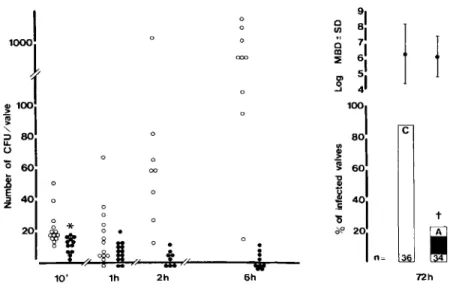
Experiments with 105S. intermedius. Although
aortic valves 10 min after bacterial inoculation, the number of streptococcal colonies attached to the valves of amoxicillin-treated animals (median, 8; range, 1-14) was slightly but significantly lower than in controls (median, 13; range, 8-50; P< .05 by Wil-coxon rank sum test; figure 1). The reduction in ber of cfu per valve was not due to different num-bers of circulating bacteria because quantitative cultures of blood performed at the time of killing yielded similar numbers of bacteria: 12.6± 9.6 cfu/ ml in control rats versus 16 ± 27 cfu/ml in amoxi-cillin-treated rats. Furthermore, killing could not ac-count for this reduction because exposure to the anti-biotic was very brief and there was no detectable serum bactericidal activity at this time. Therefore at this time of death (10 min), antibiotic-mediated inhi-bition of bacterial adherence was a likely mechanism involved in the reduction of streptococcal numbers on the heart valves of amoxicillin-treated rats when compared with the bacterial numbers in controls. One hour after bacterial challenge a slight decrease in number of cfu per valve was detected in both groups of rats compared with the number of cfu per valve in animals killed 10 min after bacterial chal-lenge. However, when animals were killed 2 or 6 hr after bacterial challenge, the number of cfu per valve in control rats had increased by 2.5- and 10-fold, respectively, compared with the number in control animals killed at 1 hr, a finding demonstrating that
the adherent bacteria had started to multiply. In con-trast, the valvular homogenates of amoxicillin-treated rats showed a further reduction in the num-ber of cfu per valve in animals killed at 2 or 6 hr, and five of nine valves were completely sterile at the latter time.
When rats were killed three days after bacterial challenge, 32 (890/0) of 36 control animals had de-veloped endocarditis due to S. intermedius,a result demonstrating that 105S. intermediusused as an
in-oculum for iv bacterial challenge were sufficient to produce endocarditis in 90070 of the rats. In contrast, in the amoxicillin-treated rats, endocarditis was pres-ent in only eight (23070) of 34 rats (P< 10-4
) ,a result
demonstrating the successful prophylaxis afforded by amoxicillin after challenge with the 90% infec-tious dose.
Effect ofamoxicillin inactivation by penicillinase injected 30 min after bacterial challenge with 105
S.intermedius. Penicillinase treatment 30 min af-ter bacaf-terial challenge completely abolished the prophylactic effect of amoxicillin. When animals were killed three days after bacterial challenge, only 11 (360/0) of 30 rats given amoxicillin alone had de-veloped endocarditis compared with 21 (840/0) of 25 rats that had received penicillinase after amoxicillin prophylaxis (P
<
.01), an incidence similar to that in control rats operated on at the same time (18 [90%] of 20). These results suggested that someantibac-10' 1h 2h o o
.
J,
6hI
1
c t 36 72hFigure 1. Natural history of en-docarditis due to S.intermediusearly after iv challenge with 105cfu. Left,
ab-solute numbers of bacteria cultured from the damaged valves during the early course of endocarditis. Each point represents the number of bacte-ria on the valve of one single rat: con-trols (0) and amoxicillin-treated rats
(.). Right,the incidence of endocardi-tis in paired control(C)and amoxicil-lin-treated(A) rats three days after iv challenge with 105S.intermedius.The
numbers at the bottom of each column are the number of rats in each group. In the upper part are given the mean
± SD (bars) bacterial densities (MBD) recovered from infected valves (ex-pressed as log cfu/g of valve). An aster-isk indicates P
<
.05compared with control rats by Wilcoxon rank sum test. A dagger indicatesP<.0001compared with controls by X2test with Yates'sterial activity in the serum was necessary for success-ful prophylaxis even after the end of the bacteremic phase after the iv challenge.
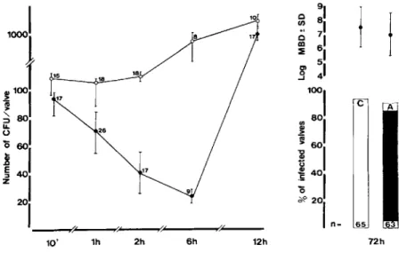
Experiments with 106 S.intermedius. Early
af-ter bacaf-terial challenge with an inoculum 10 times greater than the90070 infectious dose, the valves were
colonized with "-'10 times more bacteria than the valves in animals challenged with an inoculum of 105S. intermedius(figure 2). Ten minutes after
bac-terial challenge the amoxicillin-treated group showed a slight but nonsignificant reduction in the number of cfu per valve compared with the control group. Quantitative cultures of blood at this time of killing yielded 32 ± 25 cfu/ml of blood in controls versus 23 ± 11 cfu/ml in amoxicillin-treated rats(P> .1).
In rats killed 1 hr after bacterial challenge, the num-ber of cfu per valve was slightly reduced in both groups of rats compared with bacterial numbers re-covered at 10 min, and cultures of blood were ster-ile. At 2, 6, and 12 hr after challenge a rapid bac-terial growth was observed on cultures of the valves in control rats. In contrast, in the amoxicillin-treated group a further reduction in the numbers of cfu per valve was observed up to 6 hr. This reduction cor-responded approximately to the interval that serum inhibitory activity was measurable after amoxicillin prophylaxis. However, unlike the valves in the experi-ments with an inoculum of 105S.intermedius,eight
of nine valves were still infected at this time. As a result, when animals wert> ~..illed after 12 hr, when there was no serum inhibitory activity detectable, 13 of 14 amoxicillin-treated rats had a 1oo-fold increase in bacterial numbers, and at three days 60 (92070) of 65 control animals and 63(91070) of 68
amoxicillin-treated animals had developed full-blown endocardi-tis due to S. intermedius.Therefore the reduction in number of cfu per valve observed during the first 6 hr was not sufficient for prevention of bacterial growth to resume once serum inhibitory activity was no longer detectable.
Effect ofmultiple doses ofamoxicillin in rats chal-lenged with 106S.intermedius. In a further
inves-tigation of whether prolonged bacteriostatic activity after amoxicillin prophylaxis was a likely mechanism of protection against the development of endocardi-tis, experiments were performed in which amoxicil-lin-treated rats challenged with 106 S. intermedius
(an inoculum 10 times greater than the 90070
infec-tious dose that overcame successful single-dose amoxicillin prophylaxis) were given none, one, or two subsequent doses of amoxicillin at 6-hr intervals. In these experiments the incidence of endocarditis was
90070 (38 of 42) in rats given a single amoxicillin
prophylactic dose,69070 (11 of 16) in rats given one
subsequent dose 6 hr thereafter(P> .1), and 25070 (6 of 24) in rats given two subsequent doses at 6 and 12 hr after bacterial challenge (P
<
10-4 comparedwith the other two groups of rats). Therefore, provid-ing serum inhibitory levels of amoxicillin durprovid-ing an 18-hr period extended the prophylactic efficacy of amoxicillin to a bacterial inoculum 10 times greater than the 90070 infectious dose.
Discussion
Several recent studies performed both in vitro and in vivo [1-4] have provided indirect evidence that in-hibition of bacterial adherence might be a
mecha-A 72h
j
I
c 65 12h / ( 6h 2h,oJ
1
~1001
~ -,~
80 1i
601
~ 401
201----,,-, --..,.e~--~""'---...,..''''''---10' 1h Figure 2. Natural history ofen-docarditis due to S.intermediusafter iv challenge with 106 cfu. Left, mean ± SD (bars) numbers of bacteria cul-tured from the damaged valves during the early course of endocarditis: con-trols (0) and amoxicillin-treated rats (.). The numbers next to each point are the number of rats at each inter-val.Right,the incidence of endocardi-tis in paired control(C)and amoxicil-lin-treated(A) rats three days after iv challenge with 106S.intermedius.The
numbers at the bottom of each column are the number of rats in each group. In the upper part are given the mean ±SD (bars) bacterial densities (MBD) recovered from the infected valves (ex-pressed as log cfu/g of valve).
nism by which antibiotics prevent bacterial en-docarditis in the absence of bacterial killing. In the present experiments with a streptococcal strain
(S.
in-termedius)
tolerant to the killing effect of amoxicil-lin, we have also observed that amoxicillin-treated bacteria were less able than controls to stick to platelet-fibrin matrices in vitro and to colonize damaged valves in rats with catheter-induced vege-tations. Itis likely, however, that this early reduc-tion in the valvular colonizareduc-tion observed in treated rats at the end of the bacteremia could not entirely explain the successful protection afforded by amox-icillin, for at least two reasons. First, in rats chal-lenged with 105cfu of S.intermedius
(i.e., the mini-mal inoculum infecting 900/0 of control rats), although the average number of cfu per valve 10 and 60 min after iv challenge was slightly decreased af-ter amoxicillin prophylaxis, there was a striking over-lap in the number of bacteria present on the valves of amoxicillin-treated and control rats. In contrast, the reduction in the incidence of endocarditis con-ferred by amoxicillin in rats killed at three days was 740/0 compared with the incidence in control rats. Second, the inactivation of amoxicillin by penicil-linase 30 min after bacterial challenge completely abolished its prophylactic efficacy. Ifthe decrease in early valvular colonization due to the inhibition of bacterial adherence by amoxicillin were to play a major role in the prevention of bacterial endocardi-tis, suppression of the antibiotic activity in serum after the end of the bacteremic phase should not have abolished the prophylactic effect of amoxicillin. It appears therefore that for conferring protection, amoxicillin had to be present in rat serum after the end of the bacteremia, i.e., after the circulating micro-organisms had attached to the cardiac vegetations. During the first 6 hr after the iv challenge with both 105and 106S.intermedius
(i.e., until the serumlevels of amoxicillin became undetectable), rates of growth of bacteria recovered from the valves of amoxicillin-treated animals and controls were strik-ingly different. In control rats, bacterial multiplica-tion on the damaged valves could already be detected 2 hr after bacterial challenge, and a 10-fold increase was observed after 6 hr. In contrast, the number of cfu on the valves of amoxicillin-treated rats decreased progressively during the 6-hr interval after iv chal-lenge. During this period, the bacteria (l05 S.
inter-medius)
disappeared from the damaged valves, but this was not enough time for complete clearance af-ter challenge with 106 S.intermedius.
Several possible mechanisms might have been responsible for the decrease in the number of cfu on the valves of animals given prophylactic amox-icillin. First, the adherent bacteria might have been killed by amoxicillin. However, we have previously shown in vitro that sustained amoxicillin concentra-tions simulating peak serum levels did not kill 106
S.
intermedius
over a 24-hr incubation period [3]. Similarly, exposure of 103 and 102 S.intermedius
(simulating bacterial numbers present on vegetations early after bacterial challenge) to peak concentra-tions of amoxicillin for 24 hr did not result in sig-nificant bacterial killing (data not shown) and thus rendered unlikely the possibility that small bacterial inocula could be killed faster than large inocula. Moreover, in vivo serum bactericidal activity was not detectable in rats at the time of bacterial challenge, i.e., at the time of peak amoxicillin level. Thus, al-though the presence of active amoxicillin in serum for several hours after bacterial challenge was neces-sary for successful endocarditis prophylaxis, mech-anisms other than mere killing by amoxicillin ap-peared to be responsible for the reduction in bacterial numbers on the valves.
A second possible mechanism for diminished bac-terial numbers during antibiotic exposure might be related to amoxicillin-mediated detachment of bac-teria from the vegetations. Amoxicillin slightly but clearly interfered with the in vitro and in vivo stick-ing of S.
intermedius
to platelet-fibrin structures.Itis conceivable that the drug subsequently weakened the attachment of adherent bacteria to the vegeta-tions. If this hypothesis were true, inhibition by amoxicillin of adherence mechanisms would have had two manifestations:(1)the failure of organisms to attach initially and (2) the detachment of already adherent bacteria. However, the latter hypothesis of antibiotic-mediated detachment was not supported by in vitro experiments, which showed that prolonged exposure to and washing with amoxicillin of the platelet-fibrin matrices did not increase the detach-ment of S.
intermedius
over that seen with PBS washes. Similarly, Lowy et al. [4] did not observe in-creased detachment of streptococci adherent to platelet-fibrin matrices on exposure to inhibitory concentrations of penicillin. The detachment hy-pothesis was not supported by in vivo observations either, which showed that in untreated rats killed early after bacterial challenge there was a spontane-ous trend toward the disappearance of bacteria from the colonized valves, even in the absence ofantibi-otic prophylaxis. This finding was most striking in the experiments with 104 S. intermedius, in which
the valves were colonized with small numbers of bac-teria early after iv challenge but were sterile three days later. Similarly, when 10 and 100 times larger inocula were injected iv, which resulted in attachment of 10-100 times more bacteria on the valves, the num-ber of adherent bacteria decreased to the same ex-tent in the control and amoxicillin-treated rats dur-ing the first hour after bacterial challenge, before bacterial growth started in controls. Thus even in the absence of antibiotic, bacteria attached to the damaged valves showed a natural tendency toward disappearance. Therefore, the in vivo and in vitro ob-servations did not support the hypothesis that the inhibition of attachment, the promotion of detach-ment, or both fully accounted for successful antibi-otic prophylaxis.
Successful prophylaxis against S.intermedius
en-docarditis afforded by amoxicillin in the present ex-periments was apparently not conferred by bacterial killing mechanisms due to amoxicillin alone, or by mechanisms related to inhibition of adherence or promotion of detachment. Because the presence of bacteriostatic concentrations of amoxicillin for sev-eral hours after the bacteremic phase was crucial for prophylaxis, the mere inhibition of bacterial growth may be responsible for the successful prophylaxis in allowing the adherent bacteria to disappear from the valves. Six hours of bacterial growth inhibition provided by one dose of amoxicillin permitted bac-terial clearance and successful prophylaxis in animals challenged with lOSS. intermedius, and two
subse-quent doses (inhibiting bacterial growth during an 18-hr period) afforded effective protection in animals challenged with 106S.intermedius. Whether the
bac-terial clearance of resting organisms was solely due to hemodynamic forces, or whether it was mediated by host defense factors alone [10-12] or promoted by amoxicillin remains to be determined [5, 13].
In conclusion, the present experiments suggest that in the absence of bacterial killing, growth inhibition may be an important mechanism of successful an-tibiotic prophylaxis of endocarditis in allowing the bacteria to be cleared from the damaged valves. When large inocula are used, prolonged administra-tion of inhibitory concentraadministra-tions of antibiotics might
circumvent the limited efficacy of antibiotics in pre-venting endocarditis.
References
1. Bernard JP. Francioli P, Glauser MP. Vancomycin prophy-laxis of experimentalStreptococcus sanguis. Inhibition of bacterial adherence rather than bacterial killing. J Clin In-vest 1981;68:1113-6
2. Scheid WM, Zak0,Vosbeck K, Sande MA. Bacterial adhe-sion in the pathogenesis of infective endocarditis: effect of subinhibitory antibiotic concentrations on streptococ-cal adhesion in vitro and the development of endocarditis in rabbits. J Clin Invest 1981;68:1381-4
3. Glauser MP, Bernard JP, Moreillon P, Francioli P. Success-ful single-dose amoxicillin prophylaxis against experimental streptococcal endocarditis: evidence for two mechanisms of protection. J Infect Dis 1983;147:568-75
4. Lowy FD. Chang DS, Neuhaus EG, Horne DS, Tomasz A, Steigbigel NH. Effect of penicillin on the adherence of Streptococcus sanguis in vitro and in the rabbit model of endocarditis. J Clin Invest 1983;71:668-75
5. McDonald PJ, Wetherall BL, Pruul H. Postantibiotic leu-kocyte enhancement: increased susceptibility of bacteria pretreated with antibiotics to activity of leukocytes. Rev Infect Dis 1981;3:38-44
6. Arnold SB, Valone JA, Askenase PW, Kashgarian M, Freed-man LR. Diffuse glomerulonephritis in rabbits with Strep-tococcus viridans endocarditis. Lab Invest 1975;32:681-9 7. Heraief E, Glauser MP, Freedman LR. Natural history of aortic valve endocarditis in rats. Infect Immun 1982;37: 127-31
8. Sabath LD, Anhalt JP. Assay of antimicrobics. In: Lennette EH, Balows A, Hausler WJ Jr, Truant JP, eds. Manual of clinical microbiology. 3rd ed. Washington, DC: Amer-ican Society for Microbiology, 1980:485-90
9. The antibiotic prophylaxis of infective endocarditis. Report of a Working Party of the British Society for Antimicrobial Chemotherapy. Lancet 1982;2:1323-6
10. Francioli PB, Freedman LR. Streptococcal infection of en-docardial and other intravascular vegetations in rabbits: natural history and effect of dexamethasone. Infect Im-mun 1979;24:483-91
11. Meddens MJ, Thompson J, Eulderink F, Bauer WC, Mattie H. Van Furth R. Role of granulocytes in experimental Strep-tococcus sanguis endocarditis. Infect Immun 1982;36: 325-32
12. Meddens MJ, Thompson J, Leijh PC, Van Furth R. Role of granulocytes in the induction of an experimental endocardi-tis with a dextran-producingStreptococcus sanguis and its dextran-negative mutant. Br J Exp Pathol 1984; 65: 257-65
13. Horne D, TomaszA.Hypersusceptibility of penicillin-treated group B streptococci to bactericidal activity of human poly-morphonuclear leukocytes. Antimicrob Agents Cherno-ther 1981;19:745-53