Living patches engineered from human umbilical cord derived fibroblasts
and endothelial progenitor cells
*Do
¨rthe Schmidt
a, Anita Mol
b, Stefan Neuenschwander
a, Christian Breymann
c,
Matthias Go
¨ssi
d, Gregor Zund
a, Marko Turina
a, Simon P. Hoerstrup
a,*
aDepartment of Surgical Research and Clinic for Cardiovascular Surgery, University Hospital and University of Zurich,Raemistrasse 100, CH 8091 Zurich, Switzerland
bDepartment of Biomedical Engineering, Technical University, Eindhoven, The Netherlands cDepartment of Gynaecology and Obstetrics, University Hospital, Zurich, Switzerland
dInstitute of Polymers, Swiss Institute of Technology (ETH), Zurich, Switzerland
Received 3 September 2004; received in revised form 14 January 2005; accepted 25 January 2005
Abstract
Objective: A major shortcoming in contemporary congenital heart surgery is the lack of viable replacement materials with the capacity of growth and regeneration. Here we focused on living autologous patches engineered from human umbilical cord derived fibroblasts and endothelial progenitor cells (EPCs) as a ready-to-use cell source for paediatric cardiovascular tissue engineering. Methods: EPCs were isolated from 20 ml fresh umbilical cord blood by density gradient centrifugation and myofibroblasts were harvested from umbilical cord tissue. Cells were differentiated and expanded in vitro using nutrient media containing growth factors. Before seeding, cell-phenotypes were assessed by immuno-histochemistry. Biodegradable patches fabricated from synthetic polymers (PGA/P4HB) were seeded with myofibroblasts followed by endothelialization with EPCs. All patches were cultured in a perfusion bioreactor. A subgroup of patches was additionally stimulated by cyclic strain. Analysis of the neo-tissues comprised histology, immuno-histochemistry, extracellular matrix (ECM) analysis and biomechanical testing. Results: Endothelial phenotypes of EPCs before seeding were confirmed by Ac-Dil-LDL, CD 31, von-Willebrand-Factor and eNOS staining. Histology of the seeded patches demonstrated layered viable tissue formation in all samples. The cells in the newly formed tissues expressed myofibroblast markers, such as desmin and alpha-SMA. The EPCs derived neo-endothelia showed constant endothelial phenotypes (CD 31, vWF). major constituents of ECM such as collagen and proteoglycans were biochemically detected. Stress–strain properties of the patches showed features of native-analogous tissues. Conclusions: Living tissue engineered patches can be successfully generated from human umbilical cord derived myofibroblasts and EPCs. This new cell source may enable the tissue engineering of versatile, living, autologous replacement materials for congenital cardiac interventions.
Q2005 Elsevier B.V. All rights reserved.
Keywords: Endothelial progenitor cells; Umbilical cord; Endothelium; Patches
1. Introduction
A major shortcoming in today’s surgery of congenital cardiovascular defects is the lack of living replacement materials with the capacity of growth and regeneration. Currently, paediatric surgical treatment requires the appli-cation of synthetic or non-autologous bioprosthetic materials, such as patches and vascular conduits [1,2]. These materials are burdened with substantial disadvan-tages including obstructive tissue ingrowth and calcification
of the replacement [3]. These limitations and the lack of growth typically cause re-operations of paediatric patients with cardiovascular defects associated with increased morbidity and mortality. To address the above-mentioned problems cardiovascular tissue engineering is a new scien-tific field aiming at in vitro fabrication of living, autologous grafts with the capacity of growth, repair, and regeneration. Important functional features of ideal replacement materials for the repair of congenital defects comprise a living matrix with native-analogous biomechanical proper-ties and a thrombo-resistant surface. In previous studies, we have demonstrated the feasibility of using human umbilical cord derived myofibroblasts as a cell source for engineering of functional paediatric tissues[4]. The umbilical cord blood is a known source for endothelial progenitor cells (EPCs) differentiated from haemangioblasts, a common progenitor for both haematopoetic and endothelial cells. These cells have the potential to differentiate into mature endothelial cells and have been successfully utilized in non-tissue
www.elsevier.com/locate/ejcts
1010-7940/$ - see front matter Q 2005 Elsevier B.V. All rights reserved. doi:10.1016/j.ejcts.2005.01.064
*
Presented at the joint 18th Annual Meeting of the European Association for Cardio-thoracic Surgery and the 12th Annual Meeting of the European Society of Thoracic Surgeons, Leipzig, Germany, September 12–15, 2004.
* Corresponding author. Tel.: C41 1 255 3644; fax: C41 1 255 4775. E-mail address: [email protected] (S.P. Hoerstrup).
engineering applications such as for the repair of injured vessels, neo-vascularization or regeneration of ischemic tissue[5–8]as well as coating of synthetic vascular grafts[9]. Recently, animal derived EPCs have been used for the endothelialization of decellularized grafts in animal models [10]and for seeding of hybrid grafts[11].
Little is known about the feasibility and utility of umbilical cord derived EPCs for in vitro engineered endo-thelium in humans. Here, we investigate human umbilical cord blood derived endothelial progenitor cells (EPCs) as a source for in vitro generation of functional endothelia covering living tissue engineered patches as a simplified model for pediatric cardiovascular tissue engineering.
2. Material and methods
2.1. Isolation of myofibroblasts from human umbilical cord tissue
Cells were obtained from umbilical cords of healthy individuals after informed consent was obtained from the participants. Myofibroblasts were isolated from umbilical cord tissue of newborns. Several (z8 mm3) pieces of umbilical cord tissue were washed with PBS, placed in petri-dishes and cultured in a humidified incubator (37 8C, 5% CO2) in advanced DMEM medium (Gibco, Invitrogen)
supplemented with 10% fetal bovine serum (PAN Biotech, Germany), 2 mM Glutamax (Gibco) and Gentamycin (50 mg/ml, PAN). Myofibroblasts were expanded up to passage 11.
2.2. Isolation of EPCs from human umbilical cord blood and differentiation into endothelial phenotype
The human umbilical cord vein of newborns was punctured directly after delivery and 20 ml of fresh blood were obtained. Immediately, EPCs were isolated from mononuclear cells by density gradient centrifugation (Histopaque-1077, Sigma), followed by differential cen-trifugation as described by Asahara et al.[12]. Isolated cells were cultured in endothelial basal medium (EBMe-2, Cambrex), containing growth factors and supplements provided by the supplier: Vascular Endothelial Growth Factor (VEGF), human Fibroblasts Growth Factor (hFGF), human recombinant long-Insulin-like Growth Factor-1 (R3-IGF), human Epidermal Growth Factor (hEGF), Gentamycin and Amphotericin (GA-1000), Hydrocortisone, Heparin, Ascorbic Acid and 2% Fetal Bovine Serum. After 4 days, attached cells were reseeded and after 7 days the medium was changed to EBM-2 containing the same growth factors but 20% FBS. After 3 weeks of culturing, the endothelial phenotype was confirmed by immuno-histochemistry. The confluent monolayers were stained with antibodies for von Willebrand factor (vWF, polyclonal; DAKO A/S), CD 31 (IgG1/kappa, Clone JC/70A; DAKO A/S), CD34 (IgG1, Clone QBEND/10; Serotec) and examined for uptake of Ac-LDL (Dil-Ac-LDL, Biomedical tech.) and expression of endo-thelial nitric oxide synthase (eNOS, polyclonal; Transduc-tion Laboratories, Inc.).
2.3. Fabrication of scaffolds
Scaffolds were fabricated from a non-woven polyglycolic-acid mesh with randomized distributed fibers (PGA, thick-ness: 1.0 mm, specific gravity: 69 mg/cm3, porosity O95%, Albany Int.), cut into 2.3 cm2 patches (Fig. 1) and coated with Poly-4-Hydroxybutyric acid (10% w/v P4HB, TEPHA Inc., Cambridge, MA) by dipping. Three patches each were attached to a ring-shaped support of 20 mm diameter and 15 mm length. After drying in an exicator, the scaffolds were cold-gas sterilized.
2.4. Seeding of scaffolds
Cells were expanded as monolayers either in 175 cm2
tissue culture flasks (TRP, Milian, Geneva, Switzerland) or in roller bottles (Costar, 850 cm2/bottle) using the Cellroll
system (Integra biosciences) at a speed of 0.5–1 minK1. At confluency, cells were harvested by treatment with trypsi-n/EDTA (0.25%) and seeded onto the biodegradable PGA/P4HB patches by using fibrin as a cell carrier [13]. Briefly, 3.5!106 cells/100 mm3 of scaffold were
resus-pended in a sterile thrombin solution (Sigma, 10 U/ml medium) and kept on ice in a volume that equals half of the void volume of the scaffold. The same volume of sterile, ice-cold fibrinogen solution was added (10 mg/ml medium) and after carefully mixing, the cells were dripped onto the scaffold.
The patches were positioned into a bioreactor flow system and perfused with culture medium supplemented with 1 mM ascorbic acid-2-phosphate. After 7 days, the cultivation conditions were altered. Patches of group 1 (nZ 9) were continued to be perfused only, while patches of group 2 (nZ9) were additionally exposed to cyclic straining performed about 12 mmHg resulting in a straining of 10–15% by using a pressure applicator connected to a pressure air chamber. After another 14 days, all patches were taken out of the bioreactor flow system and were endothelialized with EPCs (1.5!106cells/cm2) on both sides. Patches were kept
at humidified incubator conditions (37 8C, 5% CO2) in EBM-2,
containing growth factors and 20% FBS. Endothelialization
was repeated after 24 h. After additional 24 h, the patches of both groups were placed back into the bioreactor flow system and exposed for 4 subsequent days to perfusion or perfusion and cyclic straining, respectively. After culturing, the patches were cut into samples and portioned out for analysis.
2.5. Tissue formation analysis 2.5.1. Histology
Samples were fixed in 4% phosphate buffered formalin (pH 7.0) and paraffin-embedded. Sections of 3–5 mm of each group were examined histologically by Eosin-Hematoxylin (H&E), Hematoxylin-Sudan (H&S), and Trichrom-Masson staining. Selected samples were characterized by immuno-histochemistry. Vimentin, a-SMA, and desmin were used for analysis of the myofibroblast phenotype and vWF, CD 31 and eNOS staining to validate the phenotype of the EPC derived endothelial cells.
2.5.2. Quantification of extracellular matrix elements 2.5.2.1. Collagen. As an indicator for collagen formation, hydroxyproline content was determined from dried samples as described by Huszar et al. [14]. Briefly, tissue samples were hydrolyzed in 50–100 ml 4 M NaOH (Fluka, Switzerland) in an autoclave at a temperature of 120 8C for 10 min. The solution was neutralized by adding an equal volume of 1.4 M citric acid (Fluka). Chloramin-T (Riedel-de Hae¨n (Fluka) 62 mM) was added and the samples were allowed to oxidize for 25 min. Aldehyde/perchloric acid solution (1 M) was subsequently added and the chromophore was allowed to develop at 65 8C for 15 min. The absorbance of the obtained solutions was determined at 570 nm. The amount of hydroxyproline present in the hydrolysates was determined from a standard curve using known amounts of trans-4-hydroxy-L-proline (Sigma).
2.5.2.2. s-Glyco-Amino-Glycans. To further specify the extra cellular matrix, s-Glyco-Amino-Glycans were detected col-orimetrically using 1,9-di-methyl-methylene blue stain[15], following complete papain digestion of the constructs in 100 mM sodium phosphate pH 6.5, 5 mM cysteine, 5 mM EDTA and 125 mg/ml papain (Sigma).
2.5.2.3. Determination of cell number. The amount of cells growing on the constructs was indirectly determined by measuring the DNA content from the same papain digested samples using Hoechst dye (Bisbenzimide H 33258, Fluka) [16]and a fluorometer (Fluostar, BMG, Offenburg, Germany, 355 nm excitation/460 nm emmission). Calf thymus DNA (Sigma) was used as a standard.
2.5.3. Testing of mechanical properties
Measurements of the mechanical properties were per-formed with freshly harvested patches (15!5!1 mm) using an uniaxial tensile tester (Instron) equipped with a 10 N load cell. Tensile stress and strain were recorded and the Young’s modulus was calculated.
3. Results
3.1. Macroscopic appearance of the patches
All patches showed homogenous tissue formation and dense surfaces (Fig. 1).
3.2. Phenotype of myofibroblasts
Myofibroblasts showed positive staining for vimentin and expression of alpha-SMA and desmin (Fig. 2a and b). 3.3. Morphology and phenotype of EPCs
EPCs could be isolated from all blood samples. After isolation, the plated cells were initially rounded. After 4 days, cells were attached and formed clusters. Two different types of EPCs were found: spindle-like shaped cells (80%) and polymorph cells (20%). Polymorph EPCs formed colonies and showed good growth under in vitro conditions. After 3 weeks, they differentiated into mature endothelial cells forming cobblestone monolayers.
Before seeding onto the scaffolds, differentiated EPCs showed an endothelial phenotype. They stained positive for vWF, CD 31, CD34, eNOS, and showed Ac-LDL-uptake. Differentiated EPCs kept this phenotype when cultivated under both cyclic strain and perfusion in the bioreactor flow system (Fig. 2e and f).
3.3.1. Histology
Histological examination of the seeded patches revealed good cell-to-polymer attachment. Myofibroblasts showed good ingrowth into the PGA/P4HB scaffolds under cyclic strain. A similar picture was observed after culturing under perfusion. EPCs demonstrated a good attachment on the neo-tissue formed by myofibroblasts in both groups of culturing conditions.
H&E staining revealed organized tissue-formation with good extracellular matrix formed by myofibroblasts in the inner part of the patches (Fig. 2c). Trichrom–Masson-staining highlighted elements of extracellular matrix (Fig. 2d). An endothelial cell lining formed by differentiated EPCs was found on surfaces of all patches. Immunohistochemistry confirmed the endothelial phenotype of the neo-tissue (Fig. 2e and f).
3.4. Quantification of extracellular matrix elements A summary of the extracellular matrix composition is given in Fig. 3. The collagen content of both groups were comparable (strained 4.06 mg/mgG1.92, nZ8, perfused 4.21 mg/mgG0.44, nZ8). The sulfated glycosaminoglycan content was higher in the strained than in the perfused patches (6.44 mg/mgG1.45, nZ4 versus 4.65 mg/mgG0.61, nZ5) and the cell number was significantly higher in the strained patches (3.14 mg/mgG1.02, nZ4 versus 1.24G0.35, nZ5 Student’s t-test p!0.05).
3.5. Tensile testing
Tissue engineered patches showed normal stress–strain profiles in both groups of culturing conditions (Table 1). Only
strain at break showed significant differences between the groups.
4. Discussion
For surgical treatment of congenital cardiac defects such as augmentation of the hypoplastic, stenotic right ventri-cular out flow tract or the pulmonary artery in tetralogy of Fallot, for the closure of a complete AVSD, enlargement of the aorta in the Norwood I procedure synthetic or
bioprosthetic replacement materials are commonly used. Such non-autologous materials have been associated with substantial complications including calcifications, obstruc-tive tissue ingrowth, infections, and thrombogenicity[1,3, 17]. The availability of living graft materials with the potential to grow, regenerate and to adapt to the changes in the developing cardiovascular system would have funda-mental advantages over currently used replacement materials.
In the present study, we investigated the feasibility to utilize human umbilical cord blood derived endothelial progenitor cells for in vitro fabrication of living autologous patches. We further investigated the influence of different in vitro culture conditions on tissue maturation and functionality of the newly formed endothelia.
As demonstrated previously [4], umbilical cord derived myofibroblasts showed exellent ingrowth into the biode-gradable scaffolds and organized tissue formation with the production of extracellular matrix proteins such as collagen and proteoglycans. This myofibroblasts based matrix for-mation represents a crucial prerequisite for the successful creation of mechanically competent, surgically implantable replacement materials. Isolation of the endothelial progeni-tor cells from umbilical cord blood samples was easily performed and the cells were expandable in sufficient amounts for tissue engineering purposes. After seeding, the endothelial progenitor cells formed functional endo-thelial layers on the surfaces of the myofibroblast derived neo-matrices of all patches. Most importantly, the EPCs showed constant endothelial phenotype during the whole tissue engineering process and expressed functional features such as eNOS. In previous studies we and others have demonstrated that mechanical conditioning of growing tissue engineered cardiovascular structures, such as heart valves and blood vessels results in enhanced and more mature tissue formation[18]. Accordingly, the patches were cultured in a custom designed bioreactor in vitro system. We have chosen two bioreactor protocols using flow and cyclic strain as mechanical stimulation to increase the production of extracellular matrix and mechanical strength of the patches. By comparing the culture conditions, we found higher contents of extracellular matrix proteins such as proteoglycans as well as cell number when patches were exposed to cyclic strain. However, collagen content was not significantly different. These results indicate the important
Fig. 3. Extracellular matrix analysis. HYP, hydroxyproline; GAG, sulfated glycosamino glycans; DNA, deoxy nucleic acid (cell number). Averages are given in mg/mg of dry weight. The error bars represent the standard deviation.
Table 1
Mechanical properties of perfused or perfused and strained tissue engineered patches Samples Tensile strength (MPa) Young’s modulus (MPa) Strain at break Strained 1 0.20 0.92 0.38 Strained 2 0.17 0.74 0.33 Strained 3 0.11 0.49 0.34 Strained 4 0.22 1.07 0.28 Strained meanGSD 0.18G0.05 0.80G025 0.33G0.04 Perfused 1 0.30 0.74 0.56 Perfused 2 0.17 0.50 0.60 Perfused 3 0.17 0.30 0.64 Perfused 4 0.08 0.24 0.50 Perfused meanGSD 0.18G0.09 0.44G0.23 0.57G0.06
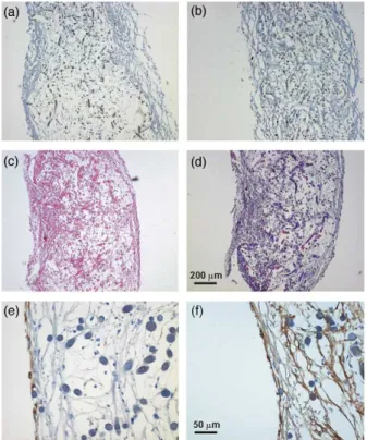
Fig. 2. Histological and immunohistological characterization. Myofibroblasts of the new tissue showed positive staining for (a) desmin and (b) a-SMA (magnification 50!). Hemalaun & Eosin staining (c) demonstrated organized tissue formation with production of extracellular matrix components; in Trichrom–Masson staining (d) collagen fibres are blue (magnifications 50!). Expression of (e) CD31 on surfaces of patches and (f) vWF, respectively confirm the endothelial phenotype of the neo-endothelium formed by EPCs (magnification 200!). (For interpretation of the reference to colour in this legend, the reader is referred to the web version of this article.)
influence of strain stimulation on early matrix formation as recently described by Mol et al.[19].
The mechanical profiles of all patches showed features of native cardiovascular tissues demonstrating a non-linear mechanical behaviour. In contrast, the scaffold material itself exhibits linear behaviour indicating that the measured mechanical properties are relating from the neo-tissues. However, the mechanical strength did not reach physiologi-cal values during the investigated in vitro time, restricting the current patches to low-pressure applications such as in reconstruction of the RVOT. Interestingly, we observed a higher Young’s modulus representing a higher stiffness in the cyclically strained compared to perfused tissues. This difference in stiffness of the patches may result from more mature extracellular matrix formation reflected by increased proteoglycans content and a higher degree of cross-links among collagen fibres to be assessed in further experiments.
In summary, in this feasibility study we created living autologous human patches based on umbilical cord derived cells, representing a versatile replacement material for congenital cardiac surgery. The endothelial progenitor cells isolated from the umbilical cord blood showed excellent expansion capacities, constant endothelial differentiation, and formation of endothelia with functional properties. Apart from being a potent cell source for differentiated endothelial cells, human endothelial progenitor cells may enable to use tissue engineered replacement materials directly after birth, since they can be harvested prenatal, e.g. by ultrasound guided chordocentesis. This approach would provide sufficient time for the in vitro generation of autologous replacement material ready to use at or shortly after birth and is currently investigated in animal models.
Acknowledgements
The authors wish to thank Sirpa Price, Laboratory for Tissue Engineering and Cell Transplantation, University Hospital Zurich, for her valuable work on cell culture. We further thank Prof. Bernhard Odermatt, Department of Pathology, University Zurich, for performing the immuno-histochemestry analysis and Astrid Morger for support by the histological examination. The authors wish to recognize the contribution by Dr Alberto Weber, Prof. Takayuki Asahara and Prof. Stefanie Dimmeler regarding cell isolation.
References
[1] Mayer Jr JE. Uses of homograft conduits for right ventricle to pulmonary artery connections in the neonatal period. Semin Thorac Cardiovasc Surg 1995;7:130–2.
[2] Schoen FJ, Levy RJ. Founder’s Award, 25th Annual Meeting of the Society for Biomaterials, perspectives. Providence, RI, April 28–May 2, 1999. Tissue heart valves: current challenges and future research perspec-tives. J Biomed Mater Res 1999;47:439–65.
[3] Endo S, Saito N, Misawa Y, Sohara Y. Late pericarditis secondary to pericardial patch implantation 25 years prior. Eur J Cardiothorac Surg 2001;20:1059–60.
[4] Hoerstrup SP, Kadner A, Breymann C, Maurus CF, Guenter CI, Sodian R, Visjager JF, Zund G, Turina MI. Living, autologous pulmonary artery conduits tissue engineered from human umbilical cord cells. Ann Thorac Surg 2002;74:46–52.
[5] Kawamoto A, Gwon HC, Iwaguro H, Yamaguchi JI, Uchida S, Masuda H, Silver M, Ma H, Kearney M, Isner JM, Asahara T. Therapeutic potential of ex vivo expanded endothelial progenitor cells for myocardial ischemia. Circulation 2001;103:634–7.
[6] Kocher AA, Schuster MD, Szabolcs MJ, Takuma S, Burkhoff D, Wang J, Homma S, Edwards NM, Itescu S. Neovascularization of ischemic myocardium by human bone-marrow-derived angioblasts prevents cardiomyocyte apoptosis, reduces remodeling and improves cardiac function. Nat Med 2001;7:430–6.
[7] Assmus B, Schachinger V, Teupe C, Britten M, Lehmann R, Dobert N, Grunwald F, Aicher A, Urbich C, Martin H, Hoelzer D, Dimmeler S, Zeiher AM. Transplantation of progenitor cells and regeneration enhancement in acute myocardial infarction (TOPCARE-AMI). Circulation 2002;106:3009–17.
[8] Pesce M, Orlandi A, Iachininoto MG, Straino S, Torella AR, Rizzuti V, Pompilio G, Bonanno G, Scambia G, Capogrossi MC. Myoendothelial differentiation of human umbilical cord blood-derived stem cells in ischemic limb tissues. Circ Res 2003;93:e51–e62.
[9] Shirota T, Yasui H, Shimokawa H, Matsuda T. Fabrication of endothelial progenitor cell (EPC)-seeded intravascular stent devices and in vitro endothelialization on hybrid vascular tissue. Biomaterials 2003;24: 2295–302.
[10] Kaushal S, Amiel GE, Guleserian KJ, Shapira OM, Perry T, Sutherland FW, Rabkin E, Moran AM, Schoen FJ, Atala A, Soker S, Bischoff J, Mayer Jr JE. Functional small-diameter neovessels created using endothelial pro-genitor cells expanded ex vivo. Nat Med 2001;7:1035–40.
[11] Shirota T, He H, Yasui H, Matsuda T. Human endothelial progenitor cell-seeded hybrid graft: proliferative and antithrombogenic potentials in vitro and fabrication processing. Tissue Eng 2003;9:127–36.
[12] Asahara T, Murohara T, Sullivan A, Silver M, van der Zee R, Li T, Witzenbichler B, Schatteman G, Isner JM. Isolation of putative progenitor endothelial cells for angiogenesis. Science 1997; 275:964–7.
[13] Mol A, Lieshout van MI, Dam-de Veen GC, Neuenschwander S, Hoerstrup SP, Baaijens FPT, Bouten CVC. Fibrin as a cell carrier in cardiovascular tissue engineering. Biomaterials 2004; 26(16): 3113–21.
[14] Huszar G, Maiocco J, Naftolin F. Monitoring of collagen and collagen fragments in chromatography of protein mixtures. Anal Biochem 1980; 105:424–9.
[15] Farndale RW, Buttle DJ, Barrett AJ. Improved quantitation and discrimination of sulphated glycosaminoglycans by use of dimethyl-methylene blue. Biochim Biophys Acta 1986;883:173–7.
[16] Kim YJ, Sah RL, Doong JY, Grodzinsky AJ. Fluorometric assay of DNA in cartilage explants using Hoechst 33258. Anal Biochem 1988;174: 168–76.
[17] Ben-Shachar G, Nicoloff DM, Edwards JE. Separation of neointima from Dacron graft causing obstruction. Case following Fontan procedure for tricuspid atresia. J Thorac Cardiovasc Surg 1981;82:268–71.
[18] Niklason LE, Gao J, Abbott WM, Hirschi KK, Houser S, Marini R, Langer R. Functional arteries grown in vitro. Science 1999;284:489–93.
[19] Mol A, Bouten CV, Zund G, Gunter CI, Visjager JF, Turina MI, Baaijens FP, Hoerstrup SP. The relevance of large strains in functional tissue engineering of heart valves. Thorac Cardiovasc Surg 2003;51:78–83.
Appendix A. Conference discussion
Mr V. Tsang (London, United Kingdom): I’m no engineer. You need to give me a simple answer.
Presumably, your living tissue patches, in your dream, you want to use it as a patch; is that correct?
Dr Schmidt: It’s a patch, but it’s also augmentation material, for example, for pulmonary arteries.
Dr Tsang: So the biodegradable platform you use for the seeding of cells would have dissolved by the time you want to use your patches; is that correct?
Dr Tsang: And you mentioned the mechanical properties of the patches, the strength. I’m sure a lot of people here do not know what that means. How strong is it?
Dr Schmidt: We characterize the stiffness of the patches, of the augmentation material, and found them approximating to native tissues.
Dr Tsang: So you think it could be used in the pulmonary artery as an augmentation patch?
Dr Schmidt: In the lower pressure system we can use it.
Dr G. Gerosa (Padova, Italy): I would like to ask you, for how long did you keep your construct in the pulse duplicator to do some, let’s say, preconditioning of the tissue?
Did the phenotype expression of the endothelial cells, change over this period?
Finally, going back to the question of one of the chairmen, how pliable and thick is this tissue?
Dr Schmidt: To your first question, we cultured the tissue and construct for 21 days, followed by the endothelialization for additional 5 days, that is 4 weeks total culture time.
And the second question, the endothelial phenotype was assessed first for the endothelial progenitor cells also stated in the literature, we have seen a phenotype change focusing on late endothelial progenitor cells with a reduction of expression of specific antigens such as VEGFR-2, Ulex Lectin, CD 34. After seeding and formation of endothelia, we checked CD 31 and von Willebrand factors detectable in mature endothelial cells.
And the last question, we measured the stiffness and elasticity of the tissue engineered patches. The dimensions were 4 to 4 cm, and a thickness of 0.5 cm. Dr Gerosa: So you did not experience any core necrosis in the middle of your patch?
Dr Schmidt: No, we did not detect necrosis in the inner layer of the patches. Obviously there was sufficient nutrient media supply in the in vitro system.
Dr M. Haw (Southampton, United Kingdom): I was just wondering, from a practical point of view, how long can you keep the patches alive supposing you were growing this for clinical use? Can you keep them indefinitely?
Dr Schmidt: In the present study we have kept them in culture in our perfusion system for 4 to 6 weeks. However, we think, if clinically necessary we can keep them longer.