© 1994 Oxford University Press
Functional recognition of in vivo processed
self antigen
B. Stockinger and B. Hausmann
1National Institute for Medical Research, Department of Molecular Immunology, The Rtdgeway, Mill Hill, London NW7 1AA, UK
1 Basel Institute for Immunology, 487 Grenzacherstrassse, CH 4005 Basel, Switzerland
Key words: antigen presentation, dendritic cells, MHC class II restriction, self proteins, setf tolerance, T cell recognition
Abstract
C5, the fifth component of complement, Is a circulating self protein which Induces complete tolerance in MHC class II restricted, CD4+ T cells due to the presentation of C5 taken up from
plasma. Functional recognition of In vivo processed C5 was monitored by activation of C5 specific T cell hybrids cultured with antigen presenting cells (APC) from C5 expressing mice. Dendritic cells Isolated from various tissues (spleen, thymus, skin) proved to be the most efficient APC, since 10-to 50-fold more macrophages and at least 100- 10-to 500-fold more B cells were needed 10-to achieve similar T cell activation. Stimulatory C5 peptide - class II complexes generated In vivo were retained on the surface of dendritic cells but not on macrophages and B cells upon prolonged culture: Dendritic cells but not macrophages from thymus presented In vivo processed C5. Taken together these findings emphasize the crucial role dendritic cells play for recognition of soluble self proteins by MHC class II restricted T cells.
Introduction
T cells recognize antigens in the form of peptides bound to MHC molecules on the surface of antigen presenting cells (APC). Recognition of self peptides at an earty stage of maturation in the thymus leads to negative selection of T cells and immundogical tolerance to the particular self antigen, whereas recognition of antigen by mature T cells in the periphery usually leads to an immune response.
It has been shown that APC do not discriminate between self and foreign antigens (1) and that self antigens are constitutively processed and presented in vivo (2,3). The spectrum of self peptides which are recognized by the two classes of T cells, MHC class I restricted cytotoxic T cells and MHC class II restricted T cells is different (4). Most peptides derived from endogenous biosynthesized proteins within an APC will bind to MHC class I molecules in the endoplasmic reticulum (ER) and be presented to class I restricted T cells (5). The major source of peptides for recognition by class II restricted T cells is derived from exogenous antigens which are degraded in endosomal/lysosorna) compartments (6) and presented by MHC class II bearing APC. It folbws that antigen recognition in class II restricted T cells is subject to additional constraints not encountered for antigen recognition by MHC class I restricted T ceils. Firstly, only a minority of cells expresses MHC class II determinants to qualify as APC for MHC class II restricted cells and secondly, exogenous antigen has to be internalized efficiently to allow access to the class II presentation pathway.
C5, the fifth component of mouse complement, is a serum self protein which induces complete T ceil tolerance in C5 expressing mouse strains. Self C5 is constitutively processed in vivo as revealed by the abflity of spleen and thymus APC from C5 expressing mouse strains to directly activate C5 specific T cell clones (raised in C5 deficient, non-tolerant mice) in vitro in the absence of additional antigen (3).
In this paper we compare the efficiency of different APC subpopulatjons to present in vivo processed setf antigen to specific T cells. Dendritic cells regardless of their tissue of origin proved to be highly potent APC for the circulating self antigen C5, by far exceeding the presentation capacity of macrophages and B cells. The efficiency of setf antigen presentation is likely to have considerable impact on induction and maintenance of tolerance. Dendritic cells therefore may be the principal cells responsible for tolerance induction in MHC class II restricted T cells.
Methods
Animals
CBA/Ca, A/J, and BALB/c mice were maintained at the National Institute for Medical research specific pathogen-free unit SP6 transgenic mice (7) were maintained on a BALB/c background at the animal facfiies of the Basel Institute for Immunology. These mice Correspondence to: B. Stockinger
express a trinitrophenyl/dinitrophenyl (TNP/DNP)-specific Ig receptor on B cells. Transgenic progeny were identified by their high titers of anti-TNP antibodies as assessed by standard ELJSA techniques.
T cell hybrids
The T cell hybrid C8-15 specific for the fifth component of mouse complement (C5) and restricted by I-Ed was derived from C5 deficient B10.D2/0 mice immunized with C5. Hybrid A18 specific for C5 in the context of I-E* was derived from C5 deficient A/J. Establishment of C5 specific clones and hybrids is described in detail elsewhere (3).
APCs
B cells were prepared by gentle mechanical dissection of spleens. Cell suspensions were centrifuged on a discontinuous PercoD gradient with densities of r - 1.078, 1.062, and 1.05 kg/I for 20 min at 1200 g. Cells from the 1.062 interphase were collected, washed twice, and then placed on 60 mm Petri dishes for two sequential rounds of adherence of 2 h each. Non-adherent cells were finally depleted of T cells by incubation with magnetic beads (Dynabeads; Dynal, Oslo, Norway) coupled with anti-Thy-1 antibody T24 (a gift from D. Gray, Royal Postgraduate Medical School, London, UK).
Macrophages were obtained by extensive washing of Petri cfishes after adherence of spleen cell suspensions described as above. They were detached from plastic by ice-cold PBS containing 20 mM EDTA and placed on a shaker for 15 min (macrophage source for Rg. 1). Alternatively, macrophages were collected as the adherent fraction after overnight adherence as part of the isolation of dendritic cells (see below, Figs 2-4).
Dendritic cells from spleen and thymus were isolated after enzyme digestion of organs with a cocktail of collagenase (1.6 mg/ml Worthington CLS4) and DNase (0.1% Sigma Fraction IX) for 60 min at 37°C. Cell suspensions were washed twice and then centrifuged on Percoll as described for B cells. The low density fraction (1.062) was placed on 60 mm Petri dishes and adhered for 2 h at 37°C in a humidified CO2 incubator. Non-adherent cells were washed off and the dishes were put back in the incubator for an overnight incubation. After this incubation step, dendritic cells have detached from the plastic surface while macrophages remain adherent Spleen dendritic cells were found to be contaminated with — 10% B cells and were therefore subjected to panning on 22 mm plastic dishes coated with goat anti-mouse Ig (10 /ig/ml) for 20 min at 4°C. The non-adherent fraction from the panning step was used as pure source of dendritic cells. Purity was assessed by staining with mAb N418 specific for a distinct integrin on the surface of dendritic cells (8) and anti-mouse Ig.
Skin Langerhans cells were isolated from mouse ears. The ears were split and dorsal and ventral halves were placed with the dermal side facing down onto culture medium and incubated for 4 - 7 days (9). Non-adherent migratory Langerhans cells were collected from the bottom of the wells and contaried - 6 0 - 7 0 % Langerhans cells as assessed by staining with anti-class II antibody M5/114 (ATCC TIB 120).
Bone marrow dendritic cells were generated according to Schleicher etal. (10) with some modifications according to Inaba et al. (11). Briefly, 5x10* bone marrow cells were placed into 150 ml Falcon flasks in 15 ml culture medium containing 250 U/ml granulocyte macrophage colony stimulating factor (GM-CSF).
Recombinant murine GM-CSF was kindly provided by Behringwerke (Marburg, Germany).
On day 2 of culture non-adherent cells were pipetted off to remove the bulk of granukxytes, fresh medium with GM-CSF was added, and from day 8 to day 14 non-adherent and loosely adherent cells were washed off and used as source of dendritic cells. About 80% of these cells expressed MHC class II.
Antigen presentation assays
About 5x10* T cell hybrid cells per well were cultured with different numbers of APC and antigen doses as indicated for 24 h in flat bottom microtjter plates (Costar) in Iscove's modified Dulbecco medium supplemented with 5% FCS, 2 x 1 0 "3 M L-glutamine, 100 ^g/ml gentamycin, and 3 x 1 0 ~5 M mercaptoethanol.
After 24 h, 100 /J aliquots of supernatant were transferred to fresh miaotter plates together with 5000/well ll-2-dependent CTLL cells. [3H]Thymidine incorporation of CTLL was measured 24 h later; 1 jiCi thymidine/well was added for the last 8 h of culture.
C5 antigen preparations
C5 was purified from ascites fluid by affinity chromatography as described (12).To allow phagocytosis of C5, Latex beads were
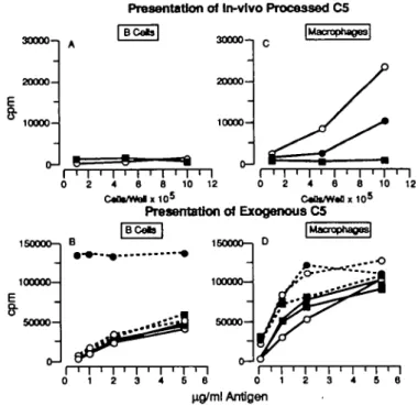
Presentation of In-vlvo Processed C5 I BCetol 30000— 10000-30000" 20000- 10000-|Macrophage3| I T 0 2 I > I ' I ' I ' I ' I • I 0 2 4 6 8 10 12 0 2 4 8 8 10 12 c « a * / w a t x i o5 c«at/w*a x 105 Presentation of Exogenous C5 1S0000-1 100000—
I
50000-B 160000-1 100000— 50000-iMacrophages] . . - • o I ' I 4 5 ng/ml AntigenFig- 1- B cells and macrophages from (O) BALB/c (C5+), ( • ) SP6
(C5+), and ( • ) DBA/2 (C5-) mice (five mice per group) were prepared from spleens as described in Methods. Serial dilutions of cells were tested for presentation of in vivo processed C5 (A and C) to the C5-spedfic T cell hybrid C8-15. A constant concentration of 2x10⁵/well B cells (B) and
2 x 1 ty/well macrophages (D) were assayed for presentation of different concentrations of C5 antigen added in vitro either soluble (solid curves) or haptenated with DNP (stipled curves in B) or immobilized in the presence of anti-C5 coupled Latex beads (stipled curves in D). The figure shows [3H]thymidine uptake by IL-2 dependent CTLL cells (mean c.p.m. of
coupled with the C5-specific antibody BB5.1 as described in detail (13) and C5 was incubated with the coupled beads for 30 min before addition of APC. DNP-C5 was a gift from Dr David Gray.
Presentation of in-vivo Processed C5 B Cells, Macrophages and Dendritic Cells
150000-, 1000005 0 0 0 0 -0 -1 o-o I 0.01 rrwy— 01 10 100
Fig. 2. B cells ( • ) , macrophages ( • ) , and dendritic cells (A) from C5+
CBA spteen (filled symbols) or C5~ A/J spleen (open symbols) were cultured in serial titrations with the C5 specific T cell hybrid A18. The figure shows pHJthymidine uptake by IL-2 dependent CTLL cells (mean c.p.m. of triplicate cultures).
Results
Comparison of C5 presentation (processed in vivo or processed in vitro) by B cells and macrophages
As shown previously whole spleen cell suspensions from C5 expressing (C5+) mice can stimulate C5 specific T cell clones and hybrids indicating the presence of in vivo processed C5 - MHC class II peptjde complexes on the surface of APC (3). To investigate which MHC class II positive APC in spleen presents C5 we fractionated spleen cells into B cells and macrophages.
Donor animals for isolation of these APC populations were C5+ BALB/c mice, SP6 mice which are transgenic for a TNP hapten specific Ig receptor on a BALB/c background and C5 deficient (C5") DBA/2 mice. As responder we used the T cell hybrid C8-15, specific for an as yet undefined epitope on the C5 a chain in the context of I-Ed.
As shown in Rg.1(A), purified B cells did not activate C8-15 whether they were derived from C5+ BALB/c and SP6 or from C5- DBA/2.
B cells from all three strains could present C5 added in vitro. The presentation efficiency was low and reminiscent of the very low C5 presentation capacity observed with B lymphoma cells as described previously (13). In that case it became obvious that uptake via non-specific endocytosis was inadequate in B cells to allow subsequent presentation.
However, extensive data in the literature emphasize the high
Turnover of the Stimulatory Peptlde/Class II Complex In vitro
9
50-i 40- 30- 20- 10-t
20- 10-o-l DayO 2.S 5 10 20oli a s i 2
Day1 90- 80- 70- 60- 50- 40- 30- 20- 1 0-2.5 5 10 20o5 0 5 i 2
0.5 50- 40- 30-20 10-0 50 40 30 20 10 0 80 80 70 60 50 40 30 20 10 0 Dsy2 2.5 10 20 0i5 0.5 1 2 X10"4 Cells/WellFig. 3. CBA (C5+) B cells and macrophages were cultured in serial titrations with T cell hybrid A18 on the day of isolation (day 0) or after incubation
for 1 and 2 days (day 1 and 2 B cells were cuftured m the presence of 10 /tg/ml Hpopolysccharide). CBA dendritic cells were added to A18 after overnight incubation for separation from macrophages (day 1) and cultured for one more day before addition of T cells (day 2). All B cells and macrophages from day 0 were isolated from mechanically teased spleen (two CBA mice), whereas dendritic cells and macrophages day 1 and 2 were isolated from enzyme digested spteen (10 CBA mice). The figure shows [3H]thymidine uptake of IL-2 dependent CTLL (mean c.p.m. of triplicate cultures).
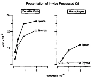
Presentation of in-vtvo Processed C5 30-1 2 0 1 0 -Thymus I Macrophapet | Spleen Thymus ceUs/welx10~
Fig. 4. Dendritic cells and macrophages were prepared from enzyme digested thymus of CBA (20 mice) as described in Methods and cultured in serial dilutions with T cell hybrid A18. The figure shows [3H]thymidlne
uptake by IL-2 dependent CTLL (mean c.p.m. of triplicate cultures). Mean c.p.m. after culture of 2 x lO/Vwell thymic macrophages in the presence of exogenous C5 (5 /ig/ml together with anti-C5 Latex) was 27,378.
antigen irttemafizatjon potential of antigen specific B cells (14). Using B cells from SP6 transgenic mice we took advantage of their TNP-specific Ig receptor (7) and haptenated C5 to allow Ig receptor mediated uptake. As shown in Fig. 1(B), non-transgenic BALBVc and DBA/2 B cells present C5 and D N P - C 5 with similar low efficiency (the SP6 receptor binds TNP and DNP with equal affinity). In contrast transgenic SP6 B cells are highly potent APC for haptenated C5. Again, these data show that while non-specific endocytosts is very inefficient in B cells, the same cells present antigen very well if they can internalize i via a specific Ig receptor. In C5 expressing, tolerant mice B cells with C5 specific receptors are either absent or of low avidity (15) so that this APC population is unlikely to play a major role in presentation of self C5.
figure 1(C and D) shows presentation by macrophages of in vivo processed C5 and in vitro processed C5 respectively. BALB/c and SP6 macrophages can both present in vivo processed C5 although the degree of activation obtained with SP6 macrophages is generally lower. This is not due to lower levels of circulating C5 since both strains have comparable levels of hemolytic activity in serum (data not shown). Macrophages of the C5~ DBA/2 strain as control population do not activate C8-15. Again all three macrophage populations can present C5 added in vitro and they do so with higher efficiency than B cells. In agreement with previous findings using bone marrow macrophages or macrophage ines presentation is enhanced if C5 is imnnobifized by incubation with anti-C5 antibody coupled Latex beads which aids internalization via phago-cytosis (13).
Presentation of in vivo processed C5 by dendritic cells
The third MHC class II expressing cell type in spleen are dendritic cells. Figure 2 shows their capacity to present in vivo processed C5 as assessed by the number of cells needed to activate T cells in comparison with B cete and macrophages. In these experiments we have used APC from the H-2* C5+ CBA strain or the C 5 " AAJ
Table 1 . Presentation of in vivo processed C5 by Langerhans cells
Cells/wells 2 x 1 0 * 1x10* 5X103 2.5X10³ 1.25X10³ APC source day 4 Langerhans C5+ 101,248 99,747 59,254 31,972 10,903 C5" 3959 3476 Day 7 C5+ 53,787 48,515 46,332 36,343 19,285 Langerhans C5" 2281 2372
Langerhans ceDs migrating out of ear skin from either C5+ C8A or C5~
AAJ (10 mice each) were cultured for 4 or 7 days in complete medium. Serial dilutions of cells were cultured with the T cell hybrid A18. The results are expressed as [3H]thymidine uptake by IL-2 dependent CTLL (mean
c.p.m. of triplicate cultures).
Table 2. C5 presentation by dendritic cells from bone marrow
cultures APC/well 4x10* 2x10* 1x10* 5X103 2x10* 2x10* 2x10* 2x10* C5 (ng/ml) 2 0.2 0.02 T cell hybrid [C5+ A18 (CBA) 3797 3522 3957 2375 4133 88,022 77,152 60,982 APC donor] C8-15 (BALB/c) 2141 2739 2548 2292 2276 81,424 54,217 25,542 Dendritic cells generated from bone marrow of C5+ CBA or C5+
BALB/c mice were cultured with T cell hybrid A18 or C8-15 respectively. Serial dilutions of cells were tested for presentation of endogenous C5 whereas a constant number of 2 x 1 0 * cells/well were tested for presentation of added C5 in 10-fold diution steps. The results are expressed as [3H]thymidine uptake by IL-2 dependent CTLL (mean c.p.m. of
triplicate cultures).
strain and the I-E? restricted T cell hybrid A18 as responder. (It should be pointed out that although A18 responds to lower concentrations of exogenous antigen than C8-15, all C5-specific T cell hybrids show the same hierarchy of activation by B celts, dendritic celts, and macrophages.)
Dendritic cells proved to be by far the most efficient APC since similar levels of T cell activation could only be achieved with 10-fold more macrophages and at least 100-fold more B cells. The fact that B cell presentation was observed in these experiments in contrast to those shown in Fig. 1 is due to the greater sensitivity of hybrid A18 compared with hybrid C8-15. In all cases APC from C5~ A/J mice served as the negative control showing that there is no C5 available in these mice to be presented to A18.
Dendritic cells isolated from various tissues (spleen, thymus, skin) of C5+ mice present in vivo processed C5 with similar efficiency.
Table 1 gives an example of C5 presentation by migratory skin Langerhans cells. In this experiment ear tissue of C5+ CBA or
C5~ A/J mice was placed in culture for either 4 or 7 days and released dendritic cells were tested for presentation of in vivo
processed C5. At both time points Langerhans cells from A/J and CBA showed the same high levels of surface MHC class II expression (not shown). Only CBA but not A/J Langerhans cells activated A18 implying that they have been in contact with circulating C5 in vivo. The levels of T cell activation were the same with CBA Langerhans cells cultured for 4 or 7 days.
It is important to point out that dendritic cells developing from C5+ CBA bone marrow precursors by culture with GM-CSF were not able to directly activate A18, but were very potent APC for exogenous C5 (Table 2). This rules out the possibility that dendritic cells themselves synthesize and secrete C5 in vitro.
Turnover of functional C5 peptide-MHC class II complexes on APC
In order to determine for how long stimulatory C5 peptjde- MHC class II complexes are expressed on the surface of APC, we cuttured B cells, macrophages, or dendritic cells for different lengths of time before adding A18 T cells. The purification of dendritic cells involves an overnight culture step so that there is no day 0 time point for this population. The day 0 group of macrophages was derived from non-enzyme treated spleen to avoid contamination with dendritic cells. The day 1 and day 2 macrophage groups consisted of firmly adherent cells after removal of dendritic cells which become non-adherent after an overnight incubation.
B cells were isolated from non-enzyme digested spleen as in the experiments shown in Figs 1 and 2, and to guarantee their sur-vival in culture were incubated in the presence of 10 /ig/ml lipopotysaccharide.
Figure 3 shows that B cells which activated A18 if added on the day of isolation rapidly lost their stimulation potential after overnight culture and were negative on day 2 of culture. They retained their capacity to present C5 added in vitro (not shown).
Macrophages retained stimulatory complexes slightly longer than B ceUs but were also negative on day 2. By that time their expression of surface MHC class II was much reduced and they showed a corresponding reduction in presentation of exogenous C5 (not shown). In contrast dendritic cells were still highly active in presenting in vivo processed C5 after 2 days of culture.
Although these data do not reflect the physiological situation h vivo where antigen is constitutively present, they serve as an indication that dendritic cells remain stirrniatory f a extended periods of time thus increasing the likelihood of T cell recognition. C5 presentation by APC from thymus
Effident presentation is necessary for tolerance induction in the thymus. To address the point of C5 presentation for tolerance induction, we tested the capacity of thymic dendritic cells and macrophages to present in vivo processed C5. As shown in Fig. 4 dendritic cells from thymus were able to activate hybrid A18 to the same degree as dendritic cells isolated from spleen. Unlike spleen macrophages, thymic macrophages did not activate A18, although they were able to present exogenously added C5 immobilized on Latex beads coupled with C5 specific antibody.
Discussion
The aim of this study was to define the relative efficiency of MHC class II bearing APC to present in vivo processed self antigen. Constitutive processing and presentation of self antigen can be detected by the ability of APC taken directly out of mice to activate
specific T cells in vitro and this phenomenon has been demonstrated for the self antigens hemoglobin (2) and C5 (3). In contrast to hemoglobin, which is sequestered in red blood cells, C5 is a representative of freely circulating self antigens. Although the global effect of in vivo processed self antigen by APC from different tissue origins has been reported previously (2,3,16), no attempt has been made to compare relative efficiencies of APC subpopulatjons for self antigen presentation. We show in this study that there is a pronounced hierarchy of C5 presentation with dendritic cells being the most potent APC, whether they are isolated from spleen, thymus, a skin, followed by macrophages and lastly B cells. The accessability of drculating C5 to peripheral rymphoid organs, the thymus, and skin is reflected in the ability of isolated APC subpopulations to present in vivo processed C5. Given the fact that constitutive processing and presentation of self antigen is a prerequisite for induction of tolerance, our findings suggest that dendritic cells, as the most efficient APC for presentation of in vivo processed C5, will play a crucial role for tolerance induction to C5. What are the variables that influence antigen presentation to MHC class II restricted cells and determine which APC might be best suited for the task? The obvious candidates are (i) antigen concentration, (ii) levels of MHC class II expression on APC, (iii) their efficiency of internalization and processing, and (iv) the nature of the antigen.
Firstly, the concentration of C5 in plasma is ~50^g/ml (10~7 M) with higher levels found in males and with increasing age (17). This concentration is sufficient to induce complete T cell tolerance. In fact very much lower doses of self antigen in the drculation as in the case of the liver F protein (10~9 M) (18) or hen egg lysozyme (10 ng/ml, - 1 0 ~9 M) (19) were shown to induce T cell tolerance .whereas incomplete tolerance is induced to thyroglobulin which is present in the drculation at a concentration of 10~10 M (20).
The second crucial factor determining antigen recognition, MHC dass II expression, is highly variable between subpopulations of APC in vivo.
B cells, with the exception of plasma cells and pre-B cells (21), express high levels of MHC class II. The level of expression on macrophages varies depending on their state of maturity, the tissue they are isolated from, and the immune status of the mice (22). Spleen macrophages in general are MHC class II positive whereas thymic macrophages express very low levels of MHC class II (23). Dendritic cells express higher levels of MHC dass II than any of these APC throughout their lifetime and in every tissue (24) giving them increased opportunity to display self and foreign peptides for T cell recognition. In addition dendritic cells may be able to retain immunogenic complexes for longer periods of time than macrophages and B cells. The is indicated by the data concerning turnover of the stimulatory complex on the surface of APC in vitro and was found both for migratory Langerhans cells and f a dendritic cells isolated from spleen. Dendritic cells, like macrophages, stop MHC class II synthesis once they are placed into culture (25). In contrast to macrophages, however, dendritic cells retain prolonged presentation capacity indicating that the turnover of residual intracellular class II and surface class II must be very slow. These observations, although difficult to demonstrate as an in vivo event would support the assumption that dendritic cells trap antigen in peripheral sites, process it and then present the immunogenic complexes within lymphdd organs (26).
In addition to the obvious differences in MHC class II expression the capacity to internalize antigen plays a crucial role in self antigen
presentation. It is furthermore likely that the nature of an antigen, whether it is in soluble or particulate form, degraded or compexed with antibody will influence which type of APC can internalize it best The difference in internalization capacity for C5 becomes clear if we compare B cells with macrophages. Although B cells in general express -5-fold more surface MHC class II, their presentation capacity is at least 10-fold lower—as judged by the number of cells needed to activate the C5-specific T cells. This was the case despite the precaution we took to enrich for low density activated rather than resting B cells which have been reported to be inefficient in antigen presentation (27). It is difficult to fully exclude low contamination with highly potent dendritic cells when preparing B cells form low density cell fractions. Therefore the inadequacy of B cells in C5 presentation may be even more pronounced than our data indicate.
Our analysis of presentation of exogenous antigen in vitro support the assumption that nonspecific endocytosis of soluble antigen is a very inefficient process in B cells. In contrast they are very potent in presentation of antigen that they can internalize via their specific Ig receptor as in the case of TNP-Ig specific SP6 B cells which presented DNP-C5 but not unmodified C5 (Fig. 1). B cells with high avidity C5-specific antigen receptors are probably deleted in C5 expressing mice so that circulating C5 cannot be internalized by Ig receptor mediated endocytosis. Macrophages, with the exception of thyme macrophages presented in vivo processed C5 with intermediate efficiency. The presentation results with exogenousV added C5 are in accordance with previously published data on presentation capacity of macrophage cell lines (13). It is clear that C5 immobilized as antigen - antibody complex on Latex beads is presented more efficiently than soluble C5 (Fig. 1D). Since C5+ mice do not have C5-specific antibodies it is unlikely that macrophages taken ex vivo could have internalized immune complexes via Fc receptor mediated endocytosis or phagocytosis. It is difficult, however, to fuDy exclude the possibility that low contamination with dendritic cells sometimes contributed to the observed ex vivo presentation capacity by macrophages.
Although spleen macrophages presented in vivo processed C5, and circulating C5 has access to thymic APC, thymic nnacrophages did not activate C5-specific T cell hybrids in vitro. Thymic macrophages differ considerably in morphology from other tissue macrophages in as much as they appear filled with phagocytosed debris, provoking the speculation that their function is geared towards disposal of negatively selected, dying T cells rather than uptake and processing of soluble self proteins. It should be noted, however, that thymic macrophages were able to present in vivo processed hemoglobin (16). It is conceivable that this difference in presentation is due to the different nature of the two self antigens. Macrophages are actively involved in degradation of senescent red blood cells which can be phagocytosed whereas uptake of the soluble self antigen C5 by non-specific endocytosis is much less efficient The combination of a relative inefficiency in internalization of soluble protein with low levels of MHC class II expression might not generate a sufficient number of MHC class II-C5 peptide complexes to allow recognition by T cells.
Antigen uptake by dendritic cells is stfll a matter of some debate (28-30). Relatively immature dendritic cells from bone marrow cultures or skin possess Fc receptors and can to some extent phagocytose antigen (31,32) whereas mature dendritic cells do not express Fc receptors, fail to present exogenous protein 'n vitro, and shut off MHC class II and invariant chain synthesis (25,33,34). If
the termination of MHC class II synthesis and presentation of exogenous antigen is a stage in a developmental pathway in vivo rather than an in vitro phenomenon, the question arises at what time in their development dendritic cells internalized and processed C5. We did not observe any difference in the C5 presentation capacity of dendritic cells isolated from spleen, thymus, or skin. Antigen uptake could have occurred in non-lymphoid dendritic cells in the skin which subsequently moved to T cell areas of lymphoid organs (reviewed in 35). This assumption receives support by the finding that immunogenic peptide - MHC class II complexes on the surface of dendritic cells appear to be long lived whether they are generated in vitro (34) or in vivo (see Fig. 3) so that retention of a stimulatory complex in the surface of a dendritic cells during transport to other sites in the body is feasible. On the other hand, it is clear that thymic dendritic cells are generated in situ from blood borne precursors (36) and must have internalized and processed C5 in the thymus. Taken together, the extraordinary presentation activity of dendritic cells is most likely due to a combination of high MHC class II levels, slow turnover of membrane class II, and adequate antigen internalization.
In the thymus dendritic cells appear to be the only MHC class II bearing APC population capable of C5 presentation. As far as non-bone marrow derived MHC class II positive cells in the thymus are concerned, we have previously tested thymic nurse cells as representatives of cortical epithelial cells and found that they could activate C5-specific T cefl hybrids to a low extent but did not activate C5-specific T cell clones (B. Kyewski and B. Stockinger, unpublished data). This finding is in agreement with data in the literature stating that T cell clones, but not T cell hybrids require additional co-stimulatory signals not released by certain APC types—like epithelial cells—for activation (37). Since we have used excusrvely T cell hybrids for the experiments in this paper, we have not addressed the question of co-stimulatory signals. It is clear, however, that dendritic cells, which appear as the most potent APC for in vivo processed C5, are fully equipped to release co-stimulatory signals upon presentation in vivo (38).
The possiblity remains that, outside the thymus, C5 presentation by cells which fail to defiver additional co-stimulatory signals provide a failsafe mechanism for tolerance (39). With this regard it has recently been shown that B cells not only fail to activate resting T cells (40), but actually tolerize them (41). C5+ mice have been shown previously to contain C5-specific B cells (42,43), although it has not been possible to determine if their Ig receptors are of suffiaent avidity to allow tolerogenic presentation of self C5 as a failsafe mechanism in the periphery.
While antigen presentation for tolerance induction in class I restricted T cells can—at least theoretically—be performed by any type of cell in the thymus, presentation for tolerance in class II restricted cells e confined to class II expressing APC. Dendritic ceOs in the thymus are found around the cortico-medullary border, but not in the cortex where the majority of CD4+CD8+ immature thymocytes reside (44). Since macrophages which are found in the cortex as well do not appear to present C5 in vivo, the onset of negative selection in class II restricted, C5-specific T cells might be at a later stage when differentiating T cells enter the medullary area. Negative selection in class I restricted cells is often observed in the CD4+CD8+ cortical population (45-47) consistent with the possibility of efficient self antigen presentation by APC in the cortex. The need for internalization by specialized APC and the restrictive tissue distribution of MHC class II molecules puts higher constraints
on tolerance induction in MHC class II restricted cells. These requirements appear to be fully met by dendritic cells which may be the most crucial APC for tolerance induction to soluble self proteins in the circulation.
Acknowledgements
We would like to thank Drs David Gray, Andy Mellor, and Ann Ager for critical reading of the manuscript and Mrs Pamela tze-lyamu for technical assistance. The Basel Institute for Immunology was founded and is supported by F. Hoffman-La Roche Co. Basel, Switzerland.
Abbreviations APC DNP ER GM-CSF TNP
antigen presenting cell dinitrophenol
endoplasmic retculum
granulocyte macrophage colony stimulating factor trinitrophend
References
1 Winchester, G., Sunshine, G. H., Nardi, N., and Mitchison, N. A. 1984. Antigen-presenting cells do not discriminate between self and nonsetf.
Immunogenetics 19:487.
2 Lorenz, R. G. and Allen, P. M. 1988. Direct evidence for functional self protein/la-molecule complexes in vivo. Proc. NatiAcad. So'. USA 85:5220.
3 Lin, R. H. and Stockinger, B 1989. T cell immunity or tolerance as a consequence of self antigen presentation. Eur. J. Immunol. 19:105. 4 Bevan, M. J. 1987. Class discrimination in the world of Immunology.
Nature 325192.
5 Townsend, A and Bodmer, H 1989 Antigen recognition by class I restricted T lymphocytes Annu. Rev. Immunol 7601.
6 Brodsky, F. M. and Guagliardi, L. E. 1991. The cell biology of antigen processing and presentation. Annu. Rev. Immunol. 9:707. 7 Rusconi, S. and Kohler, G. 1985. Transmission and expression of a
specific pair of rearranged immunoglobulin p and k genes in a transgenc mouse line. Nature 314:330.
8 Metlay, J. P., Witmer-Pack, M. D., Agger, R., Crowtey, M. T., Lawless, D., and Steinman, R. M. 1990. The distinct leucocyte mtegrins of mouse spleen dendritic cells as identified with new hamster monoclonal antibodies. J. Exp. Med. 171-1753.
9 Larsen, C. P., Steinman, R. M., WitmerPack, M., Hankins, D. F., Morris, P. J., and Austyn, J. M. 1990. Migration and maturation of Langerhans cells in skin transplants and explants. J. Exp. Med. 172:1483. 10 Schleicher, Ch , Mehlig, M., Zecher, R., and Reske, K. 1992. Dendritic
cells from mouse bone marrow: in vitro differentiation using low doses of recombinant granulocyte-macrophage colony-stimulating factor. J.
Immunol. Methods 154:253.
11 Inaba, K., Inaba, M., Romani, N., Aya, H., Deguchi, M., Ikehara, S., Muramatsu, S., and Stenman, R. M. 1992. Generation of large numbers of dendritic celts from mouse bone marrow cultures supplemented with granulocyte/macrophage colony-stimulating factor. J. Exp. Med 1761693.
12 LJn, R. H. and Stockinger, B. 1988. Purification of the fifth component of murine complement from asctes fluid. J. Immunol. Methods 115:127. 13 Stockinger, B. 1992. Capacity of antigen uptake by B cells, fibroblasts
or macrophages determines efficiency of presentation of a soluble self antigen (C5) to T lymphocytes. Eur. J. Immunol. 22:1271. 14 Lanzavecchia, A 1990. Receptor-mediated antigen uptake and its effect
on antigen presentation to class ll-restricted T lymphocytes. Annu. Rev.
Immunol. 8:773.
15 Goodnow, C. C. and Basten, A. 1989. Setf-toterance in B lymphocytes.
Semin. Immunol. 1:125.
16 Lorenz, R. and Allen, P. M. 1989. Thymic cortical epithelial cells can present self-antigens in vivo. Nature 337. 560.
17 Cinader, D., Dubiski, M. D., and Wardlaw, A C. 1964. Distribution, inheritance, and properties of an antigen, MuB1 and its relation to hemolytic complement. J. Exp. Med. 120:897.
18 Lukic, M L and Mitehison, N. A 1984. Self-and ale-specific suppressor T cells evoked by intravenous injection of F protein. Eur. J. Immunol. 14.766.
19 Goodnow, C C, Crosbie, J., Adelstein, S., Lavote, T. B., Smith-Gill, S. J., Brink, R A., Pritchard-Bnscoe, H., Wotherspoon, J. S., Loblay, R. H., Raphael, K., Trent, A. J., and Basten, A. 1988. Altered immuno-globulin expression and functional silencing of self-reactive B lymphocytes in transgenc mice. Nature 334:676.
20 Romball, C. G. and Weigle, W. O. 1984. T cell competence in heterdogous and homologous thyroglobulins during the induction of experimental autoimmune thyroiditis. Eur. J. Immunol. 14:887. 21 McKearn, J. P., Baum, C, and Davie, J. M. 1984. Cell surface antigens
expressed by subsets of pre-B cells and B cete. J. Immunol. 132:332. 22 Gordon, S., Fraser, I., Nath, D., Hughes, D., and Clarke, S. 1992.
Macrophages in tissues and in vitro. Curr. Opin. Immunol. 4:25. 23 Kyewski, B. A., Rouse, R. V., and Kaplan, H. S. 1982. Thymocyte
rosettes: multiceDular complexes of lymphocytes and bone marrow derived stromal cells in the mouse thymus. Proc. Natl Acad. Set. USA 79:5646.
24 Crowtey, M., Inaba, K., Witmer-Pack, M., and Steinman, R. M. 1989. The cell surface of mouse dendritic cells: FACS analyses of dendritic cells from different tissues induding thymus. Cell Immunol. 118108. 25 Kampgen, E., Koch, N., Koch, F., Stoger, P., Heufler, C , Schuler, G., and Romani, N. 1991. Ctass II major histocompatibility complex mdecules of murine dendritic cells: synthesis, sialytetkxi of invariant chain, and antigen processing capacity are down-regulated upon culture. Proc. Natl Acad Set. USA 88:3014.
26 Macatonia, S. E., Knight, S. C, Edwards, A. J., Griffiths, S., and Fryer, P. 1987. Localization of antigen on lymph node dendritic cells after exposure to the contact sensitizer fluorescein isothkxyanate. Functional and morphological studies. J. Exp. Med. 166:1654.
27 Krieger, J. I., Grammer, S. F., Grey, H. M., and Chesnut, R. W. 1985. Antigen presentation by splenic B cells: resting B cells are ineffective whereas activated B cells are effective accessory cells for T cell responses. J. Immunol. 135:2937.
28 Levine, T. P. and Chain, B. M. 1992. Endocytosis by antigen presenting cells: dendritic cells are as endocytteafly active as other antigen presenting cells. Proc. Natl Acad. Set. USA 89:8342.
29 Stossel, H., Koch, F., Kampgen, E., Stoger, P., Lenz, A., Heufler, C, Romani, N., and Schuler, G. 1990. Disappearance of certain acidic organelles (endosomes and Langerhans cell granules) accompanies loss of antigen processing capacity upon culture of epidermal Langerhans cells. J. Exp. Med. 172:1471.
30 De Bruijn, M. L H., Nieland, J. D., Harding, C , and Mefef, C. J. M. 1992. Processing and presentation of intact hen egg-white lysozyme by dendritic cells. Eur. J. Immunol. 22:2347.
31 Katz, D. R. and Sunshine, G. H. 1986. Comparative accessory cell function of Langerhans cells isdated from mouse skin. J. Exp. Pathd. 67:157.
32 Reis E Sousa, C. 1992. Phagocytosis of antigens by Langerhans cells. PhD thesis, University of Oxford.
33 Gnotomoni, G., Simon, J. C, Bergstresser, P. R., and Cruz, P. J. 1990. Freshly isolated spleen dendritic cells and epidermal Langerhans cells undergo similar phenotypic and functional changes during short-term culture. J. Immunol. 145:2820.
34 Pure, E., Inaba, K., Crowtey, M. T., Tardefi, L, Witmer, P. M., Ruberti, G., Fathman, G , and Steinman, R. M. 1990. Antigen processing by epidermal Langerhans cells correlates with the level of biosynthesis of major histocompalibility complex class II molecules and expression of invariant chain. J. Exp. Med. 172:1459.
35 Mettey, P. J., Pure, E., and Steinman, R. M. 1989. Control of the immune response at the level of antigen-presenting cells: a comparison of the function of dendritic cells and B lymphocytes. Adv. Immunol. 47:45. 36 Ardavin, C, Wu, L, Li, C-L, and Shortman, K. 1993. Thyme dendritic cells and T ceOs develop simultaneously in the thymus from a common precursor population. Nature 362:761.
37 Lorenz, R. G. and Alien, P. M 1989. Thymic epthelial cells lack full capacity for antigen presentation. Nature 340557.
38 Inaba, K., Metlay, J. P., Crowtey, M. T., and Steinman, R. M. 1990. Dendritic cells pulsed with protein antigens in vitro can prime antigen-specific, MHC-restricted T cells in situ. J. Exp. Med. 172:631. 39 Hagerty, D. R., Evavotd, B. D., and Allen, P. M. 1991. The processing
limited by antigen availability and costirmiator expression. J. Immunol. 147:3282.
40 Ronchese, F. and Hausmann, B. 1993. B lymphocytes in vivo fail to prime naive T cells but can stimulate antigen experienced T lymphocytes. J. Exp. Med. 177:679.
41 Fuchs, E. J. and Matzinger, P. 1992. B cells turn off virgin but not memory T cells. Science 258:1156.
42 Harris, D. E., Cairns, L, Flosen, F., and Borel, Y. 1982. A natural model for immunotogic tolerance. Tolerance to murine C5 is mediated by T cells, and antigen is required to maintain unresponsiveness. J. Exp.
Med. 156:567.
43 Stockinger, B. and Hausmann, B. 1988. Induction of an Immune
response to a setf antigen. Eur. J. Immunol. 18:249.
44 Barclay, A. N. and Mayrhofer, G. 1981. Bone •marrow origin of la-positive cells in the medulla of rat thymus. J. Exp. Med. 153:1666. 45 Kisielow, P., Bluthmann, H., Staerz, U. D., Steinmetz, M., and Boehmer, H. v. 1988. Tolerance in T-ceD-receptor transgenic mice involves deletion of nonmature CD4+8+ thymocytes. Nature 333:742.
46 Sha, W. C , Nelson, C. A., Newberry, R. D., Kranz, D. M., Russell, J. K.andLoh, D. Y. 1988. Positive and negative selection of an antigen receptor on T ceOs in transgenic mice. Nature 336:73.
47 Pircher, H., BGrki, K., Lang, R., Hengartner, H., and Zinkernagel, R. M. 1989. Tolerance induction in double specific T-ceS receptor transgenic mice varies with antigen. Nature 342:559.