Abstract The Fas receptor is involved in the regulation
of apoptosis but also can function as a non-apoptotic
sig-nal transducer. This study was mainly designed to
quanti-tate Fas proteins in rat brain during heroin addiction and
opiate withdrawal. In rat, mouse and human brains, and in
SH-SY5Y cells, similar forms of Fas were
immunode-tected with different antibodies (i.e., 35 kDa native Fas
and 48- and 51-kDa glycosylated Fas). Acute (2 h)
treat-ments with the
µ
-opioid receptor agonists heroin (10 mg/kg)
and morphine (30 mg/kg) increased the immunodensity of
native Fas (124% and 36%) but not that of glycosylated
Fas in the cerebral cortex. Chronic (5 days) heroin (5–
30 mg/kg) and morphine (10–100 mg/kg) were also
asso-ciated with increased native Fas (76% and 45%) and with
different expressions of glycosylated Fas. In
heroin-depen-dent rats, opiate withdrawal (48 h) resulted in a sustained
increase in native Fas (107%) and in up-regulation of
51 kDa glycosylated Fas (51%). Acute treatments with
selective
δ
-receptor (SNC-80, 10 mg/kg) or
κ
-receptor
(U 50488-H, 10 mg/kg) agonists did not alter the content
of native or glycosylated Fas. Chronic pentazocine (10–
80 mg/kg, 5 days), a mixed opiate drug and
σ1
receptor
ag-onist, decreased native (48%) and glycosylated (38–82%)
Fas proteins. Similarly, the selective
σ1
agonist (+)-SKF
10047 also decreased native Fas (37%) and the effect was
blocked by the
σ1
antagonist BD 1063. Brain dynamin
was up-regulated by acute and/or chronic heroin (30–39%),
morphine (47–85%), pentazocine (51%) and heroin
with-drawal (74%). The main results indicate that chronic heroin/
morphine treatment and heroin withdrawal are associated
with up-regulation of 35 kDa native Fas (and with
differ-ent expressions of glycosylated Fas), and also with
con-comitant increases of dynamin in rat brain.
Keywords Fas receptor proteins · Dynamin · Opiate
drugs ·
σ
Ligands · Naloxone · Opiate addiction · Opiate
withdrawal · Rat brain
Introduction
The Fas/Fas-L and other signaling systems are well-known
cellular pathways associated with the physiological
regu-lation of programmed cell death (apoptosis; MacEwan
2002). The Fas receptor (also known as CD95 or APO-1)
is a prototype member of the tumor necrosis factor/nerve
growth factor superfamily of death receptors (Itoh et al.
1991; Watanabe-Fukunaga et al. 1992; Nagata and
Gol-stein 1995; Nagata 1999; Orlinick et al. 1999), which is
widely expressed in normal tissues (Watanabe-Fukunaga
et al. 1992) including the brain (Bechmann et al. 1999;
Choi et al. 1999; Boronat et al. 2001). The Fas receptor
has an extracellular domain with two putative N-linked
glycosylation sites and a cysteine-rich region for ligand
binding (Fas-L), and an intracellular death domain near de
C-terminal region of the molecule (Watanabe-Fukunaga et
al. 1992; Orlinick et al. 1999). Native Fas receptor has a
relative molecular mass
≈
35 kDa, but after
post-transla-tional modifications mature Fas is mostly expressed as
glycosylated proteins of
≈
45–52 kDa (Itoh et al. 1991;
Oehm et al. 1992; Watanabe-Fukunaga et al. 1992;
Kami-tami et al. 1997).
Upon receptor activation by Fas-L, the intracellular death
domain of Fas rapidly recruits the adaptor molecule
FADD (Fas-associated death domain protein), which is
followed by activation of various caspase cascades
(cas-pase-8 proenzyme) and specific effector caspases, such as
caspase-3, leading to apoptosis (Nagata 1999; Krammer
2000; MacEwan 2002). The induction of apoptosis in
neu-M. Julia García-Fuster · Marcel Ferrer-Alcón
·
Antonio Miralles · Jesús A. García-Sevilla
Modulation of Fas receptor proteins and dynamin during opiate addiction
and induction of opiate withdrawal in rat brain
DOI 10.1007/s00210-003-0801-9
Received: 25 April 2003 / Accepted: 19 August 2003 / Published online: 3 October 2003
O R I G I N A L A RT I C L E
M. J. García-Fuster · A. Miralles · J. A. García-Sevilla (✉) Laboratory of Neuropharmacology,
Associate Unit of the Institute of Neurobiology “Ramón y Cajal” (CSIC), Department of Biology, University of the Balearic Islands,
Cra. Valldemossa Km 7.5, 07122 Palma de Mallorca, Spain Fax: +41-22-3055799,
e-mail: [email protected] M. Ferrer-Alcón · J. A. García-Sevilla
Clinical Research Unit, Department of Psychiatry, University of Geneva,
HUG Belle-Idée, 2 Chemin du Petit-Bel-Air, 1225 Chêne-Bourg/GE, Switzerland
rons has been demonstrated to share the same basic
mech-anisms with all other cell types (Sastry and Rao 2000; Yuan
and Yankner 2000). Besides the primary role of Fas/Fas-L
in apoptosis, increasing evidence also indicates the
impor-tance of this system as a non-apoptotic signal transducer
(Wajant 2002). Among these non-apoptotic functions, the
Fas receptor has been shown to transduce proliferative
signals in normal human diploid fibroblasts and T cells
(Siegel et al. 2000), to mediate cardiomyocyte
hypertro-phy in vitro and in vivo (Badorff et al. 2002), to stimulate
axonal growth in primary neurons (Desbarats et al. 2003),
and Fas-L to facilitate antigen acquisition by dendritic
cells in melanomas (Tada et al. 2002). In contrast to
Fas-mediated apoptosis events, the apoptosis-unrelated
bio-logical effects of Fas are still poorly understood.
In a recent study (Boronat et al. 2001), chronic
treat-ment of rats with morphine was shown to be associated,
through a naloxone-sensitive mechanism, with an
oppo-site modulation of two key proteins involved in the
regu-lation of the programmed cell death in the brain:
up-regu-lation of pro-apoptotic Fas receptor and down-reguup-regu-lation
of anti-apoptotic Bcl-2 oncoprotein. Similarly, morphine
also was shown to increase the expression of Fas receptor
mRNA in heart, lung, spleen and lymphocytes of mice as
well as in human lymphocytes (Yin et al. 1999). Moreover,
Fas mRNA expression was found enhanced in splenic
lymphocytes of stressed mice (i.e., with abnormal high
contents of endogenous opioids), an effect that was
antag-onized by naloxone, indicating that Fas-mediated
lympho-cyte apoptosis is dependent on endogenous opioids (Yin
et al. 2000). Recently, Fas overexpression and
Fas-medi-ated lymphocyte apoptosis in stressed mice was found
abolished in
µ
-opioid receptor deficient mice (Wang et al.
2002). Together these findings indicated that morphine,
through the activation of
µ
-opioid receptors, could
pro-mote abnormal programmed cell death in specific types of
neurons (but see Raoul et al. 2002) and other cells.
Alter-natively, the modulation of Fas receptor by morphine in
the brain could be related to the recent discovery that this
system (Fas/Fas-L) can also function as a non-apoptotic
signal transducer in various cells including neurons
(Wa-jant 2002; Desbarats et al. 2003).
Because of the potential importance of this topic, the
present study was mainly designed to assess the
modula-tion of Fas-related proteins (native and glycosylated Fas)
during heroin addiction (acute and chronic effects) and the
induction of opiate withdrawal in rat brain. For
compari-son, the acute effects of other opiate and non-opiate drugs
were also quantitated. Moreover, the effects of some
opi-ate drugs (heroin, morphine, pentazocine) on dynamin, a
recently discovered target of chronic morphine action
(Noble et al. 2000) that plays an essential role in receptor
endocytosis, were also investigated. A preliminary report
of a portion of this work was given at the XXIV congress
of the Spanish Society of Pharmacology (García-Fuster et
al. 2002).
Materials and methods
Animals and treatments. Adult male Sprague-Dawley rats (250– 300 g) were used. The rats were housed under controlled environ-mental conditions (22°C, 70% humidity, and 12-h light/dark cycle) with free access to a standard diet and tap water. For the acute drug treatments, the rats received a single intraperitoneal (i.p.) injection of heroin (10 mg/kg), morphine (30 mg/kg), SNC-80 (10 mg/kg, a selective δ-opioid receptor agonist), U 50488H (10 mg/kg, a selec-tive κ-opioid receptor agonist), pentazocine (15 mg/kg, an opiate drug and also a potent σ1receptor agonist; see Mei and Pasternak
2001, 2002), (+)-SKF 10047 (5 mg/kg, a selective σ1receptor
ago-nist; see Matsuno et al. 1995) and BD 1063 (10 mg/kg, a selective
σ1receptor antagonist; see McCracken et al. 1999). For the chronic
treatment with heroin, the rats were injected i.p. three times daily (at 08:00, 14:00 and 20:00 h) during 5 consecutive days with in-creasing doses of the opiate as follows: day 1: 5, 10, and 10 mg/kg; day 2: 10, 15, and 15 mg/kg; day 3: 15, 20, and 20 mg/kg; day 4: 20, 25, and 25 mg/kg; day 5: 25 and 30 mg/kg (Ventayol et al. 1997). After this chronic heroin treatment, naloxone (2 mg/kg i.p., 2 h)-pre-cipitated withdrawal or spontaneous (48 h) opiate withdrawal was induced which resulted in various withdrawal reactions (data not shown; Gabilondo and García-Sevilla 1995). In another series of chronic experiments, the animals were similarly injected with morphine (10 to 100 mg/kg) or pentazocine (10 to 80 mg/kg) dur-ing 5 days (Ventayol et al. 1997). Other rats were chronically treated with (+)-SKF 10047 (3–10 mg/kg for 3 days). In all series of ex-periments, control rats received 0.9% saline vehicle or DMSO (in the case of SNC-80) i.p. (1 ml/kg) at the indicated treatment times. The animals were killed by decapitation 2 h after the last dose in the acute and chronic drug treatments or at the times indicated af-ter heroin withdrawal. The brains were rapidly removed and spec-imens of the cerebral cortex were dissected on ice and stored at –80°C until assay. This study was approved by the research and ethical review board of the Dirección General de Investigación (MCT, Madrid), and the experiments in rats followed the “Princi-ples of laboratory animal care” (NIH publication No. 85–23, re-vised 1985) and were performed according to the guidelines of the University of the Balearic Islands.
Brain sample preparations and immunoblotting of Fas-related pro-teins and dynamin. The preparation of rat brain samples and the im-munodetection of target proteins were performed as described pre-viously (Ventayol et al. 1997; Boronat et al. 2001) with some mod-ifications. Briefly, 150–200 mg of cerebral cortex was homogenized (1:20, wt/vol) in cold 40 mM Tris HCl buffer, pH 7.5, containing 1% Triton X-100, 1 mM EDTA, 1 mM MgCl2, 5 mM NaCl, and
the protease inhibitors phenylmethylsulfonyl fluoride (1 mM) and leupeptin (40µg/ml). The samples were centrifuged at 40,000 ×g for 45 min, and then 200µl of the resulting supernatant (total Fas and dynamin) was mixed with an equal volume of electrophoresis loading buffer (Ventayol et al. 1997), which was then boiled (de-natured) and stored at –20°C until use. Protein concentrations were determined by the method of Bradford (Bradford 1976). In addition to rat brain, mouse (cerebral cortex) and human (prefrontal cortex) brain samples (Ferrer-Alcón et al. 2003) and human SH-SY5Y neuroblastoma cells (a clone from SK-N-SH cells which expresses high densities of µ- and δ-opioid receptors, proportion 4:1; Yu et al. 1986; López and Ferrer 2000) were also used for the immuno-detection and characterization of Fas-related proteins with differ-ent anti-Fas antibodies (see Fig. 1). For these preliminary experi-ments, total membranes were prepared similarly as above except that the detergent Triton X-100 was not included in the homoge-nization buffer and the samples were not centrifuged (total ho-mogenates). In some experiments, rat cortical membranes were in-cubated in the absence or presence of the enzyme N-glycosidase F (15 units for 3 h at 37°C) (Ozaita et al. 1999) to further ascertain the nature of the various Fas receptor proteins (Fas M-20 anti-body). In routine experiments, 40µg protein of each rat (and mouse or human) brain sample or SH-SY5Y cells (20µg protein) was subjected to SDS-PAGE on 15-well (6×8-cm gels, 1 mm thickness, Bio-Rad Laboratories, Hercules, CA, USA) 10% polyacrylamide
minigels. Proteins were electrophoretically transferred (110 V for 2–3 h) to nitrocellulose membranes (western blotting) that were in-cubated for 1 h in a phosphate-buffered saline containing 5% non-fat dry milk, 0.2% Tween 20 and 0.5% bovine serum albumin
(blocking solution) (Harlow and Lane 1999). Then, the membranes were incubated overnight at 4°C in blocking solution containing the appropriate primary antibody: anti-Fas M-20 (rabbit polyclonal antibody raised against a peptide mapping the carboxyl terminus of Fas of mouse origin; dilution 1:2,000; sc-716, batches D219 and F251, Santa Cruz Biotechnology, CA, USA), anti-Fas C-20 (rabbit polyclonal antibody raised against a peptide mapping the carboxyl terminus of Fas of human origin [amino acids 300–319, cf. Kami-tani et al. 1997]; dilution 1:2,000; sc-715, batch D172 Santa Cruz Biotechnology), anti-Fas FL-335 (rabbit polyclonal antibody raised against a recombinant protein corresponding to the full length Fas of human origin; dilution 1:1,000; sc-7886, batch C010 Santa Cruz Biotechnology) or anti-dynamin (mouse monoclonal antibody raised against a peptide mapping the carboxyl terminus of dynamin I of rat origin; dilution 1:2,000; clone 41, batch 4, Transduction Laboratories, Lexington, KY, USA). The anti-Fas antibodies used do not cross-react with other TNF superfamily transmembrane re-ceptors. In order to test the selectivity of anti-Fas antibodies (M-20 and C-20) with specific proteins, the antigenic peptides (sc-716P and sc-715P, Santa Cruz Biotechnology) were preincubated in ex-cess with the antisera to block the binding of the antibody to the specific protein species tested (Fig. 1). The secondary antibody, horseradish peroxidase-linked anti-rabbit or anti-mouse IgG, was incubated at 1:5,000 dilution in blocking solution at room temper-ature for 2 h. Immunoreactivity of target proteins was detected with the Enhanced Chemiluminiscence (ECL) Western Blot De-tection system (Amersham International, Buckinghamshire, UK) and visualized by exposure to Hyperfilm ECL film (Amersham) for 30 s to 2 min (autoradiograms).
Quantitation of immunodensities of Fas-related proteins and dy-namin. The autoradiograms were quantitated by densitometric scanning (GS-700 Imaging Densitometer, resolution: 42µm, Bio-Rad, Hercules, CA, USA). The amount of target proteins (Fas and dynamin) in the cerebral cortex of rats treated with opiate or other drugs was compared in the same gel with that of control rats which received saline or DMSO solution. Prior to analyses, the linearity of protein concentration for Western blotting was ascertained by resolution of selected concentrations of protein (i.e., total protein loaded versus integrated optical density, IOD, units, consisting of 5 points of different protein content, usually 10 to 80µg, resulting in good linear relations; data not shown, see Boronat et al. 2001). Experiments were performed by using 40µg protein known to be within the linear range for immunolabeling of Fas-related proteins and dynamin. The quantification procedure was assessed 2–4 times in different gels (each gel with different brain samples from saline/ DMSO- and drug-treated rats). Finally, percent changes in immuno-reactivity with respect to control samples (100%) were calculated for each rat treated with the specific drug in the various gels and the mean value used as a final estimate. Since opiate drugs induce alterations in the content of cytoskeletal proteins such as neurofil-aments (García-Sevilla et al. 1997; Boronat et al. 2001; Ferrer-Al-cón et al. 2003), actin (Papakonstanti et al. 1998) and dynamin (Noble et al. 2000), a specific housekeeping cytoskeletal protein was not used as a negative control. Instead, unidentified peptides, not related to Fas, were also quantitated with anti-Fas antibodies (≈65 kDa band for M-20 and ≈70 kDa band for C-20, see Fig. 1) and the various opiate treatments did not alter significantly the im-munodensity of these unknown peptides (e.g., mean ± SEM IOD values for the unidentified 65 kDa band, saline: 14.4±0.6, n=3; acute heroin: 14.1±0.3, n=3; chronic heroin: 14.0±0.3, n=3; chronic heroin plus naloxone: 14.6±0.4, n=3; chronic pentazocine: 12.8±0.6, n=3). Data analyses and statistics. All series of data were analyzed with the program GraphPad Prism, version 3.0. Results are expressed as mean ± SEM values. One-way analysis of variance (ANOVA) fol-lowed by Bonferroni’s multiple comparison test, and Student’s two-tailed t-test were used for the statistical evaluations. The level of significance was chosen as p=0.05.
Drugs and chemicals. Opiate drugs (and their sources) included heroin HCl and morphine HCl (Unión Químico-Farmacéutica S.A.E. (Madrid, Spain), (±) pentazocine HCl (Laboratorios Fides, Barce-Fig. 1 Representative autoradiographs of Western blots depicting
labeling of immunodetectable Fas-related proteins (≈35 kDa native Fas and ≈48 and 51 kDa glycosylated Fas, arrows) with various antibodies (M-20, C-20 and FL-335) in the rat brain (RB, cerebral cortex), mouse brain (MB, cerebral cortex), human brain (HB, pre-frontal cortex) and human SH-SY5Y neuroblastoma cells (SH). The samples (40µg protein for brain tissue and 20µg protein for SH cells) were subjected to SDS-PAGE, transferred to nitrocellu-lose membranes (immunoblotting), incubated with the specific pri-mary and secondary antibodies, and visualized by the Enhanced Chemiluminiscence method. The specificity of the antibodies anti-Fas M-20 and C-20 were assessed by preincubating the corre-sponding antibody with its antigenic peptide (preabsorbed anti-body), which resulted in the blockade of the immunoreaction for the specific proteins (double arrows). The apparent molecular masses of Fas-related proteins were determined by calibrating the blots with prestained molecular weight markers as shown on the left hand side. For a given antibody all samples were run in the same gel
lona, Spain), SNC-80 ((+)-4-[(αR)-α -(2S,5R)-4-allyl-2,5-dimethyl-1-piperazinyl)-3-methoxybenzyl]-N-N,diethylbenzamide) (Tocris Cookson Ltd., Avonmouth, UK), U-50488-H HCl (1S-trans)-3,4- dichloro-N-methyl-N-[2-(1-pyrrolidinyl)cyclohexyl]-benzeneacet-amide) (Sigma/RBI, St Louis, Mo, USA) and naloxone HCl (Endo Laboratories, Garden City, NY, USA). (+)-SKF 10047 HCl and BD 1063 HCl (1-[2-(3,4-dichlorophenyl)ethyl]-4-methylpiperazine) were purchased from Tocris. N-glycosidase F was from Sigma. Acrylamide (Protogel) was from BDH Brunschwig (Dorset, UK). Other materials such as the secondary antibodies, ECL reagents and autoradiography films were purchased from Amersham Inter-national (UK) or Santa Cruz Biotechnology (USA). All other chemicals were from Sigma Chemical.
Results
Immunodetection of Fas-related proteins in brain tissue
and in SH-SY5Y cells
Fas is a widely expressed cell-surface receptor of relative
molecular mass
≈
35 kDa for native Fas and
≈
45–52 kDa
for their glycosylated forms (Itoh et al. 1991;
Watanabe-Fukunaga et al. 1992). The antibodies used (M-20 and
C-20) for the immunodetection of these Fas-related proteins
(antisera directed against the cytoplasmic domain of Fas)
were first tested for their specificity on Western blots of
rat, mouse and human brain membranes as well as in
hu-man SH-SY5Y neuroblastoma cell membranes (no
deter-gent added). In rat brain, the antiserum anti-Fas M-20
la-beled specific proteins of Mr
≈
35 kDa (native Fas) and
≈
48 and 51 kDa (glycosylated Fas) (Fig. 1, top). Thus,
previous preincubation of Fas M-20 antibody with the
antigenic peptide (preabsorbed antibody) resulted in the
blockade of the immunoreaction for the specific proteins
(35-, 48- and 51-kDa), but not for other unknown
unre-lated peptide of higher molecular mass (
≈
65 kDa, Fig. 1,
top). Similar results were obtained in mouse and human
brains (including two prominent peptides of
≈
45 kDa
prob-ably related to glycosylated Fas) and in SH-SY5Y cells
(Fig. 1, top). In rat, mouse and human brain tissues as well
as in SH-SY5Y cells, the antiserum anti-Fas C-20 labeled
a specific protein of
≈
51 kDa (glycosylated Fas) which
was not immunodetected with the preabsorbed antibody
(Fig. 1, middle). In these tissue and cell preparations,
anti-Fas C-20 also immunodetected a specific band of
≈
97 kDa
(not shown) which probably represents a processed form
of Fas aggregates (
≈
110–200 kDa; Kamitani et al. 1997).
In rat, mouse and human brains and in SH-SY5Y cells,
the pattern of Fas-related proteins immunodetected with
the antiserum anti-Fas FL-335 (directed against the full
length protein; antigenic peptide not available) was
simi-lar to that of anti-Fas M-20, although the immunoblots
showed a higher background (Fig. 1, bottom). Therefore,
with the use of various antibodies raised to independent
epitopes of Fas it was possible to identify the same
spe-cific Fas-related proteins in rat brain (i.e.,
≈
35 kDa native
Fas and
≈
48 and 51 kDa glycosylated Fas).
In rat brain membranes extracted with 1% Triton X-100
and probed with anti-Fas M-20, the immunodensity of
glycosylated 48 kDa Fas was greatly increased and that of
51 kDa Fas blunted (Fig. 2A). Protein extraction with this
nonionic detergent also improved the immunodetection of
native 35 kDa Fas (antibody M-20), glycosylated 51 kDa
Fas (antibody C-20) and glycosylated 48- and 51-kDa Fas
(antibody FL-335; see Figs. 1, 3). N-deglycosylation of
Fas receptor induced a marked increase in the expression
of native 35 kDa Fas and related peptides (
≈
43/45 kDa;
Fig. 2B). Therefore, the effects of opiate and other drugs
on Fas-related proteins were assessed in brain membranes
extracted with Triton, and in which native 35 kDa Fas and
glycosylated 48 kDa Fas were quantitated with antibody
M-20, glycosylated 51 kDa Fas with antibody C-20, and
both glycosylated forms of Fas also with antibody FL-335
(Fig. 3).
Fig. 2 A Immunodetection of Fas-related proteins (≈35 kDa na-tive Fas and ≈48 and 51 kDa glycosylated Fas, double arrows) with antibody M-20 in the rat brain (cerebral cortex). The samples (40µg protein, three animals in each group) were prepared in the absence or presence of a nonionic detergent (Triton X-100). Note that protein extraction with Triton markedly improved the immuno-detection of 48 kDa glycosylated Fas and blunted that of 51 kDa glycosylated Fas. B Immunodetection of Fas-related proteins with antibody M-20 in the rat cerebral cortex after receptor deglycosyl-ation. The samples (18 and 36µg protein, prepared with Triton X-100) were incubated in the absence (G–) or presence (G+) of the enzyme N-glycosidase F (15 units for 3 h at 37°C). Note that N-deglycosylation of Fas receptor induced a marked increase in the expression of native 35 kDa Fas and related peptides (≈43/45 kDa). The apparent molecular masses of Fas-related proteins were deter-mined by calibrating the blots with prestained molecular weight markers as shown on the left hand side
Effects of heroin and other opiate drugs
on Fas-related proteins in rat brain
The acute treatment with the
µ
-opioid receptor agonist
heroin (10 mg/kg, i.p. for 2 h), compared with saline
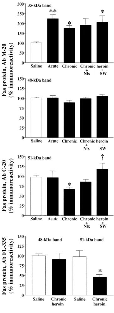
solu-Fig. 3 Representative immunoblots of Fas-related proteins (≈35 kDa native Fas and ≈48 and 51 kDa glycosylated Fas, arrows) detected with different antibodies (M-20, C-20 and FL-335) in the rat cere-bral cortex (40µg protein extracted with Triton) after various heroin and morphine treatments. Groups of treatments (two ani-mals for each group): saline, acute heroin (10 mg/kg, i.p., 2 h), chronic heroin (10–30 mg/kg i.p. for 5 days), chronic heroin plus naloxone (Nlx, 2 mg/kg i.p. for 2 h), chronic heroin plus sponta-neous withdrawal (SW, 48 h) and chronic morphine (10–100 mg/kg for 5 days; only for antibody FL-335). The apparent molecular masses of Fas-related proteins were determined by calibrating the blots with prestained molecular weight markers as shown on the left hand side. For a given antibody all samples were run in the same gelFig. 4 Effects of acute and chronic treatments with heroin and of opiate withdrawal after the chronic treatment on the immunodensi-ties of Fas-related proteins in the rat brain (cerebral cortex). Groups of treatments: saline, acute heroin (10 mg/kg, i.p., 2 h), chronic heroin (10–30 mg/kg i.p. for 5 days), chronic heroin plus naloxone (Nlx, 2 mg/kg i.p. for 2 h), chronic heroin plus spontaneous with-drawal (SW, 48 h). Quantitation of Fas-related proteins: 35 kDa na-tive Fas and 48 kDa glycosylated Fas (antibody M-20); 51 kDa glycosylated Fas (antibody C-20), and 48 kDa and 51 kDa glyco-sylated Fas (antibody FL-335). Columns are means ± SEM of five experiments per group with an animal per experiment, and expressed as percentage of saline-treated rats. One-way ANOVA detected significant differences between groups with respect to 35 kDa na-tive Fas (antibody M-20) [F(4,21)=5.77, p=0.005] and 51 kDa gly-cosylated Fas (antibody C-20) [F(4,21)=3.38, p=0.028]. *p<0.05; **p<0.01 when compared with the corresponding saline group, and †p<0.05 when compared with the chronic heroin group (ANOVA followed by Bonferroni’s test). Chronic heroin treat-ment also reduced significantly the immunodensity of 51 kDa gly-cosylated Fas (antibody FL-335) *p<0.05 when compared with the saline group (Student’s two-tailed t-test)
tion administration, induced a marked increase in the
im-munodensity of 35 kDa native Fas in the rat cerebral
cor-tex (124%, p<0.01) (Figs. 3 top, and 4). In contrast, acute
heroin treatment did not alter significantly the
immuno-densities of 48- and 51-kDa glycosylated Fas in rat brain
(Figs. 3, top and middle, 4; Table 1). Chronic treatment
with heroin (5–30 mg/kg for 5 days) also was associated
with an increase in the immunodensity of 35 kDa native
Fas in rat the cerebral cortex (76%, p<0.05) (Figs. 3 top, 4),
but this effect was less pronounced than that induced by
the acute treatment, which suggested the induction of
tol-erance. Chronic heroin treatment did not modify
signifi-cantly the immunodensity of 48 kDa glycosylated Fas
(Figs. 3, top and bottom, 4; antibodies M-20 and FL-335)
but decreased that of 51 kDa glycosylated Fas (33%,
p<0.05 with antibody C-20; and 54%, p<0.05 with
anti-body FL-335; Figs. 3, middle and bottom, 4; Table 1).
Acute (30 mg/kg, i.p. for 2 h) and chronic treatment
with morphine (10–100 mg/kg for 5 days) also increased
the content of 35 kDa native Fas in the rat cerebral cortex
(36% and 45%, p<0.05, respectively) (Table 1). Moreover,
chronic morphine induced an increase in the
immuno-density of 48 kDa glycosylated Fas (42%, p<0.01, Table 1,
antibody M-20, and 43%, p<0.01, Fig. 3 bottom, antibody
FL-335) and a decrease in that of 51 kDa glycosylated Fas
(45%, p<0.05, Fig. 3 bottom, antibody FL-335).
Acute treatments with the opiate drugs SNC-80 (a
selec-tive
δ
-receptor agonist, 10 mg/kg i.p. for 2 h) or U 50488-H
(a selective
κ
-receptor agonist, 10 mg/kg i.p. for 2 h) did
not modify significantly the immunodensity of native or
glycosylated Fas in the rat cerebral cortex (Table 1), which
indicated that the rapid modulation of Fas proteins in the
brain is related to the activation of
µ
-opioid receptors (heroin
and morphine).
Effects of heroin withdrawal after chronic opiate treatment
on Fas-related proteins in rat brain
In heroin-dependent rats (5–30 mg/kg for 5 days),
nalox-one (2 mg/kg)-precipitated withdrawal (2 h) or spontaneous
opiate withdrawal (48 h) did not modify significantly the
already increased immunodensities of 35 kDa native Fas
(91%, p=0.06, and 107%, p<0.05, when compared to
saline). Opiate withdrawal, like chronic heroin, did not
in-duce significant changes in the content of 48-kDa
glyco-sylated Fas (Figs. 3, top and middle, 4; Table 1). In these
heroin-dependent rats, however, spontaneous opiate
with-drawal (48 h) resulted in up-regulation in the density of
51-kDa glycosylated Fas (51% when compared to chronic
heroin; Figs. 3 middle, 4; Table 1).
Effects of pentazocine and other
σ
ligands
on Fas-related proteins in rat brain
The acute treatment with pentazocine (15 mg/kg, i.p. for 2 h),
a
σ1
receptor agonist and a mixed nonselective
δ
/
κ
-opioid
receptor agonist and
µ
-antagonist, did not modify
signifi-cantly the immunodensity of native or glycosylated Fas in
the rat cerebral cortex (Table 1). In contrast, chronic
treat-ment with pentazocine (10–80 mg/kg for 5 days) resulted in
marked decreases in the immunodensities of 35 kDa
na-tive Fas (48%, p<0.01), 48 kDa glycosylated Fas (38%,
p<0.01 with antibody M-20, and 46%, p<0.01 with antibody
FL-335), and 51 kDa glycosylated Fas (47%, p<0.0001
with antibody C-20, and 82%, p<0.0001 with antibody
FL-335; Figs. 5, 6) in the brain (Table 1).
The acute treatment with the selective
σ1
receptor
ago-nist (+) SKF 10047 (5 mg/kg i.p. 2 h) decreased the
con-Treatment (time) Dose Antibody M-20 Antibody C-20
(mg/kg) 51 kDa n
35 kDa 48 kDa n
Acute heroin (2 h) 10 224±22** 101±6 5 97±16 5
Chronic heroin (5 days) 5–30 176±13* 89±6 5 67±6* 5
+ Naloxone (2 h) 2 191±33 99±3 5 86±6 5
+ SW (48 h) – 207±32* 105±4 5 118±15† 5
Acute morphine (2 h) 30 136±8* 117±5 4 NT
Chronic morphine (5 days) 10–100 145±7** 142±5** 4 NT
Acute SNC-80 (2 h) 10 90±7 97±7 5 97±4 5
Acute U 50488-H (2 h) 10 119±9 102±7 4 95±2 4
Acute pentazocine (2 h) 15 95±10 118±9 3 97±5 3
Chronic pentazocine (5 days) 10–80 52±6** 62±9** 5 53±5*** 5
Acute (+) SKF 10047 (2 h) 5 74±6* 93±6 4 93±9 4
Acute BD 1063 (2.5 h) 10 101±11 99±9 3 102±10 3
Acute BD + SKF 10+5 96±6 95±5 4 89±9 4
Chronic SKF 10047 (3 days) 3–10 61±6** 106±5 5 100±2 5 *p<0.05, **p<0.01, ***p<0.0001 when compared with its saline group; †p<0.05 when compared with the chronic heroin group (ANOVA followed by Bonferroni’s test or Student’s t-test)
Table 1 Changes in Fas receptor protein immunoreactivities in-duced by various opiate drugs (heroin, morphine, µ-agonists; SNC-80, δ-agonist; U50488-H, κ-agonist) and σ1ligands
(pentaz-ocine, SKF10047, BD1063) in the rat cerebral cortex. Each value
represents the mean ± SEM (expressed as percentage of the corre-sponding saline group [100%]) of n experiments per group with one animal per experiment. See Materials and methods for experi-mental details. SW spontaneous withdrawal, NT not tested
tent of 35 kDa native Fas (26%, p<0.05), but not that of
glycosylated Fas, in the rat cerebral cortex (Table 1). This
inhibitory effect on native Fas was antagonized by BD
1063, a selective
σ1
receptor antagonist (Table 1). Chronic
treatment of rats with (+)-SKF 10047 (3–10 mg/kg for
3 days) also resulted in a sustained decrease in 35 kDa
na-tive Fas in the brain (37%, p<0.01) (Fig. 7, Table 1).
Effects of heroin, morphine and pentazocine,
and of naloxone-precipitated heroin withdrawal
on dynamin content in rat brain
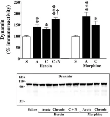
The acute treatments with heroin (10 mg/kg, i.p. for 2 h) and
morphine (30 mg/kg i.p. for 2 h), compared with saline
so-lution administration, induced marked increases in the
im-munodensity of dynamin in the rat cerebral cortex
(hero-ine: 39%, p<0.01; morph(hero-ine: 85%, p<0.001; Fig. 8). Chronic
treatments (5 days) with heroin (5–30 mg/kg) and
mor-phine (10–100 mg/kg) were also associated with
up-regu-lations in the content of dynamin in the brain (heroin:
30%, p<0.05; morphine: 47%, p<0.05), but these effects
were less pronounced, although statistically not
signifi-cant, than those induced by the acute treatments
(espe-cially in the case of morphine), which might suggest the
Fig. 5 Representative immunoblots of Fas-related proteins (≈35 kDanative Fas and ≈48 and 51 kDa glycosylated Fas, arrows) detected with different antibodies (M-20, C-20 and FL-335) in the rat cere-bral cortex (40µg protein extracted with Triton) after chronic pen-tazocine administration. Groups of treatments (three animals for each group): saline and chronic pentazocine (10–80 mg/kg i.p. for 5 days). The apparent molecular masses of Fas-related proteins were determined by calibrating the blots with prestained molecular weight markers as shown on the left hand side. For a given anti-body all samples were run in the same gel
Fig. 6 Effect of chronic treatment with pentazocine on the im-munodensities of Fas-related proteins in the rat brain (cerebral cortex). Groups of treatments: saline and chronic pentazocine (10–80 mg/kg i.p. for 5 days). Quantitation of Fas-related proteins: 48 kDa glycosylated Fas and 35 kDa native Fas (antibody M-20); 51 kDa glycosylated Fas (antibody C-20), and 48 kDa and 51 kDa glycosylated Fas (antibody FL-335). Columns are means ± SEM of five experiments per group with an animal per experiment, and expressed as percentage of saline-treated rats. *p<0.01; **p<0.0001 when compared with the corresponding saline group (Student’s two-tailed t-test)
induction of tolerance (Fig. 8). Chronic pentazocine
treat-ment (10–80 mg/kg for 5 days) also induced an increase in
the immunodensity of dynamin in the rat cerebral cortex
(51%, p<0.05) (data not shown). In heroin-dependent rats
(5–30 mg/kg for 5 days), naloxone (2 mg/kg)-precipitated
withdrawal (2 h) induced a marked up-regulation in the
immunodensity of dynamin in the brain (74%, p<0.001),
which was significantly greater (44%, p<0.01) than that
observed after chronic heroin administration (Fig. 8).
Discussion
The immunodetection of Fas-related proteins (
≈
35 kDa
native Fas and
≈
48 and 51 kDa glycosylated Fas) in rat,
mouse and human brains, as well as in SH-SY5Y
neuro-blastoma cells, was in good agreement with previous
stud-ies in various human cell lines, which used similar
anti-bodies directed against the cytoplasmic domain of Fas
(Kamitani et al. 1997). In Fas-deficient mice (disruption of
the Fas gene by deletion of exon 9 coding for the receptor
cytoplasmic region), normal size Fas mRNA and
Fas-re-lated proteins (
≈
40–45 kDa) were not expressed in
thymo-cytes (Adachi et al. 1995). The heterogeneity of Fas
ex-pression in the mammal brain is most likely due to
differ-ential glycosylation of the two N-linked glycosylation sites
in the extracellular domain of Fas (Itoh et al. 1991;
Wata-nabe-Fukunaga et al. 1992; Kamitani et al. 1997;
Kram-mer 2000), the expression of which apparently depends
on sample preparation (e.g., protein extraction with the
Fig. 7 Representative immunoblots of Fas-related proteins (≈35 kDanative Fas and ≈48 and 51 kDa glycosylated Fas, arrows) detected with antibody M-20 (batch F251) in the rat cerebral cortex (40µg protein extracted with Triton) after A acute treatment with the se-lective σ1receptor agonist (+)-SKF 10047 and its interaction with
the σ1antagonist BD 1063, and B chronic (+)-SKF 10047
admin-istration. Groups of treatments (two or three animals for each group): saline, acute (+)-SKF 10047 (5 mg/kg i.p. for 2 h), acute BD 1063 (10 mg/kg i.p. for 2.5 h) + SKF 10047, and chronic SKF 10047 (3–10 mg/kg i.p. for 3 days). The apparent molecular masses of Fas-related proteins were determined by calibrating the blots with prestained molecular weight markers as shown on the left hand side. For A and B all samples were run in the same gel. Note that batch F251 for antibody M-20 (compared with batch D219 in Figs. 2, 3, 5) detected Fas related peptides of ≈44/46 kDa that were also immunodetected in mouse and human brains (see Fig. 1, top; batch D219)
Fig. 8 Effects of acute and chronic treatments with heroin or mor-phine and of heroin withdrawal after the chronic treatment on the immunodensity of dynamin in the rat brain (cerebral cortex). Groups of treatments: saline (S), acute heroin (A, 10 mg/kg, i.p., 2 h), chronic heroin (C, 10–30 mg/kg i.p. for 5 days), chronic heroin plus naloxone (C+N, 2 mg/kg i.p. for 2 h), acute morphine (30 mg/kg i.p., 2 h) and chronic morphine (10–100 mg/kg i.p. for 5 days). Columns are means ± SEM of 4–8 (heroin) or 6–8 (morphine) ex-periments per group with an animal per experiment, and expressed as percentage of saline-treated rats. One-way ANOVA detected significant differences between groups with respect to dynamin immunodensity after heroin [F(3,16)=22.7, p<0.0001] and mor-phine [F(2,17)=10.61, p=0.001] treatments. *p<0.05; **p<0.01; ***p<0.001 when compared with the corresponding saline group, and †p<0.01 when compared with the chronic heroin group (ANOVA followed by Bonferroni’s test). Bottom: Representative immunoblots for the effects of the various heroin and morphine treatments (two animals for each group, 40µg protein extracted with Triton) on the immunodensity of dynamin in the rat cerebral cortex (all samples were run in the same gel). The apparent molec-ular mass of dynamin was determined by calibrating the blots with prestained molecular weight markers as shown on the left hand side
nonionic detergent Triton X-100 improved the
immuno-detection of glycosylated Fas).
The major findings of the current study are that chronic
heroin treatment (tolerant state) modulated differentially
Fas receptor proteins (up-regulation of 35 kDa native Fas
and down-regulation of 51 kDa glycosylated Fas) and that
spontaneous heroin withdrawal (dependent state) was
as-sociated with increases of these forms of Fas in the brain
(i.e. a sustained increase for native Fas and the induction
of up-regulation for glycosylated Fas). Moreover, chronic
morphine also induced up-regulation of 35 kDa native Fas.
Interestingly, acute treatments with the
µ
-agonists heroin
and morphine, but not with selective
δ
- and
κ
-agonists,
in-creased the content of native Fas, which clearly indicated
the involvement of
µ
-opioid receptors in the rapid
modu-lation of Fas in the brain. The results extend previous
findings on the modulation of Fas receptor (48 kDa
glyco-sylated protein) in morphine-dependent rats, which was
mediated through a naloxone-sensitive mechanism
(Boro-nat et al. 2001). The results also confirm the general
ob-servation that activation of opioid receptors (mainly the
µ
-type) induces an increase in the expression of Fas receptor
mRNA and protein in various tissues and cells (Yin et al.
1999, 2000; Chatzaki et al. 2001; Singhal et al. 2002;
Wang et al. 2002). In this context, it is of interest to note
that the basal immunodensity of 35 kDa native Fas was
found decreased (30%, n=5, p<0.05) in
µ
-opioid receptor
deficient mice (García-Fuster et al., unpublished results),
suggesting that
µ
-receptors tonically stimulate, through
endogenous opioid peptides, the activation of Fas in the
brain. The content of glycosylated Fas proteins (48- and
51-kDa peptides) was not changed in
µ
-deficient mice
(unpublished results; see also Wang et al. 2002).
More-over, the basal expression of 35 kDa native Fas was not
modified in brains of
δ
- or
κ
-opioid receptor deficient
mice (unpublished results). These data agree with the
ob-served stimulating effects of acute heroin and morphine
on 35 kDa Fas in rat brain and indicate the involvement of
µ
-opioid receptors in mediating the effects of these opiate
agonists.
In a previous study, chronic morphine was associated
with an increased content of 48 kDa Fas in the brain
(Boronat et al. 2001). In the present study, chronic
mor-phine also increased this glycosylated Fas form but
de-creased other (51 kDa protein). However, chronic heroin
(and heroin withdrawal) did no alter 48 kDa glycosylated
Fas in rat brain. The reason for this discrepancy is not
known. Although morphine and heroin (diacetyl-morphine)
may differ in some effects (Schuller et al. 1999), the in
vivo efficacies of heroin, 6-acetylmorphine and morphine
were shown to be mediated by pharmacologically similar
populations of
µ
-opioid receptors (Negus et al. 2003). In
any case, the current results indicate that heroin and
mor-phine addiction in rats (tolerant and/or dependent states)
is associated with up-regulation of 35 kDa native Fas (and
with different expressions of glycosylated Fas) in the brain.
At present, the molecular mechanisms by which heroin
and other opiate drugs modulate the Fas receptor in the
brain are not known. It is unclear whether the opioid
re-ceptors and the Fas receptor are physically associated or
influence each other at the level of their signaling
path-ways. In a recent in vitro study (Singhal et al. 2002),
mor-phine was shown to enhance the expression of FasL/Fas
in macrophages which led to cell apoptosis, and this effect
was mediated by opioid receptors via p38
mitogen-acti-vated protein kinase (MAPK) phosphorylation. The MAPK
and/or the modulation of other signaling pathways (one
possibility would be through the interaction of inhibitory
Gi/o proteins activated by opiate drugs and the Fas
adap-tor molecule FADD recruited after recepadap-tor stimulation)
could also be involved in mediating the effects of opiate
drugs on Fas in the brain.
In contrast to the acute and chronic effects of heroin and
morphine on 35 kDa native Fas (up-regulation), chronic
(±)-pentazocine induced down-regulation of native Fas in
rat brain. Chronic pentazocine, but not chronic heroin,
also reduced 48 kDa glycosylated Fas, and both
treat-ments decreased 51 kDa glycosylated Fas in the brain.
Pentazocine, a former “agonist-antagonist” opiate analgesic
agent, is also a potent
σ1
receptor agonist (Mei and
Paster-nak 2001 and other references therein). Both enantiomers
of pentazocine have good affinities for the cloned
σ1
re-ceptor (Ki: 2.5–18 nM; Mei and Pasternak 2001), but
(–)-pentazocine also has good affinity for the cloned
µ
- (Ki:
5.7 nM, antagonist),
κ
- (Ki: 7.2 nM, agonist) and
δ
- (Ki:
31 nM, agonist) opioid receptors (Raynor et al. 1994;
Craft and McNiel 2003). Among other features, the
recep-tors defined as
σ
are not longer opioid receptors and
there-fore they are naloxone-inaccessible (Quirion et al. 1992).
Since chronic treatment with a high dose of naloxone (10 mg/kg
for 13 days) did not alter the expression of Fas in the
rat brain (Boronat et al. 2001), the inhibitory effects of
chronic pentazocine on Fas cannot be related to its
µ
-an-tagonist properties. Similarly, acute treatments with
selec-tive
δ
- (SNC-80) and
κ
- (U 50488-H) agonists did not
de-crease Fas in the brain. In contrast, acute and chronic
treat-ments with the selective
σ1
receptor agonist (+)-SKF 10047
decreased, through a
σ1
receptor-specific mechanism, the
content of 35 kDa native Fas in the brain. It is of interest
to note that the density of glycosylated Fas was also
re-duced by chronic pentazocine but not by the
σ1
agonist,
suggesting that the former drug is also able to impair the
proper post-translational modification of Fas receptor. At
the functional level the
σ1
receptors comprise a potent
anti-opioid system, the activation of which by (+)-pentazocine
markedly reduces opiate analgesia in mice (Mei and
Pas-ternak 2002). In line with this concept (anti-opioid effect),
the down-regulation induced by chronic pentazocine on
native Fas is also opposite to the up-regulation induced by
heroin and morphine, and both effects are mediated trough
different receptor mechanisms.
Dynamin is a neuronal phosphoprotein and a GTPase
enzyme that plays an essential role in receptor-mediated
endocytosis via clathrin-coated pits and caveoli (McClure
and Robinson 1996). In the current study, chronic opiate
drug treatments (heroin, morphine) and opiate withdrawal
(heroin) resulted in marked up-regulations in the content
of dynamin in the brain, indicating the importance of this
molecular target in opiate addiction. These results confirm
and extend previous observations on the modulation of
dynamin in morphine addiction (Noble et al. 2000).
Con-versely, chronic treatment with naltrexone, an opioid
re-ceptor antagonist, was associated with down-regulation
(30%) of dynamin in mouse spinal cord (Patel et al. 2002).
Moreover, the present data also indicate that acute heroin
(10 mg/kg, 2 h) and morphine (30 mg/kg, 2 h) treatments
resulted in up-regulation of dynamin in the rat cerebral
cortex, which indicates its rapid modulation by opiate drugs.
Furthermore, chronic pentazocine also up-regulated
dy-namin content in rat brain, which also suggests the
in-volvement of
σ1
receptors in its regulation. The
up-regu-lation of dynamin by opiate drugs could contribute to the
plasticity of the endogenous opioid system, which is
rele-vant in the development of tolerance and dependence to
opiate drugs (see Noble et al. 2000). In this context, the
up-regulation of dynamin in specific brain regions could
regulate the level of expression of opioid receptors through
modulation of receptor internalization induced by opiate
drugs (Murray et al. 1998; Whistler and Von Zastrow 1999).
In conclusion, chronic heroin and morphine treatments
and heroin withdrawal in rats are associated with
up-reg-ulation of 35 kDa native Fas (and with different
expres-sions of glycosylated Fas) in the brain. At present, the
functional consequences of the up-regulation of Fas
re-ceptor in opiate addiction are not known. Opiate drugs
can promote, through the activation of Fas, abnormal cell
death (Yin et al. 1999, 2000; Chatzaki et al. 2001; Singhal
et al. 2002; Wang et al. 2002), but most neurons are
resis-tant to Fas-induced apoptosis (Raoul et al. 2002).
Alterna-tively, Fas could function as a non-apoptotic signal
trans-ducer (Wajant 2002; Desbarats et al. 2003), and in this case
it would represent a new component in the opioid receptor
signaling cascade potently modulated by opiate drugs.
Acknowledgements This study was supported by grant BFI2000– 0306 from MCT (Madrid, Spain) and by grant 32.57066.99 from FNSRS (Bern, Switzerland). M.J.G.-F. was supported by a predoc-toral fellowship from CSIC/MECD-Associated Units. J.A. García-Sevilla is a member of the Institut d’Estudis Catalans (Barcelona, Spain).References
Adachi M, Suematsu S, Kondo T, Ogasawara T, Tanaka T, Yoshida N, Nagata S (1995) Targeted mutation in the Fas gene causes hyperplasia in peripheral lymphoid organs and liver. Nat Gen 11:294–300
Badorff C, Ruetten H, Mueller S, Stahmer M, Gehring D, Jung F, Ihling C, Zeiher AM, Dimmeler S (2002) Fas receptor signal-ing inhibits glycogen synthase kinase 3βand induces cardiac hypertrophy following pressure overload. J Clin Invest 109: 373–381
Bechmann I, Mor G, Nilsen J, Eliza M, Nitsch R, Naftolin F (1999) FasL (CD95L, Apo1L) is expressed in the normal rat and human brain. Glia 27:62–74
Boronat MA, García-Fuster MJ, García-Sevilla JA (2001) Chronic morphine induces up-regulation of the pro-apoptotic Fas recep-tor and down-regulation of the anti-apoptotic Bcl-2 oncopro-tein in rat brain. Br J Pharmacol 134:1263–1270
Bradford MM (1976) A rapid and sensitive method for the quanti-tation of microgram quantities of protein utilizing the principle of protein-dye binding. Anal Biochem 72:248–254
Chatzaki E, Makrigiannakis A, Margioris AN, Kouimtzoglou E, Gravanis A (2001) The Fas/FasL apoptotic pathway is in-volved in κ-opioid-induced apoptosis of human endometrial stromal cells. Mol Human Reprod 7:867–874
Choi C, Park JY, Lee J, Lim J-H, Shin E-C, Ahn Y, Kim C-H, Kim S-J, Kim J-D, Choi IS, Choi I-H (1999) Fas ligand and Fas are expressed constitutively in human astrocytes and the expres-sion increases with IL-1, IL-6, TNF-α, or IFN-γ. J Immunol 162:1889–1895
Craft RM, McNeil DM (2003) Agonist/antagonist properties of nalbuphine, butorphanol and (–)-pentazocine in male vs. fe-male rats. Pharmacol Biochem Behav 75:235–245
Desbarats J, Birge RB, Minouni-Rongy M, Weistein DE, Palerme J-S, Newell MK (2003) Fas engagement induces neurite growth through ERK activation and p35 upregulation. Nat Cell Biol 5:118–125
Ferrer-Alcón M, La Harpe R, Guimón J, García-Sevilla JA (2003) Down-regulation of neuronal cdk5/p35 in opioid addicts and opiate-treated rats: relation to neurofilament phosphorylation. Neuropsychopharmacology 28:947–955
Gabilondo AM, García-Sevilla JA (1995) Spontaneous withdrawal from long-term treatment with morphine accelerates the turn-over of α2-adrenoceptors in rat brain: up-regulation of
recep-tors associated with increased receptor appearance. J Neuro-chem 64:2590–2597
García-Fuster MJ, Ferrer-Alcón M, Miralles A, García-Sevilla JA (2002) Modulation of pro-apoptotic Fas receptor proteins dur-ing heroin addiction in rat brain. Methods Find Exp Clin Phar-macol 24 [Suppl A]:161
García-Sevilla JA, Ventayol P, Busquets X, La Harpe R, Walzer C, Guimón J (1997) Marked decrease of immunolabelled 68 kDa neurofilament (NF-L) proteins in brains of opiate addicts. Neuro-report 8:1561–1565
Harlow E, Lane D (1999) Using antibodies. A laboratory manual. Cold Spring Harbor Laboratory Press, Cold Spring Harbor Itoh N, Yonehara S, Ishii A, Yohenara M, Mizushima S-I,
Same-shima M, Hase A, Seto Y, Nagata S (1991) The polypeptide encoded by the cDNA for human cell surface antigen Fas can mediate apoptosis. Cell 66:233–243
Kamitani T, Nguyen HP, Yeh ETH (1997) Activation-induced ag-gregation and processing of the human fas antigen. Detection with cytoplasmic domain-specific antibodies. J Biol Chem 272: 22307–22314
Krammer PH (2000) CD95’s deadly mission in the immune sys-tem. Nature 407:789–795
López E, Ferrer I (2000) Staurosporine- and H-7-induced cell death in SH-SY5Y neuroblastoma cells is associated with cas-pase-2 and caspase-3 activation, but not with activation of the FAS/FAS-L-caspase-8 signaling pathway. Mol Brain Res 85:61– 67
MacEwan DJ (2002) TNF ligands and receptors – a matter of life and death. Br J Pharmacol 135:855–875
Matsuno K, Senda T, Kobayashi T, Mita S (1995) Involvement of
σ1receptor in (+)-N-allylnormetazocine-stimulated
hippocam-pal cholinergic functions in rats. Brain Res 690:200–206 McClure SJ, Robinson PJ (1996) Dynamin, endocytosis and
intra-cellular signalling. Mol Membr Biol 13:189–215
McCracken KA, Bowen WD, Matsumoto RR (1999) Novel σ re-ceptor ligands attenuate the locomotor stimulatory effects of cocaine. Eur J Pharmacol 365:35–38
Mei J, Pasternak W (2001) Molecular cloning and pharmacologi-cal characterization of the rat sigma1receptor. Biochem
Phar-macol 62:349–355
Mei J, Pasternak W (2002) σ1Receptor modulation of opioid
anal-gesia in the mouse. J Pharmacol Exp Ther 300:1070–1074 Murray SR, Evans CJ, Von Zastrow M (1998) Phosphorylation is
not required for dynamin-dependent endocytosis of a truncated mutant opioid receptor. J Biol Chem 273:24987–24991
Nagata S (1999) Fas ligand-induced apoptosis. Ann Rev Gen 33: 29–55
Nagata S, Golstein P (1995) The Fas death receptor. Science 267: 1449–1456
Negus SS, Brandt MR, Gatch MB, Mello NK (2003) Effects of heroin and its metabolites on schedule-controlled responding and thermal nociception in rhesus monkeys: sensitivity to an-tagonism by quadazocine, naltrindole and β-funaltrexamine. Drug Alcohol Depend 70:17–27
Noble F, Szücs M, Kieffer B, Roques BP (2000) Overexpression of dynamin is induced by chronic stimulation of µ- but not
δ-opioid receptors: relationship with µ-related morphine depen-dence. Mol Pharmacol 58:159–166
Oehm A, Behrmann I, Falk W, Pawlita M, Maier G, Klas C, Li-Weber M, Richards S, Dhein J, Trauth B, Ponstingl H, Kram-mer P (1992) Purification and molecular cloning of the APO-1 cell surface antigen, a member of the tumor necrosis factor/ nerve growth factor receptor family. J Biol Chem 267:10709– 10715
Orlinick JR, Vaishnaw AK, Elkon KB (1999) Structure and func-tion of Fas/Fas ligand. Int Rev Immunol 18:293–308
Ozaita A, Escribá PV, García-Sevilla JA (1999) The alkylating agent EEDQ facilitates protease-mediated degradation of the human brain α2A-adrenoceptor as revealed by a
sequence-spe-cific antibody. Neurosci Lett 263:105–108
Papakonstanti EA, Bakogeorgou E, Castanas E, Emmanouel DS, Hartig R, Stournaras C (1998) Early alterations of actin cy-toskeleton in OK cells by opioids. J Cell Biochem 70:60–69 Patel M, Gomes B, Patel V, Yoburn BC (2002)
Antagonist-in-duced µ-opioid receptor up-regulation decreases G-protein re-ceptor kinase-2 and dynamin-2 abundance in mouse spinal cord. Eur J Pharmacol 446:37–42
Quirion R, Bowen WD, Itzhak Y, Junien JL, Musacchio JM, Roth-man RB, Su T-P, Tam SW, Taylor DP (1992) A proposal for the classification of sigma binding sites. Trends Pharmacol Sci 13:85–86
Raoul C, Estévez AG, Nishimune H, Cleveland DW, deLapeyrière O, Henderson CE, Haase G, Pettmann B (2002) Motoneuron death triggered by a specific pathway downstream of Fas: po-tentiation by ALS-linked SOD1 mutations. Neuron 35:1067– 1083
Raynor K, Kong H, Chen Y, Yasuda K, Yu L, Bell GI, Reisine T (1994) Pharmacological characterization of the cloned kappa-, delta-, and mu-opioid receptors. Mol Pharmacol 45:330–334 Sastry PS, Rao KS (2000) Apoptosis and the nervous system.
J Neurochem 74:1–20
Schuller AGP, King MA, Zhang J, Bolan E, Pan Y-X, Morgan DJ, Chang A, Czick ME, Unterwald EM, Pasternak GW, Pintar JE (1999) Retention of heroin and morphine-6β-glucoronide anal-gesia in a new line of mice lacking exon 1 of MOR-1. Nat Neu-rosci 2:151–156
Siegel RM, Ka-Ming Chang F, Chun HJ, Lenardo MJ (2000) The multifaceted role of Fas signaling in immune cell homeostasis and autoimmunity. Nat Immunol 1:469–474
Singhal PC, Bhaskaran M, Patel J, Patel K, Kasinath BS, Durai-samy S, Franki N, Reddy K, Kapasi AA (2002) Role of P38 mitogen-activated protein kinase phosphorylation and Fas-Fas ligand interaction in morphine-induced macrophage apoptosis. J Immunol 168:4025–4033
Tada Y, O-Wang J, Takiguchi Y, Tatsumi K, Kuriyama T, Okada S, Tokuhisa T, Sakiyama S, Tagawa M (2002) A novel role for Fas ligand in facilitating antigen acquisition by dendritic cells. J Immunol 169:2241–2245
Ventayol P, Busquets X, García-Sevilla JA (1997) Modulation of immunoreactive protein kinase C-αand βisoforms and G pro-teins by acute and chronic treatments with morphine and other opiate drugs in rat brain. Naunyn-Schmiedebergs Arch Phar-macol 355:491–500
Wajant H (2002) The Fas signalling pathway: more than a para-digm. Science 296:1635–1636
Wang J, Charboneau R, Barke RA, Loh HH, Roy S (2002) µ -Opi-oid receptor mediates chronic restraint stress-induced lympho-cyte apoptosis. J Immunol 169:3630–3636
Watanabe-Fukunaga R, Brannan CI, Itoh N, Yonehara S, Cope-land NG, Jenkins NA, Nagata S (1992) The cDNA structure, ex-pression, and chromosomal assignment of the mouse Fas anti-gen. J Immunol 148:1274–1279
Whistler JL, Von Zastrow M (1999) Dissociation of functional roles of dynamin in receptor-mediated endocytosis and mitogenic signal transduction. J Biol Chem 274:24575–24578
Yin D, Mufson RA, Wang R, Shi Y (1999) Fas-mediated cell death promoted by opioids. Nature 397:218
Yin D, Tuthill D, Mufson RA, Shi Y (2000) Chronic restraint stress promotes lymphocyte apoptosis by modulating CD95 ex-pression. J Exp Med 191:1423–1428
Yu VC, Richards ML, Sadée W (1986) A human neuroblastoma cell line expresses µand δopioid receptor sites. J Biol Chem 261:1065–1070
Yuan J, Yankner BA (2000) Apoptosis in the nervous system. Na-ture 407:802–809