Iris-Katharina Penner Klaus Opwis
Ludwig Kappos
Relation between functional
brain imaging, cognitive impairment
and cognitive rehabilitation in patients
with multiple sclerosis
Introduction
In general, cognitive impairment is defined as a decline in cognitive performance that has an impact on the sub-ject’s social and occupational life. The decline can either refer to cognitive subfunctions such as attention, mem-ory, executive functions etc. or can be a global problem where many subfunctions are affected at the same time. One of the main issues, however, is that the subject ex-periences psychological strain indicating that alterna-tive cognialterna-tive strategies are not sufficient to cope with the impairment.
■ Cognitive impairment in MS
Cognitive decline was already described by Charcot in 1877 [10] to be one of the key features of MS. Surpris-ingly, research in the following decades focussed solely
on the physical components of the disease disregarding psychological and cognitive aspects. This disregard, however, was not due to a lack of interest in psychologi-cal aspects but reflects how MS was perceived at the be-ginning of the 19th century. The obvious and visible
symptoms clearly derived from the motor system and the understanding of the disease was directed towards neuropathological changes in the white matter. Cogni-tion, on the other hand, was thought to be exclusively a function of the cortex where white matter tracts and subcortical structures play only a minor role [41]. Today, it is well-known that the fibre integrity of the white mat-ter is essential for cognitive functioning and that cor-tico-subcortical connections mainly control the infor-mation processing speed while fronto-parietal and fronto-temporal connections are mainly responsible for attention, memory and executive processes. Further-more, cortical demyelination is nowadays also discussed to be one possible feature of tissue destruction in MS.Al-though three different cortical lesion types were defined
JON 2013
■ Abstract Cognitive impairment belongs to the core symptoms in MS affecting quality of life, self-es-teem, and social as well as occupa-tional functioning. Due to this high
I.-K. Penner, PhD (쾷) · K. Opwis University of Basel
Dept. of Cognitive Psychology and Methodology
Missionsstr. 60/62 4055 Basel, Switzerland E-Mail: [email protected] L. Kappos
University Hospital Basel Dept. of Neurology Basel, Switzerland
impact on patients’ well-being effi-cient treatment concepts are re-quired. Imaging studies on cogni-tion have shown that funccogni-tional reorganisation takes place sponta-neously to compensate for deficits. In mildly to moderately impaired patients these processes may sup-port coping with emerging deficits. However, these compensatory processes seem to be limited as brain activation of cognitively se-verely impaired patients is charac-terised by decreased additional re-cruitment of brain regions. Cognitive rehabilitation concen-trates on the question whether
in-duction of brain plasticity is possi-ble for both the support of the spontaneous processes and the ini-tiation of new ones. Combining cognition, brain imaging and cog-nitive rehabilitation in MS, an in-triguing question is whether fMRI can provide further insights into the mechanisms of induced plastic-ity and serve as objective outcome measures for efficient cognitive in-tervention.
■ Key words brain imaging · fMRI · cognition · multiple sclerosis · cognitive rehabilitation
(1. cortico-subcortical compound lesions, 2. small perivascular intracortical lesions, 3. band-like subpial demyeliniation) [8, 21, 36] their contribution to cogni-tive decline has not yet been clarified. A recently pub-lished paper by Kutzelnigg and Lassmann [23] is con-cerned with the relation of cognitive decline and the different types of cortical lesions. Using sensitive im-munocytochemical techniques these specific alterations in the cortex itself are detectable post-mortem. Interest-ingly, the distribution of cortical lesions might have a di-rect relation with the cognitive domains mainly affected in MS. However, as imaging techniques are currently not sensitive enough to visualise the different types of corti-cal lesions, correlative analyses with cognitive test per-formance are not yet feasible in the living brain. Thus, the scientific focus in MS has changed remarkably by at-tending to cognitive decline as one of the main symp-toms affecting patients with MS. Particularly, this be-comes apparent in the high number of emerging publications dealing with cognition and possible correl-atives in MS.
Cognitive impairment and structural MRI
Conventional MRI is a well accepted tool in the diag-nostic assessment of patients with MS, in characterising lesions and in monitoring disease evolution [26]. How-ever, it is only of low pathological specificity and thus not a powerful correlative for cognitive alterations. This might be one reason for only weak correlations between structure and function in MS [e. g. 9, 17]. Only studies correlating regional lesion volume with specific cogni-tive functions succeeded in finding better relations [e. g. 42, 46] indicating that a general brain volume decrease can be compensated while destruction of critical and specialised brain regions results in cognitive alterations. These results are in accordance with the hypothesis for-mulated by Kutzelnigg and Lassmann [23] that the dis-tribution of cortical demyelinated plaques might ac-count for specific cognitive deficits. Disappointingly, a longitudinal study on cognitive dysfunction over 8.5 years [37] showed high correlations between temporal, occipital and frontal lesions and specific cognitive test performances at baseline, but these relations could not be confirmed in the long run. Thus, the increase in the number of regional lesions did not correspond to the evolution of cognitive deficits which means that even lo-cal brain pathology can only, in part, explain cognitive changes in MS.
More specific MRI parameters such as whole brain atrophy [1, 5] or atrophy of specific brain regions [4–6, 11, 19], magnetisation transfer [12, 15, 49] and MR spec-troscopy [16, 18, 28] show much better correlations with cognitive alterations even in the early phase of MS and even for specific cognitive functions. Thus, brain
pathol-ogy in terms of irreversible demyelination and axonal loss seems to be related more directly to cognitive im-pairment although even in this context quantitative and predictive values are still missing.
Cognitive impairment and functional
brain imaging
As structural MRI does not provide a satisfying expla-nation for cognitive decline, functional brain imaging might be a more powerful approach to characterise cog-nitive alterations in MS. Indeed, changes in functional brain organisation can already be observed in the very early stages of the disease.
PET/SPET
PET and SPET studies on cognitive decline in MS report metabolic changes. Deficits in perfusion, mainly con-centrated on the left hemisphere, were reported in the frontal and temporal lobe [31, 39, 40, 44] as well as global and regional reduction in cerebral glucose metabolism [7]. These results indicate that hypometabolism of brain regions related to specific cognitive functions might be one critical factor in the development of cognitive de-cline in MS.
MR spectroscopy
MR spectroscopy as a method to evaluate axonal in-tegrity has been used sparsely to study cognitive alter-ations in MS. In a study by Pan et al. [28] lower N-acetyl aspartate (NAA) levels in predefined regions of interest were correlated with specific cognitive functions. Ac-cordingly, a recently published study by Gadea et al. [18] reports lowest NAA/Cr levels in RRMS patients who showed the highest decrease in attention performance. Thus, NAA seems to provide a specific measure for pathological changes in the CNS that are especially re-sponsible for cognitive functioning.
fMRI
In accordance with studies on the motor system [e. g. 29, 43] fMRI data on cognition show significant changes in brain organisation compared to healthy controls. Inter-estingly, these changes are already evident at the very early stages of the disease, when cognitive deficits are not yet clinically detectable [2, 3]. Additional activation, mainly in the prefrontal cortices during the perfor-mance of a working memory task, was interpreted as a compensatory mechanism that contributes to normal
performance. In mildly to moderately impaired pa-tients, increased and additional activation was observed when patients performed the PASAT [25, 47]. In these cases, a measurable mild cognitive impairment was re-lated to a significant increase in brain activation sug-gesting that the brain uses alternative pathways to per-form the cognitive task. At this point, it is open to discussion whether, as a consequence, severe cognitive impairment will be related to even more additional re-cruitment of functional areas into a compensatory net-work. Interestingly, this does not seem to be the case as a comparison between mildly and severely impaired MS patients who performed a tonic alertness task clearly demonstrated that the increase in functional brain acti-vation is significantly reduced in severely impaired pa-tients [35].
From the fMRI studies presented so far we can con-clude that CIS patients and those with a mild to moder-ate cognitive impairment recruit additional functional brain areas meaning (1) that functional changes are al-ready present before deficits are clinically detectable and (2) that the increase in brain activation seems to be responsible for the compensation of a deficit. Further, a lack of those compensatory mechanisms might be re-sponsible for severe cognitive impairment where the MS brain is no longer capable to build up new functional networks. If so, then the relation between functional brain activation and cognitive impairment may be ex-pressed by an inversed u-shaped curve where at some stage of the disease the CNS is no longer able to deal with the widespread destruction of the brain tissue on a func-tional level.
fMRI, brain plasticity and cognitive rehabilitation
So far, fMRI studies have shown spontaneous processes of functional reorganisation in the MS brain. The re-cruitment of additional functional brain regions is as-sumed to be related to compensatory mechanisms that result in non-pathological behaviour or only moderate impairment. If these mechanisms are powerful enough to keep behavioural performance on a high level then the question arises if it is possible to use this power in cognitive rehabilitation. The rationale would be to iden-tify the cognitive functions mainly affected in a patient, to compose a training procedure for these functions, to stimulate alternative pathways by this training and thereby to improve cognitive functioning. Cognitive re-habilitation can be seen as an intervention aimed at changes in specific neuronal circuits. However, although imaging studies have provided good insight into the mechanisms of recovery and plasticity in MS [2, 3, 25, 35, 47] reliable tools to evaluate the effects of cognitive re-habilitation are still missing [32]. Studies on cognitive rehabilitation in MS are still rare and the results are hardly comparable as cognitive outcome measures, training tools and training procedures differ remark-ably [20, 24, 38, 45, 48]. Therefore, the efficacy of cogni-tive rehabilitation has not yet been proven by using ob-jective readouts.As fMRI provides direct insight into the working brain, one intriguing question is whether this method might be used as a reliable tool to evaluate the effects of cognitive rehabilitation. If cognitive rehabili-tation is seen as a procedure to induce plasticity processes in the brain, then fMRI might be useful to ver-ify this assumption. In a first pilot study [33, 34] we tried to determine if a computerised attention training in-duces alterations in functional brain organisation. For this purpose, cognitively mildly and severely impaired MS-patients were investigated on different attention functions using a computerised attention software and fMRI. For three to four weeks all patients received a computerised training on selective attention. After the
Fig. 1 Functional brain activation patterns for control subjects, mildly impaired MS patients and severely impaired MS patients while performing a tonic alertness task (p < 0.001, corrected for multiple comparisons)
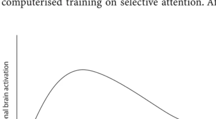
Fig. 2 Schematic illustration of the relation between the evolution of cognitive impairment and the increase in functional brain activation
Increase in impairment
Functional brain activation
training interval, all patients were reinvestigated behav-iourally and by fMRI. When comparing pre- and post-training fMRI results it became evident that in both groups of patients three attention related structures were activated in addition: the posterior cingulate cor-tex, the precuneus and the dorsal frontal gyrus [33, 34]. These three structures have specified functions in the at-tention network including focussing and inhibition [50], attentional shifting and task switching [13, 22, 27] and selection of action [30]. As a clear increase in be-havioural performance could be verified one can con-clude that the attention training caused recruitment of specific attentional areas that finally resulted in behav-ioural improvement. However, these findings cannot be generalised, since the study included only 11 patients and behavioural effects were small. Nevertheless, these data should motivate to start cognitive rehabilitation studies on more homogeneous patient cohorts and to
define a gold standard for primary outcome measures and training tools. Finally, studies that are aimed at defining the most efficient training interval are needed. At present, it is not known whether a temporally longer and more distributed intervention might be more suc-cessful than a short and concentrated one. In a study by Draganski et al. [14] a three month training on juggling even induced changes in grey matter indicating that changes in brain structure are already evident after a short period of time. If we combine these results with those of our pilot study and suppose that structural changes last longer than functional changes we can as-sume that a powerful training should last at least four to 12 weeks to induce functional changes. However, the un-certainty remains as to how intensive a training proce-dure has to be to produce an optimal outcome. Studies to clarify these questions are needed to provide efficient rehabilitation tools for MS patients in the future.
References
1. Amato MP, Bartolozzi ML, Zipoli V et al. (2004) Neocortical volume de-crease in relapsing-remitting MS pa-tients with mild cognitive impairment. Neurology 63:89–93
2. Audoin B, Au Duong MV, Malikova I et al. (2006) Functional magnetic reso-nance imaging and cognition at the very early stage of MS. J Neurol Sci 245:87–91
3. Audoin B, Ibarrola D, Ranjeva JP et al. (2003) Compensatory cortical activa-tion observed by fMRI during a cogni-tive task at the earliest stage of MS. Hum Brain Mapp 20:51–58
4. Benedict RH, Bakshi R, Simon JH, Pri-ore R, Miller C, Munschauer FE (2002) Frontal cortex atrophy predicts cogni-tive impairment in multiple sclerosis. J Neuropsychiatry Clin Neurosci 14: 44–51
5. Benedict RH, Carone DA, Bakshi R (2004) Correlating brain atrophy with cognitive dysfunction, mood disturb-ances, and personality disorder in multiple sclerosis. J Neuroimaging 14:36S–45S
6. Berg D, Maurer M, Warmuth-Metz M, Rieckmann P, Becker G (2000) The correlation between ventricular diam-eter measured by transcranial sonog-raphy and clinical disability and cogni-tive dysfunction in patients with multiple sclerosis. Arch Neurol 57: 1289–1292
7. Blinkenberg M, Rune K, Jensen CV et al. (2000) Cortical cerebral metabo-lism correlates with MRI lesion load and cognitive dysfunction in MS. Neurology 54:558–564
8. Bo L, Vedeler CA, Nyland HI, Trapp BD, Mork SJ (2003) Subpial demyelina-tion in the cerebral cortex of multiple sclerosis patients. J Neuropathol Exp Neurol 62:723–732
9. Camp SJ, Stevenson VL, Thompson AJ et al. (1999) Cognitive function in primary progressive and transitional progressive multiple sclerosis. A con-trolled study with MRI correlates. Brain 122:1341–1348
10. Charcot JM (1877) Lectures on the diseases of the nervous system. New Sydenham Society, London 11. Comi G, Filippi M, Martinelli V et al.
(1993) Brain magnetic resonance imaging correlates of cognitive impair-ment in multiple sclerosis. J Neurol Sci 115:66–73
12. Deloire MSA, Salort E, Bonnet M et al. (2005) Cognitive impairment as marker of diffuse brain abnormalities in early relapsing remitting multiple sclerosis. J Neurol Neurosurg Psychia-try 76:519–526
13. Dove A, Pollmann S, Schubert T, Wiggins CJ, Von Cramon DY (2000) Prefrontal cortex activation in task switching: An event-related fMRI study. Cogn Brain Res 9:103–109 14. Draganski B, Gaser C, Busch V,
Schuierer G, Bogdahn U, May A (2004) Neuroplasticity: changes in grey matter induced by training. Nature 427:311–312
15. Filippi M, Tortorella C, Rovaris M et al. (2000) Changes in the normal appear-ing brain tissue and cognitive impair-ment in multiple sclerosis. J Neurol Neurosurg Psychiatry 68:157–161
16. Foong J, Rozewicz L, Davie CA, Thompson AJ, Miller DH, Ron MA (1999) Correlates of executive function in multiple sclerosis: the use of mag-netic resonance spectroscopy as an in-dex of focal pathology. J Neuropsychia-try Clin Neurosci 11:45–50
17. Fulton JC, Grossman RI, Udupa J et al. (1999) MR lesion load and cognitive function in patients with relapsing-re-mitting multiple sclerosis. Am J Neuro-radiol 20:1951–1955
18. Gadea M, Martinez-Bisbal MC, Marti-Bonmati L, Espert R, Casanova B, Coret F, Celda B (2004) Spectroscopic axonal damage of the right locus coeruleus relates to selective attention impairment in early stage relapsing-remitting multiple sclerosis. Brain 127:89–98
19. Huber SJ, Bornstein RA, Rammohan KW, Christy JA, Chakeres DW, McGhee RB (1992) Magnetic resonance imag-ing correlates of neuropsychological impairment in multiple sclerosis. J Neuropsychiatry Clin Neurosci 4: 152–158
20. Jønsson A, Korfitzen EM, Heltberg A, Ravnborg MH, Byskov-Ottosen E (1993) Effects of neuropsychological treatment in patients with multiple sclerosis. Acta Neurol Scand 88: 394–400
21. Kidd D, Barkhof F, McConnell R, Algra PR, Allen IV, Revesz T (1999) Cortical lesions in multiple sclerosis. Brain 122: 17–26
22. Krause BJ, Schmidt D, Mottaghy FM et al. (1999) Episodic retrieval activates the precuneus irrespective of the im-agery content of word pair associates: A PET-study. Brain 122:255–263 23. Kutzelnigg A, Lassmann H (2006)
Cor-tical demyelination in multiple sclero-sis: A substrate for cognitive deficits? J Neurol Sci 245:123–126
24. Lincoln NB, Dent A, Harding J, Wey-man N, Nicholl C, Blumhardt LD, Play-ford ED (2002) Evaluation of cognitive assessment and cognitive intervention for people with multiple sclerosis. J Neurol Neurosurg Psychiatr 72:93–98 25. Mainero C, Caramia F, Pozzilli C et al. (2004) fMRI evidence of brain reorga-nization during attention and memory tasks in multiple sclerosis. Neuro-Image 21:858–867
26. McDonald WI, Compston A, Edan G et al. (2001) Recommended diagnostic criteria for Multiple Sclerosis: Guide-lines from the international panel on the diagnosis of Multiple Sclerosis. Ann Neurol 50:121–127
27. Nagahama Y, Okada T, Katsumi Y et al. (1999) Transient neural activity in the medial superior frontal gyrus and pre-cuneus time locked with attention shift between object features. Neuro-Image 10:193–199
28. Pan JW, Krupp LB, Elkins LE, Coyle PK (2001) Cognitive dysfunction lateral-izes with NAA in multiple sclerosis. Appl Neuropsychol 8:155–160 29. Pantano P, Iannetti GD, Caramia F et al.
(2002) Cortical motor reorganization after a single clinical attack of multiple sclerosis. Brain 125:1607–1615 30. Passingham R (1993) The frontal lobes
and voluntary action. Oxford Univer-sity Press, Oxford
31. Paulesu E, Perani D, Fazio F et al. (1996) Functional basis of memory impairment in multiple sclerosis: A [18F]FDG PET study. NeuroImage 4: 87–96
32. Penner IK, Kappos L (2006) Retraining attention in MS. J Neurol Sci 245: 147–151
33. Penner IK, Kappos L, Opwis K (2005) Induced changes in brain activation using a computerized attention train-ing in patients with Multiple Sclerosis (MS). In: Opwis K, Penner IK (eds) Proceedings of KogWis05. The Ger-man Cognitive Science Conference. Schwabe, Basel, pp 150–154
34. Penner IK, Kappos L, Rausch M, Opwis K, Radü EW (2006) Therapy-induced plasticity of cognitive functions in MS patients: Insights from fMRI. J Physiol Paris 99:455–462
35. Penner IK, Rausch M, Kappos L, Opwis K, Radü EW (2003) Analysis of impair-ment related functional architecture in MS patients during performance of different attention tasks. J Neurol 250: 461–472
36. Peterson JW, Bö L, Mörk S, Chang A, Trapp BD (2001) Transected neurites, apoptotic neurons, and reduced in-flammation in cortical multiple sclero-sis lesions. Ann Neurol 50:389–400 37. Piras MR, Magnano I, Canu ED et al.
(2003) Longitudinal study of cognitive dysfunction in multiple sclerosis: neuropsychological, neuroradiological, and neurophysiological findings. J Neurol Neurosurg Psychiatry 74: 878–885
38. Plohmann AM, Kappos L, Ammann A et al. (1998) Computer assisted retrain-ing of attentional impairments in pa-tients with multiple sclerosis. J Neurol Neurosurg Psychiatr 64:455–462 39. Pozzilli C, Gasperini C, Anzini A,
Grasso MG, Ristori G, Fieschi C (1993) Anatomical and functional correlates of cognitive deficit in multiple sclero-sis. J Neurol Sci 115:55–58
40. Pozzilli C, Passafiume D, Anzini A, Borsellino G, Koudriavtseva T, Sarlo G, Fieschi C (1992) Cognitive and brain imaging measures of Multiple Sclero-sis. Ital J Neurol Sci 2:133–136 41. Prosiegel M, Michael C (1993)
Neu-ropsychology and multiple sclerosis: Diagnostic and rehabilitative approaches. J Neurol Sci 115(Suppl): S51–S54
42. Pujol J, Vendrell P, Deus J, Junqué C, Bello J, Marti-Vilalta JL, Capdevila A (2001) The effect of medial frontal and posterior parietal demyelinating lesions on Stroop interference. NeuroImage 13:68–75
43. Rocca MA, Matthews PM, Caputo D, Ghezzi A, Falini A, Scotti G, Comi G, Filippi M (2002) Evidence for wide-spread movement-associated func-tional MRI changes in patients with PPMS. Neurology 58:866–872 44. Sheremata WA, Secush S, Knight D,
Ziajka P (1984) Altered cerebral metabolism in MS. Neurology 34 (Suppl 1):118
45. Solari A, Motta A, Mendozzi L et al. (2004) Computer-aided retraining of memory and attention in people with multiple sclerosis: a randomized, dou-ble-blind controlled trial. J Neurol Sci 222:99–104
46. Sperling RA, Guttmann CR, Hohol MJ et al. (2001) Regional magnetic reso-nance imaging lesion burden and cog-nitive function in multiple sclerosis. Arch Neurol 58:115–121
47. Staffen W, Mair A, Zauner H et al. (2002) Cognitive function and fMRI in patients with multiple sclerosis: Evi-dence for compensatory cortical acti-vation during an attention task. Brain 125:1275–1282
48. Tesar N, Bandion K, Baumhackl U (2005) Efficacy of a neuropsychologi-cal training programme for patients with multiple sclerosis – a randomised controlled trial. Wien Klin Wochenschr 117:747–754
49. van Buchem MA, Grossman RI, Arm-strong C et al. (1998) Correlation of volumetric magnetization transfer imaging with clinical data in MS. Neurology 50:1609–1617
50. Von Zerssen GC, Mecklinger A, Opitz B, von Cramon DY (2001) Conscious recollection and illusory recognition: An event-related fMRI study. Eur J Neurosci 13:2148–2156