www.impactjournals.com/oncotarget/ Oncotarget, Vol. 7, No. 10
Bevacizumab specifically decreases elevated levels of circulating
KIT
+CD11b
+cells and IL-10 in metastatic breast cancer patients
Sarah Cattin1, Benoît Fellay2, Sylvain Pradervand3, Andreas Trojan4, Thomas Ruhstaller5, Curzio Rüegg1,*, Gregor Fürstenberger6,*1 Department of Medicine, Faculty of Science, University of Fribourg, CH-1700 Fribourg, Switzerland 2 Central Laboratory, HFR Hôpital Cantonal, CH-1700 Fribourg, Switzerland
3 Genomic Technologies Facility, Center of Integrative Genomic (CIG), University of Lausanne (UNIL), CH-1015 Lausanne,
Switzerland
4OnkoZentrum Zürich, CH-8038 Zürich, Switzerland
5Breast Center, Kantonsspital St.Gallen, CH-9000 St.Gallen, Switzerland 6Tumor and Breast Center ZeTuP, CH-9006 St.Gallen, Switzerland *These authors have contributed equally to this work
Correspondence to: Curzio Rüegg, e-mail: [email protected] Keywords: breast cancer, angiogenesis, monocytes, KIT, IL-10
Received: September 28, 2015 Accepted: January 18, 2016 Published: January 31, 2016
AbstrAct
Background: Whether bevacizumab exerts its anti-tumor properties through systemic effects beyond local inhibition of angiogenesis and how these effects can be monitored in patients, remain largely elusive. To address these questions, we investigated bone marrow-derived cells and cytokines in the peripheral blood of metastatic breast cancer patients undergoing therapy with bevacizumab.
Methods: Circulating endothelial cells (CEC), circulating endothelial progenitor (CEP) and circulating CD11b+ cells in metastatic breast cancer patients before and
during therapy with paclitaxel alone (n = 11) or in combination with bevacizumab (n = 10) were characterized using flow cytometry, real time PCR and RNASeq. Circulating factors were measured by ELISA. Aged-matched healthy donors were used as baseline controls (n = 12).
Results: Breast cancer patients had elevated frequencies of CEC, CEP, TIE2+CD11b+ and KIT+CD11b+ cell subsets. CEC decreased during therapy, irrespective
of bevacizumab, while TIE2+CD11b+ remained unchanged. KIT+CD11b+ cells decreased
in response to paclitaxel with bevacizumab, but not paclitaxel alone. Cancer patients expressed higher mRNA levels of the M2 polarization markers CD163, ARG1 and IL-10 in CD11b+ cells and increased levels of the M2 cytokines IL-10 and CCL20 in plasma.
M1 activation markers and cytokines were low or equally expressed in cancer patients compared to healthy donors. Chemotherapy with paclitaxel and bevacizumab, but not with paclitaxel alone, significantly decreased IL-10 mRNA in CD11b+ cells and IL-10
protein in plasma.
Conclusions: This pilot study provides evidence of systemic immunomodulatory effects of bevacizumab and identified circulating KIT+CD11b+ cells and IL-10 as
candidate biomarkers of bevacizumab activity in metastatic breast cancer patients.
INtrODUctION
Growing experimental evidence indicates that tumors can mobilize a diverse spectrum of bone marrow derived (BMD) and inflammatory cells and recruit them to the primary and metastatic tumor microenvironments. Upon
recruitment these cells are educated to support cancer cell proliferation, survival and invasion and to induce tumor angiogenesis and matrix remodeling [1]. Mobilization and recruitment of BMD and inflammatory cells have also been associated with resistance to anti-cancer therapy, including anti-angiogenic therapy [2]. Among these cells, endothelial
progenitor cells and BMD myelomonocytic cells have received particular attention in relationship to resistance to therapy [1, 3]. It has been reported that chemotherapy mobilizes endothelial progenitor cells, thereby favoring tumor angiogenesis, while concomitant anti-angiogenic therapy blunts this mobilization and enhances response to chemotherapy [4]. Blockade of monocyte/macrophage recruitment in combination with chemotherapy was shown to reduce tumor growth and improve survival of mammary tumor-bearing mice compared to chemotherapy alone [5]. Strikingly, anti-angiogenic therapy itself was reported to mobilize BMD CD11b+ myelomonocytic cell sub-populations with pro-angiogenic properties, thereby contributing to resistance to anti-angiogenic therapy [6]. Therefore, it has been proposed that therapy-mobilized BMD myelomonocytic cells and circulating endothelial progenitor cells (CEP) could serve as biomarkers of resistance to therapy as well as therapeutic targets to elude resistance [7]. In addition, circulating endothelial cells (CEC) were shown to be present at higher levels in the peripheral blood of cancer patients and to represent a potential biomarker for angiogenic, unstable vessels and vascular disruption by therapy [3]. The role of mobilized BMD cells, in particular CD11b+ cells and CEP, in predicting response and/or mediating resistance to therapy in human cancer remains undefined. We previously reported increased levels of CEC in breast cancer patients, which decreased during chemotherapy, whereas CEP increased during chemotherapy [8]. Increase in VEGFR2+ CEP was shown to predict relapse in non-metastatic breast cancer patients and disease progression in metastatic patients [9]. Increased levels of circulating CD11b+ myeloid-derived suppressor cells were reported in breast cancer and other malignancies, and were shown to correlate with cancer stage, metastatic burden and chemotherapy treatment [10–12].
Anti-VEGF therapy with bevacizumab, alone or in combination with chemotherapy, is now standard in several types of advanced/metastatic cancers [13]. At present however the mechanisms of action of bevacizumab is still matter of investigation and no biomarkers of activity or prediction of response have been identified [14]. Both subjects are clinically relevant, as only a fraction of patients initially respond to bevacizumab and most of them eventually develop resistance thereby resulting in limited overall survival benefits [15, 16].
A few studies investigated blood-circulating factors in several cancers, such as non-small-cell lung cancer (NSCLC) [17], metastatic colorectal cancer [15, 18, 19] and advanced pancreatic cancer [20] in relationship with bevacizumab therapy, and identified candidate biomarkers of response or outcome. Additional studies explored the effect of bevacizumab on circulating CEC, CEP and CD11b+ myelomonocytic cells and the relationship to patients’ response to therapy [21–23].
A further characterization of bevacizumab effects on circulating cells might allow a more complete understanding of its mechanism of actions and possibly
defining biomarkers for predicting or monitoring the response to treatment. In this pilot study we analyzed the frequency and phenotype of CEC, CEP and selected CD11b+ cell subsets in breast cancer patients diagnosed with metastasis before and during the first cycle of therapy with paclitaxel ± bevacizumab. To further characterize CD11b+ cells we performed gene expression analysis by RNASeq and validate expression of selected genes at mRNA and protein levels by real time qPCR and ELISA.
rEsULts
Metastatic breast cancer patients have elevated frequency of cEc and cEP
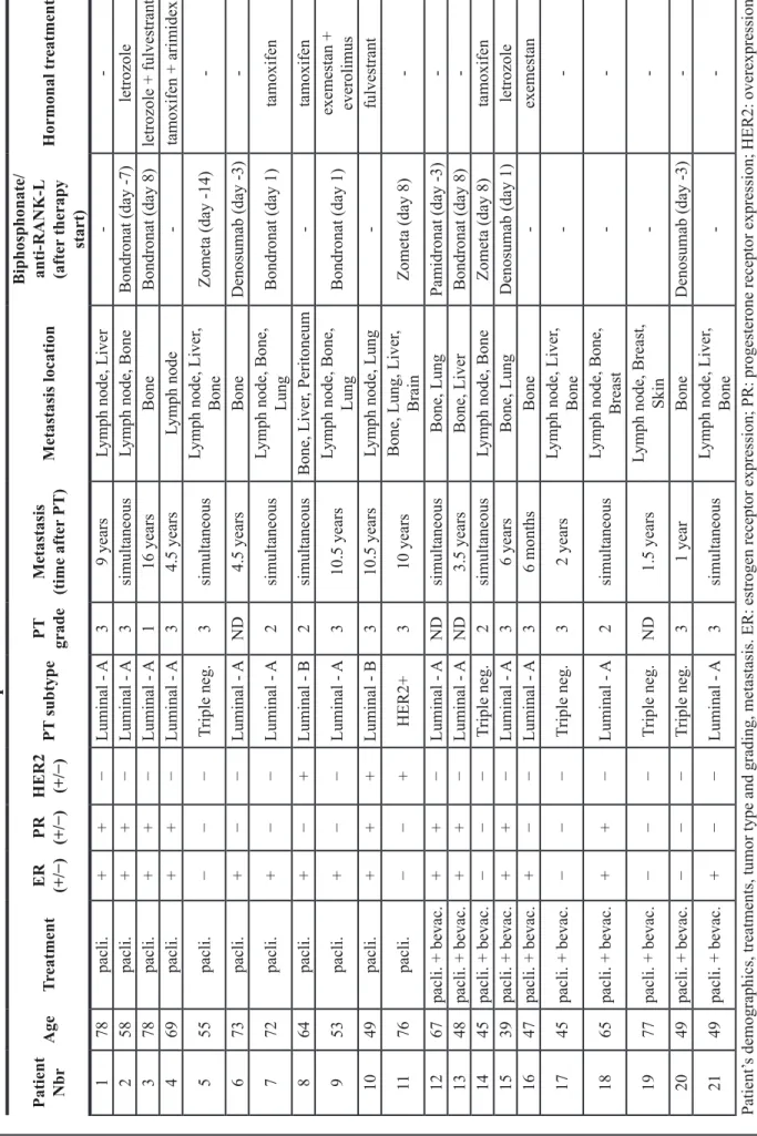
First we quantified the frequency of CEC and CEP in the blood of 21 metastatic breast cancer (mBC) patients and 12 age-matched healthy donors by cell surface staining and flow cytometry following the protocol by Bertolini et al. [24, 25] (Table 1). CEC were defined as CD45−DNA+CD146+CD31+ cells and CEP as CD45− DNA+CD133+CD34+ cells. Compared to healthy donors, breast cancer patients had elevated levels of CEC and CEP (Figure 1A–1B). CEPs in mBC patients were also significantly more positive for VEGFR2 expression compared to healthy donors (Figure 1C). Surprisingly the fraction of dying CEP, but not CEC, determined using 7AAD+ permeability staining, was significantly higher in mBC patients (Figure 1D–1E).
bevacizumab treatment has no additional effect on cEc and cEP over paclitaxel chemotherapy
To monitor the effect induced by paclitaxel ± bevacizumab on circulating CEC and CEP, we analyzed blood from 11 patients upon breast metastasis detection treated first line with one cycle of paclitaxel chemotherapy alone and from 10 patients treated with paclitaxel in combination with bevacizumab. Blood was taken before therapy start (day 0) and at days 8 and 15 immediately before the administration of the treatment. Under chemotherapy the frequency of CEC dropped by over 80% regardless of the presence of bevacizumab (Figure 1F, left panel). The number of CEP doubled at day 8 of therapy with paclitaxel and bevacizumab (although the difference is not statistically significant due to large variability in the values) and returned to normal at day 15. Therapy with paclitaxel alone had no effect.
These data demonstrate the presence of elevated levels of blood circulating CEC and CEP in patients with mBC, and that bevacizumab treatment has no effect on their frequency in addition to chemotherapy at the investigated time points.
Increased frequency of circulating tIE2+cD11b+
and KIt+cD11b+ cells in mbc patients
We previously observed elevated levels of VEGFR1+CD11b+ [26], KIT+CD11b+ [27] and JAM1+CD11b+
table 1:
c
linical data of metastatic br
east cancer
patients
Patient Nbr Age tr eatment E r (+/−) Pr (+/−) HE r 2 (+/−) Pt subtype Pt grade Metastasis (time after PT) Metastasis location b iphosphonate/ anti-r ANK-L (after therapy start) Hormonal tr eatment 1 78 pacli. + + − Luminal - A 3 9 yearsLymph node, Liver
-2 58 pacli. + + − Luminal - A 3 simultaneous
Lymph node, Bone
Bondronat (day -7) letrozole 3 78 pacli. + + − Luminal - A 1 16 years Bone Bondronat (day 8) letrozole + fulvestrant 4 69 pacli. + + − Luminal - A 3 4.5 years Lymph node -tamoxifen + arimidex 5 55 pacli. − − − Triple neg. 3 simultaneous
Lymph node, Liver
, Bone Zometa (day -14) -6 73 pacli. + − − Luminal - A ND 4.5 years Bone Denosumab (day -3) -7 72 pacli. + − − Luminal - A 2 simultaneous
Lymph node, Bone,
Lung Bondronat (day 1) tamoxifen 8 64 pacli. + − + Luminal - B 2 simultaneous Bone, Liver , Peritoneum -tamoxifen 9 53 pacli. + − − Luminal - A 3 10.5 years
Lymph node, Bone,
Lung Bondronat (day 1) exemestan + everolimus 10 49 pacli. + + + Luminal - B 3 10.5 years
Lymph node, Lung
-fulvestrant 11 76 pacli. − − + HER2+ 3 10 years
Bone, Lung, Liver
, Brain Zometa (day 8) -12 67 pacli. + bevac. + + − Luminal - A ND simultaneous Bone, Lung Pamidronat (day -3) -13 48 pacli. + bevac. + + − Luminal - A ND 3.5 years Bone, Liver Bondronat (day 8) -14 45 pacli. + bevac. − − − Triple neg. 2 simultaneous
Lymph node, Bone
Zometa (day 8) tamoxifen 15 39 pacli. + bevac. + + − Luminal - A 3 6 years Bone, Lung Denosumab (day 1) letrozole 16 47 pacli. + bevac. + − − Luminal - A 3 6 months Bone -exemestan 17 45 pacli. + bevac. − − − Triple neg. 3 2 years
Lymph node, Liver
, Bone -18 65 pacli. + bevac. + + − Luminal - A 2 simultaneous
Lymph node, Bone,
Breast -19 77 pacli. + bevac. − − − Triple neg. ND 1.5 years
Lymph node, Breast,
Skin -20 49 pacli. + bevac. − − − Triple neg. 3 1 year Bone Denosumab (day -3) -21 49 pacli. + bevac. + − − Luminal - A 3 simultaneous
Lymph node, Liver
, Bone -Patient’ s demographics, treatments, tumor type and grading, metastasis. ER: estrogen receptor expression; PR: progesterone receptor expression; HER2: overexpression of HER-2/neu; PT : primary tumor .
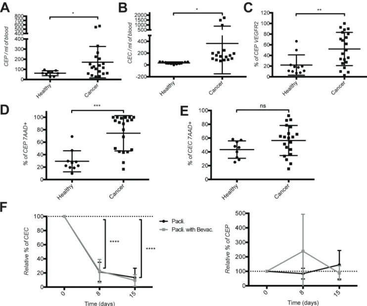
(unpublished data) in murine models of breast cancer and TIE2+CD11b+ cells in mBC cancer patients [28]. We therefore hypothesized that the frequency of CD11b+ cells and subsets thereof could potentially reflect the presence of a tumor and possibly patients’ response to the treatment. To test this hypothesis we used flow cytometry to measure CD11b+ cell subsets in healthy donors and in patients with mBC before and during therapy. Frequencies of circulating TIE2+CD11b+ and KIT+CD11b+ cells, but not VEGFR1+CD11b+ or JAM1+CD11b+ cells were significantly elevated in mBC patients compared to healthy donors (Figure 2A–2D). JAM1+ or VEGFR1+ cells represented a large fraction of the CD11b+ cell population (approx. 90% and 70%, respectively). In contrast, the relative frequency of TIE2+ cells within the CD11b+ cells was very low (< 1% in average), and that of
KIT+ cells was spread over a large spectrum, ranging from 2–5% to 60% of the CD11b+ cells. Importantly, KIT+CD11b+ cells were extremely rare in healthy donors (0.3% of CD11b+ cells).
As we observed a large variation of KIT protein expression at the cell surface of mBC patients, we tested whether KIT mRNA expression in CD11b+ blood cells was more homogeneous. CD11b+ cells were isolated from fresh blood of mBC patients and healthy donors by MACS technology and analyzed for KIT mRNA levels by qPCR. Purity of isolated CD11b+ cells was > 98% (Supplementary Figure 1). Surprisingly results revealed that KIT mRNA expression levels were very low in mBC patients and indistinguishable from healthy donors (Supplementary Figure 2).
Figure 1: Increased frequency of cEc and cEP in the blood of metastatic breast cancer patients. Quantification of (A) CEP, (b) CEC, (c) CEP expressing VEGFR2, (D) dying CEP using 7AAD staining, and (E) dying CEC using 7AAD staining in the blood of healthy donors and metastatic breast cancer patient before therapy start. (F) Quantification of CEC and CEP under chemotherapy ± bevacizumab treatments in the blood of metastatic breast cancer patients. Cell quantifications were performed by flow cytometry and data are represented as mean +/− SD.
bevacizumab reduces the frequency of circulating KIt+cD11b+ in paclitaxel-treated
patients
Next we measured the effect of therapy on CD11b+ cell subsets. Therapy with ± bevacizumab did not significantly alter the frequency of VEGFR1+CD11b+, JAM1+CD11b+ or TIE2+CD11b+ cells (Figure 2E–2G). In
contrast, therapy with paclitaxel caused a significant 2-fold increase in the frequency of circulating KIT+CD11b+ cells at day 15, while addition of bevacizumab resulted in a significant drop (60–80%) at days 8 and 15 (Figure 2H).
From these experiments we concluded that circulating TIE2+CD11b+ and KIT+CD11b+ cells are enriched in mBC, and bevacizumab decreases KIT+CD11b+, but not TIE2+CD11b+ cells.
Figure 2: Increased frequency of tIE2+cD11b+ and KIt+cD11b+ cells in the blood of metastatic breast cancer patients and decreased frequency of KIt+cD11b+ cells by bevacizumab therapy. Quantification of CD11b+ cells expressing (A) VEGFR1, (b) JAM1, (c) TIE2 and (D) KIT in the blood of healthy donors and breast cancer patients before therapy start. Quantification under both chemotherapy treatments of CD11b+ cells expressing (E) VEGFR1, (F) JAM1, (G) TIE2 and (H) KIT in the blood of metastatic breast cancer patients. Cell quantifications were performed by flow cytometry and data are represented as mean +/− SD.
circulating cD11b+ cells in mbc patients have
a distinctive gene expression profile compared to healthy donors
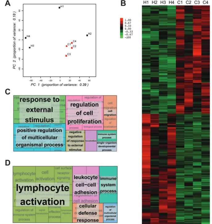
To further characterize CD11b+ cells in mBC patients we performed RNASeq analysis on CD11b+ cells isolated from 4 healthy donors and 4 cancer patients. Principal Component Analysis (PCA) revealed that the gene expression profiles of CD11b+ cells derived from mBC patients clustered together while those from CD11b+ cells derived from healthy donors were dispersed (Figure 3A). This result suggests that in healthy conditions CD11b+ cells have rather heterogeneous gene expression across individuals, while in cancer a re-programming takes place resulting in a more homogenous gene expression across patients. Self-organizing heat-maps confirmed that CD11b+ cells from cancer patients and healthy donors display different gene expression profiles (Figure 3B). Interestingly, among the most differentially expressed genes, we observed that CD11b+ cells from mBC patients expressed genes of M2 monocytes’ activation state (i.e. IL-10 and CCL20) (Supplementary Table 1). Gene ontology analysis of the differentially expressed genes (Figure 3C–3D and Supplementary Tables 2–3) reveled that expression of immune response genes was reduced in mBC, consistent with a M2 tumor-induced CD11b+ cells polarization.
cD11b+ monocytes in mbc patient display a M2
activation phenotype
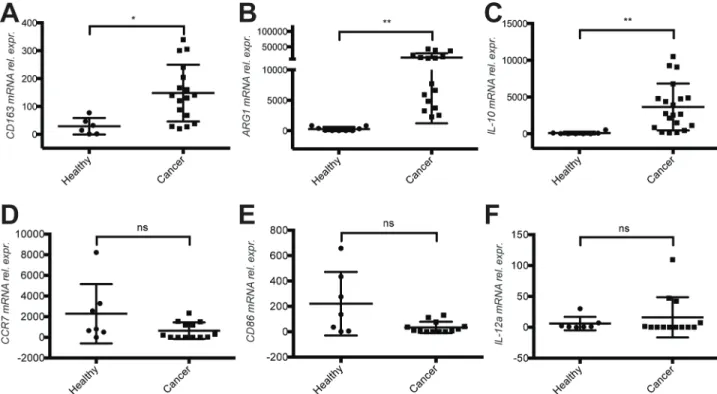
To strengthen this observation we monitored the expression of additional transcripts associated with M2 activation states in CD11b+ cells derived from healthy donors and mBC patients. IL-10, ARG1 and CD163, typical M2 activation markers, were highly up regulated in mBC patient (Figure 4A–4C). In contrast, the M1 activation markers CCR7, CD86 and IL-12a were expressed at lower levels, albeit not significantly (Figure 4D–4F). We also validated the increased expression of fibronectin mRNA in CD11b+ cells of mBC patients (Supplementary Table 1 and Supplementary Figure 3).
From these results we concluded that circulating CD11b+ cells in mBC have a different gene expression profile compared to healthy donors, and express genes associated with a M2, but not M1 activation.
Increased levels of the circulating M2 cytokines IL-10 and ccL20 in mbc patients
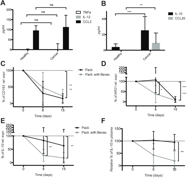
To collect additional evidence that the M2 activation in mBC was a systemic event, we measured selected M1 and M2 cytokines in the plasma of healthy donors and mBC patients. The M1 cytokines TNFα and IL-12 were below detection levels, while CCL2 was detectable, but no differences were observed (Figure 5A). In contrast,
the level of the M2 cytokines IL-10 and CCL20 was significantly higher in mBC patients compared to healthy donors (Figure 5B). These results corroborate the mRNA expression data consistent with a M2 activation state of circulating CD11b+ cells in mBC patients.
bevacizumab diminishes IL-10 mrNA
expression in cD11b+ cells and protein level in
plasma
Next we monitored the effect of therapy on the elevated M2 markers. Due to limited number of CD11b+ cells and derived mRNA recovered form treated patients, we focused the analysis on a few genes only. The mRNA expression level of CD163 and ARG1 clearly decreased in cancer patients under chemotherapy regardless of the presence of bevacizumab, although bevacizumab accelerated the decrease of ARG1 (Figure 5C–5D). Importantly, we observed a significant decrease of IL- 10 both at mRNA level in CD11b+ cells and at protein levels in the plasma of patients treated with bevacizumab in addition to paclitaxel (Figure 5E and 5F). Furthermore we observed a stronger increase in CD86 mRNA level in CD11b+ cells and CCL2 protein level (both reflecting M1 activation state) in plasma of mBC patients treated with paclitaxel + bevacizumab compared to patients treated with paclitaxel only. However due to a greater variability and the small number of patients, differences were not statistically significant (Supplementary Figure 4).
These results demonstrate that mBC patients have M2-activated circulating CD11b+ cells and elevated levels of M2 cytokines in plasma. Bevacizumab addition to chemotherapy causes a significant decrease of IL-10 mRNA levels in CD11b+ cells and IL-10 protein levels in plasma.
DIscUssION
The mode of action of bevacizumab is not yet fully understood. Beyond a rather transient anti-angiogenic effect, a possible more complex, systemic effect has been proposed [29, 30]. Additionally, immunomodulatory effects of anti-angiogenic therapies have been discussed [31]. To collect evidence supporting systemic, immunomodulatory effects of bevacizumab, and to identify candidate biomarkers to predict or monitor response to treatment, we investigated the effect of bevacizumab in addition to paclitaxel, on CEC, CEP and CD11b+ cell subsets in the blood of mBC patients.
Beyond the fact that CEC and CEP are present at higher frequency in the blood of mBC patients compared to healthy donors and that paclitaxel therapy decreases CEC independently of bevacizumab, no relationship between anti-angiogenic treatment and CEP behavior are observed. We couldn’t validate the mobilization of CEP by paclitaxel and the inhibitory effect by bevacizumab previously reported in preclinical and clinical studies
[4, 8]. Rather we observed a transient (at day 8) but statistically non-significant increase of CEP in patients treated with paclitaxel and bevacizumab compared to paclitaxel treatment alone. The discrepancy of the observation on CEP may be due to the different time points analyzed (a few hours vs. 8 and 15 days after therapy start) and to differences in the definition and analysis of CEP across these studies.
In contrast, we observed a KIT+CD11b+ population present only in the peripheral blood of cancer patients and whose frequency decreased significantly under therapy with paclitaxel and bevacizumab compared to paclitaxel only. This effect is specific, as three additional subpopulations of CD11b+ cells (VEGFR1+, JAM1+, TIE2+) were not affected. We have previously reported that circulating KIT+CD11b+ cells promote metastasis
Figure 3: cD11b+ myelomonocytic cells from metastatic breast cancer patients and healthy donors show different expression profiles. (A) PCA plot representing differential clustering of cancer patients (C1-4) and healthy donors (H1-4) based on mRNA expression profiles of CD11b+ cells. (b) Self-organizing heat-map of the top 100 genes with greatest variability across all samples, showing different expression profiles in CD11b+ cells from cancer patients compared to healthy donors. (c) Gene ontology analysis showing the most up-regulated biological processes comparing cancer patients and healthy donors. (D) Gene ontology analysis showing the most down-regulated biological processes comparing cancer patients and healthy donors.
in the 4T1 murine model of breast cancer relapse after radiotherapy [27]. Little was known, however, about the clinical and biological significance of KIT+CD11b+ cells in patients. Here we demonstrate that such cells circulate in the blood of patients with metastatic breast cancer but are virtually absent in healthy individuals. This observation raises several questions. A critical one is whether KIT+CD11b+ cells might be a potential biomarker paralleling activity of, or predicting response to, bevacizumab treatment. The broad distribution of KIT+CD11b+ frequency suggests that patients may be heterogeneous in term of KIT+CD11b+ mobilization or recruitment. The potential therapeutic significance of this observation is open at this point, and further studies including more patients and monitoring clinical outcome are necessary to address it. The origin, role and fate of these cells in cancer progression are also intriguing. The discrepancy observed between levels of KIT protein and mRNA is consistent with immature myeloid-derived cells shutting down KIT expression upon mobilization. These cells, once in the blood circulation, may be on the way to differentiation and therefore no longer express KIT mRNA, while still carry KIT protein at the cell surface. In the 4T1 model we reported that mobilized Kit+CD11b+ cells rapidly lose KIT surface expression upon adoptive transfer [27].
The gene expression profiling of CD11b+ cells demonstrated significant differences between cancer patients and healthy donors. Strikingly, expression profiles of
CD11b+ cells from cancer patients were more homogeneous compared to those of healthy donors. This is consistent with the functional heterogeneity CD11b+ cells in healthy individuals reflecting physiological individualistic behaviors vs. a tumor-driven CD11b+ reprogramming resulting in more homogenous gene expression profiles. Consistent with this notion, we observed the expression of transcripts for M2 activation markers in CD11b+ cells of cancer patients in comparison to CD11b+ cells from healthy controls. These observations are corroborated by higher levels of IL-10 and CCL20, two M2-associated cytokines, in the plasma of cancer patients. IL-10 is a well-known immune suppressive cytokine produced during cancer progression [32]. CCL20 is involved in promoting tumor growth, migration and EMT transition in breast cancer in vitro models [33] and it also plays a role in recruiting immature dendritic cells and in tumor-promoting Tregs in other cancer types [34]. We also found elevated fibronectin (FN1) transcript in CD11b+ cells of cancer patients. Interestingly, fibronectin deposition is observed in the metastatic niche in preclinical models in associated in with recruitment of CD11b+ cells [35] and with cancer progression, including of the breast, in human [36].
Unfortunately it was not possible to monitor the therapy effect on the M2 phenotype by RNASeq due to insufficient amounts and low quality material. However, we were able to measure selected transcripts for M1 and M2 markers in CD11b+ cells and M1 and M2 cytokines in the plasma of patients under therapy. Most of them
Figure 4: Evidence for a M2 activation phenotype of cD11b+ cells in the blood of metastatic breast cancer patients. (A) Quantification of the mRNA expression levels of M2 markers CD163, (b) ARG1 and (c) IL-10 in CD11b+ cells derived from the blood of healthy donors compare to cancer patients before therapy start. (D) Quantification of mRNA expression levels of M1 markers CCR7, (E) CD86 and (F) IL-12α at mRNA level in CD11b+ cells of the same healthy donors and cancer patients. Analysis was performed by real time qPCR. All data are represented as mean +/− SD.
didn’t change or were below detection level (data not show). Nevertheless we observed significant and specific decrease in the M2 markers ARG1 at mRNA level (albeit ARG1 was only significant at day 8) and IL-10 cytokine secretion under bevacizumab therapy, and a consistent, but not significant, trend in increased M1 markers CD86 and CCL2 cytokine production. These results suggest that bevacizumab could prompt an immune switch from a pro-tumoral M2 activity toward an anti-pro-tumoral M1 activity.
Tumor-induced M2 polarization and cytokine production may reach beyond local reprogramming occurring in the tumor microenvironment and may have systemic relevance [28, 37].
These observations may have practical implications to breast cancer diagnosis, treatment and monitoring. Circulating KIT+CD11b+ cells and IL-10 levels (or more generally M2/M1 marker ratio) could serve as potential biomarkers to follow patient’s response to bevacizumab
Figure 5: Increased expression of the M2 cytokines IL-10 and ccL20 in the plasma of metastatic breast cancer patients and decrease of IL-10 levels by bevacizumab. (A) Quantification of M1 cytokines TNFα, IL-12p70 and CCL2 in plasma of healthy donors and cancer patients before therapy start. (b) Quantification of M2 cytokines IL-10 and CCL20 in the plasma of the same healthy donors and cancer patients before therapy start. (c) Quantification of CD163, (D) ARG1 and (E) IL-10 mRNA expression variation in CD11b+ cells of cancer patient during therapy with paclitaxel ± bevacizumab using real time qPCR. (F) Quantification of IL-10 in the plasma of cancer patients during therapy with paclitaxel ± bevacizumab. Cytokines were measured by ELISA. All data are represented as mean +/− SD.
treatment. This might be particularly relevant in conditions where bevacizumab effects may be restricted to a subset of the patients, as it appears to occur in metastatic breast cancer. Their modulation is consistent with an immunostimulatory effect of bevacizumab. Interestingly, preclinical and clinical evidence suggest that response to chemotherapy involves the activation of the immune system and induction of an anti-tumor immune response [38, 39]. Thus bevacizumab, in addition to its anti-angiogenic effects, might provide therapeutic benefits by reversing the M2-associated state of immunosuppression. Larger studies are needed to test this hypothesis.
Most patients received bisphosphonates (or an anti-RANK-L antibody) as part of their therapies due to bone metastasis (Table 1), but no correlations were observed between marker levels and bisphosphonate (or anti-RANK-L) treatment (Supplementary Figures 5 and 6), except a transient increased mobilization of CEP at day 8 of therapy with paclitaxel and bevacizumab. Bisphosphonates have been involved in modulation of bone-resident macrophage activity including reprogramming from tumorigenic M2 status to an anti-tumoral M1 status [40]. Bisphosphonate effects on circulating CD11b+ cells cannot be confirmed by our data (data not show). Also, the limited number of patients did not allow drawing an association between a particular subtype of breast cancer and circulating cells (Supplementary Figure 7). In addition, some patients also received in parallel an endocrine therapy based on their cancer subtype (Table 1). These treatments also did not have any effect on our cells and markers levels (data not show). The fact that some patients had primary tumor surgery more then 6 months before treatment start (Table 1) also did not have any significant impact on the observed changes on CEC, CEP, KIT+ CD11b+ cells and M2 markers (data not show). It should be emphasized that considering the small number of patients analyzed, these lack of correlations do not exclude that some correlations might be observed in larger patients populations.
In summary, we demonstrate that metastatic breast cancer patients have an increased frequency of circulating KIT+CD11b+ cells, M2-polarized CD11b+ cells and elevated levels of the M2 cytokine IL-10 and CCL20. Compared to paclitaxel monotherapy only, co-treatment with bevacizumab significantly decreased KIT+CD11b+ cells and IL-10. These results warrant the further investigation of changes in circulating KIT+CD11b+ cells and IL-10 levels as candidate biomarkers of bevacizumab activity in metastatic breast cancer and possibly other cancer types. They also provide original evidence for a systemic immunomodulatory effect of bevacizumab. We are planning a larger study to address these questions.
MAtErIALs AND MEtHODs
clinical data and materialThe study was approved by the regional ethic boards of St.Gallen and Zurich, Switzerland. We studied 21 female patients (Table 1) with metastatic breast cancer who received first line treatment of palliative chemotherapy with either paclitaxel on day 1, 8 and 15 of a 28 days cycle, or paclitaxel (same schedule) in combination with bevacizumab on day 1 and 15. From these patients, blood samples were collected before administration of the first therapy at day 0, and before therapy on day 8 and 15. All donors gave written informed consent before study entry. Patients were recruited in St.Gallen Kantonspital, Zürich Oncozentrum and Tumor- und Brustzentrum in Chur upon diagnosis of breast cancer metastasis and before starting any new treatment. Samples from 12 age-matched healthy females were analyzed accordingly. Age mean for cancer patient was 56.5 years and for healthy donors was 58 years old (all patients were between 39 and 78 years old). Healthy donors were recruited at HFR Hôpital Cantonal Fribourgeois in parallel to breast cancer patient recruitment, based on the following criteria: age-matched relative to breast cancer patient, no surgical intervention in the past 10 years, no regular medications in the last 6 months, no previous cancer diagnosis, no chronic diseases and normal blood analyses at time of recruitment.
blood processing
Blood was collected using BD Vacutainer® Blood Collection EDTA Tubes (Becton Dickinson, Franklin Lakes, NJ, USA) following manufacturer’s instructions. All following analysis were performed without 24 h after blood collection to minimize cell death. Plasma and total white blood cells were isolated using BD Vacutainer® CPT™ Cell Preparation Tube (Becton Dickinson) with Sodium Heparin following manufacturer’s instructions of use. Plasma fraction was immediately frozen at −80°C. CD11b+ cells were isolated from total blood using MACS® separation technique, following manufacturer’s instructions. MS columns and anti-human/mouse CD11b micro-beads were used. All reagents were from Miltenyi Biotec (Bergisch Gladbach, Germany). Isolated CD11b+ cells were immediately lysed in RNA stable RLT lysis buffer (Qiagen, Venlo, Netherlands) and stored at −80°C.
Flow cytometry
Whole blood staining’s were performed within 24 hours after blood collection. White blood cells were counted using Cell-Dyn Sapphire Hematology System (Abbott Diagnostics, Chicago, IL, USA). For CEC and CEP staining 5 millions cells per tube were used, while
for monocyte and isotype staining’s 1 million cells was used. Directly labeled antibodies were added on total blood and incubated for 20 minutes at 4°C, followed by 10 minutes red-blood-cells lysis (Bühlmann Laboratories, Schönenbuch, Switzerland) and washing using cold PBS. All anti-human antibodies were used at the concen-tration give in manufacturer’s instructions of use: anti-CD45-V450, anti-CD146-Pe, anti-CD31-AlexaFluo 647, anti-CD34-PeCy7, anti-VEGFR2-Pe, anti-CD11b-V450, anti-CD64-FITC, anti-JAM1-Pe, anti-TIE2-AlexaFluo 647, anti-KIT-PeCy7, anti-CD195-Pe, mouse IgG2a-AlexaFluor 647, mouse Pe, mouse PeCy7, mouse IgG1-APC, mouse IgG2a-Pe, 7AAD (all from Becton Dickinson), Syto16 (Life Technologies, Carlsbad, CA, USA), anti-CD133-APC (Miltenyi), anti-VEGFR1-APC (R & D Biosystems, Minneapolis, MN, USA).
BD FACSCanto II (Becton Dickinson) instrument was used to perform experiments and FlowJo 9.8.3 (Treestar Inc., Ashland, OR, USA) software was used to analyze all data.
ELIsA
For TNF-α, IL-12p70, IL-10, CCL20 and CCL2 quantification in plasma ELISA MAX Deluxe assays from Biolegend (San Diego, CA, USA) was used following manufacturer’s instructions. Each sample was run in triplicate and absorption was measured at 450 nm (VICTOR X3 Reader, PerkinElmer, Waltham, MA, USA).
real-time qPcr
Total RNA from CD11b+ isolated cells was extracted using the RNeasy Mini kit from Qiagen. The quality and quantity of all RNA samples were examined by Agilent 2100 Bioanalyzer (Agilent Technologies, Santa Clara, CA, USA) and NanoDrop (Witec AG, Luzern, Switzerland). Total RNA was retro-transcribed using Ovation Nugen RNASeq System v2 kit following manufacturer’s instructions (Nugen, San Carlos, CA, USA) using 15 ng of total RNA. cDNA was subjected to amplification by real time qPCR with the StepOne SYBR System (Life Technologies) using the following primer pairs (Eurofins Genomics, Huntsville, AL, USA) at the indicated hybridization temperatures: GAPDH 58°C (Fw-TCTTCTTTTGCGTCGCCAGC, Rev-GATT TTGGAGGGATCTCGCTCCT), CCR7 58°C (Fw- ATG GACCTGGGGAAACCAAT, Rev- TGATAGGGAGGAA CCAGGCT), CD86 58°C (Fw- TCATTGCCGAGGAA GGCTTG, Rev- CTCCATTGTGTTGGTTCCACATT), IL-12a 60°C (Fw- TTCCCATGCCTTCACCACTC, Rev- ACT CCCATTAGTTATGAAAGAGGTC), CD163 58°C (Fw- GCGGCTTGCAGTTTCCTCAA, Rev- CTCAGAATG GCCTCCTTTTCCA), ARG1 58°C (Fw- GGAGTC ATCTGGGTGGATGC, Rev- CTGGCACATCGGGAATC TTTC), IL-10 58°C (Fw- CGAGATGCCTTCAGCA
GAGT, Rev- AATCGATGACAGCGCCGTAG), KIT 57°C (Fw- GATTATCCCAAGTCTGAGAATGAA, Rev- CGTC AGAATTGGACACTAGGA). FN1 52°C (Fw- ACTTC GACAGGACCACTTGA, Rev- TCAAATTGGAGATT CATGGGA). Real-time PCR data were then analyzed using the comparative Ct method [41].
Gene expression analysis by rNAseq
RNASeq libraries preparation, data sequencing and bioinformatics analysis was performed following previously published methodology [42]. But purity-filtered reads were adapted and quality trimmed wit Cutadapt (v.1.3), filtered for low complexity with PRINSEQ (v.0.20.3) and aligned against Homo sapiens.GRCh37.75 genome using TopHat (v.2.0.9) [43, 44]. Quality of the RNASeq data alignment was assessed using RSeQC (v.2.3.7) [45]. The number of read counts per gene locus was summarized with htseq-count (v.0.6.1) using Homo sapiens.GRCh37.75 gene annotation [46]. Data normalization and differential expression analysis were performed in R (v.3.1.1), using Bioconductor packages DESeq2 (v.1.6.2) [47]. The 15996 genes with at least 1 count per million in at least one sample were kept for statistical analysis. Gene Ontology (GO) analysis was performed using Gorilla [48] and representations were generated using REVIGO [49]. Significant enriched GO terms were identified after Bonferroni correction (P value < 4.13e-06). The complete data set is publicly available at GEO through the accession number GSE70404.
statistical analysis
Acquired data were analyzed and graphics were generated using Prism Software (GraphPad, La Jolla, CA, USA). Statistical comparisons were performed by two-way ANOVA with Bonferroni post-test. Highest and lowest values from each group were excluded. Results were considered to be significantly from p < 0.05. In the figures the various p values thresholds are presented as follow: ≤ 0.05 =*, ≤ 0.01 =**, ≤ 0.001 =***, ≤ 0.0001 =****.
AcKNOWLEDGMENts
The authors wish to thank Dr. Rudolf Morant (Tumorzentrum ZeTup, CH-8040 Rapperswil-Jona), Prof. Dr. Florian Otto (Tumorzentrum ZeTup, CH-9006 St.Gallen), Dr. Brian Meehan (Kantonsspital, CH-9007 St.Gallen), and Dr. Jean-Luc Magnin (HFR, CH-1700 Fribourg) for participating to the study, as well as Dr. Keith Harshman at the Genomic Technologies Facility (CIG, UNIL, CH-1015 Lausanne) for genomic profiling. We wish to thank all patients for taking part to this study. We also thank Dr. Francesco Bertolini and Dr. Patrizia Mancuso (European Institute of Oncology, Milano) for their generous help in establishing the flow cytometry analysis of CEC and CEP.
Authorsʼ contributions
SC: planned and designed clinical study; designed and performed experiments; analyzed data; wrote the manuscript. BF: designed experiments; recruited healthy donors. SP: performed gene expression analysis and interpretation. AT: performed clinical study; recruited breast cancer patients. TR: performed clinical study; recruited breast cancer patients. CR: planned and designed clinical study; evaluated experiments; wrote the manuscript. GF: planned and designed clinical study; evaluated experiments; recruited breast cancer patients; wrote the manuscript.
FINANcIAL sUPPOrt
This work was supported by grants from the Swiss National Science Foundation (31003A_159824), the Medic Foundation to CR and the Foundation for Tumor-diagnostics and Prevention (STIFTUP, St.Gallen) to GF.
cONFLIcts OF INtErEst
The authors declare no conflicts of interest.
rEFErENcEs
1. Laurent J, Touvrey C, Botta F, Kuonen F, Ruegg C. Emerging paradigms and questions on pro-angiogenic bone marrow-derived myelomonocytic cells. Int J Dev Biol. 2011; 55:527–534.
2. Katz OB, Shaked Y. Host effects contributing to cancer therapy resistance. Drug Resist Updat. 2015; 19:33–42. 3. Mancuso P, Bertolini F. Circulating endothelial cells as
biomarkers in clinical oncology. Microvasc Res. 2010; 79: 224–228.
4. Shaked Y, Henke E, Roodhart JM, Mancuso P, Langenberg MH, Colleoni M, Daenen LG, Man S, Xu P, Emmenegger U, Tang T, Zhu Z, Witte L, et al. Rapid chemotherapy-induced acute endothelial progenitor cell mobilization: implications for antiangiogenic drugs as chemosensitizing agents. Cancer Cell. 2008; 14:263–273. 5. DeNardo DG, Brennan DJ, Rexhepaj E, Ruffell B,
Shiao SL, Madden SF, Gallagher WM, Wadhwani N, Keil SD, Junaid SA, Rugo HS, Hwang ES, Jirstrom K, et al. Leukocyte complexity predicts breast cancer survival and functionally regulates response to chemotherapy. Cancer Discov. 2011; 1:54–67.
6. Shojaei F, Wu X, Malik AK, Zhong C, Baldwin ME, Schanz S, Fuh G, Gerber HP, Ferrara N. Tumor refractoriness to anti-VEGF treatment is mediated by CD11b+Gr1+ myeloid cells. Nat Biotechnol. 2007; 25:911–920.
7. Moschetta M, Mishima Y, Sahin I, Manier S, Glavey S, Vacca A, Roccaro AM, Ghobrial IM. Role of endothelial progenitor cells in cancer progression. Biochim Biophys Acta. 2014; 1846:26–39.
8. Furstenberger G, von Moos R, Lucas R, Thurlimann B, Senn HJ, Hamacher J, Boneberg EM. Circulating endothelial cells and angiogenic serum factors during neoadjuvant chemotherapy of primary breast cancer. Br J Cancer. 2006; 94:524–531.
9. Jain S, Ward MM, O’Loughlin J, Boeck M, Wiener N, Chuang E, Cigler T, Moore A, Donovan D, Lam C, Cobham MV, Schneider S, Christos P, et al. Incremental increase in VEGFR1(+) hematopoietic progenitor cells and VEGFR2(+) endothelial progenitor cells predicts relapse and lack of tumor response in breast cancer patients. Breast Cancer Res Treat. 2012; 132:235–242.
10. Diaz-Montero CM, Salem ML, Nishimura MI, Garrett-Mayer E, Cole DJ, Montero AJ. Increased circulating myeloid-derived suppressor cells correlate with clinical cancer stage, metastatic tumor burden, and doxorubicin-cyclophosphamide chemotherapy. Cancer Immunol Immunother. 2009; 58:49–59.
11. Solito S, Falisi E, Diaz-Montero CM, Doni A, Pinton L, Rosato A, Francescato S, Basso G, Zanovello P, Onicescu G, Garrett-Mayer E, Montero AJ, Bronte V, et al. A human promyelocytic-like population is responsible for the immune suppression mediated by myeloid-derived suppressor cells. Blood. 2011; 118:2254–2265.
12. Ohki S, Shibata M, Gonda K, Machida T, Shimura T, Nakamura I, Ohtake T, Koyama Y, Suzuki S, Ohto H, Takenoshita S. Circulating myeloid-derived suppressor cells are increased and correlate to immune suppression, inflammation and hypoproteinemia in patients with cancer. Oncol Rep. 2012; 28:453–458.
13. Ferrara N. VEGF as a therapeutic target in cancer. Oncology. 2005; 69 Suppl 3:11–16.
14. Sessa C, Guibal A, Del Conte G, Ruegg C. Biomarkers of angiogenesis for the development of antiangiogenic therapies in oncology: tools or decorations? Nat Clin Pract Oncol. 2008; 5:378–391.
15. Lohmann AE, Chia S. Patients with metastatic breast cancer using bevacizumab as a treatment: is there still a role for it? Curr Treat Options Oncol. 2012; 13:249–262.
16. Stevenson CE, Nagahashi M, Ramachandran S, Yamada A, Bear HD, Takabe K. Bevacizumab and breast cancer: what does the future hold? Future Oncol. 2012; 8:403–414. 17. Fleitas T, Martinez-Sales V, Vila V, Reganon E, Mesado D,
Martin M, Gomez-Codina J, Montalar J, Reynes G. VEGF and TSP1 levels correlate with prognosis in advanced non-small cell lung cancer. Clin Transl Oncol. 2013; 15:897–902. 18. Liu Y, Starr MD, Brady JC, Rushing C, Bulusu A, Pang H,
Honeycutt W, Amara A, Altomare I, Uronis HE, Hurwitz HI, Nixon AB. Biomarker signatures correlate with clinical outcome in refractory metastatic colorectal cancer patients receiving bevacizumab and everolimus. Mol Cancer Ther. 2015; 14:1048–1056.
19. Clarke S, Burge M, Cordwell C, Gibbs P, Reece W, Tebbutt N. An Australian translational study to evaluate the prognostic role of inflammatory markers in patients with
metastatic ColorEctal caNcer Treated with bevacizumab (Avastin) [ASCENT]. BMC Cancer. 2013; 13:120. 20. Nixon AB, Pang H, Starr MD, Friedman PN, Bertagnolli MM,
Kindler HL, Goldberg RM, Venook AP, Hurwitz HI, Alliance for Clinical Trials In O. Prognostic and predictive blood-based biomarkers in patients with advanced pancreatic cancer: results from CALGB80303 (Alliance). Clin Cancer Res. 2013; 19:6957–6966.
21. Botta C, Barbieri V, Ciliberto D, Rossi A, Rocco D, Addeo R, Staropoli N, Pastina P, Marvaso G, Martellucci I, Guglielmo A, Pirtoli L, Sperlongano P, et al. Systemic inflammatory status at baseline predicts bevacizumab benefit in advanced non-small cell lung cancer patients. Cancer Biol Ther. 2013; 14:469–475.
22. Matsusaka S, Mishima Y, Suenaga M, Terui Y, Kuniyoshi R, Mizunuma N, Hatake K. Circulating endothelial progenitors and CXCR4-positive circulating endothelial cells are predictive markers for bevacizumab. Cancer. 2011; 117:4026–4032. 23. Curigliano G, Bagnardi V, Bertolini F, Alcalay M,
Locatelli MA, Fumagalli L, Rabascio C, Calleri A, Adamoli L, Criscitiello C, Viale G, Goldhirsch A. Antiangiogenic therapy in recurrent breast cancer with lymphangitic spread to the chest wall: A randomized phase II trial of bevacizumab with sequential or concurrent oral vinorelbine and capecitabine. Breast. 2015; 24:263–271. 24. Bertolini F, Mancuso P, Benayoun L, Gingis-Velitski S,
Shaked Y. Evaluation of circulating endothelial precursor cells in cancer patients. Methods Mol Biol. 2012; 904:165–172. 25. Mancuso P, Antoniotti P, Quarna J, Calleri A, Rabascio C,
Tacchetti C, Braidotti P, Wu HK, Zurita AJ, Saronni L, Cheng JB, Shalinsky DR, Heymach JV, et al. Validation of a standardized method for enumerating circulating endothelial cells and progenitors: flow cytometry and molecular and ultrastructural analyses. Clin Cancer Res. 2009; 15:267–273. 26. Laurent J, Hull EF, Touvrey C, Kuonen F, Lan Q, Lorusso G,
Doucey MA, Ciarloni L, Imaizumi N, Alghisi GC, Fagiani E, Zaman K, Stupp R, et al. Proangiogenic factor PlGF programs CD11b(+) myelomonocytes in breast cancer during differentiation of their hematopoietic progenitors. Cancer Res. 2011; 71:3781–3791.
27. Kuonen F, Laurent J, Secondini C, Lorusso G, Stehle JC, Rausch T, Faes-Van’t Hull E, Bieler G, Alghisi GC, Schwendener R, Andrejevic-Blant S, Mirimanoff RO, Ruegg C. Inhibition of the Kit ligand/c-Kit axis attenuates metastasis in a mouse model mimicking local breast cancer relapse after radiotherapy. Clin Cancer Res. 2012; 18:4365–4374.
28. Guex N, Crespo I, Bron S, Ifticene-Treboux A, Faes-Van’t Hull E, Kharoubi S, Liechti R, Werffeli P, Ibberson M, Majo F, Nicolas M, Laurent J, Garg A, et al. Angiogenic activity of breast cancer patients’ monocytes reverted by combined use of systems modeling and experimental approaches. PLoS Comput Biol. 2015; 11:e1004050. 29. Carmeliet P, Jain RK. Molecular mechanisms and clinical
applications of angiogenesis. Nature. 2011; 473:298–307.
30. Potente M, Gerhardt H, Carmeliet P. Basic and therapeutic aspects of angiogenesis. Cell. 2011; 146:873–887.
31. Huang Y, Yuan J, Righi E, Kamoun WS, Ancukiewicz M, Nezivar J, Santosuosso M, Martin JD, Martin MR, Vianello F, Leblanc P, Munn LL, Huang P, et al. Vascular normalizing doses of antiangiogenic treatment reprogram the immunosuppressive tumor microenvironment and enhance immunotherapy. Proc Natl Acad Sci USA. 2012; 109:17561–17566.
32. Yang L, Carbone DP. Tumor-host immune interactions and dendritic cell dysfunction. Adv Cancer Res. 2004; 92:13–27. 33. Marsigliante S, Vetrugno C, Muscella A. Paracrine CCL20
loop induces epithelial-mesenchymal transition in breast epithelial cells. Mol Carcinog. 2015.
34. Zhang CY, Qi Y, Li XN, Yang Y, Liu DL, Zhao J, Zhu DY, Wu K, Zhou XD, Zhao S. The role of CCL20/CCR6 axis in recruiting Treg cells to tumor sites of NSCLC patients. Biomed Pharmacother. 2015; 69:242–248.
35. Kaplan RN, Rafii S, Lyden D. Preparing the “soil”: the premetastatic niche. Cancer Res. 2006; 66:11089–11093. 36. Oskarsson T. Extracellular matrix components in breast cancer
progression and metastasis. Breast. 2013; 22 Suppl 2:S66–72. 37. Ruffell B, Coussens LM. Macrophages and therapeutic
resistance in cancer. Cancer Cell. 2015; 27:462–472. 38. Pol J, Vacchelli E, Aranda F, Castoldi F, Eggermont A,
Cremer I, Sautes-Fridman C, Fucikova J, Galon J, Spisek R, Tartour E, Zitvogel L, Kroemer G, et al. Trial Watch: Immunogenic cell death inducers for anticancer chemotherapy. Oncoimmunology. 2015; 4:e1008866. 39. Galluzzi L, Senovilla L, Zitvogel L, Kroemer G. The secret
ally: immunostimulation by anticancer drugs. Nat Rev Drug Discov. 2012; 11:215–233.
40. Rogers TL, Holen I. Tumour macrophages as potential targets of bisphosphonates. J Transl Med. 2011; 9:177. 41. Schmittgen TD, Lee EJ, Jiang J, Sarkar A, Yang L, Elton TS,
Chen C. Real-time PCR quantification of precursor and mature microRNA. Methods. 2008; 44:31–38.
42. Knuchel S, Anderle P, Werfelli P, Diamantis E, Ruegg C. Fibroblast surface-associated FGF-2 promotes contact-dependent colorectal cancer cell migration and invasion through FGFR-SRC signaling and integrin alphavbeta5-mediated adhesion. Oncotarget. 2015; 6:14300–14317. doi: 10.18632/oncotarget.3883.
43. Schmieder R, Edwards R. Quality control and preprocessing of metagenomic datasets. Bioinformatics. 2011; 27:863–864. 44. Kim D, Pertea G, Trapnell C, Pimentel H, Kelley R,
Salzberg SL. TopHat2: accurate alignment of transcriptomes in the presence of insertions, deletions and gene fusions. Genome Biol. 2013; 14:R36.
45. Wang L, Wang S, Li W. RSeQC: quality control of RNA-seq experiments. Bioinformatics. 2012; 28:2184–2185. 46. Anders S, Pyl PT, Huber W. HTSeq—a Python
framework to work with high-throughput sequencing data. Bioinformatics. 2015; 31:166–169.
47. Love MI, Huber W, Anders S. Moderated estimation of fold change and dispersion for RNA-seq data with DESeq2. Genome Biol. 2014; 15:550.
48. Eden E, Navon R, Steinfeld I, Lipson D, Yakhini Z. GOrilla: a tool for discovery and visualization of enriched GO terms in ranked gene lists. BMC Bioinformatics. 2009; 10:48.
49. Supek F, Bosnjak M, Skunca N, Smuc T. REVIGO summarizes and visualizes long lists of gene ontology terms. PLoS One. 2011; 6:e21800.