HAL Id: tel-02117873
https://tel.archives-ouvertes.fr/tel-02117873
Submitted on 2 May 2019
HAL is a multi-disciplinary open access archive for the deposit and dissemination of sci-entific research documents, whether they are pub-lished or not. The documents may come from teaching and research institutions in France or abroad, or from public or private research centers.
L’archive ouverte pluridisciplinaire HAL, est destinée au dépôt et à la diffusion de documents scientifiques de niveau recherche, publiés ou non, émanant des établissements d’enseignement et de recherche français ou étrangers, des laboratoires publics ou privés.
Cell-based partial pulp regeneration in a porcine
preclinical model
Francesca Mangione
To cite this version:
Francesca Mangione. Cell-based partial pulp regeneration in a porcine preclinical model. Human health and pathology. Université Sorbonne Paris Cité, 2017. English. �NNT : 2017USPCB046�. �tel-02117873�
Université Paris Descartes
Ecole doctorale BioSorbonne Paris CitéLaboratoire EA2496 Pathologies, Imagerie et Biothérapie Orofaciales
Cell-based partial pulp
regeneration in a porcine
preclinical model
Par Francesca Mangione
Thèse de doctorat de Biologie Cellulaire
Dirigée par Madame le Pr S. VITAL
Présentée et soutenue publiquement le 10 Octobre 2017
Devant un jury composé de:
Pr GALLER Kerstin Rapporteur
Pr LAMBRICHTS Ivo Rapporteur
Pr CHAUSSAIN Catherine Examinateur
Pr JACOBS Reinhilde Examinateur
A Daniel
La bellezza del cosmo é data non solo dalla unità nella varietà, ma anche dalla varietà nell’unità. (Umberto Eco)
Acknowledgments
At the conclusion of my PhD, I would like to sincerely thank all the persons who directly or indirectly contributed to this important experience of my professional as well as personal life. First of all my gratitude goes to Prof. Sibylle Vital who directed this work and patiently guided my evolution as a researcher. I am very grateful to Prof. Catherine Chaussain who accepted me in her laboratory 4 years ago and constantly gave support to Sibylle and me during decisive moments of this experience. My most sincere thank you is addressed to Prof. Galler and Prof. Lambrichts for accepting to be the referees and part of the committee of my thesis. For sure I am grateful to Prof. Jacobs, Prof. Simon, Prof. Vital and Prof. Chaussain for being part of the committee: I am very honoured of your presence.
Then I would like to address a special thanks to Prof. Jacobs for her very kind welcome in her laboratory in Leuven during 3 months: it has been a honour for me and this experience has been amazing.
So I cannot forget to sincerely thank all the team of Oral and Maxillo-facial Surgery - Imaging & Pathology (OMFS-IMPATH) of the University of Leuven (KU Leuven), and especially my friends Mostafa, Laura, Jeroen and Yan: you all teached me a lot and you gave me a lot of support.
I am very thankful to the laboratory Cr2i of INRA (Jouy-en-Josas) in particular to Michel Bonneau and Chantal Kang for they very precious collaboration and constant help.
I am grateful to University Paris Descartes, National French Agency for Research (Grant PulpCell ANR-14-CE16-0006-01) and Fondation des Maladies Rares (Grant large animal experiment LAM-RD-U201140104) that supported this work with their grants.
Tchilalo Boukpessi, Elvire Le Norcy, Céline Gaucher, Marjolaine Gosset, Gaël Rochefort, Caroline Gorin, Dominique Septier, Dominique Le-Denmat, Brigitte Baroukh, Annie Llorens, Jeremy Sadoine, Sandy Ribes, Elisabeth Jimenez, Maheva Garcia, Tania Selbonne and Alexandra Benoist. A special thank to Julie Lesieur for her teaching and helping as well as for her involvement in this project.
Thank you to all my colleagues and friends from « la salle des étudiants » and from the hospital Albert Chenevier.
I am grateful to University Paris Descartes, National French Agency for Research (Grant PulpCell ANR-14-CE16-0006-01) and Fondation des Maladies Rares (Grant large animal experiment LAM-RD-U201140104) that supported this work with their grants. Finally I would like to express all my sincere gratitude to my parents Luigi and Fiorella, my brother Fabio, my sister in law Francesca, my gorgeous niece Cecilia, my grandparents Anna and Sante as well as my boyfriend Daniel for their infinite love and support in every moment of this unforgettable experience of my life.
Abstract
The dental pulp is a connective tissue, which is highly innervated and vascularized, encapsulated in a mineralized inextensible structure formed by enamel, dentin and cementum, ensuring the homeostasis and sensibility of the tooth. The pulp is often damaged by caries and trauma, resulting in infection or necrosis. In such situations, the routine clinical treatment is a root canal therapy, which consists in the elimination of the affected tissue and filling of the endodontic canal system with bioinert synthetic materials. In spite of satisfactory clinical outcomes, none of the original functions is restored and the lack of sensitivity as well as natural defence may lead to tooth fracture and reinfection.
Cell-based pulp regeneration could provide a valid alternative to traditional endodontic treatment of damaged teeth. This strategy focuses, in fact, on the preservation of the healthy pulp tissue and the regeneration of the damaged one, by combining stem cells, scaffolds and growth factors.
In case of trauma or carious lesion, as the pulp inflammatory reaction is compartmentalized in first instance, such conservative approach could be indicated. Regarding non-rodent animal model, to our knowledge, only Iohara et al. (2009) reported the achievement of partial pulp regeneration in canine tooth by implantation of subfractions of autologous pulp cells; however, in the perspective of a transfer to the human clinic, larger animal models should be developed to test the feasibility and the success of the therapy mimicking the clinical conditions of pulpotomy.
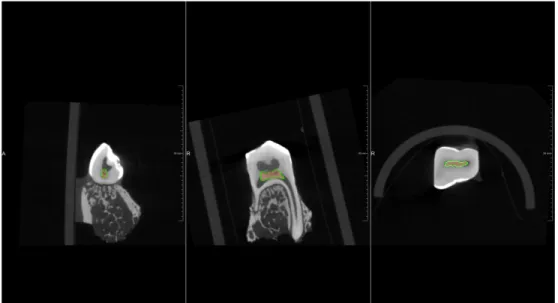
The aim of this study was to develop a preclinical model of partial dental pulp regeneration in minipig, by implanting a pulp construct, made by self-assembling nano-peptide injectable hydrogel and porcine minipig dental pulp cells (pDPCs), in artificially created pulp defects. Secondarily, in the context of this preclinical model, two different techniques of analysis of the regeneration process have been developed. In particular, an in vivo 3D subtraction angiography has been set for the visualization of dental pulp vascular network. Indeed, further developments of this modality open promising perspectives of its application for the morphometric characterization of angiogenesis process in newly formed dental tissues and bone defects.
Moreover, using specific morphometric parameters, initially developed to characterize bone, a micro-CT morphometric analysis of the mineralized reparative tissues, obtained by the partial pulp regeneration protocol, has been elaborated.
By split mouth model, pulp constructs made with self-assembling injectable nano-peptide hydrogel with and without porcine dental pulp cells (pDPCs) were implanted, after pulp chamber amputation in premolars and molars. At day 21 after surgery, three-dimensional morphometric characterization, Masson’s trichrome and immunolabeled for DSP and BSP were performed on treated teeth. 3D subtraction angiographies have been performed before and after partial pulp regeneration procedure.
Regardless of the presence of pDPCs, the implantation of pulp construct induces the formation of an osteodentin bridge, whose microarchitecture sensibly differs from the native dentin. Furthermore, the presence of pDPCs in the construct slightly impairs this reparative process. The latter was led the remaining pulp cells, instead of the pDPCs in the
scaffold. Angiographies could show entire vascularization of jaws and continuously growing teeth but blood supply of treated mature permanent teeth could not be displayed.
The failure of partial pulp regeneration cell based strategy, in these near-real clinical conditions, highlights the importance of preclinical models, to identify the factors promoting a favourable regenerative environment, in the perspective of a transfer to the human clinics.
Resumé
La pulpe dentaire est un tissu connectif innervé et vascularisé, contenu dans une structure minéralisée inextensible formée par l’email, la dentine et le cément. Ce tissu assure l’homéostasie et la sensibilité de la dent. Il est sujet à des lésions sévères faisant suite à une carie ou à un traumatisme. La thérapeutique conventionnelle préconisée alors est le traitement endodontique, qui consiste en l’exérèse de la totalité du tissu pulpaire et le comblement de l’espace évidé par un matériau synthétique bioinerte. Malgré les résultats cliniques satisfaisants, cette thérapeutique induit une fragilisation de la dent et une plus grande susceptibilité aux infections, qui peuvent conduire à terme à la perte de la dent. En se basant sur la présence de cellules souches mésenchymateuses dans la pulpe dentaire, des stratégies de régénération alternatives au traitement endodontique traditionnel sont à l’étude, afin de permettre le maintien des fonctions de nutrition et de sensibilité de la pulpe, garantes de la pérennité de la dent sur l’arcade. Elles s’inscrivent dans deux approches: la régénération de novo, en cas de nécrose du tissu pulpaire et la régénération partielle, lorsque seul le tissu pulpaire endommagé est éliminé et régénéré. Nos travaux portent sur la faisabilité de cette dernière approche dans un modèle préclinique. En effet, dans la perspective d’un transfert vers la clinique humaine, des modèles chez le gros animal doivent être développés afin de tester la faisabilité et le succès de cette thérapie, dans des conditions proches de la clinique.
Du fait de ses similitudes avec l’homme en termes d’anatomie et de physiologie, le miniporc représente un modèle de choix pour les études précliniques d’ingénierie pulpaire.
L’objectif principal de cette étude est de tester la faisabilité de la régénération pulpaire partielle, en implantant des cellules pulpaires porcines (pDPCs) contenues des hydrogels injectables dans des défauts pulpaires artificiellement créés chez le miniporc.
Au cours ce travail, différentes techniques d’imagerie d’évaluation du processus de régénération ont été développées. En particulier, un protocole d’angiographie tridimensionnelle in- pour la visualisation du réseau vasculaire pulpaire a été mise au point. Par ailleurs, en utilisant des paramètres morphométriques spécifiques, initialement développés pour caractériser l’os, une analyse tridimentionnelle par micro-CT des tissus minéralisés de réparation a été élaborée.
En appliquant un “split mouth model”, les hydrogels injectables ensemencés ou non par des pDPCs ont été implantés dans des molaires et des prémolaires, après amputation de la pulpe camérale. À 21 jours après la chirurgie, les analyses d’imagerie, d’histologie et d’immunologie ont mis en évidence, qu’indépendamment de la présence des pDPCs, l’implantation des hydrogels a induit la formation d’un pont d’ostéodentine. La caractérisation morphométrique tridimensionnelle a montré que la microarchitecture de ce pont différait largement de la dentine native. De plus, en présence des pDPCs, le processus de réparation était modifié, avec une moins bonne étanchéité du pont.
Au cours de ce travail, une technique de suivi non invasive de la régénération a tenté d’être mise au point. Une angiographie tridimensionnelle par soustraction a été réalisée avant et après la procédure de régénération pulpaire partielle. Si les angiographies ont révélé l’entière vascularisation des mâchoires et des dents à croissance continue, l’apport vasculaire des dents matures traitées, du fait de son faible flux, n’a pas pu être mis en
L’absence de régénération partielle de la pulpe dans les conditions testées souligne l’importance des modèles précliniques pour identifier les facteurs promouvant un environnement favorable à la régénération, dans la perspective d’un transfert vers la clinique humaine.
TABLE OF CONTENTS
TABLE OF ABBREVIATIONS ... 13 TABLE OF FIGURES ... 17 INTRODUCTION ... 19 AIM ... 20 RESEARCH HYPOTHESIS ... 20 OBJECTIVES ... 20 1. DENTAL PULP AND DENTAL PULP STEM CELLS (DPSCs) ... 22 1.1 DENTAL PULP STEM CELLS ... 23 1.1.1 Phenotype characteristics ... 24 1.1.2 Isolation ... 26 1.1.3 Differentiation ... 27 2. TISSUE ENGINEERING ... 28 2.1 SCAFFOLDS ... 29 2.2 GROWTH FACTORS ... 30 2.3 CELL-BASED PULP REGENERATION ... 31 2.4 OTHER DENTAL TISSUES STEM CELLS ... 32 2.4.1 Stem cells from the apical papilla ... 32 2.4.2 Dental follicle precursor cells ... 33 2.4.3 Periodontal ligament stem cells ... 33 2.5 BANKING ... 34 3. REGENERATIVE ENDODONTICS ... 36 3.1 REVASCULARIZATION ... 36 3.2 REPARATIVE DENTINOGENESIS ... 37 3.3 CELL HOMING ... 38 3.4 IN SITU PARTIAL PULP REGENERATION ... 39 3.5 DE NOVO PULP SYNTHESIS ... 39 4. MONITORING METHODS OF ANGIOGENESIS IN TISSUE ENGINEERING ... 41 5. LARGE ANIMAL PRE-CLINICAL MODELS IN ORAL AND DENTAL TISSUES ENGINEERING ... 49 5.1 SHEEP ... 50 5.1.1 Sheep models in cell-based regenerative dentistry ... 51 5.2 - DOG ... 53 5.2.1 Dog models in cell-based regenerative dentistry ... 54 5.3 PIG ... 59 5.3.1 Porcine models in cell-based regenerative dentistry ... 61 6. AIMS ... 65 7. MATERIALS AND METHODS ... 667.3 CELL PROLIFERATION IN PURAMATRIX™ ... 67 7.4 PARTIAL PULPOTOMY PROTOCOL ... 68 7.5 MINIPIG ANGIOGRAPHY ... 70 7.6 MICRO-COMPUTED TOMOGRAPHIC IMAGING AND ANALYSIS ... 71 7.6.1 Image acquisition and export ... 71 7.6.2 Image segmentation, 3D reconstruction and quantitative morphometric characterization of hard tissue formation ... 71 7.7 HISTOLOGY AND IMMUNOHISTOCHEMISTRY ... 80 7.8 STATISTICAL ANALYSIS ... 80 8. RESULTS ... 81 8.1 PURAMATRIX™ SUPPORTED THE PROLIFERATION OF PDPCs ... 81 8.2 IMPLANTATION OF PDPCS/SCAFFOLD INDUCED REPARATIVE DENTINOGENESIS AND FAILED TO INDUCE PARTIAL PULP REGENERATION ... 82 8.3 THE DENTIN BRIDGE MICROSTRUCTURE DIFFERED FROM THE NATIVE DENTIN CONTROL AND IT WAS AFFECTED BY THE PRESENCE OF PDPCS IN THE SCAFFOLD. ... 83 8.4 PULP AMPUTATION AND SCAFFOLD IMPLANTATION IMPACTED RADICULAR AREA. ... 86 8.5 MAXILLARY AND MANDIBULAR BLOOD SUPPLY DISPLAY ... 89 9. DISCUSSION ... 96 9.1 CHARACTERIZATION OF THE REPARATIVE DENTINE ... 96 9.2 SCAFFOLD ... 97 9.3 CELL SORTING ... 98 9.4 REGENERATIVE ENVIRONMENT ... 99 9.5 MINI-PIG MODEL AND NEAR-REAL CLINICAL CONDITIONS ... 101 9.6 TRIDIMENSIONAL SUBTRACTION ANGIOGRAPHY ... 102 10- CONCLUSION AND PERSPECTIVES ... 110 References ... 111 List of communications ... 137 Annexe I ... 138
TABLE OF ABBREVIATIONS
3D: three-dimensional ASCs: adipose tissue derived stem cells B-TCP: beta tricalcium phospahte BBM: bovine bone mineral bFGF: basic fibroblasts growth factor BMAC: bone marrow aspirate centrifugation BMP-7: bone morphogenetic protein 7 BMP2: bone morphogenetic protein 2 BMP4: bone morphogenetic protein 4 BMSCs: bone marrow stem cells BRONJ: bisphosphonate-related osteonecrosis of the Jaw BS/BV: specific mineralized surface BS/TV: mineralized surface density BSP: bone sialoprotein BV/TV: mineralized volume fraction CAD-CAM: computer-aided design and computer-aided manufacturing CBCT: cone beam computed tomography cBMMSC: canine bone marrow stem cells CD105: cluster of differentiation 105 CD105+: cluster of differentiation 105+ CD117: cluster of differentiation 117 CD14: cluster of differentiation 14 CD146: cluster of differentiation 146 CD166: cluster of differentiation 166 CD271: cluster of differentiation 271CD31: cluster of differentiation 31 CD34: cluster of differentiation 34 CD44: cluster of differentiation 44 CD45: cluster of differentiation 45 CD73: cluster of differentiation 73 CD90: cluster of differentiation 90 CM: conditioned medium cMSCs: canine mesenchymal stem cells DBCs: dental bud cells dBMSCs: dog bone marrow stem cells dDFCsd: dog dental follicles cells DFCs: dental follicle cells DFSCs: dental follicle stem cells DPCs: dental pulp cells dPDLSCs: dog periodontal ligament stem cells DSCs: dental stem cells DSP: dentin sialoprotein DTSCs: stem cells from deciduous teeth EDTA: ethylenediaminetetraacetic acid EMD: enamel matrix derivative EPCs: endothelial progenitor cells ESCs: embryonic stem cells ESEHT: extraction socket-derived early healing tissue FACS: fluorescence activated cell sorting FBS: fetal bovine serum FDMBB: freeze-dried mineral bone block FHA: fluorohydroxyapatite FOV: field of view G-CSF: granulocyte-colony stimulating factor GBP-L: gabapentin -lactam GCHT: gelatin/chondroitin-6-sulfate/hyaluronan tri-copolymer
HA/TCP: hydroxyapatite/ tricalcium phosphate HIF-1a: hypoxia-inducible factor 1-alpha HUVECs: human umbilical vein endothelial cells LF-153: Larry Fisher 153 LF-84: Larry Fisher -84 MDP: multi domain peptides microCT: micro computed tomography MPR: multiplanar reconstruction MRI: magnentic resonance imaging MSCs: mesenchymal stem cells mTAP: modified tri-antibiotics paste NGF: nerve growth factor oAFMC: ovine amniotic fluid mesenchymal cells oBMSC: ovine bone marrow stem cells OCC: osteoblast cell culture OCT-3: octamer-binding transcription factor 3 oDPCs: ovine dental pulp cells oPDLSC: ovine periodontal ligament stem cells PBS: phosphate-buffered saline PCL-TCP: polycaprolactone-tricalcium phosphate PCs: periostal cells PDGF: platelet derived growth factor PDLSCs: periodontal ligament stem cells pDPC: porcine dental pulp cells pDPCs: porcine dental pulp cells PDPSCs: porcine deciduous pulp stem/progenitor cells pDPSCs: porcine dental pulp stem cells PLA: polylactic acid PLGA: polyglycolic acid
pSHED: porcine stem cells from from Exfoliated Deciduous teeth rh PDGF-BB: recombinant platelet-derived growth factor-BB SCAPs: stem cells from apical papilla SDF-1a: stromal cell-derived factor-1a SDF-1: stromal cell-derived factor-1 SHED: stem cells from human exfoliated deciduous teeth SP: side population SPECT: single photon emission computed tomography SRmCT: synchrotron radiation micro computed tomography TbSp: mineralized areas separation, TbTh: mineralized areas thickness TCP/HA: tricalcium phosphate/ hydroxyapatite TDM: treated dentine matrix TMJ: temporomandibular joint TSDO: trans-sutural distraction osteogenesis VEGF: vascular endothelial growth factor vWF: von Willebrand factor β-TCP: beta tricalcium phosphate αMEM: alpha minimum essential medium
TABLE OF FIGURES
Figure 1- Primary colture of Dental Pulp Cells. ... 24 Figure 2- Immunophenotype of DPSCs. ... 26 Figure 3- Paradigm of tissue engineering ... 28 Figure 4- Techniques of visualization of blood supply. ... 43 Figure 5- MicroCT visualization of mandibular blood supply in rat after barium sulphate injection ... 45 Figure 6- Intra-arterial angiography in minipig. ... 47 Figure 7- Sheep denture. ... 51 Figure 8- Dog’s denture. ... 54 Figure 9- Pig’s denture. ... 61 Figure 10- pDPCs in colture. ... 67 Figure 11- Description of the partial pulp regeneration model. ... 70 Figure 12- Dentin bridge segmenation (MeVisLab) ... 73 Figure 13- Native dentin control segmentation (MeVisLab) ... 73 Figure 14- Thresholding of the dentin bridge (CTan) ... 74 Figure 15- Dentin bridge segmentation (CTan). ... 74 Figure 16- Thresholding of the native dentin control (CTan) ... 75 Figure 17- Native dentin control segmentation (CTan). ... 75 Figure 18- Segmentation of pores in the dentin bridge volume (Amira) ... 76 Figure 19- Segmentation of pores in the dentin bridge volume (Amira) ... 76 Figure 20- Segmentation of pores in the native dentin control volume (Amira) ... 77 Figure 21- Segmentation of pores in the native dentin control volume (Amira) ... 77 Figure 22- Segmentation of the entire tooth volume (MeVisLab) ... 78 Figure 23- Volume rendering of the dentin bridge (3Matic) ... 78 Figure 24- Volume rendering of the entire tooth (3Matic) ... 79 Figure 25- Volume rendering of the tooth showing the location of the dentin bridge within the endodontic space (3Matic) ... 79Figure 26- pDPCs proliferation in 0.2% PuraMatrix™ in vitro. ... 81
Figure 27- MicroCT analysis of reparative dentinogenesis in teeth implanted with pDPCs/scaffold and no-pDPCs/scaffold. ... 83 Figure 28- Morphometric analysis of dentin bridge structure in comparison with native dentin control and influence of pDPCs on dentin bridge microstructure. ... 84 Figure 29- Masson’s trichrome staining, DSP and BSP labeling in the pulp of teeth, at day 21 after surgery. ... 88 Figure 30- External resorptions at 3 weeks post implantation. ... 89 Figure 31- Selective injection of Patent blue V into the inferior alveolar artery. ... 90 Figure 32- Maxillary and mandibular blood supply in minipig. ... 91 Figure 33- Contrast agent collection at the apical area of mature teeth in minipig. ... 92 Figure 34- Selective injection of iodine contrast medium in minipig.. ... 92 Figure 35- Soft tissues surrounding mandible. Volume rendering of soft tissues surrounding mandible. ... 93
Figure 38- Volume rendering of mandibular blood supply. ... 95 Figure 39- Maxillary and mandibular blood supply in mouse. ... 105 Figure 40- Maxillary and mandibular blood supply in mouse. ... 105 Figure 41- Maxillary and mandibular blood supply in mouse. ... 106 Figure 42- Maxillary and mandibular blood supply in mouse. ... 106 Figure 43- Injection of iodine contrast medium into penis vein in rat. ... 107 Figure 44- Maxillary and mandibular blood supply in rat. ... 108 Figure 45- Maxillary and mandibular blood supply in rat. ... 109
INTRODUCTION
The dental pulp is the connective tissue enclosed into the mineralized inextensible structure of dental walls. It ensures the homeostasis and sensibility of the tooth. When this tissue is damaged, consecutively to caries or trauma, the routine clinical treatment is a root canal therapy. This procedure consists in the elimination of the affected tissue and filling of the endodontic canal system with bioinert synthetic materials. In spite of satisfactory clinical outcomes, this treatment does not restore the original sensitivity, neither the natural defence of the pulp.
Thus, there is an increasing desire to provide alternative options for endodontic therapy and one of the most significant challenges in modern dentistry is the regeneration of a vital dental pulp.
Cell-based pulp regeneration could provide a valid alternative to traditional endodontic treatment of damaged teeth. This strategy focuses, in fact, on the preservation of the healthy pulp tissue and the regeneration of the damaged one, by combining stem cells, scaffolds and growth factors.
In the laboratory EA2496 directed by Prof. Catherine Chaussain, research is conducted on pulp regeneration. In particular Gorin et al. have largely investigated the growth factors implicated in the angiogenic capacity of SHED seeded into hydrogels and in vivo subcutaneous implantations (Gorin et al., 2016).
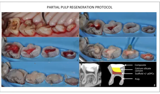
Thus, in the perspective of a preclinical application of these results and thanks to the opportunity to work with the laboratory CR2i of INRA (Jouy-en-Josas) directed by Prof. Laurent the present project has been carried on minipigs. AIM The aim of this study was to develop a preclinical model of partial dental pulp regeneration in minipig, by implanting a pulp construct, made by self-assembling nano-peptide injectable hydrogel and porcine minipig dental pulp cells (pDPCs), in artificially created pulp defects. In parallel, an interventional imaging method was explored to follow up the tissue engineering process, by interventional CBCT angiography. RESEARCH HYPOTHESIS This work relies on the hypothesis that autologous transplanted pDPCs could regenerate in vivo a functional parenchyma in pulpotomized permanent teeth and that this functionality could be detected by advanced imaging techniques OBJECTIVES
In the context of this preclinical model of pulp regeneration, three objectives were established:
1. To obtain pulp tissue regeneration after pDPCs implantation in partial pulpotomies in minipig
2. To follow-up the regenerative process by interventional CBCT angiography;
3. To characterize the newly formed tissues by non-invasive and non-destructive microCT imaging techniques.
Thanks to the collaboration with the laboratory Cr2i of INRA, the interventional CBCT angiography procedure has been set and performed. Then, during 3 months of scholarship in the departement of Oral and Maxillo-facial Surgery - Imaging & Pathology (OMFS-IMPATH) of the University of Leuven (KU Leuven), directed by Prof. Reinhilde Jacobs, different techniques of analysis have been developed. In particular, combining the high level of professionalism and competences of Prof. Jacobs’ team on CBCT and microCT image analysis and my previous experience in the same field, the data from the in vivo 3D subtraction angiography have been processed in order to analyze dental pulp vascular network. Indeed, further developments of this modality open promising perspectives of its application for the morphometric characterization of angiogenesis process in newly formed dental tissues and bone defects.
Moreover, using specific morphometric parameters, initially developed to characterize bone, a micro-CT morphometric analysis of the mineralized reparative tissues, obtained by the partial pulp regeneration protocol, has been elaborated.
This work has led to a publication accepted in the Journal of Dental Research (Mangione F, EzEldeen M, Bardet C, Lesieur J, Bonneau M, Decup F, Salmon B, Jacobs R, Chaussain C, Opsahl-Vital S. 2017. Implanted dental pulp cells fail to induce regeneration in partial
1. DENTAL PULP AND DENTAL PULP STEM CELLS (DPSCs)
The dental pulp is a unique and complex connective tissue serving to support the dentin. It is highly innervated and vascularized, encapsulated in a mineralized inextensible structure formed by enamel, dentin and cementum, assuring the homeostasis and sensibility of the tooth organ (Piette, 2001; Goldberg, 2011; Rosa et al., 2016).The pulp tissue is composed of collagen type I and type III along with a variety of noncollagenous proteins, including a large proteoglycan component (Colombo et al., 2014). The pulp contains a variety of cell types, including immune cells, fibroblasts, mesenchymal progenitor cells, vascular cells, and nerve cells (Jontell et al., 1998).
The fibroblasts are the most numerous cells in the dental pulp, they located in the central part of the parenchyma. Their main role is synthesis of collagen I and III fibers, proteoglycans and glycoproteins as well as cytokines (Mathieu et al., 2005). The odontoblasts are disposed with a palisade pattern in contact with dentin and they show the Tomes’ process, which is a cytoplasmic extension located into dentin tubules, associated with a system of fluid-filled tubules actively involved in dentin mineralization. These cells secrete the primary dentin, then the secondary and tertiary dentin (Magloire et al., 2004) and they also contribute to proprioception and nociception (Magloire et al., 2009; Magloire et al., 2010; Maurin et al., 2013). The population of immune cells, in particular tissue macrophages, hold themselves ready to respond to microbial incursion (Colombo et al., 2014).
A network of blood vessels runs throughout the pulp, perfusing the tissue and networks of nerve fibers provide enervation to the tissue, linking it with the central nervous system and providing sensory output.
Finally, as in many other dental tissues such as periodontal ligament, apical papilla and dental follicle, a resident population of mesenchymal progenitor cells provides a reservoir of pluripotent cells capable of differentiation into a variety of cells as required for tissue maintenance and repair.
In addition to cellular elements, pulp presents an extracellular matrix (ECM), mainly composed by collagen, glycosaminoglycans, glycoproteins and metallo-proteases (Bogovic et al., 2011). Such a composition of the ECM allows the maintenance of hydration by water molecules storage. They manage the transit of metabolites, nutrients and cells debris among blood vessels and pulp cells (Goldberg et al., 2009). Indeed, in the perspective of a conservative approach of the pathologies affecting the pulp, regenerative strategies are increasingly investigated. However, the peculiar heterogeneity of the tissue makes pulp regeneration challenging. 1.1 DENTAL PULP STEM CELLS Gronthos’ team has highlighted the presence of stem cells in the pulp of permanent teeth, in 2000. These cells, named Dental Pulp Stem Cells (DPSCs), showed their attitude to form the dentin-pulp complex in vitro (Gronthos et al., 2000). Few years later, the same researchers have showed that stem cells were also present within
temporary teeth, represents a unique and natural source of stem cells (Batouli et al., 2003; Kerkis and Caplan, 2012).
1.1.1 Phenotype characteristics
Dental pulp stem cells have a phenotype similar to Bone Marrow Stem Cells (BMSCs). In culture, they look like big, spindle with a big central nucleus and a voluminous cytoplasm. (fig 1).
Figure 1- Primary colture of Dental Pulp Cells. Scale bar 50 μm (EA 2496).
Some surface markers in common with BMSC have been identified (Gronthos et al., 2000) by immunohistochemistry and DPSCs are able to differentiate in vitro into numerous types of cells (tab 1 and fig 2)
Markers DPSC-1 DPSC-2 BMSC CD14 - - - CD34 - - - CD44 ++ ++ ++ CD45 - - - Integrin β1 ++/+ ++/+ ++ VCAM -1 + + ++ MyoO - - - αSM actin ++/- ++/- ++/+/- Neurofilam. - - - MUC-18 ++/- ++/+/- ++/+/- Collagen I + ++ ++/+ Collagen II - - - Collagen III ++/+ ++/+ ++/+ Osteocalcin ++/+ ++ +/- Osteonectin ++/+ ++ ++/+ BSP - - +/- Osteopontin +/- +/- +/- Alk Phos ++/+/- ++/+/- ++/+/- PPARγ - - - FGF -2 ++/+ ++ ++/+ Table 1 - Immunohistochemical analysis of human DPSCs and BMSCs in vitro. The immunostaining of DPSCs demonstrated the markers in common with BMSCs. (Gronthos et al., 2000).
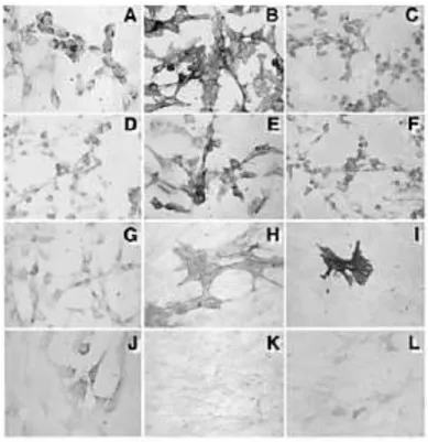
Figure 2- Immunophenotype of DPSCs. Integrin β1 (A); CD44 (B); collagen type I (C); collagen type III (D); fibroblast growth factor-2 (E); osteonectin (F); osteocalcin (G); MUC-18 (CD146) (H); α-smooth muscle actin (I); osteopontin (J); and vascular cell adhesion molecule 1 (K). Endogenous alkaline phosphatase (L) (Gronthos et al., 2000).
Phenotypically, DPSCs are similar to BMSCs (Eleuterio et al., 2013). Surface markers and proteins distribution are also comparably found (Gronthos et al., 2000; Gronthos et al., 2002; Eleuterio et al., 2013; Cao et al., 2015). Moreover, DPSCs seem to present a higher proliferation rate than BMSCs, without loosing their multipotency (Gronthos et al., 2000; Seong et al., 2010; Gong et al., 2016). 1.1.2 Isolation DPSCs can be isolated either by the enzymatic digestion or outgrowth methods (Hilkens et al., 2013). Since they have a high renewal property, Gronthos et al. in 2000 proposed to individualize cells, to seed them and to select clones having the highest proliferation rate.
This property is maintained beyond 25 passages (Rodriguez-Lozano et al., 2012).
Several markers are proposed for human dental pulp stem cells characterization (Kawashima, 2012; Gong et al., 2016): the most of them, like OCT3/4, CD90, STRO1, are in common with BMSCs. So stem cells markers can be used to sort them by FACS or by antibodies linked to spheres (Kawashima, 2012). Nevertheless, no specific DPSCs markers have been identified yet.
1.1.3 Differentiation
In vitro, DPSCs can differentiate into several germ cell lines (Kawashima, 2012). In fact, many protocols allow their differentiation into mesodermic line cells such as myocites, osteoblasts, chondrocytes, adipocytes and cardiomyocytes (Jo et al., 2007; Koyama et al., 2009; Gronthos et al., 2011; D' Alimonte et al., 2011; Dhillon et al., 2015). Endodermic line cells (hepatocytes) differentiation can also be obtained (Iohara et al., 2006; Rodriguez-Lozano et al., 2012; Dhillon et al., 2015). The differentiation into neural cells has been demonstrated (Iohara et al., 2011, Kim et al., 2012; Fang et al., 2013) but the maturation into neurons is still controversial (Gervois et al., 2015, Iohara et al., 2011).
Moreover, many authors have demonstrated that DPSCs can differentiate into endothelial cells in vitro and in vivo (Gorin et al., 2016, Iohara et al., 2011; Hilkens et al., 2013; Nakashima and Iohara, 2011; Ratajczak et al., 2016). This capacity is particularly suitable for tissue engineering.
2. TISSUE ENGINEERING
Tissue engineering relies on the simple as well as combined use of DPSCs, scaffolds and growth factors, to achieve tissue regeneration (fig 3) (Dhillon et al., 2015). Figure 3- Paradigm of tissue engineering (Malhotra and Mala, 2012).
2.1 SCAFFOLDS
In the tissue-engineering triade, the scaffold plays a very important role. In fact, it represents a three-dimensional substrate mimicking the ECM (Ma, 2008) for cell adhesion, proliferation and differentiation. At the same time the scaffold has to be biocompatible and biodegradable and it has to be mechanically, biologically and chemically adaptable for specific tissues regeneration (Galler et al., 2010; Galler et al., 2011; Yuan et al., 2011).
Scaffold can be made by natural or synthetic biodegradable polymers, inorganic materials and composites. Moreover, they can be shaped as porous scaffolds, nanofibrous materials, microparticles, and hydrogels.
The choice of an appropriate scaffold relies on the specific application and it is strongly influenced by the availability and the ease of manipulation, as well as by the balance cost/ effectiveness (Galler et al., 2010; Sun et al., 2011; Yuan et al., 2011).
Natural polymers are mechanically resistant, bioactive and biodegradable but they are not easy to be processed and they can induce immune reaction.
Synthetic polymers show good processing, manipulation and mechanical properties but they have low bioactivity and they induce high inflammatory reaction (Yuan et al., 2011). Regarding pulp regeneration, collagen type 1, PLGA and PLA have been reported as the scaffolds providing the best outcomes (Albuquerque et al., 2014).
A valid alternative is represented by hydrogels, which are characterized by high water content, viscoelastic properties matching with those of soft tissues and high capacity of cell encapsulation and delivery (Nicodemus et al., 2008; Galler et al., 2010).
being increasingly used.
Galler et al. showed that self-assembling multidomain peptides (MDP) supported pulp regeneration (Galler et al., 2008; Galler et al., 2010; Galler et al., 2012).
2.2 GROWTH FACTORS
The third factor of tissue engineering is represented by growth factors. They determine cellular fate by a signalling action within the regenerative environment (Bansal and Bansal 2011). These factors can derive from degraded dentin matrix, blood clot or be synthesized in vitro (Bessho et al., 1991).
Nakashima showed that recombinant human BMP2 and BMP4 were directly involved in dentin and osteodentin deposition in dog pulp defects capped with inactivated dentinmatrix (Nakashima, 1994). The same factors have been reported by Casagrande et al. as inducing SHEDs differentiation into odontoblasts when seeded in tooth slices treated with EDTA (Casagrande et al., 2010). VEGF is a proangiogenic factor. Its activity has been demonstrated on SHED (Sakai et al., 2010) and in co-cultures of SCAPs and HUVECs (Yuan et al., 2015). bFGF is involved in odontogenesis and in fibroblasts’ reaction to injuries (Shimabukuro et al., 2009). It has been reported that this factor inhibits differentiation and increases survivability of DPSCs (He et al., 2008; Yang et al., 2015). However, the use of growth factors is complicated because their nature, their dosage, and the delivery timing may induce controversial biological effects on cellular behaviours (Gong et al., 2016).
2.3 CELL-BASED PULP REGENERATION DPSCs have been employed in the regeneration of periodontal bone defects and dentin-pulp complex (Zheng et al., 2012, Hilkens et al., 2015). More specifically, DPSCs showed their capacity of differentiation into several types of pulp cells. Very important aspect is their capacity to differentiate into odontoblasts and synthetize dentin (Yu et al., 2006; Onyekwelu et al., 2007, Hilkens et al., 2015).
In immunocompromised mice, DPSC transplanted with hydroxyapatite/tricalcium phosphate (HA/TCP) scaffold formed a pulp tissue characterized by blood vessels and human odontoblast-like cells (Gronthos et al., 2000; Gronthos et al., 2002; Batouli et al., 2003). The same result has been achieved in more standardized model using human tooth slices and DPSC or SHED implanted in immunodeficient mice (Cordeiro et al., 2008; Prescott et al., 2008; Gorin et al., 2016).
Vascularized pulp / dentin-like tissue has been obtained by Huang et al. (2010) by implanting subcutaneously in immunodeficient mice an emptied human root (with an apical foramen of 2.5 mm) seeded with a combination of DPSCs and poly(lactic-co-glycolic acid) (PLGA) scaffold. The same results has been demonstrated in ectopic root transplantation using SHEDs associated with self-assembling peptide hydrogel (Rosa et al., 2013).
In presence of a narrower apical foramen (1 mm) Dissanayaka et al. could achieve pulp regeneration after transplantation of DPSCs or DPSCs-HUVECs co-cultures (Dissanayaka et al., 2015). This result is consistent with the study of Yang et al. based on the transplantation of DPSCs in a silk fibroin scaffold (Yang et al., 2015).
al., 2006). The same authors achieved pulp regeneration in canine pulpectomy models (Iohara et al., 2011; Ishizaka et al., 2012 ; Wang et al., 2013) and more recently they showed pulp-dentin complex regeneration by using G-CSF mobilized DPSCs in canine mature teeth (apical foramen 0.6 mm diameter) (Iohara et al., 2013 ; Nakashima et al., 2014). 2.4 OTHER DENTAL TISSUES STEM CELLS
In addition to DPSCs, many other types of stem cells from dental tissues have been then isolated and characterized: Periodontal Ligament Stem Cells (PDLSCs), Stem Cells From Apical Papilla (SCAPs) and Dental Follicle Stem Cells (DFSCs).
2.4.1 Stem cells from the apical papilla This population of stem cells isolated from the apical papilla which is a connective tissue located at the apex of developing permanent teeth is increasing the interest in pulp regeneration field (Sonoyama et al., 2006; Sonoyama et al., 2008).
In fact, SCAPs show a higher capacity of proliferation and migration in comparison with DPSCs. Already in 2006, Sonayama et al. achieved dentin pulp complex regeneration by transplanting SCAPs with HA/TCP scaffold in immunocompromised mice (Sonoyama et al., 2006).
Regarding ectopic root transplantation, better results have been showed by Huang et al. with SCAPs-PLGA than in the model with DPSCs-PLGA construct (Huang et al., 2010).
promoting root development in immature teeth affected by periodontitis or apical abscesses (Hilkens et al., 2015).
2.4.2 Dental follicle precursor cells
This population of stem cells is found in the dental follicle, the connective tissue surrounding the developing tooth and generating the periodontium.
Indeed, most of studies have investigated the capacity of DFSCs in the regeneration of periodontal tissues (Yaguu et al., 2010; Guo et al., 2012; Guo et al., 2013).
Regarding pulp regeneration, Guo et al. showed that FSCs seeded in treated dentin matrix (TDM) were able to give rise to odontoblast and dentin in the omental pouch of adult rats (Guo et al., 2009). Cementum-like, periodontal ligament and dentin-pulp complex were obtained by the same group of researchers transplanting DFSCs-TDM construct in rat alveolar sockets (Guo et al., 2012).
The ability of these cells to regenerate such a variety of dental tissues was further confirmed by implanting subcutaneously in immunocompromised mice the same construct made with human DFSCs (Yang et al., 2012; Jiao et al., 2014). 2.4.3 Periodontal ligament stem cells PDLSCs can differentiate into osteoblasts and regenerate bone (Seo et al., 2004; Gay et al., 2007; Lindroos et al., 2008; Xu et al., 2009). However, they form mineralized tissues only when cultured in tricalcium phosphate/hydroxyapatite (TCP/HA) or when a medium from
Regarding pulp regeneration, the role of these cells is controversial because they express dentin markers only when co-cultured with DPSCs (Suh et al., 2014). 2.5 BANKING Since the extraction of third molars, premolars and deciduous teeth is a common practice, dental stem cells are considered easily available. Moreover, it was demonstrated that their properties are maintained when cryopreserved (Ding et al., 2010; Lee et al., 2012). Even thought DSCs present low immunogenicity and immunomodulatory activity (Kim et al., 2010; Tomic et al., 2011; Liu et al., 2012), at present allogeneic transplantation is not possible and only autologous DSCs could be used for pulp-dentin complex regeneration. However, the small amount of cells isolated from the dental pulp needs to be processed before to be used for cell-based procedures.
DSCs banks exist, allowing a rapid access to stem cells in case of necessity of cell based therapies (Egusa et al., 2012; Huang et al., 2013). Nowadays, several private societies propose the cryopreservation of dental pulp stem cells (Genecell International®, USA) from extracted teeth. For the moment, these societies do not operate in France and in the most of European countries. Unfortunately none of the private tooth banks provide clinical-grade MSCs because of the absence of good manufacturing practice allowing the standardization of cell banking taking into account the origin and donor-associated differences among stem cells (Gong et al., 2016). In fact, many studies have investigated the effect of epigenetic and genetic modifications induced by cell culture on the regenerative potential (Duncan et al., 2016;
Gopinathan et al., 2013). Thus cell banking should apply standardized expansion of stem cells in order to provide a genetically stable population, with specific tissue regeneration ability and lack of tumorigenicity (Shahdadfar et al., 2005). Moreover these cells should not be contaminated by microorganisms: for this reason studies are conducted to develop culture media the without animal serum able to favour cell proliferation and preservation of their stemness properties (Sotiropoulou et al., 2006; Mannello and Tonti, 2007).
Finally standardized procedures should be developed for all the following steps regarding cell purification from the media, harvesting without porcine trypsin, as well as use of performing carriers (Bakopoulou and About, 2016).
3. REGENERATIVE ENDODONTICS
It is widely established that the pulp has an innate capacity for self-repair and contains all the necessary components to regenerate both the mineralized dentin and the soft tissues of the pulpal matrix.
Indeed, considering the outcomes reported in literature, pulp regeneration became a clinical reality. In fact, if techniques such as revascularization and induction of reparative dentinogenesis are already widely applied on patients, on the other hand experimental procedures including cell homing, partial and total pulp regeneration still require further development. 3.1 REVASCULARIZATION The revascularization is an approach applicable in necrotic/infected permanent immature teeth with an apical foramen of more than 1 mm. It relies on the intentional laceration of periapical area’s tissues to obtain the formation of a blood clot in a decontaminated root canal system. The clot, which acts like a scaffold, is colonized by SCAPs and growth factors and promotes root development and elongation (Diogenes et al., 2013; Albuquerque et al., 2014).
However, this procedure seems promising only in very immature teeth, because the nature of the regenerated parenchyma and the possibility of root elongation are still debated (Neha et al., 2011; Nosrat et al., 2011; Andreasen and Bakland, 2012).
2010; Cao et al., 2013; Martin et al. 2013). Thus, all these outcomes show that revascularization rather induces resolution of apical periodontitis by tissue repair (Cao et al., 2015). 3.2 REPARATIVE DENTINOGENESIS As mentioned above, the presence of a population of progenitor cells within pulp tissue is demonstrated. In case of pulp injury, these cells can differentiate into odontoblasts-like and replace damaged odontoblasts (Nakashima and Akamine, 2005; Huang, 2008; Huang, 2009; Huang et al., 2009; Scheller et al., 2009; Sun et al., 2011).
Therapeutic approaches aiming pulp vitality preservation by formation of a reparative dentin bridge, after reversible pulp injury, rely on this principle. Nevertheless, the prognosis of a direct pulp capping in case of deep caries or traumas is still uncertain and this procedure needs to be applied only on few selected cases (Bashutski and Wang, 2009). In fact, the outcomes of such therapy are strictly dependent to pulp exposure type, inflammation degree, tooth age, materials choice and restoration sealing (Mjor, 2002; Murray et al., 2002; Ward, 2002; Murray et al., 2003; Tziafas, 2004).
Furthermore, the clinical diagnosis of inflammation degree is hard to be established and at present there are no guidelines allowing the clinicians to identify the cases suitable for direct capping treatment (Huang, 2008; Sun et al., 2011; Zanini et al., 2017).
As a matter of fact, it is frequent that so treated teeth subsequently undergo endodontic therapy.
The progress into the comprehension of molecular composition and cellular mechanisms regulating dentinogenesis favours new regeneration strategies in which the damaged pulp is partially or totally eliminated and replaced by a regenerated healthy functional tissue. Thus, two different approaches are distinguished: in situ partial pulp and de novo pulp regeneration (Nakashima and Akamine, 2005; Huang et al., 2009; Sun et al., 2011).
3.3 CELL HOMING
The cell homing approach is a cell-free method to regenerate pulp-dentin complex. It represents an alternative to the revascularization and it relies on the stimulation and the migration of the endogenous MSCs on the area of the tissue to be regenerated (Mao et al., 2012). In fact, many studies have reported regeneration of a functional pulp tissue into empty endodontic spaces by combining cytokines and growth factors promoting cellular chemotaxis and cell homing.
Kim et al. showed positives outcomes in vivo using associations of bFGF, PDGF or VEGF and NGF and BMP-7 in endodontically treated teeth (Kim et al., 2010).
Suzuki demonstrated that the combination of bFGF and SDF1 recruits DPSCs and that BMP-7 promotes their differentiation (Suzuki et al., 2011). Moreover, Yang et al., obtained complete functional pulp regeneration using silk-fibroin scaffolds and SDF1 in mature canine teeth (Yang et al., 2015).
Overall, cell homing seems to be a promising economical approach for pulp regeneration and endodotic space revascularization.
3.4 IN SITU PARTIAL PULP REGENERATION
Biotechnologies focusing on partial pulp regeneration are based on the observation that in the first instance pulp inflammation is compartmentalized before that whole tissue is affected (Huang, 2008; Huang, 2009; Huang et al., 2009). Current data suggest that the healthy part of the pulp could not only be conservable but also have the potential to regenerate the lost tissue (Huang, 2009). In order to favour such regeneration, pulp constructs constituted by DPSCs and scaffolds can be implanted in the endodontic space inducing pulp-dentin complex reformation (Huang, 2009; Sun et al., 2011).
In vivo, in large animal models, two different approaches of partial pulp regeneration have been proposed. The first consists in the implantation of a pellet of autologous DPSC in tooth underwent to pulp amputation. The second relies on the use of different kinds of scaffolds containing sorted DPSCs (expressing for example CD31-, CD146- antigens).
At present, only Iohara et al. have regenerated pulp tissue in dogs by implantation of autologous DPSC pellets (Iohara et al., 2009).
3.5 DE NOVO PULP SYNTHESIS
When pulp parenchyma is completely destroyed, de novo synthesis has to be done to regenerate the tissue. The volume of mature pulp is very small (about 10-100 μl), so the regeneration of pulp should be easier than big organs or tissues. Nevertheless, this tissue is hard to be regenerated because of the following characteristics: 1) it is placed within rigid
calcified walls; 2) it has reduced blood supply due to the narrow apical foramen and 3) it is an heterogeneous tissue whose cells are distributed in several innervation layers.
Recently, many teams of researchers demonstrated the feasibility of the implantation and the survival of cells within the tooth in vivo. En 2009, Huang (Huang, 2009) showed the formation of a mineralized tissue by stem periodontal cells within pulp chamber in dog. In the same animal model, Iohara et al. (Iohara et al., 2013) obtained a functional vascularized and innervated pulp tissue by using different stem cells.
In 2017, Nakashima et al. published the first clinical trial outcomes of de novo pulp synthesis procedure and they demonstrated by MRI and CBCT functional dentin-pulp complex regeneration (Nakashima et al., 2017).
4. MONITORING METHODS OF ANGIOGENESIS IN TISSUE
ENGINEERING
Vascularization has been studied for years using several in vitro and in vivo models. Different mechanisms are distinguished: angiogenesis, arteriogenesis and vasculogenesis. Angiogenesis relies on growth of capillaries from pre-existing vessels and it is activated by tissue ischemia. Arteriogenesis is process mediated by monocytes and macrophages, which consists on pre-existing vessels dilatation and remodelling as response to augmented physiological demands. Finally, vasculogenesis is a de novo blood vessel development by bone marrow-derived endothelial progenitor cells (Duvall et al., 2004).
Angiogenesis and vasculogenesis are deemed important for regenerative strategies of all soft and calcified tissues. In fact, a key factor in tissue engineering is the possibility to obtain a functional microvascular network within constructs providing oxygen and nutrients for engraftment, differentiation, and tissue functionality. Thus, the distance of the stem cells constructs from vascular supply is critical for integration to the receiving tissue (Bolland et al., 2008; Duvall et al., 2004).
Dental pulp is a highly vascularized tissue. Blood vessels penetrate within pulp by the apical and secondary foramina. They proceed to the root canal centre in direction of the pulp chamber in which they progressively brunch out to form in the periphery a thin sub-odontoblastic capillaries net of about 5 to 10 μm diameter (Trubiani et al., 2003).
Blood vessels penetrate inside the pulp and go out from it by apical and secondary foramina in the form of one or sometimes two arterioles (35-45μm) passing through the centre of the
capillary veinules gathering to form collector veinules in the central part of the root canal. The presence of artero-venous anastomosis contributes to the regulation of blood flux as well as intra-pulp pressure and they allow blood flux drift and loop isolation in case of injury.
Regarding pulp regeneration, blood supply ensures the survival of pulp constructs and their engraftment. Nevertheless, several factors like dental anatomy, narrow diameter of root apical foramen, presence of a terminal vascular net and characteristics of calcified tissues embedding the pulp, complicate these procedures. So, the preservation of a part of pulp parenchyma allows to rely on the root remaining vascularization and to put the construct in direct contact with blood vessels, to obtain its engraftment (Bolland et al., 2008)
However, this therapeutic option is strictly dependent to the knowledge and the managing of the local blood supply, which must be precisely and reproductively quantified and visualized.
Several techniques for highlighting microvascular architecture can be employed in vivo and ex vivo, in 2-D or 3-D such as histology, immunohistochemistry, animal perfusion, casts and imaging (fig 4a, 4b, 4c)(Tab. 2). Histological staining (Hematoxylin-Eosin, Movat pentachrome) and immunohistochemistry (CD31 and vWF labelling) allow to identify vessels and their rapport with contiguous structures and tissues. Animal perfusions and the corrosion casts give a 3D high-quality representation of microvascular architecture (fig 4a)(Blery et al., 2015).
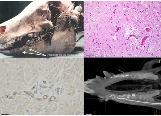
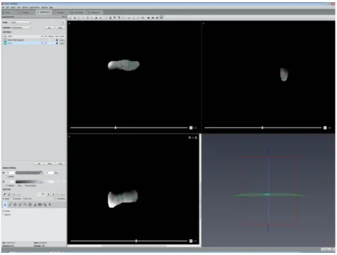
Figure 4- Techniques of visualization of blood supply. Casts of pig head vassels (a) ; haematoxylin and eosin staining of pig brain histological section showing blood vassels (scale bar 20 μm) (b) ; immunolabeling of vWF on pig skin section (scale bar 20 μm) (c) ; 3D angiography of minipig mandible (scale bar 1 cm) (d).
Among direct imaging techniques microscopy, angiography, computed tomography (CT) and micro-CT both in association or not with a synchrotron system are the most used (Blery et al., 2015)(fig 4d).
In vivo angiogenesis of tissue-engineered constructs is mainly detected by two-dimensional evaluation of microvessel concentration within histological sections (Pabst et al., 2014). The preclinical high-resolution imaging technology of micro-CT is customized to visualize mineralized tissue. However, more recently, this technique allowed a three-dimensional characterization of organs’ vascular networks (Lu et al., 2010). Besides, a little has been
published about its use for the assessment of the vascularization in implanted tissue engineering scaffold.
In combination with radiological techniques, many contrast agents are implied: iodine, liquid fatty oil, resin, gold nanoparticles, Microfil and barium sulphate (Pabst et al., 2014). Recently, some authors have assessed vascular microarchitecture parameters, deduced from bone microarchitecture analysis (Bolland et al., 2008; Jia et al., 2010; Langer et al., 2011; Prisby et al., 2011; Roche et al., 2012) using micro-CT and radio-opaque contrast agents. For this morphometric analysis, the vascular network should be entirely visualized (fig 5). By injecting barium sulphate the bone blood supply is not completely permeated, due to the heterogeneity of the network. Microfil, which is a silicon with chromate, perfuses entirely soft tissues, but it has the same gray level of mineralized structures. Thus, decalcification is required when using this contrast medium (Bentley et al., 2002; Duvall et al., 2004; Marxen et al., 2004; Bolland et al., 2008; Young et al., 2008; Mondy et al., 2009a, 2009bb; Nyangoga et al., 2011; Roche et al., 2012).
In fact, thresholding is crucial for blood supply assessment. Clearly separating the vessels from the bone is important for a good estimation of the vascular and bone microarchitecture parameters. Synchrotron-CT is considered as the gold standard for bone microarchitecture and vascularization morphometric analysis (Eppel et al., 2009; Langer et al., 2009; Chien et al., 2010; Jia et al., 2010; Lu et al., 2010; Peyrin et al., 2010; Roche et al., 2012; Matsumoto et al., 2013; Holme et al., 2014; Neldam and Pinholt, 2014).
In fact, the collimated monochromatic beam of Synchrotron radiation (SR) provides micrometric spatial resolution for the identification of different structures and it allowed the visualization of vascular canals in human and rats cortical bone (Bousson et al., 2004;
Matsumoto et al., 2011). The medical application of SRmCT is increasing and it is mainly focused on the study of osteoporotic bone and fractures (Ito, 2005; Kazakia et al., 2008; Cooper et al., 2011a), microcracks (Voide et al., 2009; Larrue et al., 2011), and alveolar bone modifications caused by orthodontic treatments (Dalstra et al., 2006). In vivo microangiography in rat hind limb has been reported to not allow visualization of capillaries (10 μm diameters) (Lu et al., 2010). Figure 5- MicroCT visualization of mandibular blood supply in rat after barium sulphate injection (Blery et al., 2016).
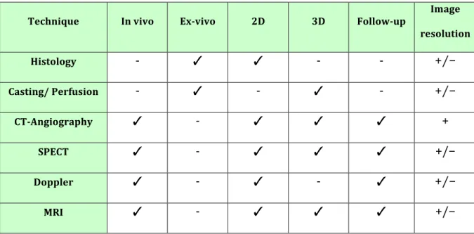
Clinically, vascularization is studied by different modalities such as Doppler flowmetry, colour Doppler ultrasonography, MRI, SPECT, computed tomography angiography (CTA) and catheter angiography (Pabst et al., 2014)(Tab 2).
Technique In vivo Ex-vivo 2D 3D Follow-up Image resolution Histology - ✓ ✓ - - +⁄− Casting/ Perfusion - ✓ - ✓ - +⁄− CT-Angiography ✓ - ✓ ✓ ✓ + SPECT ✓ - ✓ ✓ ✓ +/− Doppler ✓ - ✓ - ✓ +⁄− MRI ✓ - ✓ ✓ ✓ +/− Table 2- Techniques for blood supply visualization. ✓ possible ; - not possible ; + high ; − low ; +⁄− mean. The Doppler flowmetry is a simple modality but it is not completely reliable and the probe can reach a very limited depth. Colour doppler ultrasound gives information about both arteries and veins, but it requires high competence in performance and interpretation of the exam. Moreover it does not allow three-dimensional (3D) visualization of anatomy (Roche et al., 2012).
MRI for vascular supply visualization requires the use of a contrast agent gadolinium-chelated that is injected into a large vein. However, this technique has a limited spatial resolution (130microm).
SPECT is performed by injection in the bloodstream of a radioisotope (isotope of gallium) associated to a ligand to create a tissue specific radioligand whose concentration is registered by a gamma camera This modality presents low spatial resolution (1cm), soft tissue attenuation and exposition to ionising radiations (Johns et al., 2016; Yabuuchi et al., 2015).
Computed Tomography angiography is the main used technique for blood supply visualization, because it is less time consuming, economical and it has sufficient spatial resolution. Nevertheless, it implicates ionising radiation and injection of iodinated contrast media. The latter, mostly if injected in high volume, can induce nephropathy.
Thus, catheter angiography is more invasive and less used; in fact literature reports an important percentage of indwelling catheter malposition and vascular damage, resulting in a high risk of bleeding, thrombosis, embolism and infection. However, intra-arterial angiography requires a reduced volume of contrast medium with a satisfactory blood concentration and it provides good image quality. This modality is mainly used in the oncology field, because of the possibility to obtain a detailed map of the local vascular net allowing, at the same time, a selective action of surgical resection and injection of radio-nucleotides, as well as the evaluation of the amount of blood supply of the donor and receiving sites of reconstruction grafts (fig 6)(Sakakibara et al., 2015).
Indeed, the translation of regenerative endodontics to human clinics and the development of imaging techniques for the quali-quantitative analysis of regenerated tissues require in vivo studies in large animal models.
5. LARGE ANIMAL PRE-CLINICAL MODELS IN ORAL AND DENTAL
TISSUES ENGINEERING
The development of regenerative medicine, based on stem cells requires animal models. For oral and dental tissue engineering, literature mainly reports in vivo studies in rodent models. However, rodents display limitations, due to their size and physiology (Lee et al., 2014). The development of larger animal models is crucial in perspective to human application. The selection of the most pertinent animal species for a specific study relies on the evaluation of the several elements. First, the animal should be anatomically, physiologically as well as pathologically similar with humans and its involvement should allow the observation of a high number of subjects easily accessible, in a reasonable time period. The animal should be resistant to infections, disease and surgery and all biological information about the species should be available. In addition, one should establish in details the budget required for purchasing and caring the animals, also considering their acceptance to captivity and all housing characteristics. Finally, the use of the animal should be culturally acceptable (Pearce et al., 2007).
However, according to the purpose, in a specific field of research, several animal models can be pertinent and, in general, the use of multiple models is necessary for an extensive knowledge (Hazzard et al., 1992).
Indeed, sheep, dog and pig are the most used large animal models for oro-facial tissue engineering. Obviously, each model presents specific limits: pigs represent a good alternative in terms of size and oro-dental anatomy and physiology, but they can reach big
dimensions and be very unfriendly and uncooperative (Pearce et al., 2007; Pautke et al., 2012). 5.1 SHEEP The use of sheep in oro-facial research increased over the last decade, because of the ethical issues of employing companion animals like dogs for in vivo studies (Martini et al., 2001; Pearce et al., 2007). Sheep display the advantage to be extensively available, easy to manage and not to require expensive housing. In terms of size, sheep represent a more advantageous model than rodent models or small dog breeds, but they have a ruminant digestion, which conducts to some anatomical differences and a more basic pH of saliva in comparison with humans.
Moreover, important dissimilarities in jaw bone tissue have been reported, regarding histological properties (i.e. higher density and mechanical resistance), as well as age related changes in structure and bone remodelling (i.e. the Haversian remodelling is predominant with age) (Liebschner, 2004). These characteristics can complicate, in case of age differences, the comparison between studies, or clearly exclude this animal as model for a specific field as, for example, the research about bysphosphonates-related osteonecrosis of the jaw (BRONJ) (Pautke et al., 2012).
With regards to dental anatomy, sheep are diphyodont and in permanent denture their dental formula is composed for each quadrant of 3 premolars and molars. Incisors are present only on the lower jaw and their features are comparable with human teeth (fig 7) (Altaii et al., 2016).
Figure 7- Sheep denture. Maxillary arcade presenting lack of incisors (a-b); mandibular arcade (c-d).
5.1.1 Sheep models in cell-based regenerative dentistry
Sheep have been mostly implied in bone and periodontal regeneration studies. In 2006, Gronthos and collaborators showed ovine periodontal ligament stem cells’ (oPDLSC) regeneration potential, when seeded in HA/TCP scaffold in immunocompromised mice (Gronthos et al., 2006). Mrozik et al., in 2013 created an ovine periodontal defect model: oPDLSC seeded into a Gelfoam® scaffold were implanted in surgically created zero-wall
dehiscence periodontal defects. At 4 weeks after implantation, periodontal tissues regeneration has been histologically showed. Menicanin et al., in 2014 used an ovine
oPDLSCs using serial xenogeneic transplants of PDLSCs onto Gelfoam® scaffold from mice.
This study confirmed, at 8 weeks after transplantation, the survival of oPDLSCs in association with periodontium-related tissues regeneration, including cementum and bone-like structures. Regarding bone defects regeneration, sinus lift procedures using ovine bone marrow stem cells (oBMSC) and different types of scaffold have been extensively assessed. Gutwald et al. and Sauerbier et al., in 2010 demonstrated the bone regeneration potential of mesenchymal stem cells harvested from ovine iliac crest and combined with bovine bone mineral (BBM) in sinus lift augmentation in 6 adult sheep.
Berardinelli et al., in 2013 showed that RegenOss®, which is a MgHA/collagen-based scaffold used in bone regeneration, provided better outcomes when ovine amniotic fluid mesenchymal cells (oAFMC) were added in a sinus lift model. In 2015, Ardjomandi et al. described lower bone regeneration in sinus lift in sheep when mesenchymal stem cells, seeded onto BMM scaffold, were obtained with bone marrow aspirate centrifugation (BMAC) in comparison with FICOLL density centrifugation.
Oshima et al., in 2014 tested the new bone formation enhancement by novel gabapentin -lactam novel (GBP-L) combined with BMM and oMSCs in sinus lift procedures in sheep and they didn’t find a faster bone regeneration in comparison with sinus treated with just BMM and oMSCs.
Many sheep preclinical models have been developed also for segmental bone defects regeneration. In fact, in 2001, Schliephake et al. showed that oBMSCs seeded into cylindrical calcium phosphate scaffolds increased bone regeneration; Hosgor et al., in 2013 demonstrated the effectiveness of Osteoblast Cell Culture (OCC) associated with platelet