HAL Id: hal-02667931
https://hal.inrae.fr/hal-02667931
Submitted on 31 May 2020
HAL is a multi-disciplinary open access
archive for the deposit and dissemination of
sci-entific research documents, whether they are
pub-lished or not. The documents may come from
teaching and research institutions in France or
abroad, or from public or private research centers.
L’archive ouverte pluridisciplinaire HAL, est
destinée au dépôt et à la diffusion de documents
scientifiques de niveau recherche, publiés ou non,
émanant des établissements d’enseignement et de
recherche français ou étrangers, des laboratoires
publics ou privés.
Distributed under a Creative Commons Attribution| 4.0 International License
Potential targets of FOXL2, a transcription factor
involved in craniofacial and follicular development,
identified by transcriptomics.
Frank Batista, Daniel Vaiman, Jean Dausset, Marc Fellous, Reiner A. Veitia
To cite this version:
Frank Batista, Daniel Vaiman, Jean Dausset, Marc Fellous, Reiner A. Veitia. Potential targets of
FOXL2, a transcription factor involved in craniofacial and follicular development, identified by
tran-scriptomics.. Proceedings of the National Academy of Sciences of the United States of America ,
Na-tional Academy of Sciences, 2007, 104 (9), pp.3330-3335. �10.1073/pnas.0611326104�. �hal-02667931�
Potential targets of FOXL2, a transcription factor
involved in craniofacial and follicular development,
identified by transcriptomics
Frank Batista*, Daniel Vaiman*†, Jean Dausset‡§, Marc Fellous*¶, and Reiner A. Veitia*¶储
*De´partement de Ge´ne´tique et De´veloppement, Institut Cochin, Institut National de la Sante´ et de la Recherche Me´dicale U567, Centre National de la Recherche Scientifique Unite´ Mixte de Recherche 8104, and Faculte´ de Me´decine Rene´ Descartes, Universite´ Paris V UM 3, 75014 Paris, France; †De´partement de Ge´ne´tique Animale, Institut National de la Recherche Agronomique, 75338 Paris Cedex 07, France;‡Fondation Jean Dausset, Centre d’Etude du Polymorphisme Humain, 75010 Paris, France; and¶Universite´ Denis Diderot/Paris VII, 75005 Paris, France
Contributed by Jean Dausset, December 21, 2006 (sent for review November 16, 2006)
FOXL2 is a gene encoding a forkhead transcription factor,
whose mutations are responsible for the blepharophimosis-ptosis-epicanthus inversus syndrome that often involves premature ovar-ian failure. FOXL2 is one of the earliest ovarovar-ian markers and it offers, along with its targets, an excellent model to study ovarian development and function in normal and pathological conditions. We have recently shown that the aromatase gene is a target of FOXL2, and only three other targets have been reported so far. To detect potential transcriptional targets of FOXL2, we used DNA chips and quantitative PCR to compare the transcriptomes of granulosa-like cells overexpressing, or not, FOXL2. This analysis showed that mediators of inflammation, apoptotic and transcrip-tional regulators, genes involved in cholesterol metabolism, and genes encoding enzymes and transcription factors involved in reactive oxygen species detoxification were up-regulated. On the other hand, FOXL2 down-regulated the transcription of several genes involved in proteolysis and signal transduction and in transcription regulation. A bioinformatic analysis was conducted to discriminate between potential target promoters activated and repressed by FOXL2. In addition, the promoters of strongly acti-vated genes were enriched in forkhead recognition sites, suggest-ing that these genes might be direct FOXL2 targets. Altogether, these results provide insight into the activity of FOXL2 and may help in understanding the mechanisms of pathogenesis of FOXL2 mutations if the targets prove to be the same in the ovary.
forkhead兩 infertility 兩 premature ovarian failure 兩 ovary
B
lepharophimosis-ptosis-epicanthus inversus syndrome (BPES) is a genetic disease leading to complex eyelid malformations and other craniofacial abnormalities. Two clinical forms of the syndrome have been described. In type I BPES, eyelid and cranio-facial malformations are associated with ovarian dysfunction lead-ing to premature ovarian failure, whereas in type II BPES the craniofacial phenotype appears isolated (1). Mutations in FOXL2, a single-exon gene encoding a forkhead transcription factor, are responsible for BPES (2). Near the C terminus of the forkhead domain, the FOXL2 protein contains a conserved polyalanine tract of unknown function (3, 4). Using polyclonal anti-FOXL2 antibod-ies we had previously developed and characterized, we have shown that FOXL2 is a nuclear protein present in fetal and adult perioc-ular and ovarian follicperioc-ular cells, which is compatible with the BPES phenotype and with a role of FOXL2 as a transcription factor (3). Expression of murine Foxl2 has also been reported in the pituitary (5), which is suggestive of an implication in the hypothalamus-pituitary-ovarian axis. The expansion of the polyalanine domain of FOXL2 from 14 to 24 residues accounts for 30% of the reported mutations in the ORF (6). This mutation induces the formation of intranuclear aggregates and mislocalization of the protein due to cytoplasmic aggregation or retention (7). Moreover, FOXL2 lack-ing the polyalanine tract is not mislocalized to the cytoplasm but displays nuclear aggregation (8). Interestingly, a deletion of thepolyAla has recently been reported in a nonsyndromic (i.e., not BPES-related) case of premature ovarian failure (9).
In humans, FOXL2 is one of the earliest known markers of ovarian differentiation (3). Thus, it may play a role at an early stage of development of the ovarian somatic compartment. Because FOXL2 is still strongly expressed in postnatal and adult follicular cells, it may also play a role throughout female fertile life in follicular development and/or maintenance. In the Foxl2⫺/⫺mouse, granulosa cells (the somatic cells surrounding the oocyte) do not complete the well known morphological transition from a squa-mous to a cuboidal form. This defect leads to the absence of primary follicles. Two weeks after birth, a massive follicular activation in the presence of dysfunctional granulosa cells leads to oocyte atresia and premature follicular depletion (10). These results altogether suggest that granulosa cell function is crucial not only for oocyte growth but also for maintaining some degree of follicular quiescence in vivo. Despite the importance of FOXL2 for normal ovarian function, its target genes are not well known. Only three of them, namely the genes encoding the gonadotropin-releasing hormone receptor
(Gn-RHr), the alpha subunit of the gonadotropins (Cga), and the
steroidogenic acute regulatory protein (StAR) have been reported so far (11–13). We have recently suggested that the aromatase gene (CYP19A1) is transcriptionally activated by FOXL2 (14, 15). Thus, it is increasingly clear that FOXL2 plays a role in the regulation of steroidogenesis. In the present study, we focused on identifying part of the cellular pathways transcriptionally modulated by FOXL2 at the genome-wide scale in the human steroidogenic granulosa-like cell line KGN (16), which expresses FOXL2 (data not shown). KGN is able to secrete pregnenolone and progesterone. The aromatase activity of KGN is relatively high and is stimulated by follicle-stimulating hormone. This behavior recapitulates what happens in human steroidogenic granulosa cells. Therefore, this cell line has been considered as a useful model for understanding the regulation of steroidogenesis, cell growth, and apoptosis in human granulosa cells (16).
Author contributions: R.A.V. designed research; F.B. performed research; J.D. and M.F. contributed new reagents/analytic tools; F.B., D.V., and R.A.V. analyzed data; and F.B., D.V., and R.A.V. wrote the paper.
The authors declare no conflict of interest.
Abbreviations: BPES, blepharophimosis-ptosis-epicanthus inversus syndrome; PCA, princi-pal component analysis; qPCR, quantitative PCR; ROS, reactive oxygen species; TFBS, transcription factor binding sites.
Data deposition: The microarray data related to this paper have been deposited in the ArrayExpress data repository at the European Bioinformatics Institute, www.ebi.ac.uk/ arrayexpress (accession no. E-MEXP-985).
§To whom correspondence may be addressed. E-mail: [email protected].
储To whom correspondence may be addressed at: INSERM E21-GDPM et Universite´ Paris VII, Institut Cochin, 24, Rue du Faubourg Saint Jacques, 75014 Paris, France. E-mail: [email protected].
This article contains supporting information online at www.pnas.org/cgi/content/full/ 0611326104/DC1.
To detect direct and indirect transcriptional targets of FOXL2, we used DNA chips and quantitative PCR (qPCR) to analyze the perturbation of the transcriptome induced by the overexpression of FOXL2 in KGN cells. After functional classification, it appeared that chemokines, apoptotic and transcriptional regulators, and genes involved in cholesterol and reactive oxygen species (ROS) metabolism were up-regulated by the overexpression of FOXL2. On the other hand, FOXL2 down-regulated the transcription of several genes involved in proteolysis, signal transduction, and some transcription factors. Furthermore, by using principal component analysis (PCA), we analyzed the usage of transcription factor binding sites (TFBS) for many promoters activated and repressed by FOXL2. We found that TFBS composition possesses a predictive value for the positive or negative transcriptional response of the promoters to FOXL2. Moreover, in contrast to strongly repressed genes, strongly activated genes contain promoters enriched in forkhead recognition sites.
Results and Discussion
Chip Analysis and qPCR Validation.To identify FOXL2 targets, we transfected KGN cells (16) by using an expression vector containing the coding sequence of FOXL2 or, as a reference, the empty vector (mock transfection). The transcriptome perturbation induced by FOXL2 overexpression was then tracked by DNA chips (i.e., FOXL2- vs. mock-transfected cells) by using the platform devel-oped by NimbleGen (Madison, WI). The NimbleGen platform proposes high-density expression arrays in which every human gene is represented by several independent probes. These probes consist of 60-mers isothermal oligonucleotides, which show more robust hybridization than shorter oligonucleotides. The NimbleGen hu-man expression array involves⬇47,000 transcripts (i.e., different accession numbers corresponding to the same or different iso-forms), which represents⬇22,000 genes. In our experiment, 1,248 transcripts displayed a fold changeⱖ2 (our cutoff) in the direction of activation or repression. This set represents⬇1,200 different genes [see supporting information (SI) Table 2]. Because a gene is often represented by several transcripts, and to be as stringent as possible, we focused on those genes represented by two or more transcripts and displaying a mean fold induction/repressionⱖ2. In accordance with this criterion, we detected 118 modulated genes, 80 up-regulated and 38 down-regulated by FOXL2 overexpression. A nonexhaustive list of genes displaying a fold induction/repression ⱖ2 is given in Table 1.
To confirm our chip results, we used a qPCR approach to screen a subset of genes activated by FOXL2 (average fold inductionⱖ2; Table 1). Twenty-seven genes were analyzed by qPCR. The DNA chip and qPCR results displayed a Pearson correlation coefficient
R⫽ 0.55 (P ⬍ 0.001), demonstrating a good level of consistency
between both technologies (Table 1). Next, to assess whether our results stemmed from a nonspecific transcriptional impact of an overexpressed forkhead protein, we analyzed the ability of another overexpressed forkhead-encoding gene to modulate the same set of genes responding to FOXL2. We performed qPCR similar to that described above, using cDNA from KGN cells transfected with FOXE1 (very similar to FOXL2 in length and composition). FOXE1 was found to strongly stimulate IL11 and CXCL3, proving that the transfection was successful, yet the results obtained with the two genes showed no correlation. This outcome strengthens the idea that the observed expression modulation by FOXL2 is a specific phenomenon.
Functional Classification of Genes Regulated by FOXL2. To obtain insights about the genes whose transcription responds to FOXL2, we used the functional classification tool from the DAVID database (http://david.abcc.ncifcrf.gov/). This software provides a rapid means to organize large lists of genes into functionally related groups. Upon entering the 118 genes represented by at least two
transcripts in the array and whose mean fold induction/repression wasⱖ2, we obtained five different functional categories (Table 1). The most overrepresented class included six chemokine ligands. These genes, up-regulated by FOXL2, form a family of secreted proteins involved in immunoregulatory and inflammatory pro-cesses. Although not included in this cluster, FOXL2 increased the transcription of other immunomodulators such as IFNB1, IL12A, and 29. ICAM1 (intercellular adhesion molecule 1), which is up-regulated in response to numerous factors associated with inflammation, followed a similar trend (17). These data are in agreement with the suggestion that many biochemical events of ovulation resemble inflammatory processes (see discussion below). The second functional category contains FOXL2-stimulated genes involved in the regulation of apoptosis (three genes out of four). This is the case for BCL2A1 (BCL2-related protein A1), which was activated by FOXL2, because one of its transcripts (AY234180) showed a 4.8-fold induction level (mean induction 2.5). BCL2A1 efficiently suppresses apoptosis (18). This gene is a direct transcription target of NF-B in response to inflamma-tory mediators and is up-regulated by different extracellular signals, such as inflammatory cytokines, suggesting a cytopro-tective function (19). Recently, it has been described that oxidative stress also induces the expression of BCL2A1 and that this early-response gene protects cells from Fas-mediated apo-ptosis (20). Consistently, He et al. (21) have reported that Bcl2a1 expression is stimulated by hyperoxia in vitro and that its overexpression inhibits oxidant-induced epithelial cell apoptosis and necrosis. IER3 (immediate early response 3), also activated by FOXL2, belongs to the same functional group. IER3 protects from Fas- or TNF-␣-induced apoptosis, and its overexpression can suppress or enhance apoptosis, depending on the nature of stress (22). This gene is also thought to play a critical role in the regulation of intracellular ROS homeostasis (23). Finally, the gene encoding PPP1R15A (protein phosphatase 1, inhibitory subunit 15A) also appeared in this cluster. Induction of this gene by ionizing radiation in some cell lines is also correlated with apoptosis (24).
The third group contains genes encoding three proteases re-pressed by FOXL2 and one that was activated. MMP23A (matrix metallopeptidase 23A), whose transcription is down-regulated, is a membrane-anchored matrix metalloproteinase. Interestingly, in serum-free primary culture of rat granulosa cells, a drastic dimi-nution of MMP23 expression is observed in response to follicle-stimulating hormone (25). The same signaling activates aromatase, which is stimulated by FOXL2. The fourth group contains genes encoding different receptors, most of which were repressed by FOXL2.
The last group is enriched in genes encoding transcription factors, mainly stimulated by FOXL2 (12 genes of 14). This group includes NFATC2 (nuclear factor of activated T cells calcineurin-dependent 2), which is involved in the response to T cell receptor stimulation. However, NFATs are ubiquitously expressed, and recent evidence points to important functions in human epithelial cells. Moreover, NFAT is able to induce PTGS2/COX-2 and the synthesis of prostaglandins (26). Interestingly, the transcript level of
PTGS2, although not included in this cluster, was up-regulated by
FOXL2. Expression of PTGS2 is associated with inflammation and cell proliferation (27, 28). Interestingly, granulosa cells produce prostaglandins, and PTGS2-deficient mice show multiple female reproductive disorders related to ovulation, fertilization, implanta-tion, and decidualization (29). Moreover, treatment of rats with indomethacin, an inhibitor of PTGS2, dramatically reduces the rate of induced ovulation (30). This finding again reveals the connection between ovulation and inflammation.
This fifth group also includes TNFAIP3 (TNF-␣-induced protein 3), stimulated by FOXL2 overexpression. TNFAIP3 encodes a tightly regulated antiapoptotic Zn-finger protein (31). This finding is in agreement with the previous discussion suggesting a link
Batista et al. PNAS 兩 February 27, 2007 兩 vol. 104 兩 no. 9 兩 3331
between FOXL2 and apoptosis. Not surprisingly, NR5A2, also stimulated by FOXL2, appeared in this cluster. NR5A2 encodes an orphan nuclear receptor that controls development and cholesterol homeostasis. The down-regulation of NR5A2 induces cell cycle arrest and apoptosis (32). Thus, stimulation of NR5A2 by FOXL2 might have the opposite (i.e., antiapoptotic) effect. ATF3 (activat-ing transcription factor 3) was also stimulated by FOXL2 overex-pression. Stimulation of ATF3 may induce apoptosis; however, it
exists as two different isoforms with contrasting activities. The longer one represses transcription, whereas the shorter one, which lacks the leucine zipper, does not bind to DNA and might stimulate transcription by sequestering inhibitory factors (33). Unfortunately, for this gene the probes included in the DNA array do not allow discrimination between short and long isoforms. Interestingly, FOS was stimulated by FOXL2, although it is not included in this cluster. Fos proteins regulate cell proliferation, differentiation, and
trans-Table 1. Functional clustering of genes modulated by FOXL2 with mean fold induction/repression >2
Cluster Gene symbol Description Chip fold change qPCR fold change
Chemokine CCL3L1, 3 Chemokine (c-c motif) ligand 3-like 1 and 3 3.31/2.96
CCL20 Chemokine (c-c motif) ligand 20 2.43
CXCL2 Chemokine (c-x-c motif) ligand 2 2.58
CCL3 Chemokine (c-c motif) ligand 3 3.37 5.79
CXCL3 Chemokine (c-x-c motif) ligand 3 2.63 3.00
Apoptosis-related IER3 Immediate early response 3 2.20 4.06
BCL2A1 bcl2-related protein a1 2.51 1.52
PPP1R15A Protein phosphatase 1, regulatory (inhibitor)
subunit 15a
2.18 2.24
SERPINB2 Serpin peptidase inhibitor, clade b (ovalbumin),
member 2
3.10 2.11
Protease CPM Carboxypeptidase m 2.09
MMP23 Matrix metallopeptidase 23a ⫺2.05
PRTN3 Proteinase 3 (serine proteinase) ⫺2.13
KLK9 Kallikrein 9 ⫺2.19
Signal transduction HRH2 Histamine receptor h2 ⫺2.32
RLN3L1 Relaxin 3 receptor 1 ⫺2.03
MRGPRE mas-related gpr, member e ⫺2.36
GPRC5B G protein-coupled receptor, family c, group 5,
member b
2.01
AVPR2 Arginine vasopressin receptor 2 (nephrogenic
diabetes insipidus)
⫺2.17
TAS2R13 Taste receptor, type 2, member 13 ⫺2.01
Trnscription factor SSX2 Synovial sarcoma, x breakpoint 2 2.09
NFAC2 Nuclear factor of activated T cells, cytoplasmic,
calcineurin-dependent 2
2.06 2.77
TNFAIP3 TNF-␣-induced protein 3 2.08 3.04
NR5A2 Nuclear receptor subfamily 5, group a, member 2 2.02 2.15
ATF3 Activating transcription factor 3 2.47 3.78
SMAD6 smad, mothers against dpp homolog 6
(Drosophila)
⫺2.29
ZNF165 Zinc finger protein 165 2.32
NR4A3 Nuclear receptor subfamily 4, group a, member 3 2.12 2.54
SOX4 sry (sex determining region y)-box 4 3.18
MAFF v-maf musculoaponeurotic fibrosarcoma oncogene homolog f (avian)
2.09 4.03
TCEB3B Transcription elongation factor b polypeptide 3b
(elongin a2)
2.24
EN2 Engrailed homolog 2 ⫺2.44
SOD2 Superoxide dismutase 2, mitochondrial 2.08 2.27
Nonclustered PTGS2 Prostaglandin–endoperoxide synthase 2 4.29 6.27
FOS v-fos FBJ murine osteosarcoma viral oncogene homolog
3.00 3.01
IFNB1 Interferon,1, fibroblast 2.67
IL29 IL-29 (interferon,1) 2.54 3.19
RGS2 Regulator of G-protein signalling 2 2.42 2.53
CDKN2A Cyclin-dependent kinase inhibitor 2A 2.41
PPARGC1A Peroxisome proliferative activated receptor-␥
coactivator 1␣
2.34 3.62
CH25H Cholesterol 25-hydroxylase 2.28 2.88
LIF Leukemia inhibitory factor 2.26 2.85
SPRY1 Sprouty homolog 1, antagonist of FGF signaling 2.19 2.44
ICAM1 Intercellular adhesion molecule 1 2.16 3.92
OSR2 Odd-skipped related 2 2.13 2.81
PTHLH Parathyroid hormone-like hormone 2.09 2.06
IL11 IL-11 2.08 3.16
RSPO3 R-spondin 3 homolog (Xenopus laevis) 2.02 2.91
AMH Anti-Mullerian hormone ⫺2.24
formation, and in some cases Fos expression is associated with apoptotic cell death (34). Stimulation of apoptosis-promoting genes seems to contrast with the antiapoptotic role of several FOXL2-induced genes described so far. However, a potential dual behavior of FOXL2 is not to be excluded (see discussion below).
The only nontranscription factor included in this group was
SOD2 (mitochondrial superoxide dismutase 2/MnSOD), which
is an ‘‘antioxidant’’ gene that converts superoxide into hydrogen peroxide and oxygen. This characteristic is coherent with a potential role of FOXL2 in ROS detoxification, suggested by the activated genes appearing in the second cluster. Along the same line, PPARGC1A (peroxisome proliferator-activated receptor-␥, coactivator 1␣), although not clustered, was stimulated by FOXL2 overexpression. Its function is consistent with the ac-tivity of various genes described above. This gene is a transcrip-tional coactivator that regulates energy metabolism (35). In addition, PPARGC1A is involved in cellular cholesterol ho-moeostasis (36), as is also NR5A2. Finally, PPARGC1A induces the expression of several members of the mitochondrial ROS detoxification system (37). Interestingly, the cholesterol 25-hydroxylase gene (CH25H) was also stimulated by FOXL2, and the product of this enzyme, 25-hydroxycholesterol, inhibits cell growth and induces apoptosis (38).
Also in cluster 5 is MAFF (maf musculoaponeurotic fibrosar-coma oncogene homolog F), a gene encoding a basic leucine zipper transcription factor without a transactivation domain, which is induced by proinflammatory cytokines in myometrial cells, estab-lishing a potential link with the inflammatory response evoked above (39, 40).
Before closing this section, it is interesting to highlight the case of OSR2, a gene encoding a Zn-finger protein, which is highly expressed in the craniofacial region, particularly in the periocular mesenchyma and the developing eyelids in mouse (41). It is also strongly expressed in the adult ovary and uterus. Moreover, Lan et
al. (41) detected its expression in the mesonephros at 10.5 days
postcoitum. At this early developmental stage there may have also been expression in the genital ridge that went unnoticed in this study. In-depth phenotypic analysis of the ovaries of the mouse with disrupted Osr2 is required. Given the expression pattern of Osr2 and its striking synexpression with FOXL2, it might be a target of FOXL2 not only in the ovary but also in the craniofacial region.
The above discussion was arbitrarily based on two criteria: one concerning the fold induction/repression of the genes analyzed and the other concerning the utilization of the DAVID classification tool (pooling activated and repressed genes). Many other ways to explore the data set exist (see, for instance, SI Table 3).
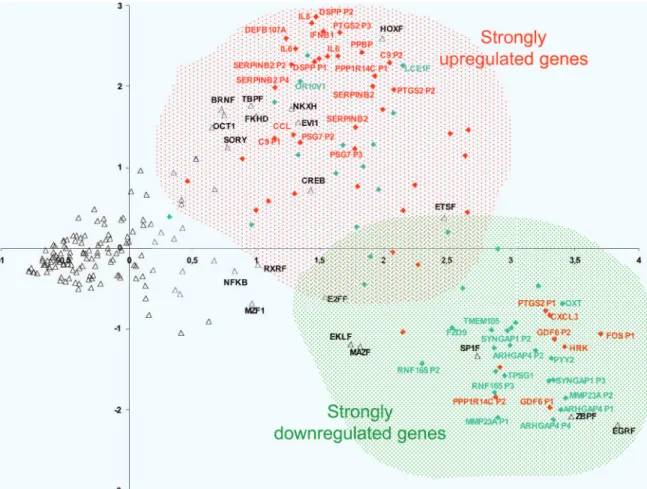
Analysis of the Promoters Responding to FOXL2 in Terms of Transcrip-tion Factor Binding Site ComposiTranscrip-tion.To obtain insights about the way the aforementioned genes respond to FOXL2, we analyzed the promoter of the 50 most induced and most repressed genes, using PCA (for details, see Materials and Methods). Promoter regions were identified by using the Genomatix software suite (http:// genomatix.de). The number of putative promoters per gene ranged from one to five. In all, 46 promoters were found for induced genes and 40 for repressed genes. Then, putative TFBS were detected. The first two axes generated by the PCA represented 25.1% and 16.8% of the total information, respectively, and were analyzed
Fig. 1. PCA enabled us to discriminate between potential target promoters activated (in red) and repressed (in green) by FOXL2. The promoters of strongly up-regulated genes were enriched in forkhead recognition sites, suggesting that they are likely to be direct FOXL2 targets. TFBS composition (open triangles) possesses a strong predictive value for positive or negative transcriptional response of the promoters to FOXL2.
Batista et al. PNAS 兩 February 27, 2007 兩 vol. 104 兩 no. 9 兩 3333
thoroughly. The inertia (information content) of the third axis dropped to 3.8%, suggesting that the analysis of the first two axes is a very good approximation for understanding the complexity encapsulated in the promoter data set.
As shown in Fig. 1, TFBS (represented by open triangles) appeared distributed as an elongated ‘‘comet’’ along the first axis. The left side of the comet involves rare sites, whereas the right side is composed of frequently occurring binding sites. Because PCA tends to give more weight to sites occurring abundantly in the promoters, it gives a strong indication about the overall ‘‘aroma’’ of a given promoter. The right part of the graph, which concerns the frequent binding sites, organizes into two statistically distinct (but overlapping) blocks. Our analysis reveals a clear opposition be-tween TFBS involved in housekeeping activities (ZBP, SP1, EGR, MAZ; low coordinates of axis 2) and TFBS generally involved in developmental networks (HOX, NKXH, BRN, TBP, FKHD, OCT; high coordinates of axis 2). The promoters whose activity is modulated by FOXL2 essentially belong to one type or the other in a rather exclusive fashion. Student’s t tests, as well as nonparametric Mann–Whitney tests, confirmed the existence of significant differ-ences between the coordinates of the promoters of genes induced or repressed by FOXL2. On axis 1, induced genes had a mean coordinate of 1.96, vs. 2.43 for the repressed genes (P⫽ 0.003). This discrimination was especially strong for the mean coordinates of the second axis (1.04 for up-regulated genes vs. ⫺0.30 for down-regulated genes; P⫽ 8.8 ⫻ 10⫺6). It is important to notice that the FKHD (ForKHeaD, the known binding consensuses of FOX factors) was located in the cluster of developmental TFBS, and therefore in the upper part of the graph, along with promoters corresponding to induced genes. This result suggests that activated genes often contain forkhead binding sites in their promoters and may be direct targets of FOXL2, or at least they respond to other forkhead factors that are in turn targets of FOXL2. We have gathered evidence for direct interactions between FOXL2 and the promoters of several genes mentioned above by chromatin immu-noprecipitation using our antibodies (see SI Materials and Methods
and SI Table 4). Several genes had promoters located in both clouds (up- and down-regulated factors). This finding suggests a possible promoter ‘‘choice,’’ enabling FOXL2 to interact directly and indi-rectly with different subsets of promoters.
General Discussion and Conclusions.In recent years, knowledge has been accumulating regarding the phenotypical effects of FOXL2 mutations in both human and mouse models. However, these analyses cannot reveal the mechanistic paths from the causal mutation to the phenotype. Consequently, identifying FOXL2 targets may help in understanding the normal and pathogenic effects of this gene. Here, we have attempted to identify targets by using a cellular model of ovarian granulosa cells (Fig. 2).
As we have shown above, FOXL2 appears to be involved in the regulation of cholesterol metabolism. We found that
PPARGC1A and NR5A2, both involved in cholesterol
ho-moeostasis (35), are stimulated by FOXL2. Previous works (13) have shown that FOXL2 represses expression of Star, a protein that controls cholesterol transport from the outer to the inner mitochondrial membranes. In agreement with this finding, FOXL2 up-regulated the cholesterol 25-hydroxylase whose product is a potent inhibitor of sterol synthesis (42). As already pointed out, FOXL2 also participates in regulation of cholesterol transformation into steroid hormones by activating aromatase (14, 15). The apparent contradiction posed by the repression of cholesterol synthesis/transport and the up-regulation of estrogen synthesis could be explained by considering that ovarian steroi-dogenesis implies close communication between theca and gran-ulosa cells. Indeed, in humans androgen biosynthesis occurs in theca cells stimulated by luteinizing hormone (43, 44). These androgens diffuse into the vascular granulosa compartment. Under follicle-stimulating hormone stimulation, the androgens
undergo aromatization to estrogens as a result of aromatase activity (45).
FOXL2 is also associated with apoptosis. Indeed, FOXL2 has been thought to be an antiapoptotic agent because its absence in the FOXL2⫺/⫺knockout mouse model leads to massive follicular loss (10). However, recent results in transfected CHO cells and in rat granulosa cells suggest a proapoptotic role through the interaction of FOXL2 with DP103, a DEAD box-containing protein (46). Our data are in agreement with this dual character of FOXL2. This ambivalent involvement in apoptosis regulation is not limited to FOXL2. It is well known that in hematopoietic cells, activation of a FOXO factor is sufficient to activate proapoptotic genes. In contrast, in most other cell types activation of FOXO blocks cellular proliferation and drives the cells to quiescence, providing protective mechanisms (47).
It is also apparent that FOXL2 is involved in the regulation of ROS homeostasis. As sketched above, PPARGC1A, stimulated by FOXL2, induces the expression of several members of the mito-chondrial ROS detoxification system. The most outstanding exam-ple is MnSOD. Interestingly, FOXO3a, another forkhead factor, also protects quiescent cells from oxidative stress by increasing the quantity of MnSOD by direct transactivation (48). The nature of the interaction between FOXL2 and MnSOD deserves further study, as does the potential involvement of FOXL2 in the regulation of ovarian senescence.
Last, but not least, the fact that PTGS2 is strongly activated by FOXL2 points to a role, more important than previously recog-nized, for prostaglandins in ovarian function. Moreover, the up-regulation of genes involved in inflammation lends credence to studies claiming that ovulation is an inflammatory-like process and suggests that FOXL2 might act very early during gonadal deter-mination and all the way through the latest stages of follicular maturation and ovulation.
In the future, the transcriptomic results outlined above must be validated at the protein level. Our discussion focused on genes with known functions; however, many unannotated genes (i.e., ‘‘Loc’’ genes) also responded to FOXL2 and deserve further attention. In addition, our study must be complemented by in vivo analyses involving animal models. Because BPES is a developmental disor-der as well, uncovering FOXL2 targets in the craniofacial region and in the fetal gonad is also an important task that needs to be addressed. Finally, it would be also interesting to systematically identify targets of FOXL2 by chromatin immunoprecipitation (given that several antibodies are available). This research is the only way to prove the existence of direct interactions. In fine, a better understanding of the regulation by FOXL2 of the genes mentioned above (and many others not discussed due to the lack of
Fig. 2. Summary of FOXL2 targets. Red, up-regulation; green, down-regulation. FSH, follicle-stimulating hormone; LH, luteinizing hormone.
space) may help in understanding the pathogenic mechanisms underlying the BPES phenotype.
Materials and Methods
Plasmid Constructs.pFOXL2 is a pCDNA3.1 vector (Invitrogen, Carlsbad, CA) containing the coding region of the human FOXL2.
Cell Culture and Transient Transfections.KGN cells (16) were seeded in DMEM-F12 medium supplemented with 10% FBS and 1% penicillin/streptomycin, at a concentration of 1.0⫻ 106cells per T25
culture flask. Cells were transfected using the calcium phosphate method and transfected again 24 h after the first transfection (49). Tandem transfections improved the efficiency of this process, as judged from control experiments with pEGFP (enhanced GFP under the control of a CMV promoter) in which the final trans-fection efficiency was⬇30% (data not shown). Transfections were performed using 12.5g of pFOXL2 per T25 culture dish (three independent transfection experiments) or 12.5g of pCDNA3.1 (empty vector/mock transfection, also n⫽ 3). Transfection effi-ciency was estimated at⬇30%, as judged from control experiments using pEGFP.
RNA Extraction and dscDNA Synthesis.Twenty-four hours after the second transfection, total RNA was extracted by using TRIzol Reagent (Invitrogen) in accordance with the manufacturer’s instructions. RNA extractions from three independent transfec-tion experiments were pooled before dscDNA synthesis. In all, 80 g of total RNA was extracted from each FOXL2- and mock-transfected condition, and dscDNA synthesis was per-formed using the SuperScript dscDNA synthesis kit (Invitrogen). This protocol was used with one modification: After the second-strand cDNA synthesis reaction was stopped with EDTA, a 10-min RNase A digestion at 37°C was included. Samples were resuspended to 250 ng/l.
Gene Expression Arrays. Four micrograms of dscDNA of each FOXL2- and mock-transfected condition were sent to the NimbleGen expression array platform. DNA end-labeling,
hy-bridization, scanning, and data normalization were performed at NimbleGen, which provided the final data file.
qPCR.We used a qPCR approach to confirm the microarray results through the screening of 27 genes affected by FOXL2 overexpres-sion. The primers were designed by using Primer-3 software (http:// frodo.wi.mit.edu/cgi-bin/primer3/primer3㛭www.cgi). For qPCRs, we used the Platinum SYBR Green qPCR SuperMix-UDG system (Invitrogen) and the Roche Light-Cycler PCR apparatus.
PCA. A set of genes presenting an average of at least 2-fold induction/repression calculated from the different probes spe-cific for each gene spotted on the NimbleGen chip were selected for promoter analysis. Among these genes, the 25 most induced genes and the 25 most repressed were chosen. Putative promoter regions were automatically identified by using Genomatix (Mu-nich, Germany) software (http://genomatix.de). The putative TFBS were detected by using the Gene2Promoter function of the Genomatix software. Most isolated promoters encompassed 501 bp upstream to the ATG initiation codon. In some cases, additional information enabled the software to extend the promoter region to a maximum of ⬇920 bp. The average promoter size was of 627 bp. One hundred forty-nine types of TFBS were identified in the complete set of promoters. The FKHD mentioned in the text corresponds to any of the 16 consensus FKH binding sites present in the Genomatix database. The number of sites was normalized by the promoter size, and the complete data set was used to generate an interpromoter correlation matrix. This matrix (i.e., 86 promoters⫻ 149 sites) was used for a multidimensional PCA (details available upon request). Among the 86 axes identified, only the first 2 were analyzed thoroughly because they represented 25.1% and 16.8% of the total information, respectively.
We thank S. Caburet, L. Moumne´, and P. Laissue for insightful
comments on the manuscript and S. Kummerfeld and S. Teichmann for advice. F.B. is supported by a Pfizer ‘‘Les Conventions Industrielles de Formation par la Recherche’’ fellowship, and R.A.V. and D.V. are
funded by Institut National de la Sante´ et de la Recherche Me´dicale,
Centre National de la Recherche Scientifique, Universite´ Paris V, and
Universite´ Paris VII.
1. Zlotogora J, Sagi M, Cohen T (1983) Am J Hum Genet 35:1020–1027.
2. Crisponi L, Deiana M, Loi A, Chiappe F, Uda M, Amati P, Bisceglia L, Zelante L, Nagaraja R, Porcu S, Pilia G (2001) Nat Genet 27:159–166.
3. Cocquet J, Pailhoux E, Jaubert F, Servel N, Xia X, Pannetier M, De Baere E, Messiaen L, Cotinot C, Fellous M, Veitia RA (2002) J Med Genet 39:916–921.
4. Cocquet J, De Baere E, Gareil M, Pannetier M, Xia X, Fellous M, Veitia RA (2003)
Cytogenet Genome Res 101:206–211.
5. Kioussi C, O’Connell S, St-Onge L, Treier M, Gleiberman AS, Gruss P, Rosenfeld MG (1999) Proc Natl Acad Sci USA 96:14378–14382.
6. De Baere E, Beysen D, Oley C, Lorenz B, Cocquet J, De Sutter P, Devriendt K, Dixon M, Fellous M, Fryns JP, et al. (2003) Am J Hum Genet 72:478–487.
7. Caburet S, Demarez A, Moumne L, Fellous M, De Baere E, Veitia RA (2004) J Med Genet 41:932–936.
8. Moumne´ L, Fellous M, Veitia RA (2005) Hum Mol Genet 14:3557–3564. 9. Gersak K, Harris SE, Smale WJ, Shelling AN (2004) Hum Reprod 19:2767–2770. 10. Schmidt D, Ovitt CE, Anlag K, Fehsenfeld S, Gredsted L, Treier AC, Treier M (2004)
Development (Cambridge, UK) 131:933–942.
11. Ellsworth BS, Burns AT, Escudero KW, Duval DL, Nelson SE, Clay CM (2003) Mol Cell
Endocrinol 206:93–111.
12. Ellsworth BS, Egashira N, Haller JL, Butts DL, Cocquet J, Clay CM, Osamura RY, Camper SA (2006) Mol Endocrinol 20:2796–2805.
13. Pisarska MD, Bae J, Klein C, Hsueh AJW (2004) Endocrinology 145:3424–3433. 14. Baron D, Cocquet J, Xia X, Fellous M, Guiguen Y, Veitia RA (2004) J Mol Endocrinol 33:705–715. 15. Pannetier M, Fabre M, Batista F, Kocer A, Renault L, Jolivet G, Mandon-Pe´pin B, Cotinot
C, Veitia R, Pailhoux E (2006) J Mol Endocrinol 36:399–413.
16. Nishi Y, Yanase T, Mu Y, Oba K, Ichino I, Saito M, Nomura M, Mukasa C, Okabe T, Goto K, et al. (2001) Endocrinology 142:437–445.
17. Son EW, Rhee DK, Pyo S (2006) J Toxicol Environ Health 69:2137–2155. 18. D’Sa-Eipper C, Chinnadurai G (1998) Oncogene 16:3105–3114.
19. Zong WX, Edelstein LC, Chen C, Bash J, Gelinas C (1999) Genes Dev 13:382–387. 20. Kim H, Kim YN, Kim H, Kim CW (2005) Oncogene 24:1252–1261.
21. He CH, Waxman AB, Lee CG, Link H, Rabach ME, Ma B, Chen Q, Zhu Z, Zhong M, Nakayama K, et al. (2005) J Clin Invest 115:828–830.
22. Wu MX, Ao Z, Prasad KV, Wu R, Schlossman SF (1998) Science 281:998–1001. 23. Shen L, Guo J, Santos-Berrios C, Wu MX (2006) J Biol Chem 281:15304–15311. 24. Hollander MC, Zhan Q, Bae I, Fornace AJ (1997) J Biol Chem 272:13731–13733. 25. Ohnishi J, Ohnishi E, Jin M, Hirano W, Nakane D, Matsui H, Kimura A, Sawa H, Nakayama
K, Shibuya H, et al. (2001) Mol Endocrinol 15:747–764.
26. Yiu GK, Toker A (2006) J Biol Chem 281:12210–12217. 27. Hla T, Neilson K (1992) Proc Natl Acad Sci USA 89:7384–7388.
28. Tazawa R, Xu XM, Wu KK, Wang LH (1994) Biochem Biophys Res Commun 203: 190 –199.
29. Lim H, Paria BC, Das SK, Dinchuk JE, Langenbach R, Trzaskos JM, Dey SK (1997) Cell 91:197–208. 30. Espey LL, Tanaka N, Okamura H (1989) Am J Physiol 256:E753–E759.
31. Liuwantara D, Elliot M, Smith MW, Yam AO, Walters SN, Marino E, McShea A, Grey ST (2006) Diabetes 55:2491–2501.
32. Wang S, Lan F, Huang L, Dong L, Zhu Z, Li Z, Xie Y, Fu J (2005) Biochem Biophys Res
Commun 333:917–924.
33. Zhang C, Gao C, Kawauchi J, Hashimoto Y, Tsuchida N, Kitajima S (2002) Biochem Biophys
Res Commun 297:1302–1310.
34. Ahrens T, Pertz O, Haussinger D, Fauser C, Schulthess T, Engel J (2002) J Biol Chem 277:19455–19460.
35. Borniquel S, Valle I, Cadenas S, Lamas S, Monsalve M (2006) FASEB J 20:1889–1891. 36. Bhalla S, Ozalp C, Fang S, Xiang L, Kemper JK (2004) J Biol Chem 279:45139–45147. 37. Puigserver P (2005) Int J Obes 29(Suppl 1):S5–S9.
38. Wang JH, Tuohimaa P (2006) Biochem Biophys Res Commun 345:720–725.
39. Ye X, Li Y, Huang Q, Yu Y, Yuan H, Wang P, Wan D, Gu J, Huo K, Li YY, et al. (2006)
Arch Biochem Biophys 449:87–93.
40. Massrieh W, Derjuga A, Doualla-Bell F, Ku CY, Sanborn BM, Blank V (2006) Biol Reprod 74:699–705.
41. Lan Y, Ovitt CE, Cho ES, Maltby KM, Wang Q, Jiang R (2004) Development (Cambridge,
UK) 131:3207–3216.
42. Bjo¨rkhem I (2002) J Clin Invest 110:725–730.
43. Ryan KJ, Petro Z (1966) J Clin Endocrinol Metab 26:46–52.
44. Sasano H, Okamoto M, Mason JI, Simpson ER, Mendelson CR, Sasano N, Silverberg SG (1989) Hum Pathol 20:452–457.
45. Bjersing L (1968) Acta Endocrinol 125:1–23.
46. Lee K, Pisarska MD, Ko JJ, Kang Y, Yoon S, Ryou SM, Cha KY, Bae J (2005) Biochem
Biophys Res Commun 336:876–881.
47. Burgering BM, Medema RH (2003) J Leukocyte Biol 73:689–701.
48. Kops GJ, Dansen TB, Polderman PE, Saarloos I, Wirtz KW, Coffer PJ, Huang TT, Bos JL, Medema RH, Burgering BM (2002) Nature 419:316–321.
49. Sambrook J, Russel D (2001) Molecular Cloning: A Laboratory Manual (Cold Spring Harbor Lab Press, Cold Spring Harbor, NY), 3rd Ed.
Batista et al. PNAS 兩 February 27, 2007 兩 vol. 104 兩 no. 9 兩 3335