HAL Id: hal-01456279
https://hal-amu.archives-ouvertes.fr/hal-01456279
Submitted on 4 Feb 2017
HAL is a multi-disciplinary open access
archive for the deposit and dissemination of
sci-entific research documents, whether they are
pub-lished or not. The documents may come from
teaching and research institutions in France or
abroad, or from public or private research centers.
L’archive ouverte pluridisciplinaire HAL, est
destinée au dépôt et à la diffusion de documents
scientifiques de niveau recherche, publiés ou non,
émanant des établissements d’enseignement et de
recherche français ou étrangers, des laboratoires
publics ou privés.
Repair and Is Not Required for Ligase IV Stimulation
Sunetra Roy, Abinadabe de Melo, Yao Xu, Satish Tadi, Aurélie Négrel, Eric
Hendrickson, Mauro Modesti, Katheryn Meek
To cite this version:
Sunetra Roy, Abinadabe de Melo, Yao Xu, Satish Tadi, Aurélie Négrel, et al.. XRCC4/XLF
In-teraction Is Variably Required for DNA Repair and Is Not Required for Ligase IV Stimulation.
Molecular and Cellular Biology, American Society for Microbiology, 2015, 35 (17), pp.3017-3028.
�10.1128/MCB.01503-14�. �hal-01456279�
Not Required for Ligase IV Stimulation
Sunetra Roy,aAbinadabe J. de Melo,c,d,e,fYao Xu,aSatish K. Tadi,c,d,e,fAurélie Négrel,c,d,e,f* Eric Hendrickson,bMauro Modesti,c,d,e,f
Katheryn Meeka
College of Veterinary Medicine and Departments of Microbiology & Molecular Genetics and Pathobiology & Diagnostic Investigation, Michigan State University, East Lansing, Michigan, USAa
; Department of Biochemistry, Molecular Biology and Biophysics, University of Minnesota Medical School, Minneapolis, Minnesota, USAb
; Centre de Recherche en Cancérologie de Marseille, CNRS, UMR7258, Marseille, Francec
; INSERM, U1068, Marseille, Franced
; Institut Paoli-Calmettes, Marseille, Francee
; Aix-Marseille Université, Marseille, Francef
The classic nonhomologous end-joining (c-NHEJ) pathway is largely responsible for repairing double-strand breaks (DSBs) in
mammalian cells. XLF stimulates the XRCC4/DNA ligase IV complex by an unknown mechanism. XLF interacts with XRCC4 to
form filaments of alternating XRCC4 and XLF dimers that bridge DNA ends in vitro, providing a mechanism by which XLF
might stimulate ligation. Here, we characterize two XLF mutants that do not interact with XRCC4 and cannot form filaments or
bridge DNA in vitro. One mutant is fully sufficient in stimulating ligation by XRCC4/Lig4 in vitro; the other is not. This
separa-tion-of-function mutant (which must function as an XLF homodimer) fully complements the c-NHEJ deficits of some
XLF-defi-cient cell strains but not others, suggesting a variable requirement for XRCC4/XLF interaction in living cells. To determine
whether the lack of XRCC4/XLF interaction (and potential bridging) can be compensated for by other factors, candidate repair
factors were disrupted in XLF- or XRCC4-deficient cells. The loss of either ATM or the newly described XRCC4/XLF-like factor,
PAXX, accentuates the requirement for XLF. However, in the case of ATM/XLF loss (but not PAXX/XLF loss), this reflects a
greater requirement for XRCC4/XLF interaction.
T
he last bona fide classic nonhomologous end-joining
(c-NHEJ) factor discovered was XLF (XRCC4-like factor) or
Cer-nunnos, discovered by two laboratories in
⬃2006 (
1–3
). Although
XLF lacks primary sequence homology with XRCC4 (X-ray cross
complementing 4), it shares remarkable structural similarity with
it, including a globular head, a C-terminal helix, and an
unstruc-tured C-terminal tail (
4
,
5
). XLF dimers can interact with XRCC4
dimers, and structural studies reveal an extended filament
com-posed of alternating XRCC4/XLF dimers (
6–9
). These filaments
bridge and stabilize DNA ends in vitro, implicating XRCC4/XLF
in bridging broken ends prior to repair (
4
,
6
). Invoking a bridging
function for XLF would be attractive, because XLF promotes the
ligation of mismatched ends, an activity that might require a
fac-tor to hold DNA ends together while end processing progressed
(
10–13
). Alternatively, XLF’s ability to stimulate ligation might be
associated with its ability to stimulate adenylation of Lig4 (
14
).
Patients with XLF mutations have microcephaly, growth
retar-dation, and progressive lymphopenia and are radiosensitive and
immunodeficient (
1
,
15
). Additionally, cells from these patients
display defective responses to DNA replication stress (
16
). In
con-trast, XLF-deficient mice are immunocompetent and have no
neurologic phenotype or growth retardation; however, different
cell types from these mice are impacted differently by XLF
defi-ciency (
17
,
18
). For example, the progressive lymphopenia
ob-served in XLF
⫺/⫺patients can be recapitulated in ageing XLF
⫺/⫺mice and is explained by a failure of hematopoietic stem cells (
19
).
Alt and colleagues have observed that mice deficient in both XLF
and either ATM (ataxia telangiectasia mutated), H2AX (histone
2A variant X), 53BP1 (p53 binding protein 1), or DNA-dependent
protein kinase catalytic subunit (DNA-PKcs) have much more
profound deficits than animals deficient in just XLF or any one of
these other factors alone (
20–24
). Importantly, some of these
fac-tors have been suggested to have functional roles in bridging or
synapsing DNA ends. Thus, these findings provide a potential
explanation for why different cell types have different
require-ments for XLF.
Here, we characterize two XLF mutants that cannot form
fila-ments with XRCC4 in vitro. One of these mutants still is proficient
in stimulating XRCC4/Lig4 activity in vitro while the other is not.
We find that all cell types tested are dependent on XLF’s ability to
stimulate XRCC4/Lig4; however, only certain cell types are
depen-dent on XLF’s stable interaction with XRCC4 (and potential DNA
end bridging). Cells that lack XLF or express an XLF that cannot
interact with XRCC4 display defective DNA-PK
autophosphory-lation, suggesting that at least one function of XRCC4/XLF
com-plexes in living cells is to stabilize DNA-PK at synapsed DNA ends.
Loss of either ATM or the newly described XRCC4/XLF-like
fac-tor, PAXX (paralog of XRCC4 and XLF) (
25
,
26
), accentuates the
cellular requirement for XLF. However, in the case of ATM/XLF
loss (but not PAXX/XLF loss), this reflects a greater requirement
for XRCC4/XLF stable interaction. Intriguingly, disruption of
Received 16 December 2014 Returned for modification 16 January 2015 Accepted 15 June 2015
Accepted manuscript posted online 22 June 2015
Citation Roy S, de Melo AJ, Xu Y, Tadi SK, Négrel A, Hendrickson E, Modesti M, Meek K. 2015. XRCC4/XLF interaction is variably required for DNA repair and is not required for ligase IV stimulation. Mol Cell Biol 35:3017–3028.
doi:10.1128/MCB.01503-14.
Address correspondence to Katheryn Meek, kmeek@msu.edu.
* Present address: Aurélie Négrel, Sigma Aldrich, Saint Quentin Fallavier, France. Supplemental material for this article may be found athttp://dx.doi.org/10.1128 /MCB.01503-14.
Copyright © 2015, American Society for Microbiology. All Rights Reserved.
XLF in XRCC4-deficient cells enhances cellular sensitivity to
DNA-damaging agents, suggesting a function for XLF that is
in-dependent of XRCC4. Finally, as with other c-NHEJ factors (
27
)
(
28
), some XLF-deficient cells display increased sensitivity to
rep-lication stress; surprisingly, neither XLF’s ability to stimulate
XRCC4/Lig4 nor its ability to stably interact with XRCC4 is
abso-lutely required for XLF in abating replication stress. These data
suggest that XLF has multiple functions in DNA repair, and they
offer potential explanations for the pleiotropic phenotypes
asso-ciated with XLF deficiency.
MATERIALS AND METHODS
Plasmids. The expression constructs for wild-type (wt) and mutant
XRCC4 have been described (29). Wild-type and mutant XLF cDNAs were cloned into the pMSCV-neo vector plasmid (Clontech Laboratories, Palo Alto, CA). Rag1 and Rag2 expression plasmids were a gift from David Roth. Construction of the I-SceI expression plasmid was described al-ready (30). The PAXX (C9orf142) expression construct was generated by PCR using IMAGE clone 3161564 as a template, engineering an NcoI restriction enzyme site at the ATG and an XhoI site after the STOP codon, and subcloned into the NcoI-XhoI sites of pHIS parallel I. The sequence is identical to that reported recently (26). DNA sequence analysis indicated that the sequence was identical to that of UniProt entry Q9BUH6. The I-SceI-trex2 fusion expression plasmid was obtained from Jeremy Stark. Fluorescent VDJ substrate plasmids have been described by others (J. Neal and K. Meek, unpublished data). Briefly, the red fluorescent protein (RFP) coding sequence was inserted between 12RSS and 23RSS (where RSS indicates recombination signal sequence), replacing the oop tran-scription terminator in both pJH290 and pJH289. The recombination cassette was cloned upstream of the cyan fluorescent protein (CFP) cod-ing sequence in peCFP-N1 (which includes the simian virus 40 [SV40] origin of replication). In these plasmids, 12RSS and 23RSS are flanked by SalI and BamHI restriction enzyme sites, respectively. RSS with the unique coding/RSS junctures depicted were generated by inserting the following oligonucleotides (and their complements) into those sites: 12RSS, CTAGCCCGCCCCACAGTGCTACAGACTGGAACAAAAACC CTGCAG; 23RSS mismatch, GATCCCACGGGTTTTTGTACAGCCAG ACAGTGGAGTACTACCACTGTGTTTATTTA; 23 RSS match, GATCC CACGGGTTTTTGTACAGCCAGACAGTGGAGTACTACCACTGTGG GGCGGGA.
Proteins. Recombinant XRCC4, XLF, XRCC4/Lig4, and Ku were
ob-tained using procedures described previously (4,31–33). PAXX was pro-duced in Escherichia coli Rosetta/pLysS cells and purified by nickel-nitri-lotriacetic acid (Ni-NTA), followed by Sepharose Q chromatography using the same method as that for XRCC4 purification (31). Bridging assays were performed as described previously (6).
Electrophoretic mobility shift assay (EMSA). The DNA substrate was
obtained by annealing the oligonucleotide 5=-GACGCTGCCGAATTCT ACCAGTGCCTTGCTAGGACATCTTTGCCCACCTGCAGGTT CACCC to its reverse complement strand. DNA (25 nM final concentra-tion) and the indicated final concentrations of proteins were mixed in a 10-l reaction adjusted to 75 mM KCl, 10 mM Tris, pH 7.5, 0.5 mM EDTA, 0.5 mM dithiothreitol (DTT), and 5% glycerol. After incubation for⬎1 h at room temperature, reaction mixtures were fractionated by 6% PAGE in Tris-borate-EDTA buffer, followed by staining with ethidium bromide. Gel images were collected with a ChemiDoc XRS⫹ system and processed with the Image Lab software (Bio-Rad).
Ligation assays. Reaction mixtures (10l) contained 100 ng of
lin-earized pUC19 plasmid (digested with XbaI for cohesive-end ligation or with SmaI for blunt-end ligation), 2 mM MgCl2, 1 mM ATP, 75 mM KCl,
10 mM HEPES, pH 8.0, 0.5 mM EDTA, 5% glycerol, and the indicated final concentrations of proteins. T4 DNA ligase (New England BioLabs) was used at a final concentration of 8 U/l. After a 30-min incubation at room temperature, the samples were deproteinized by the addition of pronase (1.25g/l final concentration) and Sarkosyl (1.25% final
con-centration) and incubated at 55°C for 30 min. The reaction mixtures were fractionated by agarose gel electrophoresis using Tris-borate-EDTA buf-fer and stained with ethidium bromide. Gel images were acquired as in-dicated for the EMSAs.
Cell culture and cell strains. 293, HCT116, and 2BN cells were
cul-tured in Dulbecco’s modified Eagle medium (Life Technologies) supple-mented with 10% fetal bovine serum (Atlanta Biologicals, GA), 2 mM
L-glutamine, 0.1 mM nonessential amino acids, 1 mM sodium pyruvate, 100 U/ml penicillin, 100g/ml streptomycin (Life Technologies), and 10 g/ml ciprofloxacin. Cells were maintained at 37°C with 5% CO2.
Em-bryonic stem (ES) cells were cultured in the medium described above further supplemented with-mercaptoethanol and 103 U/ml ESGRO (Millipore). Abl pre-B cells were grown in RPMI medium (Life Technol-ogies) supplemented with 10% fetal bovine serum, 100 U/ml penicillin, and 100g/ml streptomycin.
For the generation of cell strains stably expressing wild-type and mu-tant constructs of XLF in 293 cells, 5g of plasmid DNA was transfected with polyethyleneimine (PEI; 1g/ml; Polysciences) at 2 l/1 g DNA. Forty-eight hours posttransfection, cells were selected with medium con-taining 800g/ml G418 (Life Technologies), and single clones were ana-lyzed for expression by immunoblotting. Similarly, ES and 2BN cells were transfected using Lipofectamine (Invitrogen) and FuGENE (Roche), re-spectively, according to the manufacturers’ instructions. Stable XLF ex-pression in HCT116 cells and Abl pre-B cells was achieved by retroviral transduction. Briefly, viruses were prepared by transient transfection of 1 g of a pMSCV-XLF construct and 1 g of helper plasmids (pCGP pVSVG ratio, 4:1) in 293T cells. Forty-eight hours after transfection, the medium was collected and passed through a 0.45-m filter to remove cellular debris. The filtrate containing viral particles was treated with 20 mM HEPES and 8g/ml Polybrene (Sigma) for 10 min. The medium containing viruses then was laid over a monolayer of HCT116 cells for 4 h and then replaced with complete medium. Forty-eight hours after infec-tion, cells were selected with medium containing 800g/ml G418. Simi-larly, Abl pre-B cells were plated at a concentration of 105 cells/100 ml medium in 24-well plates. One milliliter of viral supernatant was added to each well and spun at 1,800 rpm for 90 min at room temperature. After centrifugation, the medium was replaced with fresh complete medium. Forty-eight hours after infection, cells were selected with 2 mg/ml G418 at a concentration of 1,000 cells/100l medium in a 96-well plate. Stable transfectants were screened by immunoblotting.
Immunoblot analyses. Immunoblotting was performed as described
previously (29). Antibodies used in this study are rabbit polyclonal XLF (Abcam), rabbit polyclonal XRCC4 (Abcam), goat anti-C9ORF142 (Santa Cruz Biotechnology) for PAXX, rabbit anti-ATM (Se-rotec), and rabbit polyclonal anti-DNA-PKcs-phospho-S2056 (Abcam). The DNA-PKcs antibody (42–27) was the generous gift of Tim Carter. To detect DNA-PKcs autophosphorylation, cells (plated 18 h earlier) were treated in complete media with zeocin (2 mg/ml) for 3 h. Cells were harvested and whole-cell extracts prepared for immunoblotting.
Ni-agarose pulldown. Ten micrograms of pEF vectors expressing
ei-ther the wild-type or mutant forms of C-terminal His-tagged XLF was transfected into 293 cells as described above. Forty-eight hours after trans-fection, cells were harvested and washed with phosphate-buffered saline (PBS). Pellets were lysed with 1 ml lysis buffer (50 mM Tris, pH 7.5, 120 mM sodium chloride, 0.5% NP-40, 1 mM sodium fluoride, 1 mM sodium orthovanadate, protease inhibitor cocktail) and rocked for 30 min on ice. Lysates were centrifuged and the supernatant used for pulldown assays. Fifty microliters of Ni-NTA–agarose beads (Qiagen) was added to 1 ml cell lysate containing 10 mM imidazole and rocked for 3 h at 4°C. Beads were collected by centrifugation and washed three times in the same buf-fer containing 50 mM imidazole. After washing, beads were resuspended in 30l 4⫻ SDS-PAGE buffer and analyzed by immunoblotting.
Cas9-mediated gene disruption. Cas9-targeted gene disruption was
performed using methods similar to those reported by Mali et al. (34). Briefly, guide RNAs (gRNAs) specific for XLF, ATM, XRCC4, or PAXX
were synthesized as 455-bp fragments (Integrated DNA Technologies). The synthesized fragments were cloned into pCR2.1 using a TOPO TA cloning kit according to the manufacturers’ instructions (Life Technolo-gies). Cells were transfected with 1g gRNA plasmid and 1 g Cas9 expression plasmid (Addgene). In some cases, cells were cotransfected with 0.2g of pcDNA6 (Life Technologies) or pSuper-Puro to confer blasticidin or puromycin resistance. Western blotting was used to identify clones with deletions in each of these factors; in all cases, deletion also was confirmed by PCR amplification that revealed deletions at the target site. The 19-mers specific for each factor synthesized into the 455-bp fragments are the following: ATM, TCTTTCTGTGAGAAAATAC; XRCC4, CCTGCA GAAAGAAAATGAA; XLF, GGCCTGTTGATGCAGCCAT; PAXX-1, CTG GCCTTTGACCTCTCCA; PaXX-2, TGCTTCACGCCGGACAGCCT.
Survival assays. Clonogenic survival assays were performed for ES cells,
2BN cells, and HCT116 cells. Briefly, a hundred cells were plated for each transfectant into complete medium containing the indicated dose of zeocin or hydroxyurea (HU) in 60-mm-diameter tissue culture dishes. For HU as-says, cell culture medium was changed after 24 h. After 7 to 10 days, cell colonies were stained with 1% (wt/vol) crystal violet in ethanol to measure relative survival. 3-(4,5-Dimethyl-2-thiazolyl)-2,5-diphenyl-2H-tetrazolium bromide (MTT) staining was performed to assess cell viability for 293 cells and Abl pre-B cells. Thirty thousand to 50,000 cells were plated in each well of a 24-well plate containing medium with various concentrations of zeocin. After 5 to 7 days of zeocin treatment, cells were treated with 1 mg/ml MTT (Sigma) solution for 1 h. Medium containing MTT then was removed, and the formazan crystals produced were solubilized in acidic isopropanol. Ab-sorbance was read at 570 nm to determine relative survival.
VDJ recombination assays. Extrachromosomal VDJ recombination
assays utilizing the signal joint substrate (pJH201) and coding joint sub-strate (pJH290) were performed as described previously (35). Briefly, cells plated at 20 to 40% confluence in 60-mm-diameter dishes were tran-siently transfected with 1g substrate, 4 g each of RAG1 and RAG2, and 4g of the indicated expression construct or empty vector using the Lipofectamine 2000 transfection reagent according to the manufacturers’ instructions. Forty-eight hours after transfection, substrate plasmids were isolated by alkaline lysis and subjected to DpnI restriction enzyme diges-tion for 1 h. DpnI-digested DNA was transformed into competent DH5␣ cells (Invitrogen) according to the manufacturers’ instructions. Trans-formed cells were spread onto LB agar plates containing 100g/ml am-picillin only or with 100g/ml ampicillin and 22 g/ml chloramphenicol. The percentage of recombination was calculated as the number of colo-nies resistant to ampicillin and chloramphenicol divided by the number of colonies resistant to ampicillin.
The fluorescent VDJ substrates (diagramed inFig. 3) and VDJ as-says have been described recently (Neal and Meek, unpublished). These substrates (derived from pECFP-N1; Clontech) contain the SV40 origin of replication; thus, they are efficiently replicated epi-somally in all primate cell strains; it has been reported that this origin replicates to very low copy numbers in rodent cells (36). We have directly compared VDJ proficiency in CHO cell strains with pJH290/ pJH201 and the fluorescence-based substrates with completely analo-gous results (Neal and Meek, unpublished). Briefly, extrachromo-somal fluorescent VDJ assays were performed on cells plated at 20 to 40% confluence into 24-well plates in complete medium. Cells were transfected with 0.125g substrate, 0.25 g RAG1 per well, and 0.25 g RAG2 per well using PEI (1 g/ml; Polysciences) at 2 l/1 g DNA. In experiments with additional expression plasmids, 0.25g of the expression plasmid or vector control was included. Cells were har-vested 72 h after transfection and analyzed for CFP and RFP expression by flow cytometry. The percentage of recombination was calculated as the percentage of live cells expressing CFP divided by the percentage expressing RFP. Data presented represent at least three independent experiments, each of which includes triplicate transfections.
RESULTS
XLF L115A does not interact with XRCC4 or bridge DNA in
vitro but is fully sufficient to stimulate XRCC4/Lig4-mediated
ligation in vitro. We and others have implicated L115 as being an
XLF residue that is critical for the molecule’s interaction with
XRCC4 (
4
,
37
). Whereas our biochemical data show that an
L115A substitution completely disrupts the XRCC4/XLF
interac-tion (
4
), Fattah et al. have demonstrated end joining of plasmid
substrates and VDJ intermediates of this mutant in living cells
(
38
); the relative ability of L115A to restore radioresistance was
not studied by these authors. In contrast, de Villartay and
col-leagues found that an L115D substitution (which also does not
interact with XRCC4 in vitro) does not reverse the c-NHEJ deficits
or reverse radiosensitivity associated with XLF deficiency (
37
). To
understand the basis for these differences, the biochemical
func-tion of these two mutants in vitro were directly compared.
Previ-ously, we developed a DNA bridging assay that measures end
bridging between a free 500-bp DNA fragment and a 1,000-bp
biotin-labeled fragment that can be immobilized onto
streptavi-din beads (
6
). If bridging occurs (by protein-DNA interaction),
the untagged DNA will be pulled down onto the streptavidin
beads. In the presence of wild-type XLF and XRCC4 together but
not with either alone and not with either XLF L115A or XLF
L115D (and wild-type XRCC4), the free 500-bp fragment could be
efficiently pulled down (
Fig. 1A
). As another test of DNA bridging
in vitro, the ability of XRCC4 and XLF to promote ligation and
formation of concatemers by T4 DNA ligase was assessed. Neither
XRCC4 nor XLF alone significantly enhance ligation of either
blunt or cohesive ends by T4 ligase (
Fig. 1B
). However, ligations of
both blunt and cohesive DNA ends are markedly enhanced by
wild-type XLF and XRCC4 together. Similar to the results with the
DNA bridging assay, neither the L115A nor the L115D mutant
stimulates ligation of either blunt or cohesive DNA ends by T4
DNA ligase (
Fig. 1B
). We conclude that neither mutant can
effi-ciently bridge DNA in vitro.
XRCC4 interacts with and supports the stability of Lig4 in
liv-ing cells; from this perspective, it is unclear whether the
stimula-tion of T4 ligase activity in vitro genuinely recapitulates XRCC4/
XLF’s effect on Lig4. Thus, recombinant human XRCC4/Lig4
complexes were prepared, and the ability of XLF (wild type or
mutant) to stimulate ligation was tested. As expected, wild-type
XLF markedly stimulates the activity of XRCC4/Lig4 (
Fig. 1C
). To
our surprise, the L115A mutant was just as proficient as wild-type
XLF in stimulating intermolecular ligation by XRCC4/Lig4,
whereas L115D did not stimulate ligation by XRCC4/Lig4 (
Fig. 1C
and
D
). Moreover, no differences were observed at any ratio of
XLF to XRCC4 tested (0.25, 0.5, 1, or 2:0.4). These in vitro results
accurately mirrored and corroborated the conflicting in vivo
re-ports (
4
,
37
,
38
) and allowed us to conclude that XLF’s capacity to
stimulate XRCC4/Lig4 in vitro is independent of its DNA bridging
capacity in vitro.
Previously we studied DNA bridging using XRCC4 mutants
that could not interact with XLF (
29
); however, XLF’s ability to
stimulate XRCC4/Lig4 with these mutations was not assessed.
Thus, XRCC4/Lig4 complexes were prepared with two XRCC4
mutants. Both wild-type and L115A XLFs stimulate wild-type
XRCC4/Lig4 (
Fig. 1D
), but neither wild-type XLF nor L115A
sig-nificantly stimulates the ligase activity of either mutant XRCC4/
Lig4 complex, although minimal stimulation is observed with
both. We also considered that the presence of Lig4 stabilizes a
potential weak interaction with XRCC4. Thus, we also tested (by
pulldown assays) whether these mutants transiently expressed in
293 cells interact with XRCC4 and Lig4; neither mutant interacts
with either XRCC4 or Lig4 (
Fig. 1E
). Although other explanations
are possible, we suggest that XLF (either wild type or L115A) can
stimulate XRCC4/Lig4 without stably interacting with the
com-plex. The failure of XLF L115D to stimulate ligations is less clear.
The more dramatic alteration of L115D might impede the ability
of XLF to access XRCC4/Lig4. The fact that the two mutant
XRCC4/Lig4 complexes (i.e., with XRCC4 K65/99E and K72/90/
99E) show markedly reduced stimulation by either wild-type XLF
or the L115A mutant suggests that these mutants also partially
exclude XLF from stimulating XRCC4/Lig4.
Stimulation of XRCC4/Lig4 is intrinsic to XLF’s function in
living cells; XRCC4/XLF interaction is variably required. As
noted above, XLF deficiency has a variable impact on different cell
types and in different organisms. We considered that a bridging
function of XRCC4/XLF in vivo (but not XRCC4/Lig4
stimula-tion) might be variably required in different cell types depending
on the relative expression or activity of factors that share
func-tional redundancy with XLF (ATM, 53BP1, H2AX, DNA-PKcs,
and potentially others) (
20–22
). XLF L115A appears to represent a
separation-of-function mutant that could be useful in discerning
whether XRCC4/XLF interaction and potential filament
forma-tion is important in living cells. Thus, we next derived stable clonal
transfectants of wild-type and mutant XLF in a panel of different
XLF-deficient cell strains (
Fig. 2A
). These include a human
fibro-blast cell strain (2BN) derived from an XLF-deficient patient (
39
),
XLF-deficient HCT116 cells (a human colon cancer cell strain
from which XLF was deleted by gene targeting) (
38
), human 293
cells that are XLF deficient via a Cas9/CRISPR-mediated
muta-tion, mouse XLF-deficient embryonic stem cells (
17
), and mouse
v-Abl-transformed XLF-deficient pre-B cells (
18
). Wild-type XLF
substantially reverses the zeocin (a bleomycin analogue that
in-duces double-strand breaks [DSBs])-sensitive phenotype in all
five cell strains (
Fig. 2B
to
F
). The complementation of the
zeocin-sensitive phenotype in XLF-deficient 293 cells demonstrates that
the zeocin-sensitive phenotype is the effect of XLF deletion and
not off-target effects of Cas9/CRISPR (
Fig. 2F
). Expression of the
XLF mutant L115D that cannot stimulate XRCC4/Lig4 does not
reverse zeocin sensitivity in any of the five cell strains. L115A that
is proficient in stimulating XRCC4/Lig4 (but deficient in DNA
end bridging in vitro) completely reverses the zeocin sensitivity in
the human patient fibroblast cells (2BN) and in XLF-deficient ES
cells and partially reverses zeocin sensitivity in human 293 cells
FIG 1 XLF L115A does not interact with XRCC4; thus, it does not bridge DNA in vitro but is fully sufficient to stimulate XRCC4/Lig4. (A, left) Schematic of the
DNA bridging assay. (Right) Agarose gel showing recovery of DNA fragments bound to streptavidin beads by ethidium bromide staining. Molecular size markers are indicated (kilobases). (B to D) Ethidium bromide staining of agarose gels showing ligation products obtained from in vitro ligation reactions as described in Materials and Methods. Molecular size markers are indicated (kilobases). (B) T4 DNA ligase is utilized. (C) XRCC4/Lig4 complexes (0.4M) are utilized. Four different concentrations of XLF were utilized: 0.25M, 0.5 M, 1 M, and 2 M. (D) XRCC4/Lig4 complexes (0.2 M) are utilized, with wild-type or mutant XRCC4 as indicated and with wild-type or mutant XLF (0.5M). (E, top) Immunoblot analyses of lysates from 293 cells transiently transfected with His-tagged wt and mutant forms of XLF probed with antibodies to XRCC4, XLF, or Lig4. (Bottom) Immunoblot analyses of pulldown fractions recovered from Ni-NTA– agarose beads after 3 h of incubation of cell lysates with beads and subsequent washing. The immunoblot was probed with antibodies to XRCC4, XLF, or Lig4.
and murine Abelson-transformed B cells. In contrast, L115A does
not reverse zeocin sensitivity in human XLF-deficient HCT116
cells. From these data, we conclude that XLF’s capacity to
stimu-late XRCC4/Lig4 is required in all cell types for maximal survival
following DNA DSBs, whereas XLF’s ability to stably interact with
XRCC4 is variably required in different cell types. This variation in
the cellular requirement for XRCC4/XLF interaction is not readily
attributable to differences in expression of other DNA repair
fac-tors (see Fig. S1 in the supplemental material), although HCT116
cells express less ATM than 2BN or 293 cells. Of note, HCT116
cells have been shown to have defects in both mismatch repair and
in one allele of Mre11 (
40
,
41
). Moreover, HCT116 cells display
the most sensitivity to zeocin of all the cell strains studied here.
Stable XRCC4/XLF interaction is variably required in
differ-ent cell types to support VDJ recombination of episomal
sub-strates. XLF initially was discovered by studies of a patient with
immunodeficiency (
1
,
39
). Although XLF deficiency does not
re-sult in substantial VDJ deficits in murine lymphocytes or murine
B cell lines (
18
), XLF is required in episomal VDJ recombination
assays in mouse ES cells (
17
), mouse embryonic fibroblasts (
17
,
18
), human patient fibroblasts (
37
,
39
,
42
), and XLF-deficient
HCT116 cells (
38
). Although VDJ rearrangements have not been
examined in lymphocytes from human XLF-deficient patients, the
SCID phenotype of these patients (
1
) intuits that XLF also is
re-quired for VDJ recombination in developing human
lympho-cytes. We performed episomal VDJ assays in both XLF-deficient
murine ES cells and XLF-deficient 293 cells. In ES cells, the
stan-dard Gellert assay (
35
,
43
) was utilized, whereas in 293 cells a
fluorescent substrate (
Fig. 3
, top) and a flow-cytometric assay
were utilized (described in Materials and Methods; also see the
supplemental material). Importantly, L115A, but not L115D,
sup-ports substantial levels of both coding and signal end joining in
FIG 2 Stimulation of XRCC4/Lig4 is intrinsic to XLF’s function in living cells; XRCC4/XLF interaction is variably required. (A) Immunoblot showing stable
expression of actin or tubulin and wild-type or mutant XLF in various XLF-deficient cell strains, as indicated. vect, vector. (B to F) Zeocin sensitivity of the indicated cell strains stably expressing equivalent levels of wild-type or mutant XLF. Error bars indicate standard errors of the means (SEM) from at least three independent experiments.
XLF-deficient ES cells (
Fig. 3
). Similarly, albeit not as robustly,
L115A but not L115D restores both coding and signal end joining
in XLF-deficient 293 cells.
The proficient coding end joining by XLF L115A (that does not
interact with XRCC4 and cannot form XRCC4/XLF filaments in
vitro) is at odds with our previous study showing that XRCC4
mutants that could not interact with XLF (and could not bridge
DNA or form XRCC4/XLF filaments in vitro) had modest VDJ
recombination deficits that asymmetrically impacted coding joint
formation (
44
). These previous studies were performed in the
XRCC4-deficient CHO (Chinese hamster ovary) cell strain XR-1.
Given the substantial variability in cellular phenotypes that may
reflect differences in requirements for stable XRCC4/XLF
interac-tion, the ability of these XRCC4 mutants to complement VDJ
deficits was assessed in XRCC4-deficient ES cells (generated by
conventional gene targeting [
45
]) and in XRCC4-deficient 293
cells that were prepared for these studies by a
Cas9/CRISPR-me-diated mutation. Neither XRCC4 mutant support wild-type levels
of coding end joining in 293 cells, consistent with the partial
com-plementation of XLF
⫺/⫺293 cells by the L115A mutant (see Fig.
S2 in the supplemental material). In contrast, coding joint
forma-tion is substantially complemented by both mutants in
XRCC4-deficient ES cells, again consistent with L115A’s capacity to
com-plement VDJ joining in XLF-deficient ES cells. ES clones stably
expressing wild-type XRCC4 or K65/99E were established. As with
L115A in XLF-deficient ES cells (
Fig. 2D
), K65/99E complements
the zeocin-sensitive phenotype of XRCC4-deficient ES cells (see
Fig. S2). These data underscore the variable dependence on
XRCC4/XLF interaction; 293 cells show a strong dependence on
stable XRCC4/XLF complexes compared to that of mouse ES cells,
where stable XRCC4/XLF interactions apparently are dispensable.
XLF L115A is more defective in ligation of incompatible
DNA ends than of compatible DNA ends. Two DNA ends must
be closely juxtaposed for ligation to occur, although emerging
data suggest that repair of the two strands occurs independently
(
12
,
46
,
47
). It is also well appreciated that DSBs can be maintained
in a spatiotemporal manner in large DNA repair centers (
48
).
There is evidence that XRCC4/XLF filaments affect both the
pre-cise alignment of DNA ends, in that XLF promotes ligation of
noncohesive ends in vitro (
10
,
11
) (
Fig. 1
), as well as the
mainte-nance of ends in “repair centers,” in that XLF deficiency is
exac-erbated when factors that function in assembling
␥H2AX foci are
disrupted (
20–24
). We first assessed, in living cells, whether
L115A’s deficit in resolving coding ends was exacerbated if the
ends are not compatible. The RAG endonuclease generates
blunt-ended signal ends and hairpinned coding ends (
49
). It has been
shown that coding hairpins are nicked to generate 4-bp 3=
over-hangs (
50
). Two additional fluorescence-based coding joint
sub-strates were prepared. One substrate generates 4-bp cohesive ends
and also provides additional pairing opportunities even if
nucle-otide loss (up to 7 bp at each end) occurs at either coding end (
Fig.
4
, matched coding end). A second substrate generates overhangs
with no microhomology, even if nucleotide loss should occur.
Analyses of these substrates is complicated by the fact that the
RAG nuclease is exquisitely sensitive to the sequences of the
cod-ing end-recombination signal sequence (RSS) junctures (
51
)
(which are fairly optimal in the parental substrate but cannot be
optimized in the matched/mismatched substrates). Thus, these
coding/RSS substrates are not optimal and recombine 3- to 4-fold
less efficiently in wild-type cells than the original 290/RFP/CFP
substrate (not shown). Hence, comparison of recombination
lev-els for each substrate must be normalized to the efficiency of
re-combination in cells that are c-NHEJ proficient. Although L115A
and wild-type XLF are similarly proficient in joining the substrate
that generates matched ends, L115A is less proficient in joining
coding ends with completely mismatched termini (3.2-fold)
com-pared to the optimal substrate (2.0-fold) (
Fig. 4
). We conclude
that XRCC4/XLF interaction facilitates end joining when DNA
termini lack terminal microhomology.
XRCC4/XLF interaction is required for robust DNA-PK
au-tophosphorylation. Calsou and colleagues demonstrated that
DNA damage-induced autophosphorylation of DNA-PKcs is
de-FIG 3 XRCC4/XLF interaction is variably required in different cell types to support VDJ recombination of episomal substrates. The fluorescent substrates (top
left) were utilized to detect coding and signal joints in VDJ assays in 293 cells, whereas substrates pJH290 and pJH201 (top right) were used to detect coding and signal joints in VDJ assays in ES cells. The lower panel shows percent recombination of episomal fluorescent coding and signal joint substrates in XLF-deficient 293 cells transiently expressing full-length Rag1, Rag2, and wild-type and mutant forms of XLF. Note that the decrease in percent recombination observed by L115A and L115D are statistically significant for signal joint substrate (wt versus L115A, P⫽ 0.021; wt versus L115D, P ⫽ 0.0002) and coding joint substrate (wt versus L115A, P⫽ 0.0001; wt versus L115D, P ⬍ 0.0001) according to a two-tailed Mann-Whitney test. Error bars indicate SEM from five independent experiments. The lower panel shows percent recombination of pJH290 (left) and pJH289 (right) in XLF-deficient mouse ES cells transiently expressing full-length Rag1, Rag2, and wt and mutant forms of XLF. Note that the decrease in recombination percentage observed by L115D compared to that of the wt is statistically significant (P⫽ 0.0019 for coding joint assay and P ⫽ 0.0047 for signal joint) according to two-tailed Mann-Whitney test. Error bars indicate SEM from four independent experiments.
fective in cells deficient in Lig4 (
32
). Moreover, a catalytically
in-active mutant of Lig4 reversed this defect, suggesting that the
XRCC4/Lig4 complex facilitates autophosphorylation. The 293,
HCT116 (
Fig. 5
), and 2BN (not shown) isogenic cell strain panels
were exposed to zeocin, and DNA-PKcs autophosphorylation at
serine 2056 was assessed by immunoblotting. In all three cell
types, although phosphorylation of
␥H2AX is similar, S2056
phosphorylation is markedly reduced in cells lacking XLF; neither
L115D nor L115A reverses the defect in autophosphorylation,
suggesting that XRCC4/XLF complexes promote DNA-PKcs
au-tophosphorylation (
Fig. 5
). Although other explanations are
pos-sible, since S2056 phosphorylation can occur in trans (
52
), these
data suggest that XRCC4/XLF complexes help stabilize synapsed
DNA-PK bound ends, enhancing trans-autophosphorylation.
However, this defect in autophosphorylation does not explain
dif-ferences in the cellular requirement for XRCC4/XLF interaction,
since L115A completely complements the zeocin sensitivity of
2BN cells but not of HCT116 cells (
Fig. 2
). It should be noted that
DNA-PKcs can be phosphorylated on many residues (likely more
than 60) (
53
), it has been shown that some autophosphorylations
can occur in cis (
54
,
55
), and phosphorylation at 2056 has been
shown to occur in trans (
52
).
An ATM or PAXX deficiency accentuates the c-NHEJ deficits
of L115A. As noted above, the different cellular phenotypes could
be due to the functional redundancy of XLF/XRCC4 complexes
with other proteins in certain cell types. Thus, we next generated
293 cells that lack both XLF and ATM (see Fig. S3A in the
supple-mental material). In addition, two laboratories recently have
de-scribed another XRCC4/XLF-like factor, PAXX (
25
,
26
). PAXX
interacts with DNA-bound Ku and can be targeted to sites of DNA
damage. Thus, given the DNA bridging capacity of XRCC4 and
XLF, 293 and HCT116 cells lacking PAXX or both PAXX and XLF
also were generated (see Fig. S3A). Both HCT116 and 293 cells
lacking only PAXX were resistant to zeocin to an extent similar to
that for wild-type cells at the zeocin doses used to study zeocin
sensitivity in XLF-deficient cells, although modest sensitivity was
observed using higher zeocin doses (data not shown). These data
are consistent with the mild radiosensitivity observed in
PAXX-deficient cells by others (
25
,
26
). However, the loss of PAXX
ex-pression in XLF-deficient cells (either 293 cells or HCT116)
exac-erbated zeocin sensitivity in both HCT116- and 293 XLF-deficient
cells (
Fig. 6A
). We conclude that for zeocin resistance in both
HCT116 cells and 293 cells, PAXX and XLF are not epistatic.
Al-though Jackson and colleagues concluded that PAXX is epistatic
with XLF for repair of infrared-induced damage, XLF was
de-pleted with short interfering RNA and residual XLF was present
(
26
). Moreover, Xing and colleagues observed a lack of epistasis
between PAXX and XLF in chicken DT40 cells for the repair of
some types of DNA damage but not others (
25
). Thus, different
cell types may have distinct requirements for XLF in response to
different types of DNA damage.
Episomal VDJ assays also were performed on doubly deficient
293 cells. Loss of ATM or PAXX accentuates the requirement for
XLF in rejoining both coding and signal ends (
Fig. 6B
).
Cotrans-fection of either PAXX or XLF partially reverses the VDJ deficits in
XLF
⫺/⫺PAXX
⫺/⫺cells, whereas cotransfection of both results in
more substantial complementation (see Fig. S3B in the
supple-mental material). In addition, whereas the wild type and L115A
mutant similarly reverse the deficits in XLF
⫺/⫺PAXX
⫺/⫺cells, the
VDJ deficits associated with the L115A mutant are exacerbated in
XLF
⫺/⫺ATM
⫺/⫺cells (
Fig. 6B
). Consistent with these data, the
FIG 5 XRCC4/XLF interaction is required for robust DNA-PK
phorylation. Immunoblot analysis showing levels of DNA-PKcs autophos-phorylation at S2056, total DNA-PKcs, XLF expression, and ␥H2AX phosphorylation with and without zeocin treatment of 293 cells (left) and XLF-deficient HCT116 cells (right) stably expressing wild-type and mutant forms of XLF.
FIG 4 XLF L115A is more defective in ligation of incompatible DNA ends
than in that of compatible DNA ends. (Top) Schematic showing altered coding end sequences to generate perfectly matched or mismatched overhangs in the fluorescent coding joint substrate depicted inFig. 3. (Bottom) Comparison of recombination rate of episomal substrates with matched and mismatched cod-ing ends in XLF-deficient 293 cells transiently expresscod-ing Rag1, Rag2, and wild-type and mutant forms of XLF. Error bars indicate SEM from four inde-pendent experiments. Note that the decrease in recombination rate observed by XLF mutant L115A compared to that of the wild type is statistically signif-icant for 290 (P⫽ 0.0021) and mismatched (P ⫽ 0.0404) termini according to two-tailed Mann-Whitney test. There is no significant difference observed between recombination rate of wt XLF and XLF mutant L115A in the presence of matched termini.
VDJ deficits associated with the XRCC4 mutants K65/99E and
K72/90/99E also are exacerbated by the loss of ATM (see Fig. S4).
Wild-type and mutant XLF were stably integrated into the
XLF
⫺/⫺ATM
⫺/⫺293 cells. Whereas wild-type XLF substantially
reverses the zeocin hypersensitivity, neither the L115A nor L115D
mutant reverses zeocin sensitivity whatsoever (
Fig. 6C
). This is in
marked contrast to the significant level of complementation by
L115A in XLF-deficient (but ATM-proficient) 293 cells (
Fig. 2F
).
Of note, the lower ATM expression levels in HCT116 cells provide
a potential explanation for the complete lack of complementation
of zeocin hypersensitivity by L115A (see Fig. S1). In addition, loss
of both ATM and XLF results in a further deficiency in DNA-PKcs
autophosphorylation (
Fig. 6E
). In sum, these data suggest that it is
the ability of XLF to complex with XRCC4 (and potentially form
filaments that bridge DNA) that is functionally redundant with
ATM, as proposed by others (
20
).
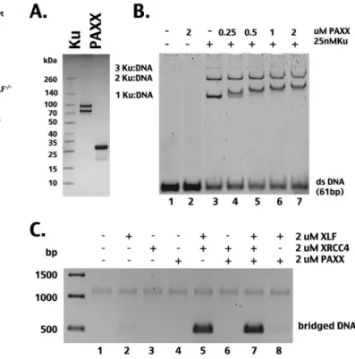
PAXX interacts with Ku-DNA complexes but does not bridge
DNA or cooperate with XRCC4/XLF to bridge DNA in vitro. In
contrast to XLF
⫺/⫺ATM
⫺/⫺cells, in XLF
⫺/⫺PAXX
⫺/⫺cells, the
L115A (but not the L115D) mutant was as proficient as wild-type
XLF in partially reversing both coding and signal joining defects.
Thus, the loss of PAXX does not accentuate the cellular
require-ment for XRCC4/XLF interaction. To understand the biochemical
basis of these differences, recombinant PAXX was generated (
Fig.
7A
). Consistent with recent reports (
25
,
26
), although it does not
itself bind DNA, PAXX interacts with Ku-bound DNA (
Fig. 7B
).
As reported by Xing et al. (
25
), we find that PAXX interacts only
with Ku/DNA complexes containing one Ku heterodimer,
whereas Ochi et al. (
26
) found that PAXX interacts exclusively
with complexes containing two Ku heterodimers. One potential
explanation for these differences is expression strategies
(baculo-FIG 6 ATM and PAXX deficiency accentuates the c-NHEJ deficits of L115A. (A)
Zeocin sensitivity of parental wild-type cells, XLF-deficient and XLF⫺/⫺PAXX⫺/⫺ 293 cells (left), and HCT116 cells (right). Error bars indicate SEM from three independent experiments. (B) Recombination percentage of episomal fluorescent coding and signal joining substrates comparing XLF-deficient, XLF/ATM doubly deficient, and XLF⫺/⫺PAXX⫺/⫺293 cells transiently expressing Rag1, Rag2, and wild-type and mutant forms of XLF as indicated. Error bars indicate SEM from at least 3 independent experiments. Note that XLF L115A shows a significantly lower coding end joining rate in XLF-deficient (P⫽ 0.0001) and XLF⫺/⫺ATM⫺/⫺(P⫽ 0.0022) 293 cells, unlike XLF⫺/⫺PAXX⫺/⫺cells. Similarly, XLF L115A performs significantly reduced signal joining in XLF-deficient (P⫽ 0.0210) and XLF⫺/⫺ ATM⫺/⫺(P⫽ 0.0022) cells, unlike XLF⫺/⫺PAXX⫺/⫺293 cells. (C) Zeocin sen-sitivity of the XLF⫺/⫺ATM⫺/⫺293 cell strain stably expressing equivalent levels of wt or mutant XLF. Data from three clones expressing L115A are presented. Error bars indicate SEM from three independent experiments. (D) Immunoblot analysis depicting XLF expression levels in XLF⫺/⫺ATM⫺/⫺cells stably transfected with wt or mutant XLF or vector control. (E) Immunoblot showing levels of DNA-PKcs autophosphorylation at S2056 with and without zeocin (Zeo) treatment of XLF/ ATM doubly deficient cells stably expressing wt and mutant forms of XLF.
FIG 7 PAXX interacts with Ku-DNA complexes but does not itself bridge
DNA or cooperate with XRCC4/XLF filaments to bridge DNA. (A) Polyacryl-amide gel electrophoresis under reducing and denaturing conditions stained with Coomassie blue showing recombinant Ku (1g) and PAXX (5 g). (B) A native polyacrylamide gel stained with ethidium bromide showing the electro-phoretic mobility shift of a 61-bp double-stranded DNA (dsDNA) after incu-bation with PAXX, Ku, or Ku and PAXX together. (C) Agarose gel stained with ethidium bromide showing recovery of DNA fragments bound to streptavidin beads after DNA bridging assays (as described forFig. 1).
virus for Ku expression for these studies and those of Xing et al.
versus bacterial expression for the studies of Ochi et al.).
More-over, PAXX by itself does not promote DNA end bridging, nor
does it alter the DNA end-bridging capacity of XRCC4/XLF (
Fig.
7C
). From data presented here, we conclude that although PAXX
can interact with Ku-bound DNA, it does not contribute to DNA
end bridging in vitro; moreover, loss of PAXX does not accentuate
the cellular requirement for XRCC4/XLF interaction (and
poten-tial DNA end bridging) in living cells.
XLF promotes cell survival after replication stress; its role in
this process is independent of its interaction with XRCC4. Cells
deficient in various c-NHEJ factors are more sensitive to agents
that induce replication stress than c-NHEJ-proficient cells (
27
,
28
,
53
). Schwartz et al. have shown previously that fibroblasts derived
from XLF-deficient patients are sensitive to low-dose aphidicolin,
display increased fragile site instability, and display cell cycle
dis-ruptions consistent with an inability to resolve DNA damage
as-sociated with replication stress (
16
). Thus, we next assessed cell
survival of these isogenic cell strains after exposure to
hy-droxyurea, which depletes nucleotide pools, resulting in
replica-tion stress. Whereas XLF-complemented and vector control 2BN
cells are similarly sensitive to HU (data not shown), XLF-deficient
HCT cells are remarkably sensitive to HU; this sensitivity is
re-versed by wild-type XLF (
Fig. 8A
). To our surprise, both L115A
and L115D substantially reverse the HU hypersensitivity, albeit
slightly less so than wild-type XLF. Of note, complementation by
L115A and L115D is equivalent, suggesting that XLF’s unexpected
role in reversing HU hypersensitivity is at least partially
indepen-dent of its ability to interact with XRCC4 or stimulate ligase IV
activity.
To test this conclusion more stringently, cells deficient in both
XRCC4 and XLF were derived and tested for their capacity to
survive HU-induced replication stress and zeocin-induced DSBs.
Three independent clones targeting XLF in XRCC4-deficient
HCT116 cells were isolated (
Fig. 8B
). The loss of XLF in each cell
line exacerbated the sensitivity of XRCC4-deficient cells to both
HU and zeocin (
Fig. 8C
and
D
). Implicit in these data is an
XRCC4-independent function for XLF.
DISCUSSION
We conclude from these studies that XLF has multiple cellular
functions that are biochemically distinct. Recent biochemical
studies have proposed that one of XLF’s functions in living cells is
to tether or bridge DNA ends with XRCC4 (potentially as
fila-ments or filament bundles) (reviewed in reference
56
). However,
evidence was lacking for formation of these DNA end-bridging
filaments in living cells. During the final revision of the
manu-script, elegant studies from Rothenberg and colleagues
unequivo-cally demonstrated that XRCC4/XLF form filaments in living cells
(with or without Lig4). Superresolution microscopy
demon-FIG 8 XLF promotes cell survival after replication stress; its role in this process is independent of its interaction with XRCC4. (A) HCT116 transfectants
expressing wild-type XLF, XLF L115A, XLF L115D, or no XLF (vector) were plated at cloning densities into complete medium with increasing doses of hydroxyurea. Colonies were stained after 7 days, and percent survival was calculated. Error bars represent the SEM. (B) XLF and XRCC4 protein expression levels were examined by Western blotting of whole-cell extracts obtained from the indicated cell strains. (C and D) HCT116 cells and HCT116 cells deficient in XLF, XRCC4, or both XLF and XRCC4 were plated at cloning densities into complete medium with increasing doses of zeocin (C) or hydroxyurea (D). Data from three clones deficient in both XLF and XRCC4 are presented.
strated the presence of these filaments adjacent to Ku-bound
DSBs, in some cases spanning Ku-bound DSBs (
57
). What is still
unclear is precisely how DNA bridging impacts end joining. Given
that the homodimerization of DNA-PK also can tether/bridge
ends together (
58
,
59
,
60
), it may seem redundant to postulate the
need for a second tethering system. However, the use of two
dis-crete tethering systems may have distinct mechanistic advantages.
The presence of a second, XRCC4/XLF-mediated tethering
pro-cess could solve a significant topological problem. The DNA-PK
complex is able to tether broken DNA ends together; this requires
both the catalytic subunit and the DNA binding subunit, Ku, a
circular-shaped protein through which the DNA is threaded (
61
,
62
). Numerous investigators have noted that Ku’s unique DNA
binding mode would leave Ku topologically locked onto the DNA
after ligation. Such an occurrence would prohibit strand
separa-tion and jeopardize subsequent transcripsepara-tion and replicasepara-tion in
that region. It has been suggested that such events can occur and
that the cell’s solution to this problem is to proteolytically degrade
the trapped Ku (
63
). The presence of an ancillary and independent
XRCC4/XLF-mediated tethering system might obviate such
path-ways. If the Ku/DNA-PKcs tethering temporally occurs first, the
presence of a subsequent XRCC4/XLF-mediated tether would
permit DNA-PKcs to undergo the significant conformational
changes that it is known to undergo upon becoming activated
(
64–66
) and would permit the release of Ku from the DNA by
sliding without the broken DNA ends becoming separated from
one another. Thus, a requirement for XRCC4/XLF-mediated
bridging might particularly impact the efficacy of joining
non-compatible ends, since these ends presumably need to be synapsed
long enough to appropriately process the mismatched ends.
Con-sistent with this prediction, the absence of XLF is clearly
deleteri-ous for the joining of mismatched ends (
Fig. 4
) (
10–12
,
67
,
68
).
Our data demonstrate that XRCC4/XLF complexes have the
abil-ity to bridge DNA in vitro, facilitating DNA-PKcs
autophosphor-ylation, suggesting that DNA ends are bridged by both synapsed
DNA-PK and XRCC4/XLF concurrently, physically promoting
DNA-PK autophosphorylation at 2056, a site which can be
phos-phorylated in trans (
52
). It also is possible that XRCC4/XLF
bridg-ing occurs prior to DNA-PK assembly; in fact, it is known that XLF
requires Ku (but not DNA-PKcs) to be appropriately targeted to
DNA damage in living cells (
65
,
66
). Our previous studies suggest
that DNA-PK phosphorylation of XRCC4/XLF disrupts filaments
(
29
). Work is ongoing to define how XRCC4/XLF filaments
pro-mote repair in living cells.
Our cellular studies show that only some cell types require
XLF’s function of forming DNA end-bridging, XRCC4/XLF
com-plexes. Although the mechanistic basis for this cell type-specific
requirement still eludes us, it is clear that it is the ability to form
these XRCC4/XLF complexes that is overlapping with ATM.
Thus, in ATM-proficient cells, ATM compensates for L115A’s
de-fect in XRCC4 stable interaction and potential DNA end bridging
(
Fig. 6
). In contrast, although loss of PAXX accentuates the
lar requirement for XLF, this effect does not accentuate the
cellu-lar requirement for XRCC4/XLF interaction and potential DNA
end bridging. This is entirely consistent with in vitro studies
show-ing that PAXX does not itself bridge DNA ends or cooperate with
XRCC4/XLF in bridging DNA ends (
Fig. 7
). Thus, although these
studies do not define how PAXX affects c-NHEJ, our studies seem
to exclude DNA end bridging as a function for PAXX in c-NHEJ.
A second function for XLF is in stimulating Lig4 catalysis.
While we noted above that XLF indirectly augments catalysis by
facilitating synapsis, we believe that this function of XLF is
inde-pendent of a stable interaction with XRCC4. Thus, XLF that only
forms homodimers and does not form XRCC4 multimers (i.e., the
XLF L115A variant) nonetheless substantially reverses NHEJ
def-icits in many cell types. We attribute this to XLF’s capacity to
promote Lig4 catalysis without forming a stable XRCC4/XLF
complex. Our cellular studies demonstrate that unlike XLF’s
sta-ble interaction with XRCC4 and potential DNA end-bridging
ac-tivity, all cell types have a requirement for XLF’s function in
stim-ulating Lig4.
In 2009, Schwartz and colleagues demonstrated that fibroblasts
derived from XLF-deficient patients are sensitive to agents that
induce DNA replication stress (
16
). These authors suggested that
this role for XLF in DNA replication explains the phenotypes
ob-served in XLF-deficient patients that cannot be attributed to
de-fective VDJ recombination. Recent studies demonstrate that
he-matopoietic stem cells from XLF-deficient mice fail at an early age
(
19
). Although this defect was not directly attributed to premature
replicative failure, other DNA repair defects phenocopy this. We
show here that a subset of XLF-deficient cells are remarkably
sen-sitive to HU-induced replication stress (
Fig. 8
). Surprisingly,
XLF’s role in abating replication stress was in large part
indepen-dent of XLF’s interaction with XRCC4. In sum, these data imply
that XLF has at least a third function that is nonoverlapping with
its other two functions. This demonstration underscores the need
for further investigation into the distinct functions of XLF and to
delineate why different cell types and different organisms have
such variable requirements for this multifunctional factor.
In summary, we propose that XLF has (at least) two distinct
functions: (i) DNA end bridging (likely in filaments with XRCC4)
and (ii) stimulation of XRCC4/Lig4 as a homodimer. Recently, the
current dogma that c-NHEJ functions in a well-defined stepwise
manner (
69–71
) has been challenged. Emerging data using both
biochemical and genetic (
12
,
46
,
72
,
73
) approaches suggest that
c-NHEJ is much more flexible. Instead, these reports propose
models whereby XRCC4/Lig4 organizes how c-NHEJ proceeds
based upon the degree of end processing that is required for repair.
Consistent with these new models, it seems intuitive that an
XRCC4/XLF bridging function occurs at an early step in DNA
DSB repair, whereas XLF’s role in stimulating ligation would be a
later step.
ACKNOWLEDGMENTS
This work was supported by a Public Health Service grant AI048758 (K.M.), by the USDA National Institute of Food and Agriculture and Michigan AgBioResearch (K.M.), by project number SFI20121205867 from the Fondation ARC pour la Recherche sur le Cancer (M.M.), and by grant PLBIO13-103 from the Institut National du Cancer (M.M.). E.H. was supported by a grant from the National Institutes of Health (GM088351) and the National Cancer Institute (CA154461).
REFERENCES
1. Buck D, Moshous D, de Chasseval R, Ma Y, le Deist F,
Cavazzana-Calvo M, Fischer A, Casanova JL, Lieber MR, de Villartay JP. 2006.
Severe combined immunodeficiency and microcephaly in siblings with hypomorphic mutations in DNA ligase IV. Eur J Immunol 36:224 –235. http://dx.doi.org/10.1002/eji.200535401.
2. Revy P, Malivert L, de Villartay JP. 2006. Cernunnos-XLF, a recently identified non-homologous end-joining factor required for the develop-ment of the immune system. Curr Opin Allergy Clin Immunol 6:416 – 420.http://dx.doi.org/10.1097/01.all.0000246623.72365.43.
3. Ahnesorg P, Smith P, Jackson SP. 2006. XLF interacts with the XRCC4-DNA ligase IV complex to promote XRCC4-DNA nonhomologous end-joining. Cell 124:301–313.http://dx.doi.org/10.1016/j.cell.2005.12.031. 4. Andres SN, Modesti M, Tsai CJ, Chu G, Junop MS. 2007. Crystal
structure of human XLF: a twist in nonhomologous DNA end-joining. Mol Cell 28:1093–1101.http://dx.doi.org/10.1016/j.molcel.2007.10.024. 5. Li Y, Chirgadze DY, Bolanos-Garcia VM, Sibanda BL, Davies OR,
Ahnesorg P, Jackson SP, Blundell TL. 2008. Crystal structure of human
XLF/Cernunnos reveals unexpected differences from XRCC4 with impli-cations for NHEJ. EMBO J 27:290 –300. http://dx.doi.org/10.1038/sj .emboj.7601942.
6. Andres SN, Vergnes A, Ristic D, Wyman C, Modesti M, Junop M. 2012. A human XRCC4-XLF complex bridges DNA. Nucleic Acids Res 40: 1868 –1878.http://dx.doi.org/10.1093/nar/gks022.
7. Hammel M, Rey M, Yu Y, Mani RS, Classen S, Liu M, Pique ME, Fang
S, Mahaney BL, Weinfeld M, Schriemer DC, Lees-Miller SP, Tainer JA.
2011. XRCC4 protein interactions with XRCC4-like factor (XLF) create an extended grooved scaffold for DNA ligation and double strand break repair. J Biol Chem 286:32638 –32650. http://dx.doi.org/10.1074/jbc .M111.272641.
8. Ropars V, Drevet P, Legrand P, Baconnais S, Amram J, Faure G,
Marquez JA, Pietrement O, Guerois R, Callebaut I, Le Cam E, Revy P, de Villartay JP, Charbonnier JB. 2011. Structural characterization of
filaments formed by human Xrcc4-Cernunnos/XLF complex involved in nonhomologous DNA end-joining. Proc Natl Acad Sci U S A 108:12663– 12668.http://dx.doi.org/10.1073/pnas.1100758108.
9. Wu Q, Ochi T, Matak-Vinkovic D, Robinson CV, Chirgadze DY,
Blundell TL. 2011. Non-homologous end-joining partners in a helical
dance: structural studies of XLF-XRCC4 interactions. Biochem Soc Trans
39:1387–1392.http://dx.doi.org/10.1042/BST0391387.
10. Tsai CJ, Kim SA, Chu G. 2007. Cernunnos/XLF promotes the ligation of mismatched and noncohesive DNA ends. Proc Natl Acad Sci U S A 104: 7851–7856.http://dx.doi.org/10.1073/pnas.0702620104.
11. Akopiants K, Zhou RZ, Mohapatra S, Valerie K, Lees-Miller SP, Lee KJ,
Chen DJ, Revy P, de Villartay JP, Povirk LF. 2009. Requirement for
XLF/Cernunnos in alignment-based gap filling by DNA polymerases lambda and mu for nonhomologous end joining in human whole-cell extracts. Nucleic Acids Res 37:4055– 4062.http://dx.doi.org/10.1093/nar /gkp283.
12. Gu J, Lu H, Tsai AG, Schwarz K, Lieber MR. 2007. Single-stranded DNA ligation and XLF-stimulated incompatible DNA end ligation by the XRCC4-DNA ligase IV complex: influence of terminal DNA sequence. Nucleic Acids Res 35:5755–5762.http://dx.doi.org/10.1093/nar/gkm579. 13. Gu J, Lu H, Tippin B, Shimazaki N, Goodman MF, Lieber MR. 2007. XRCC4:DNA ligase IV can ligate incompatible DNA ends and can ligate across gaps. EMBO J 26:1010 –1023.http://dx.doi.org/10.1038/sj.emboj .7601559.
14. Riballo E, Woodbine L, Stiff T, Walker SA, Goodarzi AA, Jeggo PA. 2009. XLF-Cernunnos promotes DNA ligase IV-XRCC4 re-adenylation following ligation. Nucleic Acids Res 37:482– 492.
15. Woodbine L, Gennery AR, Jeggo PA. 2014. The clinical impact of defi-ciency in DNA non-homologous end-joining. DNA Repair 16:84 –96. http://dx.doi.org/10.1016/j.dnarep.2014.02.011.
16. Schwartz M, Oren YS, Bester AC, Rahat A, Sfez R, Yitzchaik S, de
Villartay JP, Kerem B. 2009. Impaired replication stress response in cells
from immunodeficiency patients carrying Cernunnos/XLF mutations. PLoS One 4:e4516.http://dx.doi.org/10.1371/journal.pone.0004516. 17. Zha S, Alt FW, Cheng HL, Brush JW, Li G. 2007. Defective DNA repair
and increased genomic instability in Cernunnos-XLF-deficient murine ES cells. Proc Natl Acad Sci U S A 104:4518 – 4523.http://dx.doi.org/10.1073 /pnas.0611734104.
18. Li G, Alt FW, Cheng HL, Brush JW, Goff PH, Murphy MM, Franco S,
Zhang Y, Zha S. 2008. Lymphocyte-specific compensation for XLF/
cernunnos end-joining functions in V(D)J recombination. Mol Cell 31: 631– 640.http://dx.doi.org/10.1016/j.molcel.2008.07.017.
19. Avagyan S, Churchill M, Yamamoto K, Crowe JL, Li C, Lee BJ, Zheng
T, Mukherjee S, Zha S. 2014. Hematopoietic stem cell dysfunction
un-derlies the progressive lymphocytopenia in XLF/Cernunnos deficiency. Blood 124:1622–1625.http://dx.doi.org/10.1182/blood-2014-05-574863. 20. Zha S, Guo C, Boboila C, Oksenych V, Cheng HL, Zhang Y, Wesemann
DR, Yuen G, Patel H, Goff PH, Dubois RL, Alt FW. 2011. ATM damage
response and XLF repair factor are functionally redundant in joining DNA breaks. Nature 469:250 –254.http://dx.doi.org/10.1038/nature09604.
21. Oksenych V, Kumar V, Liu X, Guo C, Schwer B, Zha S, Alt FW. 2013. Functional redundancy between the XLF and DNA-PKcs DNA repair fac-tors in V(D)J recombination and nonhomologous DNA end joining. Proc Natl Acad Sci U S A 110:2234 –2239. http://dx.doi.org/10.1073/pnas .1222573110.
22. Oksenych V, Alt FW, Kumar V, Schwer B, Wesemann DR, Hansen E,
Patel H, Su A, Guo C. 2012. Functional redundancy between repair factor
XLF and damage response mediator 53BP1 in V(D)J recombination and DNA repair. Proc Natl Acad Sci U S A 109:2455–2460.http://dx.doi.org /10.1073/pnas.1121458109.
23. Zha S, Sekiguchi J, Brush JW, Bassing CH, Alt FW. 2008. Complemen-tary functions of ATM and H2AX in development and suppression of genomic instability. Proc Natl Acad Sci U S A 105:9302–9306.http://dx .doi.org/10.1073/pnas.0803520105.
24. Liu X, Jiang W, Dubois RL, Yamamoto K, Wolner Z, Zha S. 2012. Overlapping functions between XLF repair protein and 53BP1 DNA damage response factor in end joining and lymphocyte development. Proc Natl Acad Sci U S A 109:3903–3908.http://dx.doi.org/10.1073 /pnas.1120160109.
25. Xing M, Yang M, Huo W, Feng F, Wei L, Jiang W, Ning S, Yan Z, Li
W, Wang Q, Hou M, Dong C, Guo R, Gao G, Ji J, Zha S, Lan L, Liang H, Xu D. 2015. Interactome analysis identifies a new paralogue of XRCC4
in non-homologous end joining DNA repair pathway. Nat Commun
6:6233.http://dx.doi.org/10.1038/ncomms7233.
26. Ochi T, Blackford AN, Coates J, Jhujh S, Mehmood S, Tamura N,
Travers J, Wu Q, Draviam VM, Robinson CV, Blundell TL, Jackson SP.
2015. DNA repair. PAXX, a paralog of XRCC4 and XLF, interacts with Ku to promote DNA double-strand break repair. Science 347:185–188. 27. Lundin C, Erixon K, Arnaudeau C, Schultz N, Jenssen D, Meuth M,
Helleday T. 2002. Different roles for nonhomologous end joining and
homologous recombination following replication arrest in mammalian cells. Mol Cell Biol 22:5869 –5878.http://dx.doi.org/10.1128/MCB.22.16 .5869-5878.2002.
28. Saintigny Y, Delacote F, Vares G, Petitot F, Lambert S, Averbeck D,
Lopez BS. 2001. Characterization of homologous recombination induced
by replication inhibition in mammalian cells. EMBO J 20:3861–3870. http://dx.doi.org/10.1093/emboj/20.14.3861.
29. Roy S, Andres SN, Vergnes A, Neal JA, Xu Y, Yu Y, Lees-Miller SP,
Junop M, Modesti M, Meek K. 2012. XRCC4’s interaction with XLF is
required for coding (but not signal) end joining. Nucleic Acids Res 40: 1684 –1694.http://dx.doi.org/10.1093/nar/gkr1315.
30. Neal JA, Dang V, Douglas P, Wold MS, Lees-Miller SP, Meek K. 2011. Inhibition of homologous recombination by DNA-dependent protein ki-nase requires kiki-nase activity, is titratable, and is modulated by autophos-phorylation. Mol Cell Biol 31:1719 –1733. http://dx.doi.org/10.1128 /MCB.01298-10.
31. Junop MS, Modesti M, Guarne A, Ghirlando R, Gellert M, Yang W. 2000. Crystal structure of the Xrcc4 DNA repair protein and implications for end joining. EMBO J 19:5962–5970.http://dx.doi.org/10.1093/emboj /19.22.5962.
32. Cottarel J, Frit P, Bombarde O, Salles B, Negrel A, Bernard S, Jeggo PA,
Lieber MR, Modesti M, Calsou P. 2013. A noncatalytic function of the
ligation complex during nonhomologous end joining. J Cell Biol 200:173– 186.http://dx.doi.org/10.1083/jcb.201203128.
33. Ono M, Tucker PW, Capra JD. 1994. Production and characterization of recombinant human Ku antigen. Nucleic Acids Res 22:3918 –3924.http: //dx.doi.org/10.1093/nar/22.19.3918.
34. Mali P, Yang L, Esvelt KM, Aach J, Guell M, DiCarlo JE, Norville JE,
Church GM. 2013. RNA-guided human genome engineering via Cas9.
Science 339:823– 826.http://dx.doi.org/10.1126/science.1232033. 35. Hesse JE, Lieber MR, Gellert M, Mizuuchi K. 1987. Extrachromosomal
DNA substrates in pre-B cells undergo inversion or deletion at immuno-globulin V-(D)-J joining signals. Cell 49:775–783.http://dx.doi.org/10 .1016/0092-8674(87)90615-5.
36. Piechaczek C, Fetzer C, Baiker A, Bode J, Lipps HJ. 1999. A vector based on the SV40 origin of replication and chromosomal S/MARs replicates episomally in CHO cells. Nucleic Acids Res 27:426 – 428.http://dx.doi.org /10.1093/nar/27.2.426.
37. Malivert L, Ropars V, Nunez M, Drevet P, Miron S, Faure G, Guerois
R, Mornon JP, Revy P, Charbonnier JB, Callebaut I, de Villartay JP.
2010. Delineation of the Xrcc4-interacting region in the globular head domain of cernunnos/XLF. J Biol Chem 285:26475–26483.http://dx.doi .org/10.1074/jbc.M110.138156.