HAL Id: hal-02388062
https://hal.uca.fr/hal-02388062
Submitted on 30 Nov 2019
HAL is a multi-disciplinary open access
archive for the deposit and dissemination of
sci-entific research documents, whether they are
pub-lished or not. The documents may come from
teaching and research institutions in France or
abroad, or from public or private research centers.
L’archive ouverte pluridisciplinaire HAL, est
destinée au dépôt et à la diffusion de documents
scientifiques de niveau recherche, publiés ou non,
émanant des établissements d’enseignement et de
recherche français ou étrangers, des laboratoires
publics ou privés.
Morphological and immunohistochemical study of
ovarian and tubal dysplasia associated with tamoxifen
G. Chêne, Nina Radosevic-Robin, A. Tardieu, A. Cayre, I Raoelfils, P.
Dechelotte, J. Dauplat, F. Penault-Llorca
To cite this version:
G. Chêne, Nina Radosevic-Robin, A. Tardieu, A. Cayre, I Raoelfils, et al.. Morphological and
im-munohistochemical study of ovarian and tubal dysplasia associated with tamoxifen. European Journal
of Histochemistry, PAGEpress, 2014, 58, �10.4081/ejh.2014.2251�. �hal-02388062�
Morphological and
immunohistochemical study
of ovarian and tubal dysplasia
associated with tamoxifen
G. Chene,1,2N. Radosevic-Robin,1A.S. Tardieu,1A. Cayre,1I. Raoelfils,1
P. Dechelotte,3J. Dauplat,4
F. Penault-Llorca1
1Department of Histopathology, Centre
Jean Perrin, ERTICA Research Team, EA 4677, Clermont-Ferrand
2Department of Gynecology, HFME, CHU
Lyon
3Department of Histopathology, CHU
Estaing, Clermont-Ferrand,
4Department of Surgery, Centre Jean
Perrin, Clermont-Ferrand, France
Abstract
Ovarian epithelial dysplasia was initially described in material from prophylactic oophorectomies for BRCA mutation. Similar histopathological abnormalities have been revealed after ovulation stimulation. Given that tamoxifen (TAM) has a clomid-like effect and is sometimes used to induce ovulation, we studied the morphological features and immunohistochemical expression patterns of neoplasia-associated markers in adnexec-tomies previously exposed to TAM for breast cancer. We blindly reviewed 173 histopatholog-ical slides of adnexectomies according to three groups – oophorectomies associated with TAM exposure (n=42), oophorectomies associated with Clomiphene exposure (n=15) and a spon-taneously fertile non cancerous control group (n=116). Morphological features (with an ovarian and tubal dysplasia scoring system) and immunohistochemical expression pat-terns of Ki-67, p53 and Aldehyde dehydroge-nase 1 (ALDH1 is an enzyme significantly associated with early-stage ovarian cancer) were evaluated and correlated. Mean tubal dys-plasia score was significantly higher in the TAM group and Clomiphen group than in con-trols (respectively 7.8 vs 3.5, P<0.007 and 6.8
vs 3.5, P=0.008). There is no statistical
differ-ence for the ovarian score in TAM group in comparison with the control group whereas we found a significant score for Clomiphen group (6.5, P=0.009). Increased ALDH1 expression was observed in the two exposed group where-as expression patterns of Ki67 and p53 were moderate. Interestingly, ALDH1 expression was low in non-dysplastic epithelium, high in dysplasia, and constantly low in the two carci-noma.
Furthemore, we confirm our previous results showing that ALDH1may be a useful tissue biomarker in the subtle histopathologi-cal diagnosis of tubo-ovarian dysplasia.
Introduction
Tamoxifen (TAM) is commonly used as an adjuvant therapy in the antihormonal treat-ment of choice in premenopausal breast can-cer patients. It is a non-steroidal selective estrogen receptor modulator (SERM) and, like clomiphene citrate, it has also been used to induce ovulation. It is also known that Tamoxifen increases the incidence of ovarian
follicular cysts.1,2 On the other hand,
histopathological study of material from pro-phylactic oophorectomies performed for a genetic predisposition to ovarian cancer can reveal cytological and architectural abnormali-ties considered to be pre-cancerous manifesta-tions. These abnormalities are termed
dyspla-sia by analogy with the pre-invasive lesions
described for the genital tract (vulva, vagina,
cervix, endometrium).3-9 Similarly, serous
tubal intraepithelial lesions (STILs, a spec-trum of epithelial changes ranging from nor-mal appearing tubal epithelium to lesions with cytologic atypia and dysplasia) have recently been described in prophylactically removed Fallopian tubes of women predisposed to devel-op ovarian cancer.10,11 Moreover, several
stud-ies have found similar ovarian dysplasia lesions after stimulation of ovulation in infer-tile patients.12-14 We sought to identify
dyspla-sia lesions in salpingo-oophorectomies associ-ated with Tamoxifen and to assess the expres-sion of proliferation and differentiation-relat-ed proteins (Ki67, p53, and Aldehyde dehydro-genase 1 or ALDH1).2,3 To the best of our
knowledge, this is the first study to assess tubo-ovarian lesions under TAM on the one hand, and on the other to underline the accu-racy of ALDH1 staining as for tubo-ovarian pre-cursor lesions.
Materials and Methods
Patients
Between January 1995 and December 2000, we selected three groups of patients:
Group A: salpingo-oophorectomies associated with tamoxifen exposure (TAM group). We
included 42 removed ovaries and tubes associ-ated with tamoxifen exposure for previous breast cancer without BReast CAncer gene (BRCA) mutation. Two occult ovarian carcino-mas were excluded from the dysplasia evalua-tion study because morphological dysplasia
analysis was designed to identify potential pre-malignant lesions.
Group B: salpingo-oophorectomies associat-ed with clomiphene citrate. We selectassociat-ed an
infertile population who had adnexectomies after ovulation induction by clomiphene sever-al years later and whose ovaries and tubes were reported as normal on routine histologi-cal examination: 15 cases were included.
Group C: Control group. We selected a
spon-taneously non cancerous and fertile population of matching age, with no personal nor family history of gynaecologic neoplasia (breast, ovary, endometrium), who underwent adnex-ectomy for which the histopathological exami-nation concluded that the ovaries and tubes did not show any sign of cancerous or border-line pathology or salpingitis: 116 controls were included in the study.
Histopathological criteria
Evaluation of morphological features Morphological studies were processed on 3 micron paraffin sections stained with standard haematoxylin phloxin safran (HPS). The num-ber of sections available for review for each case (ovary and tube) ranged from 7 to 11 in all groups.
The histopathology slides for the 40 patients who had used Tamoxifen, 15 patients who had used Clomiphene citrate and the 116 control were all scored blinded by two spcialists in gynecopathology (FPL and IR) who were spe-cialists in onco-gynaecology pathology, in order to obtain an average score. As initialy described in ovarian dysplasia studies,12,13,15
when several slides were available, the one
Correspondence: Gautier Chene, Department of Histopathology, Centre Jean Perrin, Clermont-Ferrand, 63000 France.
Tel. +33.6.07081786 - Fax: +33.4.77828956. E-mail: chenegautier@yahoo.fr
Key words: ovarian carcinogenesis, ovarian dys-plasia, prophylactic oophorectomy, ALDH1, p53. Conflict of interests: the authors declare no con-flict of interests.
Funding: La ligue contre le cancer Foundation. Received for publication: 29 July 2013. Accepted for publication: 3 March 2014. This work is licensed under a Creative Commons Attribution NonCommercial 3.0 License (CC BY-NC 3.0).
©Copyright G. Chene et al., 2014 Licensee PAGEPress, Italy
European Journal of Histochemistry 2014; 58:2251 doi:10.4081/ejh.2014.2251
with the highest dysplasia score was retained. For the ovarian cystectomies, the slides were read to confirm the histopathological diagno-sis and to inspect associated ovarian tissue to determine its dysplasia score. Absence of asso-ciated ovarian tissue was an exclusion criteri-on. In the event of obvious disagreements between pathologists, a further examination was carried out to reach a consensus. Ovarian dysplasia
There is no consensual dysplasia scoring system, as the histopathological characteris-tics of dysplasia are difficult and subtle to determine. The definition of ovarian dysplasia was thus based on cytological and architectur-al criteria described in previous studies of ovarian dysplasia, i.e., dysplasia found in
pro-phylactic oophorectomy for BRCA mutations,3-5
in areas adjacent to Stage 1 ovarian carcino-ma,6,7in the contralateral ovary in case of
uni-lateral ovarian cancer,8,9and in relation to
ovu-lation induction.12 We pooled the criteria to
design an eleven criteria (listed below) scor-ing system used in our previous studies on the relationship between ovarian dysplasia, ovula-tion inducovula-tion, and genetic risk:13-15
- epithelial pseudostratification
- epithelial proliferation : pilling up the cells, increased cellularity and Ki67 expression, as previously published13-16
- surface papillomatosis
- irregular nuclear chromatine pattern - irregular nuclear contour
- cellular pleiomorphism
- increase in nuclear size (nuclear-cytoplas-mic ratio and comparison to the normal nuclei)
- inclusion cysts (presence of more than three contiguous cysts)
- deep epithelial invaginations - psammoma
- stromal hyperplasia.
In each case, the abnormal areas were scored between 0 and 2 (0, normal; 1, moder-ately abnormal; 2, severely abnormal), whether located on the surface or in an inclusion cyst. An overall dysplasia ovarian score was then obtained for each patient by simply adding the scores for each of the 11 items (total range: 0 to 22).
Tubal dysplasia
As for the ovary, the difficulty of analysing tubal dysplasia can be explained by the lack of well established morphological criteria for pre-cursors of Fallopian tube carcinoma. Our defi-nition was based on previous studies of tubal precursor lesions (named Serous Tubal
Intraepithelial Lesions, STILs) described in
Fallopian tubes from patients with a genetic risk (prophylactic oophorectomy for BRCA1/2 mutations), and we designed a scoring system with seven histopathological criteria:10,11,16-18
- epithelial pseudostratification - tufting
- loss of nuclear polarity - increase in nuclear density - nuclear atypia
- nucleomegaly - loss of ciliation
In each case, the abnormal areas were scored between 0 and 2 (0, normal; 1, moder-ately abnormal; 2, severely abnormal). An over-all dysplasia tubal score was then obtained for
each patient by simply adding the scores for each of the 7 items (total range: 0 to 14).
Evaluation of immunostaining
Immunohistochemistry was performed on 3 micron sections, on silanised slides dried overnight at 56°C. Ki67, P53 and ALDH1 immunostaining was performed with a
BenchmarkXT immunostainer (Ventana
Medical Systems, Illkirch, France) as indicated in Table 1. For Ki67 and P53, immunostaining
Original Paper
Table 1. Immunohistochemical characteristics.
Antibody Clone Manufacturer Dilution Incubation Detection
Ki67 MIB-1 DAKO 1/100 32 min Ultraview
P53 DO-7 DAKO 1/200 32 min Ultraview
ALDH1 44/ALDH BD Biosciences 1/400 60 min Ultraview
Table 2. Comparison of respective frequencies of the eleven histopathologic abnormali-ties in our ovarian dysplasia scoring system. P1, statistical differences between group A and C; P2, statistical differences between group B and C; P3, statistical differences between group A and B.
Histopathological items Group A Group B Group C P
(n= 40) (n=15) (n=116) Epithelial pseudostratification 23 (57.5%) 10 (66.6%) 54 (46.5%) P1=0.240 P2=0.032 P3=0.750 Epithelial proliferation 12 (30%) 11 (73.3%) 35 (30.1%) P1=0.930 P2=0.002 P3=0.003 Surface papillomatosis 14 (35%) 9 (60%) 27 (23.2%) P1=0.1200 P2=0.0070 P3=0.0074
Irregular nuclear chromatine pattern 13 (32.5%) 7 (46.6%) 21 (18.1%) P1=0.054
P2=0.0072 P3=0.07
Irregular nuclear contour 7 (17.5%) 7 (46.6%) 35 (30.1%) P1=0.371
P2=0.060 P3=0.0071
Cellular pleiomorphism 13 (32.5%) 8 (53.3%) 36 (31%) P1=0.515
P2=0.006 P3=0.007
Increase in nuclear size 5 (12.5%) 7 (46.6%) 22 (18.9%) P1=0.5130
P2=0.0038 P3=0.0025
Inclusion cysts 24 (60%) 10 (66,6%) 60 (51.7%) P1=0.196
P2=0.12 P3=0.82
Deep epithelial invaginations 17 (42.5%) 13 (86.6%) 42 (36.2%) P1=0.324
P2=0.012 P3=0.019 Psammoma 22 (55%) 6 (40%) 13 (11.2%) P1<0.001 P2=0.0052 P3=0.74 Stromal hyperplasia 28 (70%) 13 (86.6%) 29 (25%) P1<0.0001 P2<0.00001 P3=0.24
was evaluated semiquantitatively and inde-pendently by two pathologists (IR, FPL) using a scoring protocol described in studies of immunohistochemistry and ovarian
dyspla-sia:19an immunoreactive score (IRS) ranging
from 0 to 12 was defined as the product of staining intensity (0 to 3) and the percentage of cells with nuclear staining (0 to 4). Scores between 0 and 3 were defined as low, scores between 4 and 7 were defined as moderate and scores between 8 and 12 were defined as high. ALDH1 is an original marker for early differen-tiation of stem cells and has never been assessed in a context of tubo-ovarian dyspla-sia: we propose a comprehensive description according to the cellular type (epithelium/stro-ma), cyctoplasmic/nuclear localization, and comparing morphologic dysplastic epitheli-um/normal epithelium. According to Chang et
al.,20 the degree of staining was quantified
using a four-score grading system: cores with <5% ALDH1- positive cells were scored as 0, those with 5-20% ALDH1-positive cells were scored as 1, those with 20-50% ALDH1-positive cells were scored as 2 and those with >50% ALDH1-positive cells were scored as 3. For the statistical analysis, we divided cases into two groups as described by Chang et al.:20 low
expression (scores between 0 and 1) and high expression (scores between 2 and 3).
This study was approved by the institutional review boards at the Centre Jean Perrin.
Statistical analysis
Our main measurement was the mean ovar-ian/tubal dysplasia score. Student’s test was used to compare the dysplasia scores of the three groups. For evaluation of immunostains,
non-parametric Wilcoxon statistics were applied.
Results
The comparison was made between 42 removed ovaries and tubes associated with TAM, 15 cases from the clomiphen group and 116 normal salpingo-oophorectomies with respect to morphological and immunohisto-chemical criteria. The indication for adnexec-tomy was represented in 80% and 70% of cases by the discovery of a cyst at ultrasound (respectively for group A and group B). In the remaining cases, the adnexectomy was associ-ated with hysterectomy for metrorrhagia and/or the discovery of a thickening of the
Figure 1. Ki67, p53 and ALDH1 immunoreactivity in ovary; left column, after treating with tamoxifen; center column, after treating with clomid; right column, untreated control group. a-c) Immunoreactivity for Ki67; note the absence of Ki67 expression in all the three representative groups. d-f ) Immunoreactivity for p53; p53 expression is low in the ovarian epithelium exposed to tamoxifen; in contrast, there is no p53 expression in ovary exposed to clomid and in the control group. g-i) immunoreactivity for ALDH1; the expres-sion of ALDH1 is strong in ovaries exposed to tamoxifen and clomid, whereas no immunopositivity for ALDH1 was observed in the control group; intense staining for ALDH1 was noted in stroma (internal control).
endometrium. In group C, surgical indication was metrorrhagia and/or pelvic pain. There were only two ovarian cancers in group A and no endometrial cancer was present. The ovar-ian cysts were always benign in groups A and B. All the adnexectomies were performed between 4 and 7 years after the introduction of tamoxifen.
Morphological analysis
Ovarian abnormalities
The cytological and architectural abnormali-ties of the ovarian epithelium described by our score were always assessed in the ovarian tis-sue. The histopathological abnormalities in all groups are described in Table 2.
In group A, the most frequent abnormalities were represented by inclusion cysts, psammo-ma, epithelial pseudostratification and stromal hyperplasia. In group B, the most frequent abnormalities were represented by epithelial pseudostratification, epithelial proliferation, surface papillomatosis, cellular pleiomor-phism, inclusion cysts, deep epithelial invagi-nations, and stromal hyperplasia. In group C, we can note that there were 51.7% of inclusion cysts.
Based on this data, a mean ovarian dyspla-sia score was determined for each of the three groups: 3.69 for group A, 6.5 for group B and 3.62 for the controls (no statistical difference between the three groups). Mean ovarian dys-plasia score was significantly higher in the clomiphene group than in group A or in con-trols (P=0.009 ).
Tubal abnormalities
Cytological and architectural abnormalities of the tubal epithelium were more frequent in group A and B compared with controls. The histopathological abnormalities in all groups are described in Table 3.
Mean tubal dysplasia score was significant-ly higher in the TAM group and clomiphene group than in controls (respectively 7.8 vs 3.5, P<0.007; and 6.8 vs 3.5, P=0.008). No signs of salpingitis were detected in these women. Histopathological fallopian tube dysplastic changes were mainly seen in the secretory cells lining.
Immunohistochemical analysis
The expression patterns differed according to the immunohistochemical markers studied: Ovarian analysis
Ki67 and p53 were low and infrequent in dysplastic and non-dysplastic areas in groups A (Figure 1a,d) and B (Figure 1b,e) compared with group C (Figure 1c,f) (P=0.82). Positivity for these two markers was found predominant-ly in the inclusion cysts and invaginations rel-ative to the ovarian surface epithelium.
Tubal analysis
Expression of Ki67 and p53 appeared to be moderate in groups A (Figure 2 a,d) and B (Figure 2 b,e) in comparison with group C (Figure 2 c,f) (P=0.0047).
ALDH1 analysis
ALDH1 was highly expressed in the tube and ovary, in the presence of dysplasia. In non-dys-plastic and in normal ovarian and tubal tissues in groups A and B, expression of ALDH1 was low and comparable to that in normal tissues of the control group (Figure 1i and Figure 2i). However, in morphologically dysplastic ovarian or tubal tissues, its expression was high and its localization was cytoplasmic. Immunohi -stologic fallopian tube dysplastic changes were mainly seen in the secretory cells lining. In
stroma, high cytoplasmic and nuclear staining of ALDH1 was noted and thus constituted our internal control.
Noteworthy, ALDH1 expression was con-stantly low in the 2 carcinomas in group A (cytoplasmic staining) whereas p53 and Ki67 expression was high. Tamoxifen and Clomid similarities and differences compared to the controls are shown in Figure 1 g-i and Figure 2 g-i, and summarized in Table 4.
Discussion
Ovarian dysplasia was initially described in ovaries with a genetic risk of cancer.4,5,21By
analogy with pre-invasive cervical lesions, the
Original Paper
Table 3. Comparison of respective frequencies of the seven histopathologic abnormalities in our tubal dysplasia scoring system. P1, statistical differences between group A and C; P2, statistical differences between group B and C; P3, statistical differences between group A and B.
Histopathological items Group A Group B Group C P
(n= 40) (n=15) (n=116) Epithelial pseudostratification 36 (90%) 13 (86.6%) 56 (48.2%) P1=0.0001 P2=0.0002 P3=0.82 Tufting 32 (80%) 13 (86.6%) 13 (11.2%) P1<0.0001 P2<0.0001 P3=0.9100
Loss of nuclear polarity 36 (90%) 12 (80%) 26 (22.4%) P1<0.0001
P2=0.003 P3=0.76
Increase in nuclear density 35 (87.5%) 7 (46.6%) 14 (12%) P1<0.0001
P2=0.0015 P3=0.011 Nuclear atypia 20 (50%) 12 (80%) 11 (9.4%) P1<0.0001 P2<0.0001 P3=0.021 Nucleomegaly 34 (85%) 10 (66.6%) 14 (12%) P1<0.0001 P2<0.0001 P3=0.12 Loss of ciliation 34 (85%) 12 (80%) 7 (6%) P1<0.0001 P2<0.0001 P3=0.89
Table 4. Tamoxifen and Clomid similarities and differences.
Group A: TAM Group B: Clomid Group C: Control
Ovarian dysplasia score 3.69 6.5 3.62
Ovarian Ki67 expression Low Low Low
Ovarian p53 expression Low Low Low
Ovarian ALDH 1 expression High High Low
Tubal dysplasia score 7.8 6.8 3.5
Tubal Ki67 expression Moderate Moderate Low
Tubal p53 expression Moderate Moderate Low
generic term dysplasia was proposed. The fact that these ovaries could evolve towards malig-nancy in absence of prophylactic ovariectomy led to the idea that ovarian epithelial dysplasia was the missing link prior to neoplasia. More recently, similar ovarian lesions described as dysplasia were detected in ovaries stimulated during in vitro fertilization (IVF) treatment. Nieto et al.12firstly found significant
abnor-malities in stimulated ovaries compared to a control population. Our previous studies con-firmed these results (mean dysplasia score 7.64) and ovarian dysplasia seemed to be linked to the intensity and number of stimula-tions (dose-effect) and after a sufficient lapse of time (time-effect).13 However, the long term
evolution is unknown.14,22Animal experiments
have given some interesting conclusions. Ovulation in rats resulted in dysplastic abnor-malities in the ovarian epithelium with a rela-tionship between the number of induced ovu-lation cycles and the severity of ovarian
dyspla-sia.23-26 Tamoxifen has a clomid-like effect and
has sometimes been used in ovulation stimu-lation protocols.1,2In addition, the deleterious
effects of tamoxifen on the endometrium, such as endometrial polyps, endometrial carcinoma
and endometrial hyperplasia27,28are now
well-known. However, results about the relation-ship between ovarian cancers and Tamoxifen are contradictory: in some studies, Tamoxifen treatment for breast cancer does not appear to increase the risk of ovarian cancer,29,30
where-as other studies have indicated ovarian cancer arising with prolonged use of Tamoxifen.31,32
We found few morphological ovarian abnor-malities in TAM group whereas there were more abnormalities in ovaries associated with Clomiphen; Madhavi et al.22 proposed in his
recent review a possible causal link between induction of ovulation and ovarian tumors because Clomiphen appear to increase the risk of borderline ovarian tumors.
Results in tubal dysplasia associated with the administration of TAM or Clomiphen are very interesting because of the similar pro-files. It is true that the link between cause and effect is difficult to establish since these patients had all had breast cancer for which this hormone therapy was the final treatment. However, all the adnexectomies took place after at least four years of tamoxifen use; except for two patients excluded from the dys-plasia scoring analysis, no patient had a BRCA 1 or 2 mutation and no patient had any other cancer with an epidemiological connection. The literature reports only two cases of bilater-al atypicbilater-al tubbilater-al hyperplasia in
tamoxifen-treated women.33 Hyperplasia of the oviduct
has also been described in female offspring of CD-1 mice that were prenatally exposed to
tamoxifen.34. An even more recent animal
study found ovarian dysplasia in female rats given tamoxifen but the slight increase in dys-plasia was not significant: the authors raised
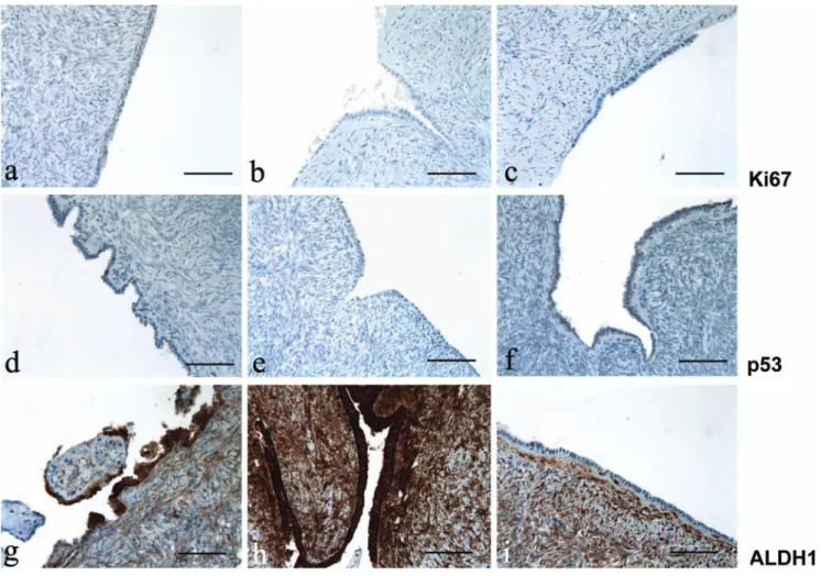
Figure 2. Ki67, p53 and ALDH1 immunoreactivity in fallopian tube; left column, after treating with tamoxifen; center column, after treating with clomid; right column, untreated control group. a-c) Immunoreactivity for Ki67; note the moderate expression of Ki67 in the tubes exposed to tamoxifen and clomid, and the low expression in the control group. d-f ) Immunoreactivity for p53; the expres-sion of p53 is weak in the tubes exposed to tamoxifen and clomid, and not present in the control group. g-h) ALDH1 immunoreactiv-ity; note the strong expression of ALDH1 in the tubes exposed to tamoxifen and clomid, and the low or absent expression in the con-trol group; also note the on/off effect of ALDH1 expression in panel h, where ALDH1 is mainly detected in the secretory cells lining but not in the ciliated cells. Scale bars: 100 μµm.
the question of a longer duration of Tamoxifen administration.35 Another animal study found
histopathological dysplasia in ovaries and tubes exposed to tamoxifen with a high Ki67
expression and a low p53 expression.36
Moreover, these results raise the question of the pathogenesis of tubo-ovarian carcinogen-esis. More recently, during systematic histopathological examination of tissues from prophylactic adnexectomies for a genetic risk, the revelation of occult tubal cancer (about 10%, and between 50 and nearly 100% of these cases have exhibited involvement of the fim-briated end of the fallopina tube) gave rise to the postulate of a tubal origin for ovarian can-cer.37-41 In view of these results, the
combina-tion of a systemic oestrogenic effect with the clomid-like effect of tamoxifen could be evoked on the epithelium of the tubes (such as in the
ndometrium).42
The immunohistochemical study added an extra-parameter for describing tubo-ovarian dysplasia. The two immunohistochemical markers p53 and Ki67 are described as being significantly expressed in ovarian cancer tis-sues compared with controls. The postulate that these markers would be expressed in dys-plastic tissues could thus confirm their pre-invasive nature. Schlosshauer et al.19found in
their study a gradual increase in immunoreac-tivity comparing normal ovarian epithelium (lowest expression), dysplastic epithelium (stronger expression) and ovarian cancer (highest expression), which would represent a molecular argument for the existence of dys-plasia. Concerning tubal dysplasia, Piek et al.18
found distinct expression of these two markers in dysplastic tubes.
In our study, expression of Ki67 and p53 appeared to be weak in ovaries whereas they were more marked in tubes of groups A and B: this could be another argument for the impor-tance of tubes in the tubo-ovarian dysplasia pathogenesis. Histopathological and immuno-histochemical fallopian tube dysplastic changes were mainly seen in the secretory cells lining, as they are the potential signifi-cant precursors of tubal and ovarian cancer. Last but not the least, the originality of our study also lies in the assessment of the mark-er for early stem cell diffmark-erentiation, ALDH1. Several scientific proofs could support the can-cer stem cell hypothesis for which some human cancers could originate in tissue stem and/or progenitor cells.43The true function of
ALDH1 is not well known and could involve in early stem cell differentiation. The high ALDH1 expression in normal human and mouse ovary, its potential role in cancer stem cells in other tumors (especially breast cancer where it may be a predictor of poor clinical out-come) suggest that ALDH1 could have an implication in ovarian tumors. This enzyme is
involved in the metabolism of retinoic acids, which plays an important role in regulation of
pathways like AKT/b-cathénine, WNT,
p21/p53.44-48
Immunohistochemical staining of ALDH1 expression was analysed in 442 primary
ovari-an carcinoma.20Unlike the case for breast
can-cer, ALDH1 would be a favourable prognostic factor in ovarian carcinoma because high expression was significantly associated with early-stage disease (P=0.006). In another study, ALDH1 expression and enzyme activity
were lower in malignant ovarian tumors.48
ALDH1 expression was constantly low in the 2 carcinomas of our study. The expression of ALDH1 appears to be exclusively found and strongly expressed in dysplastic ovarian and tubal epithelia. ALDH1 may have a different function in ovarian cancer than it does in breast cancer, where it is usually overex-pressed.49-51 We found a similar expression of
ALDH1 in a series of prophylactic ovariec-tomies for BRCA mutations (unpublished
results). ALDH1 seems to have a on/off effect
(overexpression in dysplasia area) and may be highly restricted to epithelial tubal dysplasia and could be help in their identification (Figure 1c). Auersperg et al.52 recently
com-pared the stem-cell profile of ovarian surface epithelium and fallopian tube with 5 stem-cell markers (NANOG, SFRP1, LHX9, ALDH1A1 and ALDH1A2). The authors found that ALDH1 was significantly increased in ovarian inclusion cysts and in the distal parts of the fallopian tube. They concluded that ovarian surface epithelium and tubal epithelium may have the capacity to undergo neoplastic transformation. Another study with different stem-cell markers (epithelial cell adhesion molecule, CD44, inte-grin a6) confirmed that stem cells are located in the distal end of the fallopian tubes.53
However, the exact meaning and the mecha-nism of this strong expression remain to be investigated, and further studies are neces-sary.
Conclusions
This morphological and immunohistochem-ical study found significant dysplastic epithe-lial lesions in a series of adnexectomy tissues in women exposed to tamoxifen. One of the possible clinicopathological explanations would be the combination of a systemic oestro-genic effect with the clomid-like effect of tamoxifen. However, the reason why the lesions are predominantly found in the tubes rather than in the ovaries remains question-able. This could point out the importance of the newly tubal theory of ovarian carcinogene-sis. There are many elements of bias that could
make the involvement of tamoxifen in the gen-esis of the dysplasia debatable. However, these results could encourage a fully comprehensive pathological analysis of adnexectomy tissues to be carried out for all patients with tamoxifen exposure.54
References
1. Brown J, Farquhar C, Beck J, Boothroyd C, Hugues E. Clomiphene and anti-oestro-gens for ovulation induction in PCOS. Cochrane Database Syst Rev 2009; 4:CD002249.
2. Badawy A, Gibreal A. Clomiphene citrate versus tamoxifen for ovulation induction in women with PCOS : a prospective ran-domized trial. Eur J Obstet Gynecol Reprod Biol 2011;159:151-4.
3. Salazar H, Godwin AK, Daly MB, Laub PB, Hogan M, Rosenblum N, et al. Microscopic benign and invasive malignant neoplasms and a cancer-prone phenotype in prophy-lactic oophorectomies. J Natl Cancer Inst 1996;88:1810-20.
4. Werness BA, Afify AM, Bielat KL, Eltabbakh GH, Piver MS, Paterson JM. Altered sur-face and cyst epithelium of ovaries removed prophylactically from women with a family history of ovarian cancer. Hum Pathol 1999;30:151-7.
5. Stratton JF, Buckey CH, Lowe D, Ponder
BAJ. Comparison of prophylactic
oophorectomy specimens from carriers and non carriers of a BRCA1 or BRCA2 gene mutation. J Natl Cancer Inst 1999;91:626-8.
6. Resta L, Russo S, Colucci GA, Prat J. Morphologic precursors of ovarian epithe-lial tumors. Obstet Gynecol 1993;82:181-6. 7. Plaxe S, Deligdish L, Dottino P, Cohen C. Ovarian intraepithelial neoplasia demon-strated in patients with stage I ovarian carcinoma. Gynecol Oncol 1990;38:67-72. 8. Mittal KR, Jacquotte AZ, Cooper JL,
Demopoulos R. Controlateral ovary in uni-lateral ovarian carcinoma: a search for preneoplastic lesions. Int J Gynecol Patho 1993;12:59-63.
9. Tressera F, Grases PJ, Labastida R, Ubeda A. Histological features of the controlater-al ovary in patients with unilatercontrolater-al ovarian cancer: a case control study. Gynecol Oncol 1998;71:437-41.
10. Chene G, Rahimi K, Mes Masson AM, Provencher D. Surgical implications of the potential new tubal pathway for the ovari-an carcinogenesis. J Minim Invasive Gynecol 2013;20:153-9.
11. Carcangiu ML, Radice P, Manoukian S, Spatti G, Gobbo M, Pensotti V, et al.
Atypical epithelial proliferation in Fallopian tubes in prophylactic salpingo-oophorectomy specimens from BRCA 1 and BRCA 2 germline mutation carriers. Int J Gynecol Pathol 2003;23:35-40. 12. Nieto JJ, Crow J, Sundaresan M,
Constantinovici N, Perret CW, Mc Lean AN, Hardiman PJ. Ovarian epithelial dysplasia in relation to ovulation induction and nul-liparity. Gynecol Oncol 2001;82:344-9. 13. Chene G, Penault-Llorca F, Le Bouedec G,
Mishellany F, Dauplat MM, Jaffeux P et al. Ovarian epithelial dysplasia after ovula-tion inducovula-tion: time and dose effect. Human Reprod 2009;24:132-8.
14. Chene G, Dauplat J, Radosevic-Robin N, Cayre A, Penault-Llorca F. Tu-be or not tu-be: that is the question...about serous ovarian carcinogenesis. Crit Rev Oncol Hematol 2013;88:134-43.
15. Chene G, Dauplat J, Raoelfils I, Bignon YJ, Cayre A, Jaffeux P, et al. Ovarian epithelial dysplasia: Description of a dysplasia scor-ing scheme. Ann Pathol 2011;31:3-10. 16. Chene G, Raoelfils I, Cayre A, Dauplat J,
Bignon YJ, Jaffeux P, et al. Don't forget fal-lopian tubes! A morphologic and immuno-histochemical study about Fallopian tubes with genetic risk (BRCA mutation). Gynecol Obstet Fertil 2012;40:14-8. 17. Yanai-Inbar I, Siriaunkgul S, Silverberg
SG. Mucosal epithelial proliferation of the fallopian tube: a particular association with ovarian serous tumor of low malig-nant potential? Int J Gynecol Pathol 1995;14:107-13.
18. Piek JM, van Diest PJ, Zweemer RP. Dysplastic changes in prophylactic removed fallopian tubes of women predis-posed to developping ovarian cancer. J Pathol 2001 2001 ; 195 : 451-6.
19. Schloosshauer PW, Cohen CJ, Penault-Lllorca F, Miranda CR, Bignon YJ, Dauplat J, et al. Prophylactic oophorectomy. Cancer 2003;98:2599-606.
20. Chang B, Liu G, Xue F, Rosen DG, Xiao L, Wang X, et al. ALDH1 expression corre-lates with favorable prognosis in ovarian cancers. Modern Pathol 2009;22:817-23. 21. Deligdish L, Gil J, Kerner H, Wu HS, Beck
D, Gershoni-Baruch R. Ovarian dysplasia in prophylactic oophorectomy specimens: cytogenetic and morphometric correla-tions. Cancer 1999;86:1544-50.
22. Mahdavi A, Pejovic T, Nezhat F. Induction of ovulation and ovarian cancer: a critical review of the literature. Fertil Steril 2006;85:819-26.
23. Corakci A, Filiz S, Caliskan E, Dalcik C, Ozeren S, Dalcik H. The effects of ovula-tion inducovula-tion on ovarian epithelium dys-plasia scores and Ki67 expression: an experimental study on rats. Int J Gynecol
cancer 2005;15:866-71.
24. Celik C, Gezginc K, Aktan M, Acar A, Yaman ST, Gungor S, et al. Effects of ovu-lation induction on ovarian morphology: an animal study. Int J Gynecol Cancer 2004;14:600-6.
25. Ozcan Z, Celik H, Gurates B, Ozercan HI, Nanay F, Nalbant M, et al. Effects of ovula-tion inducovula-tion agents on ovarian surface epithelium in rats. Reprod Biomed Online 2009;19:314-18.
26. Lacoste CR, Clemenson A, Lima S, Lecointre R, Peo'ch M, Chene G. Tubo-ovarian dysplasia in relatioship with ovu-lation in rats. Fertil Steril 2013;99:1768-73. 27. Williams-Brown MY, Salih SM, Xu X, Veenstra TD, Saeed M, Theiler SK, et al. The effect of tamoxifen and raloxifen on estrogen metabolism and endometrial cancer risk. J Steroid Mol Biol 2011;126:78-86.
28. Neri F, Maggino T. Surveillance of endome-trial pathologies, especially for endometri-al cancer, of breast cancer patients under tamoxifen treatment. Eur J Gynaecol Oncol 2009;30:357-60.
29. Vicus D, Rosen B, Lubinski J, Domchek S, Kauff ND, Lynch HT, et al. Tamoxifen and the risk of ovarian cancer in BRCA1 muta-tion carriers. Gynecol Oncol 2009;115:135-7.
30. Swerdlow AJ, Jones ME. Ovarian cancer risk in premenopausal and perime-naopausal women treated with Tamoxifen: a case-control study. Br J Cancer 2007;96:850-5.
31. Cohen I, Beyth Y, Tepper R, Shapira J, Zalel Y, Figer A, et al. Ovarian tumors in postmenopausal breast cancer patients treated with tamoxifen. Gynecol Oncol 1996;60:54-8.
32. Lavie O, Longacre T, Segev Y, Husain A. Ovarian carcinosarcomas associated with prolonged use of tamoxifen: case reports. Int J Gynecol Cancer 2009;19:1521-3. 33. Pickel H, Reich O, Tamussino K. Bilateral
atypical hyperplasia of the fallopian tube associated with tamoxifen : a report of two cases. Int J Gynecol Pathol 1999;17:284-5. 34. Diwan BA, Anderson LM, Ward JM.
Proliferative lesions of oviduct and uterus in CD-1 mice exposed prenatally to tamox-ifen. Carcinogenesis 1997;18:2009-14. 35. Ting AY, Kimler BF, Fabian CJ, Petroff BK.
Tamoxifen prevents premalignant changes of breast, but not ovarian cancer in rats at high risk for both diseases. Cancer Prev Res 2008;1:546-53.
36. Lima S, Clemenson A, Trombert B, Lecointre R, Lacoste CR, Peo'ch M, et al. Morphological and immunohistochemical analysis in ovaries and tubes of tamoxifen, letrozole and clomifene-treated rats. Arch
Gynecol Obstet 2014; in press.
37. Folkins AK, Jarboe EA, Saleemuddin A, Lee Y, Callahan MJ, Drapkin R, et al. A candi-date precursor to pelvic serous cancer (p53 signature) and their prevalence in ovaries and fallopian tubes from women with
BRCA mutations. Gynecol oncol
2008;109:168-73.
38. Crum CP, Drapkin R, Kindelberger D, Medeiros F, Miron F, Lee Y. Lessons from BRCA: the tubal fimbria emerges as an ori-gin for pelvic serous cancer. Clin Med Res 2007;5:35-44.
39. Powell CB, Kenley E, Chen LM, Crawford B, McLennan J, Zaloudek C, et al. Risk-reducing salpingo-oophorectomy in BRCA mutation carriers: role of serial sectioning in the detection of occult malignancy. J Clin Oncol 2005;23:127-32.
40. Finch A, Shaw P, Rosen B, Murphy J, Narod SA, Colgan TJ. Clinical and pathologic
findings of prophylactic
salpingo-oophorectomies in 159 BRCA1 and BRCA2 carriers. Gynecol Oncol 2006;100:58-64. 41. Nik NN, Vang R, Shih IM, Kurman RJ.
Origin and pathogenesis of pelvic (ovari-an, tubal, and primary peritoneal) serous carcinoma. Annu Rev Pathol 2014;9:27-45. 42. Emons G, Fleckenstein G, Hinney B, Huschmand A, Heyl W. Hormonal interac-tions in endometrial cancer. Endoc Relat Cancer 2000;7:227-42.
43. Ponnusamy MP, Batra SK. Ovarian cancer: emerging concept on cancer stem cells. J Ov Research 2008;4:1-9.
44. Liu S, Ginestier C, Charafe-Jauffret E, Foco H, Kleer CG, Merajver SD, et al. BRCA1 regulates human mammary stem/progenitor cell fate. Proc Natl Acad Sci USA 2008;105:1680-85.
45. Ginestier C, Wicinski J, Cervera N, Monville F, Finetti P, Bertucci F, et al. Retinoid signaling regulates breast cancer stem cell differentiation Cell Cycle 2009;8: 3297-302.
46. Heerma van Voss MR, Van der Groep P, Bart Joost, Van der Wall E, Van diset PJ. Expression of the stem cell marker ALDH1 in the normal breast of BRCA1 mutation carriers. Breast Cancer Res Treat 2010; 123:611-2.
47. Ginestier C, Hur MH, Charafe-Jauffret E, Monville F, Dutcher J, Brown M, et al. ALDH1 is a marker of normal and malig-nant human mammary sterm cells and a predictor of poor clinical outcome. Cell Sterm Cell 2007;1:555-67.
48. Penumatsa K, Edassery SL, Barua A, Bradaric MJ, Luborsky JL. Differential expression of aldehyde dehydrogenase 1a1 (ALDH1) in normal ovary and serous ovar-ian tumors. J Ov Research 2010;28:1-13. 49. Charafe-Jauffret E, Ginestier C, Iovino F,
Tarpin C, Diebel M, Esterni B, et al. Aldehyde dehydrogenase 1 positive cancer stem cells mediate metastasis and poor clinical outcome in inflammatory breast cancer. Clin Cancer Res 2010;16:45-55. 50. Deng S, Yang X, Lassus H, Liang S, Kaur S,
Ye Q et al. Distinct expression levels and atterns of sterm cell marker, Aldehyde Dehydrogenase Isoform 1 (ALDH1), in human epithelial cancers. Plos One 2010;5:e10277.
51. Kryczek I, Liu S, Roh M, Vatan L, Szeliga W, Wei S et al. Expression of aldehyde dehy-drogenase and CD133 defines ovarian can-cer stem cells. Int J Cancan-cer 2012;130:29-39. 52. Auersperg N. The stem-cell profile of ovar-ian surface epithelium is reproduced in the oviductal fimbriae, with increased stem-cell marker density in distal parts of the fimbriae. Int J Gynecol Pathol 2013;32:444-53.
53. Paik DY, Janzen DM, Schafenacker AM,
Velasco VS, Shung MS, Cheng D, et al. Stem-like epithelial cells are concentrated in the distal end of the fallopian tube: a site for injury and serous cancer initiation. Stem cells 2012;30:2487-97.
54. Chene G, Penault-Llorca F, Le Bouedec G, Mishellany F, Dauplat MM, Jaffeux P, et al. Ovarian epithelial dysplasia and prophy-lactic oophorectomy for genetic risk. Int J Gynecol Cancer 2009;19:65-72.