HAL Id: hal-01850544
https://hal.uca.fr/hal-01850544
Submitted on 29 Sep 2019
HAL is a multi-disciplinary open access archive for the deposit and dissemination of sci-entific research documents, whether they are pub-lished or not. The documents may come from teaching and research institutions in France or abroad, or from public or private research centers.
L’archive ouverte pluridisciplinaire HAL, est destinée au dépôt et à la diffusion de documents scientifiques de niveau recherche, publiés ou non, émanant des établissements d’enseignement et de recherche français ou étrangers, des laboratoires publics ou privés.
The cap size and shape of Arabidopsis thaliana primary
roots condition responses to an increase in medium
strength.
J Roué, Hugo Chauvet-Thiry, Nicole Brunel-Michac, F Bizet, B. Moulia,
Valerie Legue, Eric Badel
To cite this version:
J Roué, Hugo Chauvet-Thiry, Nicole Brunel-Michac, F Bizet, B. Moulia, et al.. The cap size and shape of Arabidopsis thaliana primary roots condition responses to an increase in medium strength.. Journal of Experimental Botany, Oxford University Press (OUP), In press. �hal-01850544�
The cap size and shape of Arabidopsis thaliana primary
roots affect responses to an increase in the physical strength
of the growth medium
J. Roué, H. Chauvet, N. Brunel-Michac, F. Bizet, B. Moulia, E. Badel and V. Legué* Université Clermont Auvergne, INRA, PIAF, F-63000 Clermont-Ferrand, France * Correspondence: valerie.legue@uca.fr
Highlight
Analysis of the growth and orientation of roots of Arabidopsis mutants with differing root cap sizes and shapes indicates that the form of the cap affects root responses to variations in the strength of the growth medium.
Abstract
During the progression of root in soil, root cap cells are the first to encounter obstacles and are known to sense environmental cues, thus making the root cap a potential
mechanosensing site. In this study, a two-layered growth medium system was developed in order to study root responses to variations in the physical strength of the medium and the importance of the root cap in the establishment of these responses. Root growth and trajectory of primary roots of Arabidopsis seedlings were investigated using in vivo image analysis. After contact with the harder layer of the medium, the root either penetrated it or underwent rapid curvature, thus enabling reorientation of growth. We initially hypothesized that the root-cap structure would affect apex penetration and reorientation, with pointed caps facilitating and domed caps impeding root penetration. This hypothesis was investigated by analysing the responses of Arabidopsis mutants with altered root caps. The primary root of lines of the fez-2 mutant, which has fewer root-cap cell layers and a more pointed root cap than wild-type roots, showed impaired penetration ability. Conversely, smb-3 roots, which display a rectangular-shaped cap, showed
enhanced penetration abilities. These results, which contradict our original hypothesis, reveal a role for resistance to buckling in determining root penetration abilities.
Keywords: Curvature, medium strength, Phytagel medium, root biomechanics, root buckling, root cap, root growth.
Introduction
Roots grow in a complex soil environment that exhibits spatial and temporal
heterogeneities, for example arising from the presence of sand particles, stones, or areas of hardpan. The ability of roots to penetrate the soil and to overcome physical obstacles depends either on their ability to find a path of least resistance, or on their ability to generate sufficient force to go through the obstacle (Bengough et al., 2010; Vollsnes et
al., 2010; Colombi et al., 2017a). The force needed to penetrate soil, which is provided by root growth, is determined by the soil strength. In natural conditions, soil strength— commonly estimated by penetrometer measurements—is of the order of 1 MPa and may increase as the result of soil compaction or soil drying (Jin et al., 2013; Kolb et al., 2017).
In a few laboratory studies, changes in soil strength have been mimicked using simplified models such as two-layer Phytagel-based growth media, in which the layers contain different concentrations of Phytagel (Yamamoto et al., 2008; Yan et al., 2017). It has been shown that adjusting the concentration modulates the physical strength of the growth medium (Schiavi et al., 2016). In such experiments where roots are consecutively faced with a compliant and a stiff layer, the tips are submitted to increasing axial stress. Roots that do not manage to generate sufficient force to penetrate the lower layer underg buckling, which dramatically changes the growth trajectory and the straightness of the root (Massa and Gilroy, 2003; Silverberg et al., 2012; Bizet et al., 2016). The maximal axial force that a root is able to apply before buckling, given by Euler’s law, depends on the rigidity, the length of the growth zone, and the diameter of the root to the power of four (Timoshenko and Goodier, 1970). In accordance with Euler’s law, species with a greater increase in root diameter in response to the strength of the medium are more efficient in penetrating strong layers (Materechera et al., 1992). The shape of the root tip is also an important feature that modulates the responses to the strength of the medium (Ruiz et al., 2016; Colombi et al., 2017b). In compacted soils, root tips with an acute opening angle grow faster than root tips with a blunted geometry (Iijima et al., 2003a, 2003b; Colombi et al., 2017b). Colombi et al. (2017b) showed that the shape of the tip influences the penetration stress experienced by roots by modulating the distribution of the local compaction concentrated around the tip.
The shape of the root tip arises from the shape of the cap that covers and protects the root apical meristem. In Arabidopsis, the root cap is organized in 3–5 cell layers of increasing age, and is composed of the columella (COL) and the lateral root cap (LRC) (Arnaud et al., 2010; Kumpf and Nowack, 2015). Every formative division of root-cap stem cells generates a new differentiating cell layer that pushes the older layers towards the root periphery. COL mature cells are known to sense gravity whereas LRC mature cells principally act as secretory cells (Kumpf and Nowack, 2015; Sato et al., 2015). At the periphery of the root cap, LRC mature cells undergo programmed cell death (PCD) and are degraded on the root surface in the elongation zone whereas COL mature cells are sloughed off, releasing border-like cells into the rhizosphere (Vicré et al., 2005; Fendrych et al., 2014). The balance between cell production and cell sloughing controls the changes in root-tip size and shape (Barlow, 2003). The study of root-cap formation has resulted in the identification of the molecular factors involved in cell production, differentiation, and sloughing. Among these, the FEZ and SOMBRERO (SMB)
transcription factors from the NAC family (NO APICAL MERISTEM, ARABIDOPSIS TRANSCRIPTION ACTIVATION FACTOR, CUP-SHAPED COTYLEDON) have been identified. FEZ specifically promotes periclinal divisions of COL and LRC stem cells (Willemsen et al., 2008). The fez-2 loss-of-function mutant exhibits a lower rate of periclinal division of the root-cap stem cells, which leads to a reduction of COL and LRC cell layers compared to wild-type roots (Willemsen et al., 2008; Bennett et al., 2014). SMB prevents the division of the differentiating root-cap daughter cells and promotes their differentiation. Additional COL and LRC cell layers are observed in the smb-3 loss-of-function mutant (Willemsen et al., 2008; Bennett et al., 2010). Moreover, SMB seems to indirectly regulate the PCD of LRC mature cells by targeting PCD- and cell clearance-specific genes (Fendrych et al., 2014; Huysmans et al., 2018). In the smb-3 mutant, LRC cell death is delayed, causing an increase in LRC size so that it reaches into the
elongation zone. After cell death, unprocessed dead cells accumulate on the smb-3 root surface (Fendrych et al., 2014).
The objective of this study was to investigate the role of the structure of the root cap in the establishment of root responses to the physical strength of the growing medium. We hypothesized that pointed caps would facilitate root penetration whilst domed caps would make penetration harder. We studied the root responses of the fez-2 and smb-3
Arabidopsis mutant lines because of their contrasting cap structure. We characterized the cap morphology of the roots in order to determine whether the loss or gain of cell layers affected root-cap size and shape. Col-0, fez-2, and smb-3 roots were then grown in two-layer Phytagel media of increasing mechanical resistance. Using a spatio-temporal
analysis, we characterized the ability of the roots to penetrate the harder medium, the root growth rate, and the root curvature. We found that penetration ability and the
reorientation in fez-2 and smb-3 primary roots were affected in the opposite way to how we had hypothesized.
Material and methods
Plant material and growth conditions
The wild-type strain of Arabidopsis thaliana (Col-0) and the fez-2 and smb-3 null-mutants used for the experiments were provided by the laboratory of B. Scheres (University of Utrecht, The Netherlands; Willemsen et al., 2008; Bennett et al., 2010, 2014). For all the experiments, Phytagel-based growth media were developed. The growth medium consisted of half-strength Murashige and Skoog (MS/2) medium
adjusted to pH 5.7 and containing Phytagel at different concentrations. Two-layer media were made of an upper layer of medium containing 0.2% (w/v) Phytagel and a lower layer containing 0.2%, 0.3%, or 0.5 % Phytagel (Supplementary Fig. S1 at JXB online), hereafter referred to as 0.2/0.2, 0.2/0.3 and 0.2/0.5 medium. The two-layer media were poured into specific culture chambers that consisted of polycarbonate boxes measuring 100×20 mm and 60 mm deep, which enabled roots to grow vertically inside the growth medium. The two-layer media were prepared as follows. Sterilized MS/2 medium containing 0.2%, 0.3%, or 0.5% Phytagel was poured under aseptic conditions into the culture chambers to a height of 30 mm. After 10 min, sterilized MS/2 medium containing 0.2% Phytagel was poured onto the lower layer to a height of 10 mm.
Arabidopsis seeds were surface-sterilized for 10 min with 30% (v/v) sodium hypochlorite solution added to 2% (v/v) Triton X-100. After five washes in sterile distilled water, 10 seeds were gently placed on the surface of the growth medium. The culture chambers were sealed, kept at 4 °C in the dark for 2 d, and then placed at 23 °C under a 16-h photoperiod.
Mechanical properties of Phytagel growth medium
We performed compressive and penetration tests on the Phytagel media to determine their elastic modulus and mechanical penetration resistance.
Measurement of elastic modulus.
Growth media containing 0.2–1.2% (w/v) of Phytagel were poured into 10-mm deep Petri dishes. Then, 20×20×10 mm3 samples were cut and placed in a universal testing device (Instron 5565). A uniaxial compressive load was applied on the surface of the gel by displacement of the upper plate with a constant loading rate of 0.5 mm min−1 (Fig. 1A). A 100-N load cell recorded the force needed to deform the samples. After a typical contact step, the loading force increased with the compression of the sample before decreasing notably when the sample slipped or ruptured. Real-time load and displacement recordings provided characteristic mechanical stress–strain curves (Fig. 1B). In the linear portion of the curve, i.e. when stress is proportional to strain, deformation is considered to be elastic (Schiavi et al., 2016) and the value of the elastic modulus (E) of the Phytagel samples was determined from the ratio between the incremental stress Δσ and the incremental strain rate Δε according to Hooke’s law:
E = Δσ/Δε (1)
where
σ = F/A (2)
ε = l/l0 (3)
and F is the compression force (N), A is the surface area of the cross-section of the sample (m2) on which the force is applied, l0 is the initial thickness of the sample (m),
and Δl is the change in thickness, i.e. the displacement of the plate (m). Compressive tests were repeated for three samples of each Phytagel concentration.
Measurement of resistance to penetration.
Penetration tests were conducted for the 0.2/0.2, 0.2/0.3, and 0.2/0.5 two-layer media using Phytagel medium samples that were prepared in the same culture chambers as used for root growth. A 2.5-mm diameter needle was attached to a load cell (Instron, 100 N max) and moved downward into the gel at a rate of 1 mm min−1. Real-time recording of the force-displacement curve enabled the determination of the maximal force required for the rupture of the interface between the upper and the lower layer.
Root-cap shape and determination of sharpness index
Col-0, fez-2, and smb-3 lines were grown in a 0.2% single-layer medium and at 5 d old primary roots were extracted and placed on a microscope slide. The roots were stained with 10 µg ml−1 propidium iodide (PI, Sigma Aldrich) and observed using a LSM 800 laser-scanning microscope (Zeiss) coupled to an Apochromat X20 lens.
To characterize the root-cap size and shape of each genotype, the length of the vertical section, LR, the diameter of the root at the quiescent centre, DR, and the area of the radial
section of the root cap, AR, were measured in longitudinal sections of the medium using
the ImageJ software (Schneider et al., 2017) (Fig. 2). An inverse Sharpness Index (iSI) of the root cap was calculated according to the following equation:
( * ) / 2 SI * /2 R R R R R A L D i L D (4)
The closer iSI is to 0, the more the root cap is pointed. Conversely, the closer SI is to 1, the more the root cap has a rectangular shape.
Length of the root growth zone
Col-0, fez-2, and smb-3 lines were grown in a 0.2% single-layer medium and at 5 d old primary roots were gently extracted and stained with 10 µg ml−1 PI. Using confocal optical sections of the roots, the length of the cortical cells was measured from the quiescent centre up to 2 mm. Assuming the roots were in a steady state with a constant cell flux, the local velocity over the root apex (i.e. the rate at which a cell moves away from the quiescent centre) was calculated from the cell length measurements (for details see Silk and Erickson, 1979; Silk, 1992; Liu et al., 2013). The velocity profile was used to calculate the length of the growth zone of the roots, which corresponded to the length of the root section from the quiescent centre to the point where the velocity reached 94% of the maximal value (Supplementary Fig. S2).
Maximal root growth forces
The maximal axial force the roots were able to apply on the two-layer medium interface before reorientation of the root apex was estimated for Col-0, fez-2, and smb-3 roots grown in 0.2/0.3 medium. After contact between the root and the interface, root axial growth induced a deflection of this interface until the root curved or the root apex deviated. The shape of the interface just before root apex reorientation was traced using ImageJ software and the maximal deflection, d, of the interface was measured with regards to its initial position (i.e. before contact with the root). The shape of the tip of the root cap was fitted by a circle of radius R. The maximal force before root reorientation,
Fcrit, applied on the interface by the root apex was then estimated using the theory of
1 3 2 2 4 3 * crit F E R d (5) where (6) and E1 and E2 are the elastic moduli of the lower medium layer and of the root apex, respectively, and ν1 and ν2 are the Poisson ratios of the lower layer and the root apex,
respectively. Assuming that E2 was much larger than E1, the second part of the equation
was neglected, and the value of ν1 was estimated as 0.5 (Ahearne et al., 2005).
Time-lapse photography and image processing
Root growth and orientation were subjected to time-lapse imaging from 5 d after sowing. Two Nikon D7100 cameras equipped with macro lenses (AF-S micro NIKKOR 60mm f/2.8G ED) and controlled by DigiCamControl software were placed on a rack in front of the culture chambers. Images of the roots growing in the two-layer media were recorded every 5 min for at least 24 h. Only the roots remaining in the focal plane of the objectives were analysed. All images were pre-treated using the Rawtherapee software (v4.2.1; https://rawtherapee.com/) and were analysed using the RootStem Extractor software developed by the PIAF laboratory (Chauvet et al., 2016). Briefly, the software enabled extraction of the topological skeleton of each root by screening the pictures
perpendicularly to the root axis with a step of 0.3 pixels. The length of the root on each picture and the local inclination as a function of the curvilinear abscissa can be computed from the coordinates of the skeleton. The root length as a function of time enabled us to calculate root growth rate by linear fit. Curvature intensity along the root was obtained by deriving the local inclination as a function of time and space. The curvature intensity data are presented in graphs composed of three dimensions: a vertical axis corresponding to root length from the beginning of the measurement, a horizontal axis corresponding to time, and a colour bar that depicts curvature intensity. On the spatio-temporal graphs, the green colour indicates a straight section of the root, while the red and blue colours depict the highest positive and negative curvatures, respectively.
Gravitropic responses of the roots
Col-0, fez-2, and smb-3 roots were grown vertically into a 0.5% Phytagel single-layer medium for 5 d. The culture chambers were tilted by 90° (i.e. horizontal) and images of the primary roots were then recorded every 10 min for 22 h. Analysis of the time-lapse images with RootStem Extractor was used to measure root length and root-tip angle over time (Supplementary Fig. S3). These data were used to calculate the gravitropic response by applying the equation described by Chauvet et al. (2016), which determines relative differential growth in dimensionless units.
All data were analysed using R software (RStudio, R.3.3.4; https://www.r-project.org/) and are presented as means (±SD) in the Results. The Shapiro–Wilk test was used to test for normal distribution of the data and the Bartlett test was used to test for
homoscedasticity. One- or two-way ANOVA were used to determine whether the effect of each factor was significant. When significant differences were observed, post-hoc analyses based on Tukey’s test were applied to the data.
Results
Phytagel concentration affects both the rigidity and penetration
resistance of the growth medium
The elastic modulus, E, obtained from compressive tests, was measured for Phytagel concentrations ranging from 0.2–1.2% (Fig. 3A, B). The 0.2%, 0.3%, 0.5%, and 1.2% media had mean (±SD) E values of 3.1±0.2 kPa, 7.6±1.4 kPa, 31.9±6.6 kPa, and 158.3±18.3 kPa, respectively (Fig. 3B), indicating that E was highly dependent on the Phytagel concentration.
Penetrometer tests were performed in 0.2/0.2, 0.2/0.3, and 0.2/0.5 two-layer media (Fig. 3C, D). The curves of the compression force versus needle displacement were divided into two steps: (I) after the first contact with the lower layer surface, the compression force increased markedly until reaching a maximal value; and (II) as the needle penetrated the lower layer, the loading force rapidly collapsed (Fig. 3C). The specific pattern of the displacement-force curves illustrated the presence of the interface between the two medium layers, the rupture of which required a specific force, called the maximal strength, which may have reflected a slight drying out of the surface of the lower layer during the 10 min preceding the pouring of the upper layer. The maximal strength was 0.03±0.01 N in the 0.2/0.2 medium, 0.05±0.02 N in the 0.2/0.3 medium, and 0.11±0.02 N in the 0.2/0.5 medium (Fig. 3D). Thus, the maximal strength increased significantly with the Phytagel concentration of the lower medium layer.
The Arabidopsis fez-2 and smb-3 mutations affect root-cap size and
shape in primary roots
Several morphological traits were examined in the Col-0, fez-2, and smb-3 primary roots in order to specify the impact of the mutations on root-cap size and shape (Fig. 4). Compared to the wild-type (WT), the fez-2 root cap was shorter and narrower at the base, whereas the smb-3 cap was longer and larger at the base (Fig. 4D, E). However, mean root diameter measured at 500 µm from the tip of the cap was not significantly different between the three genotypes (Supplementary Fig. S4). Estimation of the inverse
Sharpness Index indicated that fez-2 root caps (iSI=0.30±0.07) were significantly more pointed than WT caps (0.41±0.06) whilst smb-3 displayed a more rectangular shape (0.5±0.13; Fig. 4F). Our morphological characterizations thus showed that fez-2 and
The smb-3 mutation affects root growth and the gravitropic
response whereas the fez-2 mutation does not
From a kinematic analysis, the mean length of the growth zone from the quiescent centre to the end of the elongation zone was estimated to be 917±194, 846±303, and 898±299 µm in the WT, fez-2, and smb-3 lines, respectively (Fig. 5A, Supplementary Fig. S2), suggesting that the mutations may not affect the size of the zone. Analysis of root growth in a 0.2% Phytagel single-layer medium showed that the mean growth rate of WT
(3.2±0.8 µm min−1) and fez-2 (3.0±0.7 µm min−1) roots was not significantly different, whereas smb-3 roots grew more slowly (1.6±0.3 µm min−1) (Fig. 5B). Moreover, WT and
fez-2 roots exhibited a similar gravitropic response (0.17±0.05 and 0.17±0.02,
respectively), whereas smb-3 roots exhibited a significantly higher response (0.36±0.15; Fig. 5C, Supplementary Fig. S3). Our preliminary studies thus showed that the fez-2 mutation mainly affects the size and shape of the root cap whereas the smb-3 mutation also affects the root growth and gravitropic responses.
The fez-2 and smb-3 mutations affect the ability of primary roots to
penetrate the harder layer of the growth medium
In order to understand the role of cap structure in the root response to an increase in the strength of the growth medium, the penetration abilities of the primary roots of the three genotypes were compared in two-layer Phytagel media. In the 0.2/0.2 medium, where the increase in strength was the weakest, 100% of the WT and smb-3 roots crossed the
interface to penetrate the lower layer, whereas only 83% of the fez-2 roots were able to do so (Table 1). In the 0.2/0.3 medium, 60% of the WT primary roots penetrated the 0.3% lower layer whilst 80% of the smb-3 and 28% of the fez-2 roots were able to do so. Finally, only 1.7 % of the WT roots and 18.3% of the smb-3 roots penetrated the 0.5% lower layer in the 0.2/0.5 medium, and none of the fez-2 roots were able to do so. Thus, compared with the WT, the fez-2 roots exhibited impaired penetration ability in response to an increase in the strength of the growth medium, whereas the smb-3 roots exhibited enhanced penetration ability. In order to determine the cause of these
differences, we estimated the critical force that each root was able to apply on the two-layer medium interface before apex reorientation occurred.
The critical axial growth force of primary roots is impaired in fez-2
and enhanced in smb-3
The critical axial force was estimated for primary roots of the three genotypes growing in the 0.2/0.3 medium, in which the greatest differences in penetration ratio were observed between the three lines (Table 1). Upon contact, root axial growth induced a deflection of the interface until either the interface ruptured or a zone of curvature in the root was initiated. We measured the deflection of the interface before curvature initiation for roots that did not penetrate the lower layer (Fig. 6A). Wild-type roots displaced the interface to a mean maximal depth, d, of 0.37±0.03 mm, whilst for fez-2 roots d=0.27±0.01 mm and
for smb-3 roots d=0.49±0.03 mm (Fig. 6B). The values of d together with the radius of the tip of the root cap and the rigidity of the lower layer of the medium (E=7.6 kPa) made it possible to estimate the critical axial growth force of the primary roots (Fig. 6C), which were found to be 1.12±0.14 10−3 N for the WT, 0.66±0.07 10−3 N for fez-2, and 1.77±0.18 10−3 N for smb-3. This confirmed that smb-3 roots were able to apply a higher axial force on the interface compared with then WT, whilst fez-2 roots applied a weaker force.
The fez-2 and smb-3 roots exhibit altered patterns of curvature in
response to an increase in the strength of the growth medium
Roots that did not apply a sufficient axial force to induce rupture of the interface between the two layers of media reorientated their growth as the result of initiation of zones of curvature. This initiation was monitored as a function of the increase in the strength of the medium and as a function of the genotype (Supplementary Videos S1–S7).
Representative examples of WT, fez-2, and smb-3 roots reorientating their growth in 0.2/0.3 and 0.2/0.5 media are presented in Figs 7–9.
Following contact with the 0.3% interface, non-penetrating roots first grew downwards against the interface and deformed it until a first zone of curvature was formed (Fig. 7). This first zone appeared at 515±98, 603±33, and 398±53 µm from the tip of the root cap in WT, fez-2, and smb-3 roots, respectively. Once established, the curvature covered a zone of 657±121, 977±58, and 414±73 µm in WT, fez-2, and smb-3 roots, respectively (Fig. 7, Supplementary Table S1). The length of the zone was significantly greater in
fez-2 roots than in smb-3 roots, but no significant differences were observed between the WT
and mutant roots. Initiation of the first zone of curvature induced lateral deviation of the
fez-2 root apex whereas the WT and smb-3 apexes continued to grow downwards. These
different behaviours led to the establishment of two distinct root shapes (Fig. 8,
Supplementary Videos S1–3). In WT and smb-3 roots, several similar zones of curvature were initiated successively, leading to the formation of one or more curls (Fig. 8A, Supplementary Videos S1, S2); thus, these roots developed a ‘curly’ shape in the 0.2/0.3 medium. In fez-2 roots, the lateral deviation of the apex was followed by the initiation of a second zone of curvature with characteristics distinct from the first zone (Figs 7B2, 8B). Whereas the first zone was maintained after its establishment, the second one moved and kept a constant distance from the tip of the root cap (Fig. 7). The establishment of the two distinct zones of curvature in fez-2 roots led to the formation of a ‘step-like’ shape (Fig. 8B).
In the 0.2/0.5 medium, all the WT and fez-2 roots developed a step-like shape following contact with the interface (Fig. 9, Supplementary Videos S4, S5). For the smb-3 line, three distinct root shapes were observed: ‘curly’, ‘wavy’, and ‘step-like’ shapes
(Supplementary Videos S6, S7). The spatio-temporal establishment of the step-like shape of primary roots was compared between the three genotypes. After contact with the 0.2/0.5 interface, a first zone of curvature was formed at 540±50, 664±64, and 407±32 µm from the tip of the cap in WT, fez-2, and smb-3 roots, respectively (Fig. 9). In fez-2 roots, curvature was initiated significantly further from the tip than in smb-3 roots, but no significant differences were observed between the WT and the mutant roots
(Supplementary Table S2). Once established, the curvature extended over a zone of 657±121, 977±58, and 414±73 µm in the WT, fez-2, and smb-3 roots, respectively (Fig. 9). Thus, the zone of curvature was significantly longer in fez-2 roots than in WT and
smb-3 roots. Moreover, the maximal curvature intensity was significantly weaker in fez-2
roots (2.3±0.5 mm−1) than in WT (4.7±0.5 mm−1) and smb-3 roots (4.8±0.6 mm−1). As the apex reorientated its growth, the roots of all three genotypes showed a second zone of curvature, the establishment of which showed common characteristics except for the maximal intensity, which was significantly weaker in fez-2 roots (Fig. 9, Supplementary Table S2).The second zone of curvature remained at a constant distance from the tip of the cap during root growth along the 0.2/0.5 interface.
These two-layer media experiments demonstrated that root apex reorientation and the establishment of zones of curvature were affectd by both the mechanical resistance of the interface and by the size and shape of the root cap.
Root growth rates in the three genotypes respond differently to
increases in the strength of the medium
Growth rates were determined for WT, Col-0, fez-2, and smb-3 roots in media with Phytagel interfaces of 0.2/0.2, 0.2/0.3, and 0.2/0.5.
In the 0.2/0.2 medium only the growth rates of penetrating roots were measured, whilst in the 0.2/0.3 medium the growth rates were measured for both penetrating and reorientating roots (Supplementary Tables S3, S4). In wild- reorientating roots of the WT, the contact between the root and the interface was followed by a significant 60% decrease in mean growth rate (Fig. 10A). Unfortunately, our experimental system based on 2D
visualization of the roots prevented measurement of the root growth rate through to the end of the reorientation of the apex. A significant 60% decrease in mean root growth rate was also observed in reorientating roots of smb-3 whereas no significant decrease was observed in reorientating roots of fez-2 (Fig. 10A, Supplementary Table S4).
In the 0.2/0.5 medium, the growth rate of WT roots decreased by 37% after contact with the interface (upper layer 3.0±0.2 µm min−1, lower layer1.9±0.3 µm min−1; Fig. 10B), and the initial growth rate did not recover when reorientation of the apex was complete. The reorientation of the apex of smb-3 roots after contact with the interface was also accompanied by a significant 44% decrease in mean growth rate (Fig. 10B,
Supplementary Table S4). In contrast, fez-2 roots showed no variation in growth rate after contact with the interface.
These results showed that the increase in the strength of the medium in the two-layer system affected the growth rate of WT roots, and this effect seemed to be altered in the roots of the mutants, especially for fez-2.
Discussion
Our experimental design proved to be suitable for investigating root responses to
differences in the strength of the growing medium. Precise characterization of mechanical properties of the media showed strong correlations between rigidity, penetration
resistance, and Phytagel concentrations (Fig. 3). In addition, precise spatio-temporal analyses of root growth rate and root apex orientation allowed us to discriminate between the passive and active mechanisms involved in the reorientation. By using three
genotypes of Arabidopsis with contrasting root-cap morphologies, namely the Col-0 wild-type and the mutants fez-2 (pointed cap) and smb-3 (rectangular-shaped cap), our study has resulted in a better understanding of the role of the root-cap structure in the responses to mechanical stresses.
Root apex reorientation involves both passive and active
mechanisms
In our experimental system, roots grew first through a layer of medium of low strength before arriving at an interface with a layer of higher strength. In response to intermediate and high increases in strength at the interface, the growth rates of wild-type (WT) roots decreased significantly (Fig. 10, Supplementary Tables S3, S4).These decreases, which were observed for both penetrating and non-penetrating roots, were comparable to those observed in poplar roots confronted with a physical obstacle (Bizet et al., 2016). This suggests the existence of a biological process that senses the increase in the strength of the medium and that subsequent responses were triggered in the WT roots (Bizet et al., 2016).
Root growth against an interface in strength generates an increasing compression reaction force on the root (Kolb et al., 2017). If the compression force reaches a critical value while the root is still in contact with the obstacle, buckling can occur as predicted by Euler’s law (Bizet et al., 2016). Buckling was observed for non-penetrating roots in all three genotypes and resulted in two distinct principal shapes being observed depending on the genotype and on the strength of the interface. When encountering an intermediate increase in in the strength of the medium, non-penetrating roots mostly developed a curly shape (Fig. 8A). This shape has previously been observed in Medicago truncatula roots growing in a two-layer Phytagel medium (Silverberg et al., 2012) and, from a
mathematical model based on the theory of buckling rods, it was attributed to a
combination of helical buckling and simultaneous twisting near the root tip. For most of the curly roots, the first curvature was initiated in a zone between 400–700 µm from the tip of the root cap (Fig. 7). According to our kinematic analyses performed on roots growing in a compliant single-layer medium, this seemed to correspond to the zone where the maximal elongation rate occurred (Fig. 5, Supplementary Fig. S2). Thus, the zone of curvature initiation may correspond to the zone of mechanical weakness
described by Bizet et al. (2016). To confirm this, in vivo kinematic analyses would need to be carried out on roots growing in two-layer media. Taken together with previous studies, our results suggest that the curly shape is mainly driven by the mechanical
response of the root acting as a ‘growing’ rod, the end of which is blocked by an obstacle. The second distinct shape had a step-like appearance and was observed in roots mostly in response to the highest increase in the strength of the mediumacross the interface (Fig. 8B). In this case, the first curvature induced deviation of the root apex, which implied a change from axial to asymmetric radial forces being applied on the apex. The kinetics and the position of curvature initiation suggested that it may be interpreted as buckling.
The first curvature was followed by the initiation of a second one, closer to the root apex, that allowed the root tip to regain a vertical position (Fig. 9). Since the second zone of curvature remained at a constant distance from the tip of the root cap after its
establishment, it would have provided the root tip with a steady form from changing cells (Silk, 1992). The second curvature has previously been described as the result of the interaction between gravitropic and thigmotropic responses (Massa and Gilroy, 2003). The deviation of the root apex was preceded by a decrease in root growth rate that did not recover after relaxation of the axial mechanical stresses (Fig. 10). The initiation of the second curvature and the continuation of the reduced growth rate suggest that, even if the first curvature was initially provoked by buckling, the direction of the subsequent growth may have been reorientated in an active way.
The different patterns of the establishment of curvature suggest that the mechanical strength of the interface determines the subsequent root response (buckling or bending). An intermediate-strength interface may allow the root apex to sink into it, preventing its deviation after root buckling. In contrast, a strong interface seemed to be barely deformed by the root apex, which may then be free to deviate and may trigger active root
reorientation.
The structure of the root cap affects the root response to an
increase in the strength of the growth medium in the opposite way
to that expected
Based on previous studies (Vollsnes et al., 2010; Ruiz et al., 2016; Colombi et al., 2017b), we hypothesized that the shape of the root cap would influence the penetration ability of the root and its response to the strength of the medium by changing the
distribution of mechanical stresses that result from the axially orientated external force. It has been suggested that a pointed root tip increases the axial stresses at the forefront of the tip rather than around it, resulting in enhanced initiation of cracks in the medium and enhanced penetration ability (Colombi et al., 2017b). However, contrary to this
hypothesis, roots of the fez-2 mutant that have pointed tips (Fig. 4) exhibited reduced penetration abilities in the two-layer media compared to WT roots, whereas roots of
smb-3 that have dome-shaped tips exhibited enhanced penetration abilities. In the roots of the
mutants, especially those of fez-2, given that the morphological alterations mainly relate to the structure of the cap, our results suggested that it is this that has an impact on the establishment of the responses to the strength of the medium, but in a way that
contradicted our hypothesis. This suggests either that the shape of the root cap is not the key feature that determines the root responses, or that another factor could influence the establishment of the responses. In light of this, attention should be paid to the the relative cap rigidity of the WT and mutant roots. The loss or accumulation of root-cap cell layers, modulating its dimensions, could modify the local bending rigidity and compression behaviour that drive the ability of root apex to go forward in the medium. In fez-2 roots, the potential benefit of a more pointed cap shape with regards to penetration ability could be reduced by low cap rigidity, meaning that the root could not experience high
The differences in penetration abilities between the mutant and WT roots may be attributable to their different critical buckling forces. Our results showed that the fez-2 roots applied a lower critical axial force on the interface before buckling compared with the WT whilst the smb-3 roots applied a higher critical force (Fig. 6). These differences may be explained either by differences in diameter and/or rigidity at the buckling zone of the root, or by differences in the length of the growth zone, as defined by Euler’s law. Our characterizations of the root morphologies of the mutants did not reveal any differences in the diameter or length of the growth zone compared to the WT roots in conditions where the strength of the medium was low (Fig. ?)-. Our results suggest that either the rigidity of mutant roots was different compared to that of the WT, or that the length of the growth zone, tissue rigidity, and/or root diameter may have been modulated differently between the genotypes in response to an increase in the strength of the
medium.
Further experiments need to be conducted to confirm either of these hypotheses, such as atomic force microscopy assays in order to accurately characterize local rigidity in the root tissues. In particular, the lower and higher root diameters at the quiescent centre, resulting from fewer and more lateral root-cap cell layers flanking the fez-2 and smb-3 root apexes, respectively, could constitute an advantage for penetrating harder layers in the medium by affecting local rigidity in the roots (Bennett et al., 2010; Fendrych et al., 2014). In addition, in vivo kinematic analyses could be performed on roots growing in two-layer media in order to define the effect of the increase in strength on the length of the growth zone. At present, if the second hypothesis turns out to be true, we may
speculate that increases in the strength of the medium are perceived by root-cap cells and that signal transduction triggers responses in the elongation zone. In fez-2 roots, the reduction in the number of cell layers in the cap may alter the mechanosensitivity and thus affect the establishment of root responses to an increase in the strength of the medium. The fact that the growth rate of fez-2 roots was not affected by the strength of the medium supports this hypothesis. A properly formed root cap may be required to trigger the signal perception and transduction processes and thus to allow the root to respond to an increase in the strength of the medium.
Conclusions
During their progression in soil, roots frequently face physical heterogeneities that they must either move or cross by generating mechanical forces, or must circumvent by reorientating their growth. Our study provides new insights into the role of the root cap in root penetration and reorientation in response to variations in the strength of the growth medium. When Arabidopsis primary roots encounter layers in the medium with
increasing strength, they adopt three main types of response: root penetration, root buckling without apex deviation that leads to a curly shape, or root buckling followed by apex deviation that leads to a step-like shape. These responses were different in the roots of the fez-2 and smb-3 mutants, which possess more pointed and more rectangular cap shapes, respectively. The differences in penetration ability, reduced in fez-2 roots and enhanced in smb-3 roots, seems to result from different resistances to root buckling.
These results suggest that the structure of the cap could affect root responses to the strength of the growth medium by influencing root resistance to buckling.
Supplementary data
Fig. S1. Images of the two-layer Phytagel media that were used to study root responses. Fig. S2. Cell length and velocity profiles of roots growing in a single-layer medium. Fig. S3. Gravitropic responses of roots growing in a single-layer medium.
Fig. S4. Diameter of the roots at 500 µm from the tip of the root cap when grown in a single-layer medium.
Table S1. Characteristics of the zones of curvature formed in reorientating roots in the 0.2/0.3 medium.
Table S2. Characteristics of the zones of curvature formed in reorientating roots in the 0.2/0.5 medium.
Table S3. Detailed growth rates of penetrating roots in the 0.2/0.2 and 0.2/0.3 media. Table S4. Detailed growth rates of reorientating roots in the 0.2/03 and 0.2/0.5 media. Video S1. Time-lapse movie of a reorientating wild-type root in the 0.2/0.3 medium. Video S2. Time-lapse movie of a reorientating smb-3 root in the 0.2/0.3 medium. Video S3. Time-lapse movie of a reorientating fez-2 root in the 0.2/0.3 medium. Video S4. Time-lapse movie of a reorientating wild-type root in the 0.2/0.5 medium. Video S5. Time-lapse movie of a reorientating fez-2 root in the 0.2/0.5 medium.
Video S6. Time-lapse movie of a reorientating smb-3 root in the 0.2/0.5 medium forming a step-like shape.
Video S7. Time-lapse movie of a reorientating smb-3 root in the 0.2/0.5 medium forming a wavy shape.
Acknowledgements
We would like to thank Ben Scheres and Viola Willemsen (University of Utrecht, The Netherlands) for donating the mutant lines. This research received financial support from the French Space Agency (CNES) and INRA programs. JR received a doctoral grant from the French National Ministry of Education and Research. HC received a Postdoctoral grant from the Auvergne-Rhône Alpes Council with co-funding from FEDER. FB received a Postdoctoral grant from the CNES.
References
Ahearne M, Yang Y, El Haj AJ, Then KY, Liu KK. 2005. Characterizing the viscoelastic properties of thin hydrogel-based constructs for tissue engineering applications.Journal of the Royal Society, Interface2, 455–463.
Arnaud C, Bonnot C, Desnos T, Nussaume L. 2010. The root cap at the forefront.
Comptes Rendus Biologies333, 335–343.
BarlowPW. 2003. The root cap: cell dynamics, cell differentiation and cap function.
Journal of Plant Growth Regulation21, 261–286.
Bengough AG, Hans J, Bransby MF, Valentine TA. 2010. PIV as a method for quantifying root cell growth and particle displacement in confocal images.
BennettT, Sanchez-perezGF, CampilhoA, WillemsenV, SnelB, ScheresB. 2010. SOMBRERO, BEARSKIN1, and BEARSKIN2 regulate root cap maturation in Arabidopsis.The Plant Cell22, 640–654.
BennettT, van den ToornA, WillemsenV, ScheresB. 2014. Precise control of plant stem cell activity through parallel regulatory inputs.Development141, 4055–
4064.
Bizet F, Bengough AG, Hummel I, Bogeat-Triboulot MB, Dupuy LX. 2016. 3D deformation field in growing plant roots reveals both mechanical and biological responses to axial mechanical forces.Journal of Experimental Botany67, 5605–
5614.
Chauvet H, Pouliquen O, Forterre Y, Legué V, Moulia B. 2016. Inclination not force is sensed by plants during shoot gravitropism.Scientific Reports6, 35431.
Colombi T, Braun S, Keller T, Walter A. 2017a. Artificial macropores attract crop roots and enhance plant productivity on compacted soils.The Science of the Total Environment574, 1283–1293.
Colombi T, Kirchgessner N, Walter A, Keller T. 2017b. Root tip shape governs root elongation rate under increased soil strength.Plant Physiology174, 2289–2301.
Fendrych M, Van Hautegem T, Van Durme M, et al. 2014. Programmed cell death controlled by ANAC033/SOMBRERO determines root cap organ size in
Arabidopsis.Current Biology24, 931–940.
HanaorDAH, GanY, EinavI. 2015. Contact mechanics of fractal surfaces by spline assisted discretisation.International Journal of Solids and Structures59, 121–131.
Huysmans M, Buono RA, Skorzinski N, Radio MC, De Winter F, Parizot B, Mertens J, Karimi M, Fendrych M, Nowack MK. 2018. NAC transcription factors ANAC087 and ANAC046 control distinct aspects of programmed cell death in the Arabidopsis columella and lateral root cap.The Plant Cell30, 2197–
2213.
IijimaM, BarlowPW, BengoughAG. 2003a. Root cap structure and cell production rates of maize (Zea mays) roots in compacted sand.New Phytologist160, 127–
134.
Iijima M, Higuchi T, Barlow PW, Bengough AG. 2003b. Root cap removal increases root penetration resistance in maize (Zea mays L).Journal of Experimental Botany54, 2105–2109.
Jin K, Shen J, Ashton RW, Dodd IC, Parry MA, Whalley WR. 2013. How do roots elongate in a structured soil?Journal of Experimental Botany64, 4761–4777.
Kolb E, Legué V, Bogeat-Triboulot MB. 2017. Physical root–soil interactions.
Physical Biology14, 065004.
Kumpf RP, Nowack MK. 2015. The root cap: a short story of life and death.Journal of Experimental Botany66, 5651–5662.
LiuY, LaiN, GaoK, ChenF, YuanL, MiG. 2013. Ammonium inhibits primary root growth by reducing the length of meristem and elongation zone and decreasing elemental expansion rate in the root apex in Arabidopsis thaliana.PLoS ONE8,
1–11.
MassaGD, GilroyS. 2003. Touch modulates gravity sensing to regulate the growth of primary roots of Arabidopsis thaliana.The Plant Journal33, 435–445.
MaterecheraSA, AlstonAM, KirbyJM, DexterAR. 1992. Influence of root diameter on the penetration of seminal roots into a compacted subsoil.Plant and Soil144,
297–303.
RuizS, StraubI, SchymanskiSJ. 2016. Experimental evaluation of earthworm and plant root soil penetration – cavity expansion models using cone penetrometer analogs.Vadose Zone Journal15, vzj2015.09.0126.
Sato EM, Hijazi H, Bennett MJ, Vissenberg K, Swarup R. 2015. New insights into root gravitropic signalling.Journal of Experimental Botany66, 2155–2165.
Schiavi A, Cuccaro R, Troia A. 2016. Strain-rate and temperature dependent material properties of Agar and Gellan Gum used in biomedical applications.Journal of the Mechanical Behavior of Biomedical Materials53, 119–130.
SchneiderCA, RasbandWS, EliceiriKW. 2017. NIH image to ImageJ: 25 years of image analysis.Nature Methods9, 671–675.
SilkWK. 1992. Steady form from changing cells.International Journal of Plant Science
153, 49–58.
Silk WK, Erickson RO. 1979. Kinematics of plant growth.Journal of Theoretical Biology76, 481–501.
SilverbergJL, NoarRD, PackerMS, HarrisonMJ, HenleyCL, CohenI, Gerbode SJ. 2012. 3D imaging and mechanical modeling of helical buckling in Medicago truncatula plant roots. Proceedings of the National Academy of Sciences, USA
109, 16794–16799.
TimoshenkoSP, GoodierJN. 1970. Theory of elasticity. New York: McGraw-Hill.
Vicré M, Santaella C, Blanchet S, Gateau A, Driouich A. 2005. Root border-like cells of Arabidopsis. Microscopical characterization and role in the interaction with rhizobacteria.Plant Physiology138, 998–1008.
VollsnesAV, FutsaetherCM, BengoughAG. 2010. Quantifying rhizosphere particle movement around mutant maize roots using time-lapse imaging and particle image velocimetry.European Journal of Soil Science61, 926–939.
Willemsen V, Bauch M, Bennett T, Campilho A, Wolkenfelt H, Xu J, Haseloff J, Scheres B. 2008. The NAC domain transcription factors FEZ and SOMBRERO control the orientation of cell division plane in Arabidopsis root stem cells.
Developmental Cell15, 913–922.
Yamamoto C, Sakata Y, Taji T, Baba T, Tanaka S. 2008. Unique ethylene-regulated touch responses of Arabidopsis thaliana roots to physical hardness.
Journal of Plant Research121, 509–519.
Yan J, Wang B, Zhou Y. 2017. A root penetration model of Arabidopsis thaliana in phytagel medium with different strength.Journal of Plant Research130, 941–950.
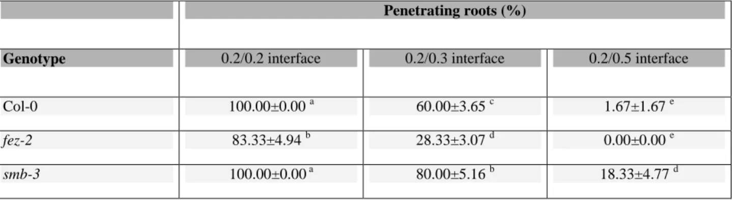
Table 1. Penetration ability of Arabidopsis Col-0, fez-2, and smb-3 primary roots in two-layer Phytagel media
Penetrating roots (%)
Genotype 0.2/0.2 interface 0.2/0.3 interface 0.2/0.5 interface
Col-0 100.00±0.00 a 60.00±3.65 c 1.67±1.67 e
fez-2 83.33±4.94 b 28.33±3.07 d 0.00±0.00 e
smb-3 100.00±0.00 a 80.00±5.16 b 18.33±4.77 d
Penetration ability is represented by the percentage of roots that were able to penetrate the 0.2%, 0.3%, and 0.5% lower layers of the Phytagel media. The upper layer was 0.2% in each case. For each genotype and interface, 10 roots in each of six replicate preparations of growth medium were observed 8 d after germination, and the data are presented as means (±SD), n=60. Different letters show significant differences as determined using two-way ANOVA followed by Tukey’s test (P<0.05).
Fig. 1. Instrument used for compressive tests and an example of a characteristic stress–
strain curve obtained. (A) A 1.2% Phytagel cube placed in an Instron device and
compressed by an upper plate at a loading rate of 0.5 mm min−1. (B) Typical stress–strain curve resulting from the compressive test. The elastic modulus, E, corresponds to the slope of the linear portion of the curve, which is the zone of elastic deformation. (This figure is available in colour at JXB online.)
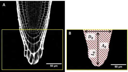
Fig. 2. Characterization of the size and shape of the root cap in Arabidopsis. (A)
Confocal microscope image of a 5-d-old root of Col-0 stained with propidium iodide. The box indicates the section of the root cap for which the size and shape were measured. (B) Root cap section threshold using the Image J software. The morphology of the root cap was characterized by its length, LR (from the quiescent centre, QC, to the tip of the cap),
the diameter of the root at the QC, DR, and the area of the root tip section, AR
(corresponding to the hatched area). These three measures enabled calculation of the
Fig. 3. Mechanical properties of Phytagel growth media. (A) Stress–strain curves
resulting from compressive tests performed on cubes of growth medium containing Phytagel at concentrations ranging from 0.2–1.2%. The data shown correspond to individual measurements. (B) The elastic modulus, E, was estimated using the slope of the linear portion of the stress–strain curve (Fig. 1). Data are means (±SD), n=3. (C, D) Penetrometer tests performed to examine the mechanical resistance of the interface in two-layer media. (C) Compression force during penetration of the needle into the lower layer of medium of 0.2%, 0.3%, and 0.5% Phytagel. The reference starting point for displacement corresponds to the first contact between the needle tip and the interface. The data shown correspond to individual measurements. (D) Maximum strength required for the needle to induce rupture of the interface as a function of the Phytagel
Fig. 4. Morphology of Arabidopsis Col-0, fez-2, and smb-3 root caps. (A–C)
Representative confocal microscope images of 5-d-old roots that were grown in compliant medium containing 0.2% Phytagel. The roots are stained with propidium iodide. Asterisks indicate the quiescent centre (QC). The scale bar is 100 µm. (D) Diameter of the roots at the QC. (E) Length of the root cap from the QC to the tip of the root cap, LR (Fig. 2). (F) The inverse Sharpness Index (iSI) of the root tips (eqn 4). Data
are means (±SD), n=20. Different letters indicate significant differences as determined using ?-way ANOVA followed by Tukey’s test (P<0.05).
Fig. 5. Growth and gravitropic responses of Arabidopsis Col-0, fez-2, and smb-3 root
apexes. (A) Length of the growth zone from the quiescent centre to the end of the elongation zone, estimated using a kinematic analysis. (B) Growth rates of roots growing in compliant 0.2% Phytagel single-layer medium. (C) Gravitropic response of roots after tilting by 90°. The data are expressed in dimensionless units (Chauvet et al. (2016). Data are means (±SD), 14≤n≤18 (A), n=15 (B), and 14≤n≤16 (C). Different letters indicate significant differences as determined using ?-way ANOVA followed by Tukey’s test (P<0.05).
Fig. 6. Critical buckling force of Arabidopsis Col-0, fez-2, and smb-3 roots. (A)
Schematic diagram of a root apex growing against the 0.2/0.3 % Phytagel interface with elastic modulus E1=7.6 kPa. The curve represents the shape of the interface just before
initiation of root curvature: d is the corresponding maximal deflection of the interface and
R is the radius of the root tip in contact with the surface. The dashed line corresponds to
the initial shape of the interface before contact with the root apex. (B) The shapes of the interface (E=7.6 kPa) as deformed by the root tips of the three genotypes just before initiation of curvature. The solid lines are means of n=10 replicates and the shading represents ±SD. The dashed line corresponds to the initial shape of the interface before contact with the root apex. (C) Evaluation of the critical force, Fcrit, that the roots were
able to apply on the interface before reorientating (eqn 5). Data are means (±SD), n=10.. Different letters indicate significant differences as determined using ?-way ANOVA followed by Tukey test’s (P<0.05).
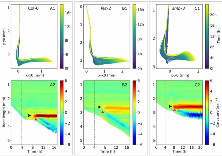
Fig. 7. Spatio-temporal characterization of apex reorientation of Arabidopsis Col-0,
fez-2, and smb-3 roots in the 0.2/0.3 % Phytagel medium. (A1, B1, C1) Skeleton and (Afez-2,
B2, C2) spatio-temporal analyses of primary roots reorientating after contact with the 0.2/0.3 interface for (A) Col-0, (B) fez-2, and (C) smb-3. In the spatio-temporal graphs, root length is presented as a function of time and the colours represent curvature intensity: negative for right-handed curvature and positive for left-handed curvature. Green corresponds to the straight zones of the roots, while red and blue depict the highest positive and negative curvatures, respectively. The dashed lines indicate the time of initial contact between the root apex and the interface. The black and purple arrowheads indicate the initiation of the first and the second zones of curvature, respectively.
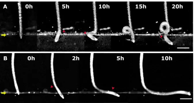
Fig. 8. Major time-steps in the establishment of the curly (A) and step-like (B) root
morphologies after contact between the Arabidopsis root apex and the interface of the 0.2/0.3 % Phytagel medium. Representative images are shown of root apex reorientation and curvature of roots that did not penetrate the lower layer in the 0.2/0.3 medium at 0– 20 h after contact with the interface. (A) A wild-type (Col-0) root that developed a characteristic curly morphology. (B) A fez-2 root that developed a characteristic step-like morphology. The yellow arrows indicate the 0.2/0.3 interface. Red arrowheads indicate the formation of zones of curvature. to the scale bars are 500 µm.
Fig. 9. Spatio-temporal characterization of apex reorientation of Arabidopsis Col-0, fez-2,
and smb-3 roots in the 0.2/0.5 % Phytagel medium. (A1, B1, C1) Skeleton and (A2, B2, C2) spatio-temporal analyses of primary roots reorientating after contact with the 0.2/0.5 interface for (A) Col-0, (B) fez-2, and (C) smb-3. In the spatio-temporal graphs, root length is presented as a function of time and the colours represent curvature intensity: negative for right-handed curvature and positive for left-handed curvature. Green corresponds to the straight zones of the roots, while red and blue depict the highest positive and negative curvatures, respectively. The dashed lines indicate the time of initial contact between the root tip and the interface. The black and purple arrowheads indicate the initiation of the first and the second zones of curvature, respectively.
Fig. 10. Growth rate of Arabidopsis Col-0, fez-2, smb-3 roots in two-layer media before
and after contact with the interface. Growth rates are shown for roots that reorientated their growth after contact with the interface in (A) 0.2/0.3 % Phytagel medium and (B) 0.2/0.5 medium. Data are means (SD), 8<n<12. The dashed lines indicate the time of initial contact between the root tip and the interface.