HAL Id: inserm-01558632
https://www.hal.inserm.fr/inserm-01558632
Submitted on 9 Jul 2017
HAL is a multi-disciplinary open access
archive for the deposit and dissemination of
sci-entific research documents, whether they are
pub-lished or not. The documents may come from
teaching and research institutions in France or
abroad, or from public or private research centers.
L’archive ouverte pluridisciplinaire HAL, est
destinée au dépôt et à la diffusion de documents
scientifiques de niveau recherche, publiés ou non,
émanant des établissements d’enseignement et de
recherche français ou étrangers, des laboratoires
publics ou privés.
immunomonitoring
Tanja Scheikl-Gatard, Caroline Tosch, François Lemonnier, Ronald Rooke
To cite this version:
Tanja Scheikl-Gatard, Caroline Tosch, François Lemonnier, Ronald Rooke. Identification of new
MUC1 epitopes using HLA-transgenic animals: implication for immunomonitoring. Journal of
Trans-lational Medicine, BioMed Central, 2017, 15 (1), pp.154. �10.1186/s12967-017-1254-0�.
�inserm-01558632�
RESEARCH
Identification of new MUC1 epitopes
using HLA‑transgenic animals: implication
for immunomonitoring
Tanja Scheikl‑Gatard
1, Caroline Tosch
2, François Lemonnier
3and Ronald Rooke
4*Abstract
Background: The success of immunotherapeutics in oncology and the search for further improvements has prompted revisiting the use of cancer vaccines. In this context, knowledge of the immunogenic epitopes and the monitoring of the immune response cancer vaccines generate are essential. MUC1 has been considered one of the most important tumor associated antigen for decades.
Methods: To identify HLA‑restricted MUC1 peptides we used eight human MHC class I transgenic mouse lines, together covering more than 80% of the human population. MUC1 peptides were identified by vaccinating each line with full length MUC1 coding sequences and using an IFNγ ELIspot restimulation assay. Relevant peptides were tested in a flow cytometry‑based tetramer assay and for their capacity to serve as target in an in vivo killing assay.
Results: Four previously identified MUC1 peptides were confirmed and five are described here for the first time. These nine peptide‑MHC combinations were further characterized. Six gave above‑background tetramer staining of splenocytes from immunized animals and three peptides were induced more than 5% specific in vivo killing.
Conclusions: These data describe for the first time five new HLA class I‑restricted peptides and revisit some that were previously described. They also emphasize the importance of using in vivo/ex vivo models to screen for immunogenic peptides and define the functions for individual peptide‑HLA combinations.
Keywords: Cancer, Immunology, MHC class I, MUC1, Tumor antigen, Immunomonitoring
© The Author(s) 2017. This article is distributed under the terms of the Creative Commons Attribution 4.0 International License (http://creativecommons.org/licenses/by/4.0/), which permits unrestricted use, distribution, and reproduction in any medium, provided you give appropriate credit to the original author(s) and the source, provide a link to the Creative Commons license, and indicate if changes were made. The Creative Commons Public Domain Dedication waiver (http://creativecommons.org/ publicdomain/zero/1.0/) applies to the data made available in this article, unless otherwise stated.
Background
After many years of mitigated results, cancer immunother-apy approaches have spawned great enthusiasm because of their capacity to generate significant improvement in
patients’ status in a number of pathologies [1]. One of these
advances exploits immunological checkpoints for which commercially approved molecules prevent the dampen-ing of the immune response arisdampen-ing in the tumor environ-ment. These immune checkpoint inhibitors (ICI) have been successfully used as stand-alone in early clinical trials which indicates that effector T cells are present, that they are the main players in tumor control and that their inca-pacity to control cancer growth is due to tumor-related
immunosuppressive mechanisms [2, 3]. It may also explain
why cancer vaccines have met with limited success so far in that the tumor antigen-specific T cells they generate are incapable of fulfilling their task in face of an inhibitory
tumor environment [4, 5]. Notwithstanding the
signifi-cance of ICI in the treatment of signifi-cancer, only a proportion of patients respond. The mechanisms underlying the absence of response are multiple and many are currently
being investigated [3]. One possible reason why some
patients do not respond is the mere absence of an antigen-directed immune response by lack of stimulation of appro-priate T cell clones. It is thereby reasonable to assume that the combination of cancer vaccines with ICI will increase the proportion of responding patients. Although impor-tant developments of sequencing technologies allow to foresee the use of patients idiotypic epitopes as source of antigens, the development path and regulatory hurdles of
Open Access
*Correspondence: [email protected]
4 Institut de Recherche Servier, 125 Chemin de Ronde, 78290 Croissy,
France
such technology jeopardizes their commercial success. Conversely, the use of a broadly distributed tumor antigen would justify the development of an “off the shelf” prod-uct as well as establishing the proof-of-concept that the immune responses stimulated by cancer vaccines are effec-tive if the tumor-associated immune suppression is relaxed. MUC1 is one of the most studied tumor associated
anti-gen [6]. This mucin protein is highly distributed among
cancers of epithelial origin and the cancer-associated post-translational modifications render it recognizable by the adaptive arm of the immune response. While it has been repeatedly identified as a major tumor-associ-ated antigen, MUC1-targeting cancer vaccines have met
with limited success in terms of patients’ benefit [7–11].
The immunomonitoring and biomarker identification programs that accompanied many studies have
identi-fied responder sub-populations in various cohorts [12,
13]. However, no consistent pattern of responders can
be established. Moreover, while MUC1-specific immune responses have been seen in healthy donors, cancer patients and MUC1-vaccinated individuals by various means, to date, no correlation with the identified response
and clinical outcome can be established [14, 15].
This may be accounted for by variations in the clini-cal protocols, in the choice of antigen and its delivery systems as well as differences in the monitoring meth-ods. Because CD8+ T cells are believed to be the main effectors in tumor control and elimination, the iden-tification of major histocompatibility complex class I (MHC I)-restricted peptides impacts on vaccine design and remains essential for monitoring purposes. Various in vitro and in silico methods have been developed to identify such peptides but their efficacy has been ham-pered by the heterogeneity of the human leukocyte anti-gen (HLA) distribution in the human population and the complexity of the antigen presentation machinery.
To identify MUC1 antigenic peptides, we made use of eight different HLA-transgenic mouse lines representing
the most common human MHC class I alleles and
cover-ing approximately 80% of the human population [16]. We
describe here for the first time, five HLA class I immu-nogenic peptides, each restricted to a specific HLA allele. Most of these peptides would not have been selected for immunomonitoring purposes by HLA-restricted peptide predicting algorithms. While these peptides were iden-tified by their capacity to restimulate IFNγ production in vitro, only six out of nine corresponding peptide-HLA tetramers could detect CD8+ T cells after immunization. Further, when tested in an in vivo killing assay, only three peptides gave more than 5% specific cytotoxicity. The present work demonstrates that HLA transgenic animals are instrumental in identifying novel human epitopes that can then be used as source of antigen and/or for immunomonitoring. Each peptide performed differ-ently in functional assays with no systematic qualitative or quantitative correlation across all assays, suggesting that they may be playing different roles in the immune response. These data warrant the use of HLA transgenic animals in combination with functional assays to better select immunogenic peptides for immunomonitoring or immunization approaches.
Methods Mice
The monochain homozygous HLA-transgenic mice have
been described previously [16–19]. Four digit alleles used
to create each line are listed in Table 1. Mice were kept
under specific pathogen-free conditions with water and food ad libitum. This study was conducted in compliance with European Union (EU) directive 2010/63/EU for ani-mal experiments. An institutional ethical committee has approved the experiments performed in this study. MUC1 peptide pool library
Peptides were synthesized by ProImmune (Oxford, UK) or ProteoGenix (Schiltigheim, France) to a minimum Table 1 HLA transgenic mouse strains used and associated MUC1 peptides
Anchor residues are in bold
a Peptide numbering is based on the Uniprot sequence P15941‑1 refered to as the canonical sequence
Mouse HLA Identified peptide AA position in the MUC1 proteina Identification method References
HLA‑A*01 isEmflqiY 1123–1131 Binding assay [13]
HLA‑A*02 stappvhnV 950–958 Prediction program [22]
sLsytnpaV 1240–1248 Binding assay [13] lLltvltV 14–21 Prediction program [23] vLvcvlvaL 1165–1173 New
HLA‑B*07 aPdnrpaL 941–948 New
HLA‑B*27 rRknygqldiF 1187–1197 New
HLA‑B*35 fPardtyhpM 1197–1206 New
purity of 95%. The identity of each peptide was confirmed by mass spectral analysis. The peptide libraries cover the entire MUC1 protein sequence and are either composed of 11mers overlapping by 8 amino acids or 15mers over-lapping by 11 amino acids. All peptides were suspended in DMSO at a concentration of 50 µg/mL. To identify HLA-specific antigenic peptide, peptides of the same length were pooled to a final concentration of 50 µg/mL per peptide (25 pools of 12 or 13 peptides for the 11mers and 24 pools of 11 or 12 peptides for 15mers). Pools were
used in a matrix format as described in Tobery [20].
MUC1‑immunizing vectors
MUC1 plasmid was generated by introducing a modified sequence of the human MUC1 cDNA (NCBI Nucleotide database# NM_002456.5) into the pcDNA3.1ΔHygro expression vector. The plasmid preparation and puri-fication was done by Geneart (ThermoFisher, Courta-boeuf, France). The plasmid was stored at 4 °C in TE buffer (10 mM TRIS, 0.1% EDTA, pH 8) and diluted in PBS prior to use. A Modified Vaccinia Ankara (MVA) recombinant virus expressing the MUC1 protein (NCBI Nucleotide database# NM_002456.5) was generated by homologous recombination between the two expression cassettes and the empty vector MVATGN33.1 in primary chicken embryo fibroblasts (CEFs) as described
ear-lier [21]. The purified virus was suspended in S08 buffer
(10 mM Tris–HCl, pH 8, 5% (wt/vol) sucrose, 10 mM sodium glutamate, and 50 mM NaCl) and stored at 80 °C.
Virus stocks between 5 × 10⁸ and 109 PFU/mL as
deter-mined by CEF-plaque assay. Viruses were diluted in S08 buffer to the concentrations required for the in vivo stud-ies immediately prior to use.
Immunizations
For DNA immunization, anesthetized mice were first injected intramuscularly in both tibialis anterior mus-cles with cardiotoxin (50 µL at 10 µM, Latoxan, Rosans, France) then, 5–7 and 17–21 days later, they were injected at the same site with 50 μg per leg of purified recombinant MUC1 plasmid DNA in 50 µL. For MVA immunization, mice were injected intravenously with
5 × 10⁷ PFU of recombinant MVA-MUC1 in a final
vol-ume of 100 μL. MVA was injected twice or thrice with a 7-day interval between injections. Epitope-specific CD8+ T cell responses were analyzed 7–9 days after the last injection of either MVA or DNA.
ELISpot assay
CD4-depleted (mouse CD4 MicroBeads, Miltenyi Bio-tec, Paris, France) or CD8-enriched (CD8α T cell isola-tion kit II; Miltenyi Biotec, Paris, France) splenocytes
(2–5 × 10⁵ cells) were seeded in triplicate wells in 96-well
polyvinylidene difluoride (PVDF) membranes (Mul-tiScreen HTS; Millipore, Fontenay Sous Bois, France) previously coated with a rat anti-mouse anti-IFNγ mAb (15 μg/mL, AN-18; Mabtech, Paris, France) and cultured in RPMI-1640 supplemented with 10% Fetal Calf serum in the presence of 1 µg/mL of MUC1 peptide pools or individual peptides. After 18 h culture, IFNγ-producing cells were revealed using biotinylated rat mouse anti-IFNγ detection mAb (1 μg/mL, R4-6A2-biotin; Mabtech, Paris, France), ExtrAvidin-alkaline phosphatase (1:5.000; Sigma-Aldrich, Paris, France) and BCIP/NBT solution (Sigma-Aldrich, Paris-France). Spots were counted using Bioreader 4000 PRO-S and analyzed with the ImmunoS-pot software (BIO-SYS, Karben, Germany). Background values were defined as mean number of spots obtained in absence of antigenic peptides + 2× the standard devia-tion and subtracted from the values obtained with anti-genic peptides.
Flow cytometry
1–2 × 10⁶ total splenocytes or CD8-enriched T cells
(CD8α T-cell isolation kit II; Miltenyi Biotec, Paris, France) were incubated for 30 min at room temperature with 2.5 μL of phycoerythrin (PE)-conjugated HLA-MUC1-peptide tetramer (TC Metrix, Epalinges, Switzer-land) or chimeric (HLA-A2 α1 + 2, H2Kb α3 or HLA-B7 α1 + 2, H2Db α3) MUC1-peptide containing tetramer (S. Buus, Copenhagen University, Denmark). Cells were then stained with NearIR LIVE/DEAD marker (Molecu-lar Probes, Paris, France) for 15 min. After washing, cells were incubated for 30 min at 4 °C with anti-CD8 (RM4-5), anti-CD4 (53–6.7), anti-NK1.1 (PK136), anti-B220 (30-F11) and anti-CD11b (M1/70) antibodies (all BD Bio-sciences, Pont-de-Claix, France). Except for the anti-CD8 Ab which was APC-conjugated, all Ab were FITC conju-gated and used as dump-channel.
Data was acquired using a FACS Aria III (BD Bio-sciences) and analyzed using FlowJo 7.6.1 software (Tree Star, Ashland, OR, USA) or Kaluza (Beckman Coulter, Villepinte, France).
In vivo cytotoxicity assay
For in vivo CTL activity, splenocytes from naive HLA-matched animals were divided into several groups; unpulsed or pulsed for 1 h at 37 °C with 10 μM of rel-evant MUC1 peptides and labeled with 0.4 or 6.4 μM car-boxyfluorescein succinimidyl ester (CFSE) (Invitrogen, Paris, France) for 10 min at 37 °C and/or 30 min at 37 °C
with 1 µM CellTracker™ Orange CMTMR
(6-(((4-chlo-romethyl)benzoyl)amino)tetramethylrhodamine) (1:1000, Molecular Probes, Paris, France). After wash-ing, the different fractions were mixed in equal propor-tions for intravenous injection into recipient mice with a
maximum of 3 × 10⁷ cells per mouse. Splenocytes were harvested 18 h later, prior to flow cytometry acquisition. The percentage of specific lysis was calculated using the formula: % specific lysis = 100 – [100 × (R in immunized mice/mean R in naive mice)], where R is the ratio of the number of pulsed cells/number of unpulsed cells.
Data were acquired either on a FACSCanto, a FAC-SAria III flow cytometer (Becton, Dickinson) or a Navios cytometer (Beckman Coulter). Accordingly, analyses were performed with Diva or Kaluza (Beckman Coulter) software.
Statistical analyses
Mann–Whitney tests were performed for individual comparisons of two independent groups. Wilcoxon tests were performed for individual comparisons of paired groups. Statistical analysis was performed with Graph-Pad Prism (version 5) software. Differences were consid-ered significant at P values of <0.05.
Results
Identification of novel HLA‑restricted MUC1 specific peptides
To identify MHC class I-restricted peptides, mice from each HLA-transgenic line were immunized by intra-muscular injection of a plasmid encoding the entire MUC1 sequence. Following the immunization proto-col, the frequency of IFNγ-producing splenocytes was determined after stimulation with pools of 15mer pep-tides overlapping by 11 amino acids. The peptide pools used for the stimulation were composed and displayed in a matrix format allowing identification of individual
peptides (Additional file 1: Figure S1) [20]. Six out of the
eight mouse lines immunized with the MUC1-expressing plasmid gave an “above background” number of IFNγ-producing cells suggesting that some of the 15mer com-prise the human MHC-restricted peptide (not shown). As described by Boucherma et al. all mouse lines have a broad T cell receptor (TCR) repertoire representative of a normal CD8 compartment exclusively selected and main-tained by the transgenic human MHC class I molecule
since the H2 MHC class I locus is inactivated [16].
Not-withstanding this observation, the proportion of CD8+ T cells present in the lymphoid compartment in the HLA-transgenic lines is lower than the one seen in inbred but otherwise wild type animals. To enhance the proportion of peptide pool-stimulated cells, we either enriched for the CD8+ cells or depleted the CD4+ cells prior to the ELISpot assay. Although this generated more interpret-able results, the use of 15mers was often associated with
inconsistencies in the matrix analysis (Additional file 1:
Figure S1a). Fifteen-mer peptides must be randomly trimmed by proteases present in the medium to fit in the
groove of MHC class I molecules or can be presented by MHC class II molecules, thereby stimulating CD4+ T cell. We reasoned that to better discriminate the MUC1 peptides capable of stimulating a MHC class I response, splenocytes from immunized animals were screened with the pools of 11mer peptides, overlapping by eight amino acids and covering the entire MUC1 sequence
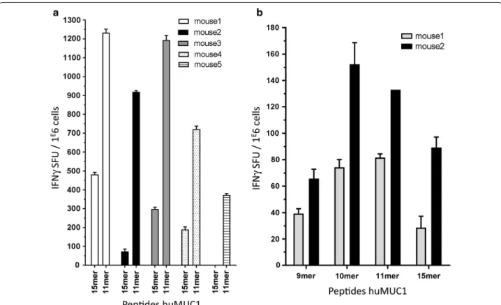
(Fig. 1a). The combination of cell enrichment and use of
11mers increased the proportion of responding cells per seeded cells and limited the detection of mouse class
II-restricted response respectively (Additional file 2: Figure
S2).
In any case, for every 15 or 11mer peptide generating an above-background response, a restimulation assay was done with peptides of various lengths (8–11mers) span-ning the suspected MUC1 region considering the
HLA-specific anchor residues (Fig. 1b). Table 1 summarize
the peptides that generated the strongest responses per transgenic mouse line. We confirmed in the HLA-A*01 mouse line a peptide originally identified using a
bind-ing assay [13]. Genetic immunization of HLA-A*02 mice
also confirmed three peptides previously described
iden-tified either using a binding assay (SLSYTNPAV) [13] or
Peptide Prediction Algorithms (STAPPVHNV,
LLLTV-LTVV) [22, 23]. Interestingly, a hereto forth undescribed
HLA-A*02-restricted peptide was identified with our approach (VLVCVLVAL). Since we focused on the pep-tides that generated the strongest ELISpot responses, many other HLA-A*02-restricted peptides described in the literature were not further analyzed as they only generated marginal responses. Finally, we identified one novel HLA class I-restricted peptide for each of the addi-tional mouse lines listed in table I. No MUC1-specific response could be generated in the A*24 and
HLA-B*08 mouse lines (data not shown) [16].
HLA‑specific tetramers staining and limitations of peptide‑HLA predicting algorithms
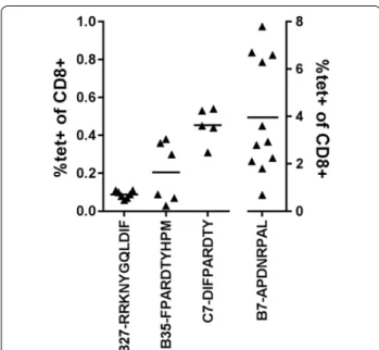
Fluorescence-conjugated peptide-HLA tetramers are important tools for monitoring the evolution of an anti-gen-specific immune response. To determine if direct detection of the peptide-MHC responding CD8+ cells was possible, tetramers were produced for each combi-nation and tested on splenocytes of immunized animals
(Fig. 2). Globally, of the nine tetramers made with fully
human MHC molecules tested, only three did not give any staining. Of those, the HLA-A*01-restricted peptide (ISEMFLQIY) which was originally identified using a
HLA binding assay [13] was predicted as the best binder
by several peptide-prediction algorithms (not shown) and it systematically gave a high frequency of IFNγ-producing cells in our restimulation assay. Two out of the four HLA-A*02 tetramers did not detect CD8+ cells
from immunized animals although each peptide gen-erated a significant IFNγ response in the ELIspot assay. One of these peptides (STAPPVHNV) has been previ-ously shown to induce a cytotoxic response in a
num-ber of studies [15, 22, 24]. To our knowledge, only one
study used STAPPVHNV-HLA-A*02 tetramers to look at MUC1-specific CD8+ cells in the blood of healthy individuals and cancer patients and found very low
fre-quencies [25]. Interestingly, four out of five different
pep-tide-HLA binding prediction algorithms predicted low
binding capacity for this peptide (Additional file 3: Table
S1). On the other hand, the same algorithms predict a higher probability for the novel VLVCVLVAL peptide to bind HLA-A*02 although we were incapable of detecting any significant tetramer staining. In the HLA-B*07 mouse line, the peptide identified (APDNRPAL) has never been described before. This is not surprising since only 2 out of 5 peptide-binding prediction algorithms could ana-lyze the binding capacity of 8mers and did not rank it as the one showing the highest affinity. This is also in sharp contrast with the high intensity and high frequency of
staining obtained with this tetramer on CD8+
spleno-cytes from immunized animals (Figs. 2, 3). Similarly, only
two programs predicted the binding capacity of 11 mers. However, they both ranked the novel HLA-B*27 binding peptide (RRKNYGQLDIF) as the peptide with the high-est affinity which correlated with the tetramer staining. Finally, the HLA-B*35 and HLA-C*07 restricted peptides (FPARDTYHPM and DIFPARDTY, respectively) are both described for the first time. The HLA-B*35-binding pep-tide was identified by all algorithms in the top six high affinity peptides, ranging from 1st to 6th and correlated with tetramer staining. In contrast, the HLA-C*07-restricted peptide was described as a low affinity by the algorithms but nonetheless showed good tetramer stain-ing. In conclusion, the use of HLA-expressing transgenic animals allowed the identification of novel HLA-binding peptides which would not have been identified using binding algorithms. Even if some predicted peptides map to the repeated sequences in the MUC1 sequence, all the peptides identified as immunodominant in the assay are
unique in the protein sequence (Table 1).
Fig. 1 Representative results for IFNγ Elispots showing CD8‑specific MUC1 responses. a Splenocytes from five immunized HLA‑B*27 mice restimu‑ lated with 11mer RRKNYGQLDIF gave a stronger response than the 15mer RRKNYGQLDIFPARD (anchor residues in bold). b CD4‑depleted spleno‑ cytes from two different immunized HLA‑B*27 mice were restimulated with 9mer RKNYGQLDI, 10mer RRKNYGQLDI, 11mer RRKNYGQLDIF and the identified 15mer RRKNYGQLDIFPARD from the peptide pool. The 11mer was chosen for the tetramer construction as it gave a strong INFγ response and contains both anchor residues for HLA‑B*27 (marked in bold)
Tetramer avidity impacts on the detection of CD8+ cells From the data described above, it appears that some pep-tide-MHC complexes stimulate the production of IFNγ but that the tetramers made of the same constituents are incapable of staining specific T cells. Many parameters impact on the interaction between clonotypic TCRs and their cognate peptide-MHC heterodimer which have led
to the concept of functional avidity (see [26] for review).
An important component influencing the TCR-peptide-MHC (pTCR-peptide-MHC) avidity, both in vitro and in vivo, is the CD8 heterodimeric co-receptor binding to the α3 domain
of MHC class I molecules [26–30]. One possible
expla-nation for the discrepancy between the positive ELIspot data and the absence of tetramer staining observed for some peptides may be related to the fact that the CD8+ T cells in HLA-transgenic mouse lines have been selected on chimeric MHC class I molecules harboring a mouse
Db α3 portion while the MHC class I molecules
mak-ing up the tetramer are fully human. The contribution of the CD8-MHC class I interaction has been estimated to affect the TCR avidity by a factor of three to fourfold
[28]. To address this possibility, tetramers made of
chi-meric HLA-A*02 and HLA-B*07 molecules correspond-ing precisely to the ones expressed in the animals were synthetized, loaded with the appropriate peptides and used to stain splenocytes from immunized animals.
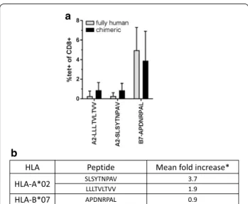
Fig-ure 4 shows the percentages of stained CD8+ cells of
immunized animals with the corresponding tetramers that could be successfully assembled. The proportion of CD8+ splenocytes stained by the fully human or chimeric HLA-B*07-APDNRPAL tetramers were not significantly different which argues in favor of a strong affinity between the TCR and this pMHC. On the other hand, while the four fully human HLA-A*02 tetramer were synthesizable, only two out of four chimeric HLA-A*02 tetramers could be obtained. The chimeric HLA-A*02 tetramers gave systematically a higher percentage of stained cells and in some mice, they allowed measuring a response otherwise undetectable with the fully-human tetramer. These latter Fig. 2 Tetramer staining. Splenocytes of immunized mice were
stained with the respective HLA‑tetramer. Each triangle represents the percentage of tetramer positive CD8+ cells per mouse. Horizontal bars represents the mean of all animals. A1‑ISEMFLQIY, A2‑STAP‑ PVHNV and A2‑VLVCVLVAL gave no tetramer staining (not shown)
0 2.8
3.6
mouse 1 mouse 2 naive mouse
CD8
Tetramer 2.3 2.9
mouse 3 mouse 4
Fig. 3 Representative tetramer staining of HLA‑B*07 splenocytes. Splenocytes of immunized HLA‑B*07 mice (mouse 1–4) or 1 naive mouse were analysed for B7‑APDNRPAL tetramer staining. Lymphocytes were gated as described in “Methods”. Numbers represent proportion of tetramer+ in the total CD8+ population
observations suggest that the increase in avidity generated from the α3-CD8 interaction is important in the detection of a HLA-A*02 responses.
Novel peptides as target for a cytotoxic response
We then evaluated the cytotoxic capacity of the CD8+ cells generated in immunized animals towards peptide-loaded target cells in an in vivo cytotoxic assay. Since background killing, defined as the difference between the percentages of killing in untreated splenocytes in immunized and naïve animal, was negligible in all experiments (no non-specific killing), we considered 5% cytotoxicity as the threshold for positivity. From this standpoint, three newly identified MUC1 peptides (HLA-A*02-VLVCVLVAL, HLA-B*07-ADPNRPAL and HLA-B*27-RRKNYGQLDIF) were capable of
induc-ing cell killinduc-ing in vivo (Fig. 5). For each mouse line,
vari-ation in cytotoxicity was important between animals (5 < N < 8, N = number of mice evaluated) but most val-ues remaining within the same quartile. However, cyto-toxicity varied greatly between mouse lines with mean values ranging from <1% (HLA-A*02-LLLTVLTVV) to >75% (HLA-B*07-ADPNRPAL). In the latter case, some animals eliminated the peptide loaded target and dem-onstrated 100% cytotoxicity. Many potentially additive mechanisms can be responsible for these differences such as peptide half-life on the surface of loaded cells, the functionality of the amplified CD8+ T cell clone and/ or the in vivo effector to target ratio. It is important to note that we established positivity on the mean cyto-toxicity value obtained from many animals (5 < N<8)
HLA Peptide Mean fold increase*
HLA-A*02 SLSYTNPAVLLLTVLTVV 3.71.9
HLA-B*07 APDNRPAL 0.9
a
b
Fig. 4 Fully human vs chimeric tetramer staining. Fully human HLA molecules or human‑mouse chimera (human α1 + α2 + mouse Db α3) were loaded with the indicated peptide and used to stain spleno‑ cytes from immunized animals. a Mean percentage of stained CD8+ cells (n = 5 mice for fully human tetramer, n = 4 for chimeric con‑ structs). b Fold increase in percentage of chimeric tetramer stained CD8+ cells/percentage of fully human tetramer stained CD8+ cells
HLA Mean SD n A1-ISEMFLQIY 3.47 2.88 6 A2-LLLTVLTVV 0.86 0.89 5 A2-SLSYTNPAV 0.75 1.50 4 A2-STAPPVHNV 0.60 1.13 4 A2-VLVCVLVAL 7.88 7.71 5 B7-APDNRPAL 76.48 32.90 5 B27-RRKNYGQLDIF 2.62 3.51 5 B35-FPARDTYHPM 18.14 13.03 8 C7-DIFPARDTY 1.25 1.49 6 %k illi ng A1 -ISEMFLQI Y A2-LL LTV LTV V A2 -SL SY TNPA V A2 -S TAP PVHNV A2 -VLVCV LVA L B7-A PD NR PA L B2 7-RRKN YG QLDI F B 35 -F PA R D TY H PM C7-DIFPA RDTY 0 10 20 30 40 50 60 70 80 90 100 a b
Fig. 5 In vivo cytotoxicity. Fluorescence‑labelled splenocytes of HLA‑matched mouse line were loaded with the indicated peptide and injected in either immunized or naive mice. Animals received concomitantly untreated splenocytes (no peptide) labelled with a different concentration of the fluorescent label. Killing was determined as described in “Methods”. a Each triangle represents the percentage of specific killing per mouse. Hori-zontal bar mean of all mice. b Table summarizing mean percentage of cytotoxicity and standard deviation. n number of animals analyzed. Peptides B7‑ADPNRPAL, B35‑FPARDTYHPM and A2‑VLVCVLVAL showed a mean specific killing above 5% (shown in bold)
which includes animals which may not have been suc-cessfully immunized. The data presented may thus be underestimated.
Discussion
Cancer immunotherapy has been the subject of research and speculations for over a century and it is only in the first half of this decade that clinical data has dem-onstrated its efficacy. Numerous ways to use patient’s immune system against his/her cancer have been put
forth and tested (reviewed in [31]). To date, the most
successful approach is based on antibodies that block signals naturally used by the immune system to control the breadth of the immune response and prevent auto-immunity. Four such ICI antibodies, targeting two main, complementary inhibitory pathways are approved by the FDA in various indications: ipilimumab blocks the interaction between the cytotoxic T lymphocyte asso-ciated protein 4 (CTLA-4) on T cells and the CD80/ CD86 molecules on the antigen presenting cells (APC) while nivolumab, pembrolizumab and atesolizumab are inhibitors of the interaction between the programmed death 1 (PD-1) receptor and its ligand (PD-L1). The other FDA-approved approach in cancer immunotherapy
con-sists in vaccinating patients against their cancer [32]. It
is based on the delivery of peptides or proteins that are specific to the cancer cells in a context that stimulates an immune response against the antigen-expressing
cells [4]. Conceivably, the combination of ICI with
vac-cination approaches are complementary and should result in improved responses in the ongoing clinical
tri-als [33]. This concept is exemplified by data stemming
from clinical trials which show that patients respond-ing to treatment with ICI have high mutation rates. This is interpreted as the demonstration that patients with tumors displaying a broad range of neo-epitopes are more likely to develop effector T cell responses since they have not been subject to central tolerance. Moreo-ver, these responses tend to be more efficient when the neoepitope-encoding mutations are homogeneously
dis-tributed across the tumor [34]. In this context, improved
cancer vaccines may be designed and used to favor
thera-peutic benefit [4].
Evaluating the immune response of cancer patients has been highly instrumental for our understanding of the interplay between the immune system and patients’ can-cers. The paradigm being established changes our view in the staging of patients and impacts on treatment choice
[35]. It also places CD8+ effector T cells as a central
component of an effective anti-cancer response further emphasizing the role therapeutic vaccination could play
in conditions of relieved immunosuppression. In clini-cal trials, many therapeutic vaccination schemes were shown to generate specific responses to the antigen but correlation with an objective tumor control was seldom
reported [36]. Great efforts were invested to identify the
relevant response and ensure the robustness and com-parability of the method to evaluate it through
interna-tional proficiency panels [37, 38]. Flow cytometry offers
the possibility of timely quantitative and qualitative assessments of immune responses throughout the course of diseases and/or treatments. The use of fluorescent pMHC multimers in combination with antibody cock-tails that detect surface markers and secreted molecules allows enumerating antigen-specific T cells with specific phenotypes. However, the knowledge of the patients’ MHC haplotype, the determination of the peptide pre-sented by specific HLA molecules and the capacity to synthesize and validate the pMHC multimer are para-mount to this endeavor. The main constraint in achiev-ing this goal has been the limited capacity of laboratory and bio-informatics tools to recapitulate all the steps involved in antigen presentation and thereby predict-ing and/or identifypredict-ing antigenic peptides recognized by specific T cells. Here we show that an ensemble of eight mouse strains, each expressing a single HLA molecule together present in more than 80% of the human popula-tion, is crucial for the proper identification of antigenic peptides. In addition, it allows the evaluation/validation of the tools required to measure the antigen-specific response. We used the MUC1 protein as a model TAA since it is one of the most commonly expressed protein on tumor of epithelial origin and is the target of many immunotherapeutic vaccination protocols. In addition to its increased expression level and the loss of its apical expression pattern on tumor cells, MUC1 is recognized as a tumor-associated antigen following post-transla-tional modifications rather than mutations in its coding
sequence [7]. This characteristic makes it a public
anti-gen more compatible with an “off-the shelf” therapeutic vaccine development scheme. The changes in the glycan moieties contribute to the enhanced immunogenicity of MUC1 by exposing the core protein to the humoral response and by modifying the interaction of cancer cells with APC. It is interesting to note that all the peptides we have identified lie outside of tandem repeat sequence that comprises all the O-linked glycosylation sites.
The method presented here identified five new anti-genic peptides that have not been identified by antianti-genic peptide predicting algorithms nor by in vitro methods. Moreover, it allows rapid and unequivocal identification of peptides that best fit the MHC groove with limited
steps to define the proper length. This is best exempli-fied by the identification of a novel HLA-A*02 peptide (VLVCVLVAL) despite the extensive work and the spec-trum of tools used to identify HLA-A*02-restricted pep-tides. The demonstration that the most immunogenic peptide was a HLA-B*07-restricted 8mer (APDNRPAL) unpredictable by three out of five bio-informatic tools is another illustration of the usefulness of this method.
The use of HLA-transgenic animals also offers the pos-sibility of evaluating the performance of pMHC-tetram-ers prior to their use in humans. Immunomonitoring of patients accrued in clinical trials represents important additional logistics and associated costs. This justifies upstream validation of the tools and methods. HLA-transgenic animals may be an important asset to achieve this. Indeed, our results show that of the nine peptides identified for their capacity to induce IFNγ produc-tion in a restimulaproduc-tion assay, only six pMHC-tetramers could detect Ag-specific CD8+ cells. The HLA mole-cule expressed in the animals are chimeras made of the α1 and α2 domains from the human sequence fused
the α3 domain of the mouse H2 Db molecule. This
con-struct was shown to impact favorably on positive selec-tion and maintenance of CD8+ cells in HLA-expressing animals by permitting a better interaction between the transgenic protein and the mouse CD8 molecule with negligible impact on the structure of the peptide-binding
pocket [39, 40]. The loss of this interaction impacts on
the avidity of the TCR-pMHC interaction and may be a possible explanation as to why cells induced to produce IFNγ in an ex vivo assay remained undetectable when exposed to a MHC tetramer containing the stimulatory peptide. To address this point, we loaded the same pep-tide in either tetramers made of fully human or chimeric HLA molecules and compared their capacity to detect the splenocytes coming from the same animals. Although results did not reach significance, for each combination studied, the chimeric tetramer detected a higher percent-age of CD8 cells than the tetramers made of fully human HLA. These results are in line with the ones published by Choi et al. justifying the use of chimeric tetramers when using HLA transgenic animals to monitor the Ag-specific
immune response [41].
HLA-transgenic mice offer the possibility to further characterize the immune response generated by immu-nogenic peptides by performing functional assays. Here, we have examined the cytotoxic response in immunized animals independent of our capacity to detect a
tetramer-specific population. The results summarized in Table 2
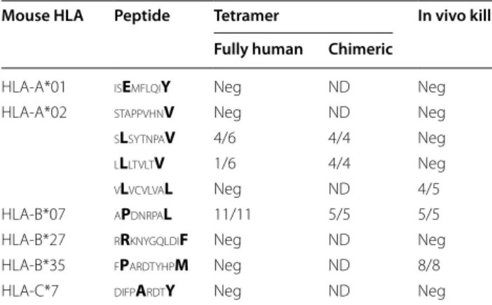
show that it was not possible to establish a correlation between tetramer staining and in vivo cytotoxicity.
Of the nine peptides identified for their capacity to stimulate splenocytes of immunized mice in an IFNγ ELIspot assay, six could specifically stain CD8+ T cells in a tetramer assay and three were recognized as target by cytotoxic T cells in vivo. Conversely, one of these peptides induces killing even though the tetramer was unsuccessful at recognizing a specific T cell population. Some techni-cal constraints may explain, at least in part these results, namely the fact that some mouse strains have only a par-tially reconstituted CD8+ compartment and that the ratio of target to effector cell may be important in the in vivo killing assay even though no trend was seen.
In terms of product development, the use of a tumor-specific antigen common to multiple tumor types and demonstrating little polymorphism between individuals offers advantages over personalized vaccination schemes that are currently in development.
Conclusions
The results presented here describe novel MUC1 pep-tides that should be included in the immunomonitoring of patients. We also demonstrate the superiority of HLA-transgenic mouse lines over in vitro or in silico methods to identify novel peptides of well-studied tumor associ-ated antigen. One important advantage is the possibility of performing functional assays. The fact that peptide-MHC complexes generate a range of response by clono-typic T cells emphasizes the importance of performing multiple assay to better define the role played by immu-nogenic peptides and these points are best addressed using HLA-transgenic animals.
Table 2 Compilation of peptide identification and valida-tions methods
All pMHC gave above background IFNg‑Elispot in an in vitro recall response in at least three independent experiments. For tetramer staining and in vivo kill assay, the number of positive results (number of mice positive/total number of mice analyzed). Anchor residues are in bold
Neg negative results, ND not done
Mouse HLA Peptide Tetramer In vivo kill
Fully human Chimeric
HLA‑A*01 isEmflqiY Neg ND Neg
HLA‑A*02 stappvhnV Neg ND Neg
sLsytnpaV 4/6 4/4 Neg lLltvltV 1/6 4/4 Neg vLvcvlvaL Neg ND 4/5
HLA‑B*07 aPdnrpaL 11/11 5/5 5/5
HLA‑B*27 rRknygqldiF Neg ND Neg
HLA‑B*35 fPardtyhpM Neg ND 8/8
Abbreviations
APC: allophycocyanine; CEF: chicken embryo fibroblasts; CFSE: carboxy‑ fluorescein succinimidyl ester; CMTMR: 6‑(((4‑chloromethyl)benzoyl)amino) tetramethylrhodamine; CTL: cytotoxic T lymphocyte; FITC: fluoresceine iso‑thiocyanate; HLA: human leukocyte antigen; ICI: immune checkpoint inhibitors; IFNγ: interferon gamma; mAb: monoclonal antibody; MHC: major histocompatibility complex; MUC1: mucin 1; MVA: Modified Vaccinia Ankara Strain; PBS: phosphate buffered saline; PE: phycoerythrine.
Authors’ contributions
TG performed all the experiments. CT performed flow cytometry. FL gener‑ ated the transgenic animals and the plasmid vectors and gave technical advice. RR designed the study and wrote the manuscript. All authors read and approved the final manuscript.
Author details
1 SONOGEN AG, Badenerstrasse 808, 8048 Zurich, Switzerland. 2 Transgene SA,
400 Bld Gonthier d’Andernach, 67400 Illkirch Graffenstaden, France. 3 Unité
INSERM 1016, Département Endocrinologie, Métabolisme et Diabète. Equipe Immunologie des Diabètes, Bâtiment Cassini, 123 Bd Port Royal, 75014 Paris, France. 4 Institut de Recherche Servier, 125 Chemin de Ronde, 78290 Croissy,
France.
Acknowledgements
The authors wish to thank S. Buss (Copenhagen University, Denmark) for sup‑ plying the chimeric MHC molecules.
Competing interests
The authors declare that they have no competing interests. Availability of data and materials
The datasets used and/or analyzed during the current study available from the corresponding author on reasonable request.
Additional files
Additional file 1: Figure S1. Representative IFNγ Elispot restimulation results and peptide pool matrix analysis. HLA‑C*07 mice were immunized with full length MUC1 coding sequences and assay was performed on splenocytes. (a) 15mer. CD8‑enriched cells (over 44% CD8+ cells) pooled from four immunized mice (HLA‑C*07) were restimulated with pool of 15mers 1–24. The Elsipot is shown above, the matrix of 15mers 1–137 from the 24 pools covering the whole MUC1 protein is shown below. Pools above background in the Elispot (framed) are highlighted in the matrix. (b) 11mer. CD8‑enriched cells (over 65% CD8+ cells) pooled from 7 immunized mice (HLA‑C*07) were restimulated with pool of 11mers 1–25. The Elispot is shown above, the matrix of 11mers 1–156 from the 25 pools covering the whole MUC1 protein is shown below. Pools above background in the Elispot (framed) are highlighted in the matrix.
Additional file 2: Figure S2. CD8‑specific IFNγ response. Total splenocytes (grey with 23% of CD8+ and 14.4% CD4+), CD8‑enriched cells (black with 84% CD8+, 0.2% CD4+) and CD8‑depleted cells (white with 1.6% CD8+ and 19.6% CD4+) pooled from four immunized mice (HLA‑B*35) were restimulated with 10mer FPARDTYHPM and 11mer IFPARDTYHPM. Only cells containing CD8+ cell showed an IFNg response (grey and black), the CD8‑depleted cells (white, not visible) showed no IFNγ response.
Additional file 3: Table S1. Prediction of antigenic peptide by algo‑ rithms. The referenced MUC1 sequence was submitted to five algorithms for antigenic peptide prediction. Default settings were used for each of them. Numbers represent the rank for each indicated peptide. “‑” indicates that the algorithm could not predict the peptide. Colored box indicate that the algorithm lacked one or more conditions for analysis (peptide length and/or allele).
Ethics approval
This study was conducted in full compliance with European Union (EU) direc‑ tive 2010/63/EU relating to the protection of animals used for experimental or other scientific purposes and in compliance to the French law (décret no 2013‑118 of February 1st 2013) and approved by the Comité National de réflexion éthique sur l’expérimentation animale (CNREEA) and approved by an institutional ethical committee (TG number 93).
Funding
This work was supported by a Grant (ANR 2010 BIOT 008 01) from l’Agence National pour la Recherche (France).
Publisher’s Note
Springer Nature remains neutral with regard to jurisdictional claims in pub‑ lished maps and institutional affiliations.
Received: 15 January 2017 Accepted: 24 June 2017
References
1. Whiteside TL, Demaria S, Rodriguez‑Ruiz ME, Zarour HM, Melero I. Emerg‑ ing opportunities and challenges in cancer immunotherapy. Clin Cancer Res. 2016;22(8):1845–55.
2. Sharma P, Allison JP. Immune checkpoint targeting in cancer ther‑ apy: toward combination strategies with curative potential. Cell. 2015;161(2):205–14.
3. Munn DH, Bronte V. Immune suppressive mechanisms in the tumor microenvironment. Curr Opin Immunol. 2015;20(39):1–6.
4. van der Burg SH, Arens R, Ossendorp F, van Hall T, Melief CJM. Vaccines for established cancer: overcoming the challenges posed by immune evasion. Nat Rev Cancer. 2016;16(4):219–33.
5. Pol J, Bloy N, Buqué A, Eggermont A, Cremer I, Sautès‑Fridman C, et al. Trial watch: peptide‑based anticancer vaccines. Oncoimmunology. 2015;4(4):e974411.
6. Hossain M, Wall K. Immunological evaluation of recent MUC1 glycopep‑ tide cancer vaccines. Vaccines. 2016;4(3):25.
7. Cheever MA, Allison JP, Ferris AS, Finn OJ, Hastings BM, Hecht TT, et al. The prioritization of cancer antigens: a national cancer institute pilot project for the acceleration of translational research. Clin Cancer Res. 2009;15(17):5323–37.
8. Haen SP, Rammensee H‑G. The repertoire of human tumor‑associated epitopes—identification and selection of antigens and their application in clinical trials. Curr Opin Immunol. 2013;25(2):277–83.
9. Lakshminarayanan V, Supekar NT, Wei J, McCurry DB, Dueck AC, Kosiorek HE, et al. MUC1 vaccines, comprised of glycosylated or non‑glycosylated peptides or tumor‑derived MUC1, can circumvent immunoedit‑ ing to control tumor growth in MUC1 transgenic mice. PLoS ONE. 2016;11(1):e0145920.
10. Mitchell P, Thatcher N, Socinski MA, Wasilewska‑Tesluk E, Horwood K, Szczesna A, et al. Tecemotide in unresectable stage III non‑small‑cell lung cancer in the phase III START study: updated overall survival and biomarker analyses. Ann Oncol. 2015;26(6):1134–42.
11. Thomas A, Giaccone G. Why has active immunotherapy not worked in lung cancer? Ann Oncol. 2015;26(11):2213–20.
12. Wurz GT, Kao C‑J, Wolf M, DeGregorio MW. Tecemotide: an antigen‑ specific cancer immunotherapy. Hum Vaccines Immunother. 2014;10(11):3383–93.
13. Ramlau R, Quoix E, Rolski J, Pless M, Lena H, Lévy E, et al. A phase II study of Tg4010 (Mva‑Muc1‑Il2) in association with chemotherapy in patients with stage III/IV non‑small cell lung cancer. J Thorac Oncol. 2008;3(7):735–44.
14. Rivalland G, Loveland B, Mitchell P. Update on mucin‑1 immuno‑ therapy in cancer: a clinical perspective. Expert Opin Biol Ther. 2015;15(12):1773–87.
• We accept pre-submission inquiries
• Our selector tool helps you to find the most relevant journal
• We provide round the clock customer support
• Convenient online submission
• Thorough peer review
• Inclusion in PubMed and all major indexing services
• Maximum visibility for your research Submit your manuscript at
www.biomedcentral.com/submit
Submit your next manuscript to BioMed Central
and we will help you at every step:
15. Rentzsch C, Kayser S, Stumm S, Watermann I, Walter S, Stevanoviç S, et al. Evaluation of pre‑existent immunity in patients with primary breast cancer: molecular and cellular assays to quantify antigen‑specific T lymphocytes in peripheral blood mononuclear cells. Clin Cancer Res. 2003;9(12):4376–86.
16. Boucherma R, Kridane‑Miledi H, Bouziat R, Rasmussen M, Gatard T, Langa‑ Vives F, et al. HLA‑A*01:03, HLA‑A*24:02, HLA‑B*08:01, HLA‑B*27:05, HLA‑ B*35:01, HLA‑B*44:02, and HLA‑C*07:01 monochain transgenic/H‑2 class I null mice: novel versatile preclinical models of human T cell responses. J Immunol. 2013;191(2):583–93.
17. Firat H, Garcia‑Pons F, Tourdot S, Pascolo S, Scardino A, Garcia Z, et al. H‑2 class I knockout, HLA‑A2.1‑transgenic mice: a versatile animal model for preclinical evaluation of antitumor immunotherapeutic strategies. Eur J Immunol. 1999;29(10):3112–21.
18. Pascolo S, Bervas N, Ure JM, Smith AG, Lemonnier FA, Pérarnau B. HLA‑ A2.1‑restricted education and cytolytic activity of CD8(+) T lymphocytes from β2 microglobulin (β2m) HLA‑A2.1 monochain transgenic H‑2Db β2m double knockout mice. J Exp Med. 1997;185(12):2043–51. 19. Rohrlich P‑S. HLA‑B*0702 transgenic, H‑2KbDb double‑knockout mice:
phenotypical and functional characterization in response to influenza virus. Int Immunol. 2003;15(6):765–72.
20. Tobery TW, Wang S, Wang XM, Neeper MP, Jansen KU, McClements WL, et al. A simple and efficient method for the monitoring of antigen‑ specific T cell responses using peptide pool arrays in a modified ELISpot assay. J Immunol Methods. 2001;254(1–2):59–66.
21. Wreschner DH, Hareuveni M, Tsarfaty I, Smorodinsky N, Horev J, Zaretsky J, et al. Human epithelial tumor antigen cDNA sequences. Differen‑ tial splicing may generate multiple protein forms. Eur J Biochem. 1990;189(3):463–73.
22. Brossart P, Heinrich KS, Stuhler G, Behnke L, Reichardt VL, Stevanovic S, et al. Identification of HLA‑A2‑restricted T‑cell epitopes derived from the MUC1 tumor antigen for broadly applicable vaccine therapies. Blood. 1999;93(12):4309–17.
23. Carmon L, El‑Shami KM, Paz A, Pascolo S, Tzehoval E, Tirosh B, et al. Novel breast‑tumor‑associated MUC1‑derived peptides: characteriza‑ tion in Db−/−x β2 microglobulin (β2m) null mice transgenic for a chimeric HLA‑A2.1/Db‑beta2 microglobulin single chain. Int J Cancer. 2000;85(3):391–7.
24. Gückel B, Rentzsch C, Nastke M‑D, Marmé A, Gruber I, Stevanović S, et al. Pre‑existing T‑cell immunity against mucin‑1 in breast cancer patients and healthy volunteers. J Cancer Res Clin Oncol. 2006;132(4):265–74. 25. Kokowski K, Harnack U, Dorn DC, Pecher G. Quantification of the CD8+
T cell response against a mucin epitope in patients with breast cancer. Arch Immunol Ther Exp. 2008;56(2):141–5.
26. Hebeisen M, Allard M, Gannon PO, Schmidt J, Speiser DE, Rufer N. Iden‑ tifying individual T cell receptors of optimal avidity for tumor antigens. Front Immunol 2015;6.
27. Cole DK, Laugel B, Clement M, Price DA, Wooldridge L, Sewell AK. The molecular determinants of CD8 co‑receptor function. Immunology. 2012;137(2):139–48.
28. Wooldridge L, van den Berg HA, Glick M, Gostick E, Laugel B, Hutchinson SL, et al. Interaction between the CD8 coreceptor and major histocom‑ patibility complex class I stabilizes T cell receptor‑antigen complexes at the cell surface. J Biol Chem. 2005;280(30):27491–501.
29. Dutoit V, Guillaume P, Ayyoub M, Hesdorffer CS, Luescher IF, Valmori D. Decreased binding of peptides‑MHC class I (pMHC) multimeric complexes to CD8 affects their binding avidity for the TCR but does not significantly impact on pMHC/TCR dissociation rate. J Immunol. 2003;170(10):5110–7.
30. Pittet MJ, Rubio‑Godoy V, Bioley G, Guillaume P, Batard P, Speiser D, et al. 3 domain mutants of peptide/MHC class I multimers allow the selective isolation of high avidity tumor‑reactive CD8 T cells. J Immunol. 2003;171(4):1844–9.
31. Farkona S, Diamandis EP, Blasutig IM. Cancer immunotherapy: the begin‑ ning of the end of cancer? BMC Med. 2016;14(1):73.
32. Gulley JL, Mulders P, Albers P, Banchereau J, Bolla M, Pantel K, et al. Perspectives on sipuleucel‑T: its role in the prostate cancer treatment paradigm. Oncoimmunology. 2016;5(4):e1107698.
33. Butterfield LH. Cancer vaccines. BMJ. 2015;350(apr22 14):h988. 34. McGranahan N, Furness AJS, Rosenthal R, Ramskov S, Lyngaa R, Saini SK,
et al. Clonal neoantigens elicit T cell immunoreactivity and sensitivity to immune checkpoint blockade. Science. 2016;351(6280):1463–9. 35. Kirilovsky A, Marliot F, El Sissy C, Haicheur N, Galon J, Pagès F. Rational
bases for the use of the immunoscore in routine clinical settings as a prognostic and predictive biomarker in cancer patients. Int Immunol. 2016;28(8):373–82.
36. Kissick HT, Sanda MG. The role of active vaccination in cancer immuno‑ therapy: lessons from clinical trials. Curr Opin Immunol. 2015;35:15–22. 37. Britten CM, Janetzki S, van der Burg SH, Huber C, Kalos M, Levitsky HI,
et al. Minimal information about T cell assays: the process of reaching the community of T cell immunologists in cancer and beyond. Cancer Immunol Immunother. 2011;60(1):15–22.
38. Britten CM, Janetzki S, Ben‑Porat L, Clay TM, Kalos M, Maecker H, et al. Harmonization guidelines for HLA‑peptide multimer assays derived from results of a large scale international proficiency panel of the Cancer Vac‑ cine Consortium. Cancer Immunol Immunother. 2009;58(10):1701–13. 39. Vitiello A, Marchesini D, Furze J, Sherman LA, Chesnut RW. Analysis of
the HLA‑restricted influenza‑specific cytotoxic T lymphocyte response in transgenic mice carrying a chimeric human‑mouse class I major histo‑ compatibility complex. J Exp Med. 1991;173(4):1007–15.
40. Nojima H, Kanou K, Kamiya K, Atsuda K, Umeyama H, Takeda‑Shitaka M. Dynamic influence of the two membrane‑proximal immunoglobulin‑like domains upon the peptide‑binding platform domain in class I and class II major histocompatibility complexes: normal mode analysis. Chem Pharm Bull. 2009;57(11):1193–9.
41. Choi EM, Palmowski M, Chen J, Cerundolo V. The use of chimeric A2K(b) tetramers to monitor HLA A2 immune responses in HLA A2 transgenic mice. J Immunol Methods. 2002;268(1):35–41.