HAL Id: hal-02646622
https://hal.inrae.fr/hal-02646622
Submitted on 29 May 2020
HAL is a multi-disciplinary open access
archive for the deposit and dissemination of
sci-entific research documents, whether they are
pub-lished or not. The documents may come from
teaching and research institutions in France or
abroad, or from public or private research centers.
L’archive ouverte pluridisciplinaire HAL, est
destinée au dépôt et à la diffusion de documents
scientifiques de niveau recherche, publiés ou non,
émanant des établissements d’enseignement et de
recherche français ou étrangers, des laboratoires
publics ou privés.
Copyright
The Rickettsia Surface Cell Antigen 4 Applies Mimicry
to Bind to andActivate Vinculin.
Hajeung Park, Jun Hyuck Lee, Edith Gouin, Pascale Cossart, Tina Izard
To cite this version:
Hajeung Park, Jun Hyuck Lee, Edith Gouin, Pascale Cossart, Tina Izard. The Rickettsia Surface
Cell Antigen 4 Applies Mimicry to Bind to andActivate Vinculin.. Journal of Biological
Chem-istry, American Society for Biochemistry and Molecular Biology, 2011, 286 (40), pp.35096 - 35103.
�10.1074/jbc.M111.263855�. �hal-02646622�
The Rickettsia Surface Cell Antigen 4 Applies Mimicry to Bind
to and Activate Vinculin
*
□SReceived for publication, May 23, 2011, and in revised form, August 10, 2011Published, JBC Papers in Press, August 13, 2011, DOI 10.1074/jbc.M111.263855
HaJeung Park‡, Jun Hyuck Lee‡1, Edith Gouin§¶储, Pascale Cossart§¶储, and Tina Izard‡2
From the‡Cell Adhesion Laboratory, Department of Cancer Biology, The Scripps Research Institute, Jupiter, Florida 33458 and §Unite´ des Interactions Bacte´ries-Cellules, Institut Pasteur, France F-75015,¶INSERM, U604, Paris F-75015, and储INRA,
USC2020, Paris F-75015
Background:Rickettsiaeinfect the host cell by co-opting the actin cytoskeleton.
Results:The Rickettsiae invasin sca4 harbors two␣-helices that bind and activate vinculin; the binding mode of one of these ␣-helices is unique.
Conclusion:Rickettsiaeuse mimicry of talin to subvert vinculin functions.
Significance:The unique nature of the sca4-vinculin interaction suggests it can be targeted by small molecules.
Pathogenic Rickettsia species cause high morbidity and mor-tality, especially R. prowazekii, the causative agent of typhus. Like many intracellular pathogens, Rickettsia exploit the cyto-skeleton to enter and spread within the host cell. Here we report that the cell surface antigen sca4 of Rickettsia co-localizes with vinculin in cells at sites of focal adhesions in sca4-transfected cells and that sca4 binds to and activates vinculin through two vinculin binding sites (VBSs) that are conserved across all
Rick-ettsia. Remarkably, this occurs through molecular mimicry of
the vinculin-talin interaction that is also seen with the IpaA invasin of the intracellular pathogen Shigella, where binding of these VBSs to the vinculin seven-helix bundle head domain (Vh1) displaces intramolecular interactions with the vinculin tail domain that normally clamp vinculin in an inactive state. Finally, the vinculin䡠sca4-VBS crystal structures reveal that vin-culin adopts a new conformation when bound to the C-terminal VBS of sca4. Collectively, our data define the mechanism by which sca4 activates vinculin and interacts with the actin cyto-skeleton, and they suggest important roles for vinculin in
Rick-ettsia pathogenesis.
Pathogenic Rickettsia are non-motile, Gram-negative, obli-gate intracellular parasites that are the causative agents of epi-demic typhus and spotted fevers (1, 2). Rickettsia rickettsii causes rocky mountain spotted fever with more than 20% mor-tality and no effective antibiotics (3), and Rickettsia prowazekii causes epidemic typhus, one of the most severe infectious
dis-eases, with more than 3 million deaths during the last century and a mortality rate of 10 – 60%. Although several rickettsial genomes have been sequenced, including that of R. rickettsii, these pathogens are, unfortunately, intractable to classical genetics. Accordingly, our understanding of Rickettsia is in its infancy. For example, there are only six Rickettsia entries in the Protein Data Bank, and all are from R. prowazekii, the cause of typhus.
R. rickettsii and other spotted fever Rickettsia are dissemi-nated to humans by tick bites in the skin (2) and then grow in epithelial and endothelial cells, triggering localized dermal and epidermal necrosis (4) and a dermal rash (4, 5). Rickettsia uses lipid raft-associated Ku70, an ATP-dependent DNA helicase, to facilitate its internalization (6), and their entry and spread within the host cell requires the cytoskeletal regulators Arp2/3 (actin-related protein 2/3), Cdc42 (cell division control protein 42), and cofilin (7–10). Rickettsia escape from a phagosomal compartment into the host cell cytosol and propel themselves using the actin network. Furthermore, the RickA protein, a
Rickettsia conorii protein with similarity to the Wiskott-Al-drich syndrome protein family, directs migration of the patho-gen within the cytoplasm by polymerizing actin filaments (8, 11).
Phylogenetic analysis of nine Rickettsia spp. showed strong positive selection on surface cell antigen (sca) genes, indicating the requirement of sca genes in Rickettsia life cycle (12). A few recent studies reported the involvement of sca proteins in the bacterial infection; sca0 (rOmpA (rickettsial outer-membrane protein A)) and sca1 are involved in the attachment to mam-malian cells, sca5 (rOmpB) is involved in both attachment and the entry process of the bacteria into nonphagocytic mamma-lian cells (13, 14), and sca2 functions as a formin mimic that is responsible for actin-based motility of Rickettsia in the host cell cytosol (15).
Precisely how Rickettsia binds to the actin cytoskeleton is not known. Building on our findings in Shigella (16 –18), we rea-soned that Rickettsia encodes an invasin that would function either as an activator or mimic of vinculin. Vinculin is a globular helix bundle protein that is normally clamped in its inactive state via hydrophobic interactions of its N-terminal seven-helix
*This work was supported, in whole or in part, by the National Institutes of Health. This is publication 20902 from The Scripps Research Institute.
□S The on-line version of this article (available at http://www.jbc.org) contains asupplemental table and Figs. S1–S9.
The atomic coordinates and structure factors (codes 3tj5 and 3tj6) have been deposited in the Protein Data Bank, Research Collaboratory for Structural Bioinformatics, Rutgers University, New Brunswick, NJ (http://www.rcsb.org/).
1A fellow of the American Heart Association. Present address: Division of
Polar Life Sciences, Korea Polar Research Institute, Incheon 406-840, South Korea.
2Supported by grants from the National Institutes of Health and by startup
funds to Scripps Florida from the state of Florida. To whom correspond-ence should be addressed. Tel.: 561-228-3220; Fax: 561-228-3068; E-mail: mkernick@scripps.edu.
at INRA Institut National de la Recherche Agronomique on June 13, 2018
http://www.jbc.org/
bundle head (Vh1)3domain with its five-helix bundle tail (Vt)
domain (19 –22). Vinculin activation requires severing the head-tail interaction, and studies initially from our laboratory (23–25) and then by others (26 –30) have established that talin is a physiologic activator of vinculin, where it binds to the
vin-culin Vh1 domain via amphipathic␣-helical vinculin binding
sites (VBSs) present in its central talin rod domain. Notably, the VBSs of talin are normally buried within helix bundle domains (26, 29, 31), and talin needs to be force-activated via integrin receptors to release these VBSs so they can bind to and activate vinculin (26, 30, 32). In contrast, no “pre-activation” of the IpaA invasin of Shigella that binds to vinculin is necessary, as full-length IpaA binds to and activates vinculin (33, 34).
A search for VBSs encoded by Rickettsia led to the discovery that the cell surface antigen sca4, a 112-kDa protein (1024 res-idues) of Rickettsia, harbors two such VBSs in the C-terminal half of this protein that are conserved across Rickettsia and which are separated by 400 residues. Our studies show that full-length sca4 binds to and activates vinculin and that it co-localizes with vinculin in cells. Finally, the crystal structures of
vinculin䡠sca4-VBS complexes reveal unique features of this
interaction.
EXPERIMENTAL PROCEDURES
Cloning, Expression, and Purification—Chromosomal DNA of R. rickettsii, Iowa strain, was generated by polymerase chain reaction (PCR) and cloned into the pEGFP-N2-sca4 expression vector. The fragment was amplified with primers
RCsca4-for-ward (CCGGAATTCATGAGTAAAGACGGTAACCTAG)
and RCsca4-reverse (GCGGTACCGGCGTTGTGGAGGG-GAAGAC), cloned in pCRBlunt, verified by sequencing using M13 and REV primers, and subcloned into the EcoRI-KpnI-digested pEGFP-N2 (Clontech) to generate pEGFP-N2-sca4. This construct was then used as a PCR template for all the
subsequent sca4 constructs. The primers (supplemental table)
were designed, and target genes were amplified to generate sca4 residues 412– 434, 774 –1008, 411–585, 21–360, and 21–1008. The amplified PCR products were digested with NdeI and XhoI and ligated into the pET-28a vector (Novagen) encoding a poly-peptide with an N-terminal hexahistidine tag with a thrombin cleavage site for purification.
The human vinculin head domain (Vh1; residues 1–258) and full-length human vinculin were purified as described (19, 24). Recombinant sca4 proteins (residues 774 –1008, 411–585, 21–360, or 21–1008) were expressed in Escherichia coli strain BL21(DE3) (Invitrogen) and grown at 37 °C in Luria-Bertani medium containing kanamycin (20 mg/l). Bacterial cultures
were induced at A600 of 0.8 by adding isopropyl 1-thio--D
-galactopyranoside to 0.5 mMand incubating at 30 °C for 24 h.
Cells were lysed by sonication, and the lysate was loaded onto a HisTrap chelating nickel affinity chromatography column (GE
Healthcare) and washed with 25 column volumes of 50 mM
NaH2PO4, 300 mMNaCl, and 5 mMimidazole. The protein was
eluted over a gradient of 1Mimidazole (pH 8). The eluted
frac-tions were pooled and concentrated, and the buffer was
exchanged to thrombin cleavage buffer (150 mMNaCl, 20 mM
Tris-HCl, pH 8, 1 mMDTT). The hexahistidine tag was cleaved
at 4 °C overnight with 2.5 units of bovine thrombin (Sigma) per mg of each protein. The proteins were further purified on a Superdex 75 or Superdex 200 gel filtration sizing
chromatogra-phy column (Amersham Biosciences) equilibrated in 20 mM
Tris-HCl (pH 8) supplemented with 150 mMNaCl.
For the actin co-sedimentation assays, sca4-VBS-N (residues 412– 434) and sca4-VBS-C (residues 812– 835) were synthe-sized and HPLC-purified (GenScript Corp.). The same regions
were also PCR-amplified (supplemental table) and cloned into
NheI/EcoRI of pMAL-c2e (New England Biolabs) to produce maltose-binding protein (MBP) fusions. The two constructs, pMAL-sca4-VBS-N and pMAL-sca4-VBS-C, were trans-formed into E. coli BL21(DE3) and expressed similarly as described above and purified by amylose chromatography col-umn (New England Biolabs) following the manufacturer’s instructions.
The pET-28a plasmid carrying the sca4-VBS-N construct (having a His tag and kanamycin resistance) was co-expressed with the pET-22b plasmid carrying the Vh1 construct (with no tag and ampicillin resistance) in E. coli BL21(DE3). The binary
complex of Vh1䡠sca4-VBS-N was purified similarly as described
previously (24).
Sca4 residues 20 –1024, 20 –360, 406 –585, and 772–1008
were PCR amplified (supplemental table) and cloned into the
mammalian expression vector pEGFP-N1 (Clontech) using the
EcoRI restriction site. The PCR product for sca4 residues 772–
1008 was cloned into pEGFP-C1 (Clontech) using the EcoRI and BamHI sites. For efficient translation of sca4 genes, with the exception of the 772–1008 construct, a Kozak sequence of GCCACC was added via the forward primers before the start codon.
Crystallization of the Vh1䡠Sca4-VBS-N and Vh1䡠Sca4-VBS-C Complexes—The purified Vh1䡠sca4-VBS-N complex was con-centrated to 36 mg/ml and crystallized by hanging drop vapor
diffusion. The Vh1䡠sca4-VBS-N complex was crystallized at
room temperature by hanging-drop vapor diffusion. Crystals
were grown on a siliconized coverslip by equilibrating a 1l of
Vh1䡠sca4-VBS-N solution (36.4 mg ml⫺1Vh1䡠sca4-VBS-N in
20 mMTris-HCl (pH 8) and 150 mMNaCl) with 1l of reservoir
solution (0.1 MMES monohydrate (pH 6.5), 14% [w/v] PEG
20,000) against 0.5 ml of reservoir solution. Single crystals formed after 2 days and grew to a largest dimension of 0.4 mm. The most suitable cryoprotectant was determined to be reser-voir solution plus 20% (v/v) glycerol. The crystals were flash-cooled by plunging in liquid nitrogen for 1 min.
Sca4-VBS-C was dissolved in 20 mMTris-HCl (pH 9) and 150
mMNaCl to a final concentration of 15 mM. For crystallizations,
an excess of various sca4-VBS-C concentrations was added to Vh1 (purified as described) (24). Initial crystallization hits were
obtained in a high salt condition using 100 mMsodium citrate
tribasic dihydrate (pH 5.6) and 2.5Mammonium phosphate,
but these crystals diffracted poorly (to about 8 Å Bragg
spac-ings). We therefore varied the Vh1䡠sca4-VBS-C ratio ranging
3The abbreviations used are: Vh1, vinculin seven-helix head domain; VBS,
vinculin binding site; eGFP, enhanced green fluorescent protein; F-actin, filamentous actin; IpaA, invasion plasmid antigen; MBP, maltose-binding protein; PDB, Protein Data Bank; sca, cell surface antigen; Vt, vinculin five-helix bundle tail domain.
Vinculin
䡠sca4 Interactions
at INRA Institut National de la Recherche Agronomique on June 13, 2018
http://www.jbc.org/
from 1:5 to 1:3 and obtained a new cubic crystal form under
different crystallization conditions. The final Vh1
䡠sca4-VBS-C crystals (obtained at 1:3 molar ratio) were grown at
room temperature by hanging drop vapor diffusion (0.5 mM
Vh1 at 14.4 mg/ml and 1.5 mMsca4-VBS-C) against a
reser-voir solution of 2% (v/v) tacsimate (pH 5), 100 mMsodium
citrate tribasic dihydrate (pH 5.6), and 16% (w/v) polyethyl-ene glycol (3350). Successful flash-freezing was achieved when the crystals were transferred directly to the reservoir solution containing 20% glycerol and were allowed to equil-ibrate for 1 min.
X-ray Data Collection and Reduction—Diffraction data were
collected for Vh1䡠sca4-VBS-N crystals at beamline 11-1
(wave-length of 1 Å) of the Stanford Synchrotron Radiation Labora-tory. To reduce x-ray absorption and increase the spot quality,
we focused the x-ray beam around the edge of the 0.6⫻ 0.6 ⫻
0.4 mm3crystal. The data were integrated and scaled using
autoProc (35), which uses either XDS (36) or MOSFLM (37) as the data reduction engine. The crystals belonged to space group
P212121with the unit cell dimensions a⫽ 54.4 Å, b ⫽ 76.3 Å,
and c⫽ 114.6 Å with one heterodimer in the asymmetric unit,
resulting in a solvent content of 68% (Table 1).
Vh1䡠sca4-VBS-C x-ray diffraction data were collected at
beamline 11-1 (wavelength of 0.97867 Å) of the Stanford Syn-chrotron Radiation Laboratory to 2.8 Å Bragg spacings and
pro-cessed using autoProc (35). The crystals belonged to the I213
space group with unit cell dimensions a⫽ b ⫽ c ⫽ 141.4 Å and
with one heterodimer in the asymmetric unit, resulting in a solvent content of 68% (Table 1).
Crystal Structure Determination and Crystallographic Refinement—Phases for Vh1䡠sca4-VBS-N were obtained by molecular replacement using MOLREP running in the CCP4 program interface with Vh1 from PDB entry 1rkc (24) as a search model. Molecular replacement using 30 to 2.4 Å data resulted in one top solution with an initial R-factor, correlation coefficient, and translation contrast of 0.48, 0.73, and 7.2, respectively, whereas the values of the second unrelated peak solution were 0.68, 0.39, and 2.2, respectively.
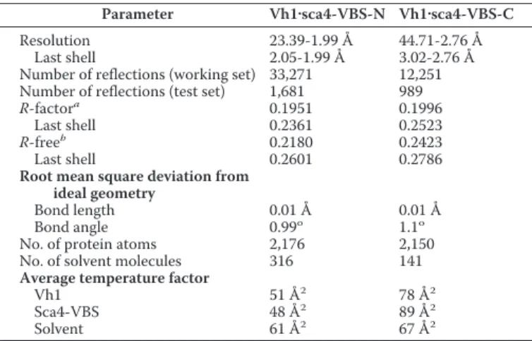
Crystallographic refinement of Vh1䡠sca4-VBS-N was
per-formed using autoBUSTER with the TLS (translation, libration, and screw-motion) and water update options (38). After six cycles of refinement, the free and crystallographic R-factors dropped to 0.25 and 0.23, respectively. Further rounds of refinement were performed after manual adjustment and visual inspection of the model using the program COOT (39). The final crystallographic and free R-factors were 0.2 and 0.22, respectively. A Ramachandran plot analysis shows that 98.9% of all residues fall in the favored regions and that the remaining 1% fall in allowed regions. MolProbity (40) analysis resulted in a score of 1.45 (97th percentile) with a clash-score of 8.38 (89th percentile) (40). Crystallographic refinement statistics are
sum-marized in Table 2. The final Vh1䡠sca4-VBS-N structure
com-prises Vh1 residues 2 to 255 and sca4 residues 411 to 434 and residues GSHM resulting from a cloning artifact at the N ter-minus of sca4 as well as six glycerol, one tetraethylene glycol, and one 2-(N-mopholino)-ethanesulfonic acid molecules and 316 ordered water molecules.
Phases for Vh1䡠sca4-VBS-C were determined by molecular
replacement using MOLREP (41) with the unliganded Vh1 structure (PDB entry 1rkc) as a search model. Molecular replacement using 44 to 3.2 Å data led to one correct solution with the initial R-factor, correlation coefficient, and translation contrast of 0.39, 0.79, and 10.1, respectively. The second unre-lated peak produced the aforementioned values of 0.60, 0.46, and 3.1, respectively. This resulted in an unambiguous solution for the position of C-terminal four-helix bundle subdomain of Vh1. However, because of conformational change, the initial electron density map using phases of the starting model showed large discrepancies for the N-terminal four-helix bundle sub-domain, which contains the binding site for VBSs. Even at this stage, strong continuous electron density for the omitted sca4-VBS-C was observed in the omit map. The bulky electron den-sity of Tyr-815 provided clear direction of sca4-VBS-C for ini-tial model building.
Multiple rounds of iterative manual fitting of sca4-VBS-C
were carried out according to the 2Fobs⫺ Fcalcand Fobs⫺ Fcalc
electron density maps by using COOT (39). Crystallographic refinement was performed using autoBUSTER (38) and resulted in crystallographic and free R-factors of 0.2 and 0.24, respectively (Table 2). The last five residues of Vh1 and the last residue of sca4-VBS-C were not included in the model due to disorder. In the Ramachandran plot, 96.3 and 3.3% of all residues fall in favored and allowed regions, respectively. Pro-tein geometry analysis by Molprobity (40) resulted in a score of 2.16 (98th percentile) with a clash score of 8.98 (99th percentile) and with 3.7% violation in rotamers and bonds and 0% violation
in angles. The final Vh1䡠sca4-VBS-C structure comprises Vh1
residues 1 to 30, 34 to 257, and sca4 residues 812 to 834 as well as 141 ordered water molecules.
Native-PAGE Shift Mobility Assays—To examine the inter-action between sca4 proteins and Vh1, native-PAGE was per-formed using a Phastgel system (GE Healthcare) according
to the manufacturer’s instructions. 10MVh1 was incubated
with various sca4 proteins in⬃1:2 molar ratios at room
tem-perature for 10 min before being run on an 8% to 25% gradi-ent native gel. Vt displacemgradi-ent assays were performed as described (16).
F-actin Co-sedimentation Assay—Full-length human
vincu-lin was prepared as described (19) and filtered with a 0.02-m
syringe filter and diluted with phosphate-buffered saline (PBS;
137 mMNaCl, 2.7 mMKCl, 4.3 mMNa2HPO4, and 1.47 mM
KH2PO4(pH 7.4)) buffer to a final concentration of 15M. Each
of the VBSs of sca4 was dissolved in PBS and then added to vinculin at 1:2 (vinculin:sca4) molar ratio and incubated for 20 min at room temperature. F-actin co-sedimentation was per-formed as described (42).
Transient Transfection and Confocal Imaging—The sca4 expression vectors and pEGFP alone as a control were trans-fected into NIH 3T3 cells grown overnight on glass coverslips with Lipofectamine 2000 (Invitrogen) following the manufac-turer’s protocol. After 16 h, the transfected NIH 3T3 cells were removed from the media and washed with PBS buffer. The cells were then fixed with PBS supplemented with 4% formaldehyde. The fixed cells were permeabilized and blocked in 3% BSA, 0.2%
Triton X-100, 1 mMNaN3in PBS. Immunostaining of
at INRA Institut National de la Recherche Agronomique on June 13, 2018
http://www.jbc.org/
nous vinculin was performed with an anti-vinculin mouse monoclonal antibody (V9131, 1:1000, Sigma) and an anti-mouse IgG conjugated with Alexa Fluor 568 (A10037, 1:1000, Molecular Probes) as a secondary antibody. The endogenous F-actin was stained with phalloidin-Alexa Fluor 633 (Invitro-gen). Images were acquired on an Olympus FluoView 1000 Confocal Microscope, and the acquired images were processed using FluoView software (Olympus).
RESULTS
Identification of VBSs in the Cell Surface Antigen (Sca4) of Rickettsia—We identified a 19-residue consensus motif for VBS binding to the vinculin Vh1 domain by aligning six VBSs of
talin, the two VBSs of IpaA, and the VBS of␣-actinin,
(ILM)-(Y/L/V/T/A)XXA(K/R)X(V/L/I)XX(A/S/K/V)(L/T/V/E/I)(S/ A/N/K)X(V/M/L/I)(L/V/I)XX(I/A/L/M), where X denotes any residue. Searches with this motif identified a potential VBS near the C terminus of the cell surface antigen sca4 (sca4-VBS-C) of
R. rickettsii, where 8 of 19 residues of this putative VBS are identi-cal to those of IpaA-VBS2 (32% sequence identity). Notably, this putative VBS was highly conserved in Rickettsia, with eight of 19 residues identical across six Rickettsia species (Fig. 1A).
The VBSs of the vinculin binding partners bind to the N-ter-minal seven-helix bundle domain of vinculin (Vh1) (16, 23, 24).
Based on preliminary binding studies,4we suspected that the
C-terminal sca4 domain (residues 411–1008) had two binding sites for Vh1. Based on secondary structure predictions and domain predictions, we therefore constructed several sca4 deletions and found that residues 21–1008, 411–585, and 775– 1008 all bound to Vh1, whereas residues 21–360 did not (Fig. 1B). Several other constructs did not result in soluble protein
and were not pursued further. Secondary structure predictions4
suggested that the 411–585 sca4 domain that binds Vh1 is
com-posed of five␣-helices, and alignment with the VBSs of talin
showed that sca4-VBS-N shares 32% sequence identity with talin-VBS1 (Fig. 1A) and 16% and 18% identity to talin-VBS2 and talin-VBS3, respectively, with 13 of 23 residues identical across six Rickettsia species.
Both of the VBSs of sca4 bound to Vh1, as did nearly full-length sca4 protein (residues 21–1008, Fig. 1C). Furthermore, recombinant sca4 protein having both VBSs (residues 411–
1008) formed a 2:1 complex with Vh1 in solution (supplemental
Figs. S1 and S2). Thus, sca4 harbors two vinculin binding sites.
Sca4 Activates Vinculin—Activation of vinculin requires sev-ering of the head-tail interaction, which exposes the cryptic actin binding sites within the Vt domain (30, 43– 47). The sequence similarity between sca4-VBS-N and sca4-VBS-C and between talin-VBS1 and IpaA-VBS2, respectively, suggested that the VBSs of sca4 have similar functions. To test if the domains harboring the sca4-VBSs and full-length sca4 were sufficient to activate vinculin, we titrated the sca4-VBSs into
preformed Vh1䡠Vt complexes. Indeed, native gel analyses
showed that the nearly full-length sca4 (residues 21–1008) as well as the domains harboring the VBSs (residues 411–585 and 774 –1008) readily displaced the Vt tail domain from
pre-exist-ing Vh1䡠Vt complexes by forming a new Vh1䡠sca4-VBS
com-plex (Fig. 2A).
A hallmark of vinculin activation is the exposure of cryptic F-actin binding sites (48, 49) that allow activated vinculin to bind to the cytoskeleton. We, therefore, tested whether the VBSs of sca4 could activate the latent F-actin binding functions of vinculin by co-sedimentation assays. Although the
sca4-VBSs did not pellet full-length vinculin, the vinculin䡠sca4-VBS
complexes co-sedimented with F-actin (Fig. 2B). Thus, both
4J. H. Lee, personal communication.
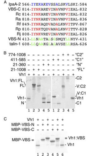
FIGURE 1. The cell surface antigen sca4 of R. rickettsii harbors two VBSs.
A, sequence alignment of all 13 known VBSs (not shown) showed that
sca4-VBS-C shares eight identical residues with IpaA-VBS2, shown in blue in the IpaA-VBS2 sequence. Sca4-VBS-C is highly conserved among Rickettsia, with eight identical residues (shown in red) among six species, including the spot-ted fever and the epidemic typhus group. Sca4-VBS-N is most similar to talin-VBS1, sharing eight residues, shown in green. IpaA-2, IpaA-VBS2; VBS-C, sca4-VBS-C; VBS-N, sca4-VBS-N; Rc, R. conorii; Ra, Rickettsia africae; Rf, Rickettsia felis;
Rp, R. prowazekii; Rj, R. japonica. B, the sca4-VBS domains bind to the vinculin
head domain Vh1 in solution. Native gel shift mobility assays (8 –25% gradient native gel) showing that nearly full-length sca4 protein, residues 21–1008 (FL,
lanes 2 and 3, left gel), sca4 residues 411–585 (C1, lanes 1 and 2, right gel), and
sca4 residues 774 –1008 (C2, lanes 3 and 4, right gel), bind to Vh1 as seen by the retarded (FL, C1, and C2) new complex bands appearing compared with unbound Vh1 (lanes 1 and 5, left and right gels, respectively). However, the N-terminal domain of sca4, residues 21–360 (N, lanes 4 and 5, left gel), does not bind to Vh1. C2 migrates poorly into the native gel probably due to its pI of 8.6. C, the VBSs of sca4 bind to Vh1 in solution. Native gel shift mobility assays show that binding of MBP fusions of VBS-N (lane 3) or sca4-VBS-C (lane 5) retards migration of Vh1 (lanes 1 and 6) or the MBP-sca4-VBSs (lanes 2 and 4). MBP-sca4-VBS-C (MBP-VBS-C) runs slightly farther (lane 2) than sca4-VBS-N (VBS-N, lane 2) and is indicated by
MBP-VBS⫽ due to their similar position. Vh1 in complex with MBP-sca4-VBS-C
runs slightly farther (lane 5) versus MBP-sca4-VBS-N (lane 3), and both com-plexes are indicated by⫽Vh1:VBS due to their similar position.
Vinculin
䡠sca4 Interactions
at INRA Institut National de la Recherche Agronomique on June 13, 2018
http://www.jbc.org/
VBSs of sca4 are sufficient to activate vinculin, establishing their functions as bona fide vinculin binding sites.
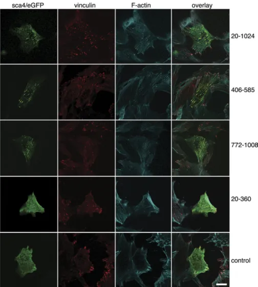
Sca4 Co-localizes with Vinculin at Sites of Focal Adhesions— To assess the sca4-vinculin interaction in cells, we transfected NIH 3T3 fibroblasts with eGFP alone or with in-frame eGFP fusions of sca4 residues 21–1008 harboring both VBSs, sca4 residues 406 –585 harboring sca4-VBS-N, sca4 residues 774 – 1008 containing sca4-VBS-C, or sca4 residues 21–360 that do not bind to vinculin in vitro (Fig. 3). Endogenous vinculin and F-actin were also visualized by fluorescence labeling. Expres-sion of the various sca4 constructs did not induce morpholog-ical changes of the transfected cells. As shown previously by others (50, 51), vinculin was observed primarily at sites of focal adhesions. Confocal microscopy established that eGFP-sca4, residues 406 –585, 776 –1008, or 21–1008 were present in fibrous-like structures and at punctate that overlapped with focal adhesions. Notably, both nearly full-length sca4s and the domains harboring the sca4-VBSs co-localized with endoge-nous vinculin. In addition, the punctate patterns were predom-inantly at the inner basal surface of the cells. Only small amounts of sca4 co-localized with vinculin at the leading edge
of the cell. The fibrous expression patterns of sca4 overlap with F-actin staining. By contrast, eGFP alone and e-GFP-sca4 (res-idues 21–360) did not co-localize with vinculin. Therefore, sca4 and vinculin can interact in cells.
Vh1 Adopts a Novel Conformation When Bound to
Sca4-VBS-C—To define the molecular details of the sca4-vinculin
interac-tion, we determined the crystal structures of the vinculin Vh1 domain in complex with either sca4-VBS to 2 and 2.8 Å,
respec-tively (Tables 1 and 2, Fig. 4, A–C;supplemental Fig. S3–S6).
Although we obtained several crystallization hits for the
Vh1䡠sca4-VBS-N complex, these crystals diffracted only to
about 8 Å Bragg spacings. However, by decreasing the molar ratio of Vh1 to sca4, we obtained cubic crystals that allowed complete data collection to 2 Å Bragg spacing on beamline 11-1 at the Stanford Linear Acceleration Center at Stanford Syn-chrotron Radiation Laboratory.
The Vh1䡠sca4-VBS-N crystal structures revealed that this
VBS activates vinculin in a conventional fashion, akin to that of
the VBSs of talin (24, 25), IpaA (16, 18) (Fig. 4A-C;
supplemen-tal Fig. S3–S6), or␣-actinin even though the VBS of ␣-actinin has opposite directionality (23). Vh1 is composed of two four-helix bundle subdomains that are connected by a large shared
central␣-helix (␣4). Binding of sca4-VBS to the N-terminal
Vh1 four-helix bundle subdomain (residues 1–130) creates a
five-helix bundle. As seen first in the Vh1䡠talin-VBS3 crystal
structure (24), the Vh1䡠sca4-VBS interfaces are predominantly
hydrophobic (supplemental Fig. S7) with buried
solvent-acces-sible surface areas of 1152 and 1110 Å2for sca4-VBS-N and
sca4-VBS-C, respectively, upon complex formation. Further-more, there are three hydrogen bond formations involving Vh1 residues Ile-12, Gln-19, and His-22 and sca4-VBS-N residues
Ser-421, Gln-425, and Asn-432. In the Vh1䡠sca4-VBS-C
struc-ture only two hydrogen bond interactions are formed that are mediated by Vh1 residues Asp-127 and His-22 and sca4-VBS-C residues Tyr-815 and Glu-833, respectively.
Each of the two subdomains can be superimposed
individu-ally onto other known Vh1䡠VBS structures with root mean
square deviations less than 1 Å, indicating that the subdomain structure is conserved. Indeed, a structural similarity search using the DALI server (52) for the N-terminal four-helix bundle Vh1 subdomain in complex with either sca4-VBS found the
Vh1䡠talin-VBS1 structure (PDB entry 1syq; Z-scores of 1.2 and
2.2, respectively) (25) as the best match followed by the
Vh1䡠IpaA-VBS2 structure (PDB entry 2ibf; Z-scores of 1.5 and
1.8, respectively) (42). Thus, like Shigella flexneri, R. rickettsii applies molecular mimicry of talin to bind to and activate vinculin.
Notably, the Vh1䡠sca4-VBS-C structure shows a novel
sub-domain movement of more than 35° relative to the sca4-VBS-N bound structure (as judged using the program Dyndom) where the hinge involves residues 8 –13 and 126 –129 and where the two 4-helix bundle Vh1 subdomains move into a unique and
indeed more “closed” Vh1 conformation (Fig. 4A;supplemental
Figs. S3, S8, and S9). A structure-based sequence alignment (Fig. 4D) reveals that the N terminus of sca4-VBS-C is acidic (Asp-811 and Asp-812) compared with all other VBSs, which might neutralize the helix dipole and stabilize the sca4-VBS-C ␣-helix. More importantly, these two aspartates could be FIGURE 2. Sca4 of R. rickettsii activates vinculin. A, native gel shift mobility
assays are shown (8 –25% gradient native gel) of Vh1 alone (lane 1), the pre-formed Vh1䡠Vt complex alone (lane 2), the Vh1䡠Vt complex with the addition of nearly full-length sca4 (residues 21–1008, lane 3), or of the sca4-VBS domains (lanes 5 and 7 for sca4-VBS-N and sca4-VBS-C domains, respectively). All three sca4 proteins displace Vt from the Vh1䡠Vtcomplexreadilyatequimolarratiosand form a Vh1䡠sca4 complex (lanes 3–8) that is retarded compared with Vh1 alone (lane 1). Unbound Vt does not show on a native gel because of its basic pI (24).
B, actin co-sedimentation analyses (8 –25% gradient native gel) of full-length
vin-culin in the presence (lanes 5–10) and absence (lanes 1– 4) of the minimal sca4-VBSs are shown. Vinculin alone does not aggregate (lanes 1– 4) and does not sediment with F-actin (lanes 1–2). The addition of a 2-fold molar excess of either sca4-VBSs to full-length vinculin does not lead to aggregates (lanes 5 and 6) but triggers co-sedimentation of vinculin with F-actin (lanes 7–10). F-act, F-actin; M, molecular weight markers; S, supernatant; P, pellet; VBS-C, sca4-VBS-C, residues 814 – 832; VBS-N, sca4-VBS-N, residues 413– 431; vinc, vinculin.
at INRA Institut National de la Recherche Agronomique on June 13, 2018
http://www.jbc.org/
responsible for the dramatic movement of Vh1 Lys-173 that may trigger subdomain movement in Vh1.
A second, somewhat subtler difference in the Vh1
䡠sca4-VBS-C crystal structure compared with all other known
Vh1䡠VBS structures is a movement of␣-helix ␣1, which is
prob-ably caused by Pro-827 (Fig. 4C;supplemental Fig. S4). Indeed,
mutating the talin-VBS1, which has a glutamate in the equiva-lent position (Glu-621; Fig. 4D) to a proline, abolishes its bind-ing to Vh1 (28). The sca4-VBS-C superimposes well with the other VBSs up to residue Pro-827, which causes a kink in the
sca4-VBS-C that probably pushes the Vh1␣-helix ␣1
some-what further away from Vh1␣-helix ␣2 and almost half a turn
closer to the C-terminal four-helix Vh1 subdomain, thus affect-ing its orientation relative to the N-terminal four-helix bundle
subdomain (Fig. 4C,supplemental Figs. S4, S8, and S9). The
distinct␣-helix ␣1 conformation results in different
interac-tions of residues residing on its N terminus with the C-terminal four-helix subdomain, in particular the C-terminal region of
Vh1␣-helix ␣5. Superposition with full-length vinculin shows
that␣-helix ␣5 moves deeper into the core of vinculin (
supple-mental Figs. S8 and S9). For example, the side chain of His-5 that points toward the side chain of Leu-182 and is rotated TABLE 1
X-ray data collection and reduction statistics
Parameter Vh1䡠sca4-VBS-N Vh1䡠sca4-VBS-C
Space group P212121 I23 Unit cell dimensions
a 54.4 Å 141.4 Å
b 76.4 Å 141.4 Å
c 114.6 Å 141.4 Å
␣ ⫽  ⫽ ␥ 90o
90o
Asymmetric unit 1 heterodimer 1 heterodimer Matthews coefficient 3.82 3.85 Solvent content 0.678 0.68 Resolution 114 Å-1.99 Å 100 Å-2.76 Å
Last shell 2.1 Å-1.99 Å 2.91 Å-2.76 Å Total measurements 196,687 182,498 Number of unique reflections 33,356 12,290
Last shell 4,774 1,759 Wilson B 37 Å2 71 Å2 Wavelength 1 Å 0.97867 Å R-mergea 0.034 0.059 Last shell 0.499 0.49 I/(I) 26.3 36.4 Last shell 3.3 6.1 Completeness 0.999 1.0 Last shell 1.0 0.998 Redundancy 5.9 14.8 Last shell 5.3 15 a
R-merge⫽ ⌺hkl⌺i兩Ii(hkl)⫺ Ii(hkl)兩/⌺hkl⌺iIi(hkl).
FIGURE 3. Sca4 and vinculin co-localize at focal adhesions in NIH 3T3 cells. Transient transfection of sca4 residues 20 –1024 harboring both VBSs (top row), 406 –585 harboring sca4-VBS-N (second row), 772–1008 harboring sca4-VBS-C (third row), the N-terminal non-binding 20 –360 domain (fourth row), and pEGFP blank vector (bottom row) were analyzed on a fluorescence microscope. Endogenous vinculin (second column) and F-actin (third column) were stained with a fluorescence-conjugated vinculin antibody and phalloidin-Alexa Fluor 633, respectively. All images are at the same magnification. The scale bar represents 20m.
Vinculin
䡠sca4 Interactions
at INRA Institut National de la Recherche Agronomique on June 13, 2018
http://www.jbc.org/
almost 180° in Vh1䡠sca4-VBS-C to point toward the N-terminal four-helix bundle. This space is filled by Phe-4 (interacting with
Ala-17 on␣1 and Leu-116 on ␣4) in the other structures. In
Vh1䡠sca4-VBS-C, Phe-4, rather, resides close to His-184,
resid-ing on Vh1␣-helix ␣6.
DISCUSSION
Obligate intracellular pathogens enter and spread within the host cell by co-opting the F-actin cytoskeleton, yet how these events are orchestrated is generally poorly understood. Our findings with sca4 suggest that this protein functions as an essential invasin for rickettsial species and that, in a remarkable case of convergent evolution, sca4 functions as an invasin akin to that of the IpaA protein of Shigella, where both invasins function as talin mimics that bind to and activate vinculin to provide connections of the respective pathogens with the F-ac-tin network. Furthermore, both IpaA and sca4 invasins harbor at least two VBSs, suggesting they both employ a mechanism that amplifies vinculin activation and actin organization at sites of pathogen entry. Finally, both of the VBSs of sca4 are con-served across all rickettsial species, suggesting their essential roles in pathogenesis.
The mechanism by which sca4 activates vinculin occurs in a fashion described for IpaA (16), where the VBSs of both invasins bind to and activate vinculin. This scenario bypasses pre-activation steps that are required for the binding of talin or ␣-actinin to vinculin, where force-induced unraveling of helix bundles is needed to expose their VBSs to allow them to bind to and activate vinculin (24, 30, 42). This strategy employed by these pathogens ensures that they effectively compete with
talin and␣-actinin for vinculin and that they effectively
seques-FIGURE 4. Sca4 induces a novel conformation in vinculin. A, superposition of all known talin-VBSs and IpaA-VBSs in their Vh1-bound states (white) onto our 2 Å Vh1䡠sca4-VBS-N (green) and 2.8 Å Vh1䡠sca4-VBS-C (magenta) crystal structures. Vh1 is shown as a C␣ trace and the VBSs as a schematic. Vh1 ␣-helices ␣4-␣7 are labeled. All VBSs (talin-VBS1, residues 608 – 627; talin-VBS11, residues 822– 841; talin-VBS2, residues 855– 874; talin-VBS33, residues 1525–1544; talin-VBS36, residues 1633–1652; talin-VBS58, residues 2345–2364; IpaA-VBS1; IpaA-VBS2) were aligned to talin-VBS3 (residues 1949 –1968) with root mean square deviations ranging from 0.5 Å to 1.1 Å. Sca4-VBS-C induces a significant movement of the C-terminal Vh1 four-helix bundle subdomain, which is unique versus all other Vh1䡠VBS structures. B, a zoomed view of the superposition of sca4-VBS-N (green) representative of all Vh1䡠VBS structures onto the sca4-VBS-C (magenta) in their Vh1-bound states. Unlike other VBSs, sca4-VBS-C has two acidic residues on its N terminus (Asp-812 is shown in sticks and is labeled), which might stabilize the sca4-VBS-C␣-helix by neutralizing its dipole, and the large movement of the C-terminal Vh1 subdomain is indicated by the movement of Lys-173 and the arrow. C, a zoomed-in view showing the kink at sca4 Pro-827 (shown in black sticks, labeled) in sca4-VBS-C (magenta) compared with the sca4-VBS-N (green); the latter is representative of all other Vh1䡠VBS structures. Residues N-terminal to Pro-827 moved, whereas the C-terminal end superimpose well. This movement causes Vh1␣-helix ␣1 to shift down (in this view) and toward the C-terminal Vh1 subdomain as indicated by the arrow. D, structure-based sequence alignment of the talin-VBSs (first six black PDB entries 1rkc, 1syq, 1zvz, 1u6h, 1zw3, 1zw2), IpaA-VBSs (next two blue PDB entries 2gww, 2hsq), and the sca4-VBSs (last two black sequences). Residue numbers are indicated. The two acidic N-terminal residues found only in sca4-VBS-C and the Pro-827 found only in sca4-VBS-C are highlighted in cyan. Residues in lowercase are not present in the structure.
TABLE 2
Crystallographic refinement statistics
Parameter Vh1䡠sca4-VBS-N Vh1䡠sca4-VBS-C
Resolution 23.39-1.99 Å 44.71-2.76 Å Last shell 2.05-1.99 Å 3.02-2.76 Å Number of reflections (working set) 33,271 12,251 Number of reflections (test set) 1,681 989
R-factora 0.1951 0.1996 Last shell 0.2361 0.2523 R-freeb 0.2180 0.2423 Last shell 0.2601 0.2786
Root mean square deviation from ideal geometry
Bond length 0.01 Å 0.01 Å Bond angle 0.99o
1.1o No. of protein atoms 2,176 2,150 No. of solvent molecules 316 141
Average temperature factor
Vh1 51 Å2 78 Å2 Sca4-VBS 48 Å2 89 Å2 Solvent 61 Å2 67 Å2 a
R-factor⫽ ⌺hkl储Fobs(hkl)兩 ⫺ 兩Fcalc(hkl)储/⌺hkl兩Fobs(hkl)兩, where 具兩Fcalc兩典 is the
ex-pectation of兩Fcalc兩 under the error model used in maximum likelihood
refinement.
b
The free R-factor is a cross-validation residual calculated using 5% (sca4-VBS-N) and 8.5% (sca4-VBS-C) reflections, which were randomly chosen and excluded from the refinement.
at INRA Institut National de la Recherche Agronomique on June 13, 2018
http://www.jbc.org/
ter this essential F-actin binding protein for the uses of the pathogen in entry, motility, and spread.
Our Vh1䡠sca4-VBS crystal structures are the first reported
structures from R. rickettsii, with only a handful structures determined from another Rickettsia species. Although both sca4-VBSs and IpaA-VBSs are talin-VBS mimics, it is notable that the sca4-VBS-C induces novel structural alterations in vin-culin, in particular to the C-terminal Vh1 four-helix bundle subdomain that does not bind to sca4-VBS, a scenario unique
among all vinculin䡠VBS structures. Two features of sca4-VBS-C
appear to trigger this change. First, this is the only known VBS that has two negatively charged side chains on its N terminus,
which we posit might stabilize the sca4-VBS-C␣-helix. Second,
sca4-VBS-C harbors a proline residue that likely contributes to
the shift of␣-helix ␣1 and to the observed contortions in the
C-terminal four-helix bundle subdomain.
The fact that Rickettsia uses sca4 to bind to and activate
vin-culin suggests that this vinvin-culin䡠sca4 interaction is involved in
invasion and might be targeted as a therapeutic strategy for combating this deadly pathogen. In this regard, the unique
interactions of sca4 and vinculin revealed in the vinculin䡠sca4
crystal structures may provide inroads for developing small molecule inhibitors that selectively disable the functions of sca4.
Acknowledgments—We are indebted to our colleagues at Scripps Florida, John L. Cleveland for discussions and critical review of the manuscript, Erumbi S. Rangarajan for fruitful discussions, and Philippe R. Bois and Zhen Wu for sequencing. We are grateful to the staff at Stanford Synchrotron Radiation Laboratory for synchrotron support.
REFERENCES
1. Walker, D. H., and Ismail, N. (2008) Nat. Rev. Microbiol. 6, 375–386 2. Hackstadt, T. (1996) Infect Agents Dis. 5, 127–143
3. Dantas-Torres, F. (2007) Lancet Infect. Dis. 7, 724 –732
4. Walker, D. H., Occhino, C., Tringali, G. R., Di Rosa, S., and Mansueto, S. (1988) Hum. Pathol. 19, 1449 –1454
5. Hand, W. L., Miller, J. B., Reinarz, J. A., and Sanford, J. P. (1970) Arch.
Intern Med. 125,879 – 882
6. Martinez, J. J., Seveau, S., Veiga, E., Matsuyama, S., and Cossart, P. (2005)
Cell 123,1013–1023
7. Martinez, J. J., and Cossart, P. (2004) J. Cell Science 117, 5097–5106 8. Jeng, R. L., Goley, E. D., D’Alessio, J. A., Chaga, O. Y., Svitkina, T. M.,
Borisy, G. G., Heinzen, R. A., and Welch, M. D. (2004) Cell. Microbiol. 6, 761–769
9. Tran Van Nhieu, G., Bourdet-Sicard, R., Dume´nil, G., Blocker, A., and Sansonetti, P. J. (2000) Cell. Microbiol. 2, 187–193
10. Cossart, P. (2000) Cell. Microbiol. 2, 195–205
11. Gouin, E., Egile, C., Dehoux, P., Villiers, V., Adams, J., Gertler, F., Li, R., and Cossart, P. (2004) Nature 427, 457– 461
12. Blanc, G., Ngwamidiba, M., Ogata, H., Fournier, P. E., Claverie, J. M., and Raoult, D. (2005) Mol. Biol. Evol. 22, 2073–2083
13. Chan, Y. G., Cardwell, M. M., Hermanas, T. M., Uchiyama, T., and Mar-tinez, J. J. (2009) Cell. Microbiol. 11, 629 – 644
14. Riley, S. P., Goh, K. C., Hermanas, T. M., Cardwell, M. M., Chan, Y. G., and Martinez, J. J. (2010) Infect. Immun. 78, 1895–1904
15. Haglund, C. M., Choe, J. E., Skau, C. T., Kovar, D. R., and Welch, M. D. (2010) Nat. Cell Biol. 12, 1057–1063
16. Izard, T., Tran Van Nhieu, G., and Bois, P. R. (2006) J. Cell Biol. 175, 465– 475
17. Ramarao, N., Le Clainche, C., Izard, T., Bourdet-Sicard, R., Ageron, E.,
Sansonetti, P. J., Carlier, M. F., and Tran Van Nhieu, G. (2007) FEBS Lett. 581,853– 857
18. Nhieu, G. T., and Izard, T. (2007) EMBO J. 26, 4588 – 4596
19. Borgon, R. A., Vonrhein, C., Bricogne, G., Bois, P. R., and Izard, T. (2004)
Structure 12,1189 –1197
20. Rangarajan, E. S., Lee, J. H., Yogesha, S. D., and Izard, T. (2010) PLoS ONE 5,e10679
21. Bakolitsa, C., Cohen, D. M., Bankston, L. A., Bobkov, A. A., Cadwell, G. W., Jennings, L., Critchley, D. R., Craig, S. W., and Liddington, R. C. (2004)
Nature 430,583–586
22. Bakolitsa, C., de Pereda, J. M., Bagshaw, C. R., Critchley, D. R., and Lid-dington, R. C. (1999) Cell 99, 603– 613
23. Bois, P. R., Borgon, R. A., Vonrhein, C., and Izard, T. (2005) Mol. Cell. Biol. 25,6112– 6122
24. Izard, T., Evans, G., Borgon, R. A., Rush, C. L., Bricogne, G., and Bois, P. R. (2004) Nature 427, 171–175
25. Izard, T., and Vonrhein, C. (2004) J. Biol. Chem. 279, 27667–27678 26. Fillingham, I., Gingras, A. R., Papagrigoriou, E., Patel, B., Emsley, J.,
Critch-ley, D. R., Roberts, G. C., and Barsukov, I. L. (2005) Structure 13, 65–74 27. Gingras, A. R., Vogel, K. P., Steinhoff, H. J., Ziegler, W. H., Patel, B., Emsley,
J., Critchley, D. R., Roberts, G. C., and Barsukov, I. L. (2006) Biochemistry 45,1805–1817
28. Gingras, A. R., Ziegler, W. H., Frank, R., Barsukov, I. L., Roberts, G. C., Critchley, D. R., and Emsley, J. (2005) J. Biol. Chem. 280, 37217–37224 29. Papagrigoriou, E., Gingras, A. R., Barsukov, I. L., Bate, N., Fillingham, I. J.,
Patel, B., Frank, R., Ziegler, W. H., Roberts, G. C., Critchley, D. R., and Emsley, J. (2004) EMBO J. 23, 2942–2951
30. del Rio, A., Perez-Jimenez, R., Liu, R., Roca-Cusachs, P., Fernandez, J. M., and Sheetz, M. P. (2009) Science 323, 638 – 641
31. Gingras, A. R., Ziegler, W. H., Bobkov, A. A., Joyce, M. G., Fasci, D., Himmel, M., Rothemund, S., Ritter, A., Grossmann, J. G., Patel, B., Bate, N., Goult, B. T., Emsley, J., Barsukov, I. L., Roberts, G. C., Liddington, R. C., Ginsberg, M. H., and Critchley, D. R. (2009) J. Biol. Chem. 284, 8866 – 8876 32. Grashoff, C., Hoffman, B. D., Brenner, M. D., Zhou, R., Parsons, M., Yang, M. T., McLean, M. A., Sligar, S. G., Chen, C. S., Ha, T., and Schwartz, M. A. (2010) Nature 466, 263–266
33. Tran Van Nhieu, G., Ben-Ze’ev, A., and Sansonetti, P. J. (1997) EMBO J. 16,2717–2729
34. Park, H., Valencia-Gallardo, C., Sharff, A., Tran Van Nhieu, G., and Izard, T. (2011) J. Biol. Chem. 286, 23214 –23221
35. Vonrhein, C., and Bricogne, G. (2008) Acta Crystallogr. A 64, C78 36. Kabsch, W. (1993) J. Appl. Crystallogr. 26, 795– 800
37. Leslie, A. G. (2006) Acta Crystallogr. 62, 48 –57
38. Smart, O. S., Brandl, C., Flensburg, P., Keller, W., Paciorek, C., Vonrhein, C., Womack, T., and Bricogne, G. (2008) Abstract of the Annual Meeting of
the American Crystallogr. Association, Knoxville, TN.Abstr. TP139 39. Emsley, P., and Cowtan, K. (2004) Acta Crystallogr. D60, 2126 –2132 40. Chen, V. B., Arendall, W. B., 3rd, Headd, J. J., Keedy, D. A., Immormino,
R. M., Kapral, G. J., Murray, L. W., Richardson, J. S., and Richardson, D. C. (2010) Acta Crystallogr. D66, 12–21
41. Vagin, A., and Teplyakov, A. (1997) J. Appl Crystallogr. 30, 1022–1025 42. Bois, P. R., O’Hara, B. P., Nietlispach, D., Kirkpatrick, J., and Izard, T.
(2006) J. Biol. Chem. 281, 7228 –7236
43. Cohen, D. M., Kutscher, B., Chen, H., Murphy, D. B., and Craig, S. W. (2006) J. Biol. Chem. 281, 16006 –16015
44. Gilmore, A. P., and Burridge, K. (1996) Nature 381, 531–535 45. Johnson, R. P., and Craig, S. W. (2000) J. Biol. Chem. 275, 95–105 46. Johnson, R. P., Niggli, V., Durrer, P., and Craig, S. W. (1998) Biochemistry
37,10211–10222
47. Winkler, J., Lu¨nsdorf, H., and Jockusch, B. M. (1996) J. Struct. Biol. 116, 270 –277
48. Critchley, D. R. (2004) Biochem. Soc. Trans. 32, 831– 836
49. Critchley, D. R., Holt, M. R., Barry, S. T., Priddle, H., Hemmings, L., and Norman, J. (1999) Biochem. Soc. Symp. 65, 79 –99
50. Geiger, B. (1979) Cell 18, 193–205
51. Geiger, B., Tokuyasu, K. T., Dutton, A. H., and Singer, S. J. (1980) Proc.
Natl. Acad. Sci. U. S. A. 77,4127– 4131
52. Holm, L., and Rosenstro¨m, P. (2010) Nucleic Acids Res. 38, W545–W549
Vinculin
䡠sca4 Interactions
at INRA Institut National de la Recherche Agronomique on June 13, 2018
http://www.jbc.org/
HaJeung Park, Jun Hyuck Lee, Edith Gouin, Pascale Cossart and Tina Izard
doi: 10.1074/jbc.M111.263855 originally published online August 13, 2011 2011, 286:35096-35103.
J. Biol. Chem.
10.1074/jbc.M111.263855
Access the most updated version of this article at doi: Alerts:
When a correction for this article is posted
•
When this article is cited
•
to choose from all of JBC's e-mail alerts
Click here
Supplemental material:
http://www.jbc.org/content/suppl/2011/08/13/M111.263855.DC1 http://www.jbc.org/content/286/40/35096.full.html#ref-list-1This article cites 51 references, 14 of which can be accessed free at
at INRA Institut National de la Recherche Agronomique on June 13, 2018
http://www.jbc.org/