ContentslistsavailableatScienceDirect
Journal
of
Neuroscience
Methods
j o ur na l ho me p ag e:w w w . e l s e v i e r . c o m / l o c a t e / j n e u m e t h
Basic
Neuroscience
Refined
methodology
for
implantation
of
a
head
fixation
device
and
chronic
recording
chambers
in
non-human
primates
夽
F.
Lanz
a,
X.
Lanz
b,d,
A.
Scherly
d,
V.
Moret
a,
A.
Gaillard
a,
P.
Gruner
c,
H.-M.
Hoogewoud
e,
A.
Belhaj-Saif
a,
G.
Loquet
a,
E.M.
Rouiller
a,∗aDepartmentofMedicine,UnitofPhysiology,FribourgCognitionCenter,UniversityofFribourg,Switzerland
bDepartmentofMecanics,Ecoled’Ingénieurs,Fribourg,Switzerland
cMedicoatAG,Mägenwil,Switzerland
dS+DScherly,LaRoche,Switzerland
eHôpitalfribourgeois(HFR),HôpitalCantonal,DepartmentofRadiology,Fribourg,Switzerland
h
i
g
h
l
i
g
h
t
s
•Aheadfixationdeviceandachronicrecordingchambercanbeimplantedwithoutusingdentalresinororthopediccement. •Completeosseous-integrationofimplantcanbeobtainedthankstoahydroxyapatitecoating.
•Aperfectmatchingoftheimplantswithindividualskullsurfacecanbeensuredwithaplasticreplicateoftheskull(3Dprinting). •Implantingsurgeriescanbegreatlyfacilitatedbytheuseofpersonalizedimplantsand3Dprinting.
•Outstandinglongevityoftheimplantsused:4yearsforheadfixationdeviceand1.5yearsforchronicrecordingchamber.
a
r
t
i
c
l
e
i
n
f
o
Articlehistory: Received20June2013 Accepted24July2013 Keywords: Macaquemonkey Skullimplants Osseous-integrationChronicbrainelectrophysiology
Hydroxyapatite
a
b
s
t
r
a
c
t
Thepresentstudywasaimedatdevelopinganewstrategytodesignandanchorcustom-fittedimplants, consistingofaheadfixationdeviceandachronicrecordingchamber,ontheskullofadultmacaque monkeys.Thiswasdonewithouttheuseofdentalresinororthopediccement,asthesemodesoffixation exertadetrimentaleffectonthebone.Theimplantsweremadeoftitaniumortekapeekandanchored totheskullwithtitaniumscrews.Twoadultmacaquemonkeyswereinitiallyimplantedwiththehead fixationdeviceseveralmonthsprevioustoelectrophysiologicalinvestigation,toallowoptimal osseous-integration,includinggrowthoftheboneabovetheimplant’sfootplate.Inasecondstep,thechronic recordingchamberwasimplantedabovethebrainregionofinterest.Thepresentstudyproposestwo originalapproachesforbothimplants.First,basedonaCTscanofthemonkey,aplasticreplicateofthe skullwasobtainedintheformofa3Dprint,usedtoaccuratelyshapeandpositionthetwoimplants.This wouldensureaperfectmatchwiththeskullsurface.Second,thepartoftheimplantsincontactwiththe bonewascoatedwithhydroxyapatite,presentingchemicalsimilaritytonaturalbone,thuspromoting excellentosseous-integration.Thelongevityoftheimplantsusedherewas4yearsfortheheadfixation deviceand1.5yearsforthechronicchamber.Therewerenoadverseeventsanddailycarewaseasy.This isclearevidencethatthepresentimplantingstrategywassuccessfulandprovokeslessdiscomforttothe animals.
© 2013 The Authors. Published by Elsevier B.V. All rights reserved.
Abbreviations: HA,hydroxyapatite;CT,computedtomography;MRI,magnetic
resonanceimaging;VPS,vacuumplasmaspraying.
夽 Thisis anopen-accessarticledistributedunderthetermsoftheCreative
CommonsAttributionLicense,whichpermitsunrestricteduse,distributionand
reproductioninanymedium,providedtheoriginalauthorandsourcearecredited.
∗ Correspondingauthor.Tel.:+41263008609.
E-mailaddress:[email protected](E.M.Rouiller).
1. Introduction
Inthefieldofneurosciences,themacaqueisamodelofchoice (scientificallyandethicallyjustified;seeWeatherallreport,2006). Thismonkeyishighlyadaptedforneuronalinvestigationsdueto itslargesimilaritytothehumanbrainfromananatomical and a functionalpoint of view.Inmodern neurosciences,there is a largerangeofapproachestoinvestigatebrainfunction,also appli-cable,tosomeextent,tonon-humanprimates:functionalbrain imaging(fMRI),electroencephalography(EEG),positronemission tomography(PET),transcranialmagneticstimulation(TMS),single
0165-0270/$–seefrontmatter © 2013 The Authors. Published by Elsevier B.V. All rights reserved.
neuronrecording,etc.Thequalityoftheresultingdatadepends onthelevelofinterferencescausedbyartifacts,whichmaybe pro-duced,forinstance,bymuscularcontractionsrelatedtoheadand/or eyesmovementsormastication.Afurtherchallengeinthistypeof researchliesinthefactthattheanimalhastobeawake,andthe headmustbekeptfixed.Indeed,anyheadmovementwouldcreate recordingartifacts.Furthermore,inthecaseof electrophysiologi-calrecordings,thereisariskthat,inanon-headfixedsystem,the recordingelectrodesmoveandcausebraininjuries.Thatiswhy itispreferablethattheanimal’sheadbeimmobilizedwhenitis performingbehavioraltasks.Tothis aim,untilrecently, numer-ouslaboratoriesusedaheadfixationdeviceanchoredtotheskull withdentalacryliccement(FuchsandLuschei,1970;Lisbergerand Westbrook,1985;GuoandLi,1997;Kermadietal.,1997,1998; LiuandRouiller,1999;ChurchlandandLisberger,2000)or ortho-pediccement(Durifetal.,2003;Peetersetal.,2009;Kaeseretal., 2010,2011).Suchanapproachallowedcreationofafirmpointof fixation,buttheinterfacebetweenthedentalresinorthe orthope-diccementandthebonewasnotoptimal.Itwasobservedthatthe cementadheredtotheboneinasuperficialwaywithoutintegration betweenthetwocomponents(boneandcement).Thisrepresented aconsiderableriskoffracture.Althoughvariablefromoneanimal toanother,thepresenceofcement(dentalororthopedic)exerted adetrimentalimpactontheboneinthemid-andlong-termrun. Inparticular,theriskofinfection,inflammation,growthof granu-lationtissueandsofteningofthebonewasincreased.Theseeffects wereofteninitiatedbythehightemperaturegeneratedwhenthe cementwasappliedtothebonesurface, andas aconsequence increasedtheriskofheadfixationdevicelossesofovertime.
Inlinewithrecentreports(Adamsetal.,2007,2011;McAndrew etal.,2012),thegoalofthepresentstudywastointroducearefined methodtoanchorabiocompatibleheadfixationdeviceandchronic recordingchambersonmacaque’sskull,withouttheuseofdental resinororthopediccement.Thisaimwasachievedherebytaking advantageofnewlydevelopedmaterialsandcoatingswhichare usedfororthopedicsurgery.Thesearegenerallyassimilatedbythe boneinsteadofbeingrejectedbyit.However,foraperfect inte-grationbetweenimplantsandbone,aperfectmatchoftheshape oftheimplants(headfixationdeviceorrecordingchamber)with theskullsurfaceofeachindividualmonkeyisrequired.A3D repli-cateoftheskullofthelivingmonkeywasobtainedbasedonCT andMRIdata.Thisreplicatewasusedtoaccuratelyguidethe posi-tioningoftheimplantsontheskullaswellastoderivetheirshape sothattheywouldperfectlymatchthecontouroftheskullatthe calculatedtargetposition.
2. Methods
2.1. Subjects
ThepresentexperimentswereconductedontwoadultMacaca fascicularis, originating from our own breeding colony. At the time of headpost fixation (see Fig. 3), one animal (Mk-LI)was 9-years-oldandweighedabout8.0kg,whereasthesecondanimal (Mk-JZ) was 7-years-old and weighed about 8.0kg. The body weightwas checkeddaily.In case of a 10% lossof weight,the experimentwouldbeinterrupteduntilweightwasregained(an interruptioncriterionthatwasnotmetinthecourseofthepresent study).Betweendailyexperimentalsessionstheanimalsshared livingquarterswithothermonkeys(groupsof2to5animals)inan enclosureof45m3(15m3until2010;seee.g.(Kaeseretal.,2011)).
Theycouldfreelymoveandhadfreeaccesstowater.The experi-mentswereconductedaccordingtotheguidelinesoftheNational InstituteofHealth(GuidefortheCareandUseoflaboratory Ani-mals,1996),oftheEuropeanCommunity(GuidelinesforAnimals
ProtectionandUseforExperimentation)andtheARRIVEguidelines (http://www.nc3rs.org.uk) (Animal Research: Reporting In Vivo Experiments),aswellastheSwissveterinaryauthorities(cantonal andfederal)whoapprovedtheexperimentalprocedures.
2.2. 3Dreplicate(print)ofthemonkey’sskull
The first stage was to obtain such a 3D replica of the liv-ing monkey’s skull. The acquisition of the skull morphology involvedusinga computedtomography scan(CTscan) (Depart-ment of radiology atHôpital Fribourgeois [HFR]).The obtained CT scan was processed with the Osirix software (64 bits) in order tofabricate a 3D reconstructionof the skull. Thismodel was transferredto theEngineering Schoolof Fribourg for final processing. The final 3D print was performedwiththe follow-ing equipment: 3D printer, 3D uPrint Plus which uses Fused DepositionModeling(FDM)Technologytobuild3Dreplicawith ABSplus thermoplastics. The principle of the 3D replica of the skull is illustrated in the supplementary video sequence #1 (http://www.unifr.ch/neuro/rouiller/research/multi/lanz/l1.html). Thepresent3Dreplicationtookapproximately25h.Itwasthen polished,includingremovalofunwantedplasticpartsbyovernight treatmentinachemicalbath.Becausethe3Dreplicawasbased onCT data,theskullsurface andthebone thicknesswasa 1:1 representation ofthemonkey’sskull.Althoughthethicknessof theskullcouldbedeterminedbytheCTimages,the3Dmodelwas usedinsteadduringsurgeryandwasadvantageous.
2.3. Headfixationdevice
Similar to other recent studies (Adams et al., 2007, 2011; McAndrewetal.,2012)theaimherewastodevelopastableand solidimplantwithoutusingdentalororthopediccement.Inthis studytheheadfixationdeviceinitiallydevelopedbyAdamsetal. (2007,2011) waschosenasa baseand wasmodifiedaccording toexperimentalneeds. Thematerialusedtoelaboratethehead fixationdevicewastitanium,whichhasbeenusedformorethan 30yearsinthemedicalindustry.Titaniumpresentstheadvantage ofbeing,alongwithgoldandplatinum,oneofthemost biocom-patiblemetals,andisresistanttobodyfluids(RubodeRezende andJohansson,1993).Titaniumdemonstrateshighcorrosion resis-tanceandthehigheststrength-to-weightratioofanyknownmetal. Oneofthemostimportantadvantagesassociatedwiththeuseof titaniumwasthatboneadhereswelltoitandyieldsgood osseous-integration(Brånemarketal.,1969;AlbrektssonandAlbrektsson, 1987;RubodeRezendeandJohansson,1993;Augatetal.,1995; Betelaketal.,2001).Theheadfixationdevicesweremanufactured (AteliersClémentS.A.CH–1731Ependes)fromapuretitanium cube(CP, Grade2)asmono-blocks,allowingexcellent osseous-integration(notehoweverthatGrade5wouldberecommended ifonewantedtoreduceartifactsforsubsequentMRI).Becausethe headfixationdeviceneedednowelding,abreakattheweldline betweenthepostandthefootplatewasprevented(Adamsetal., 2007).
Theheadfixationdeviceusedinthepresentstudyisillustrated inFig.1A.Fromamechanicalpointofview,it couldbedivided into two different parts. The base of the implant presented a “K-shaped”footplatedesignedforattachmenttothemostrostral partof the skull (Fig. 1B) with12 or 16 bone-titanium screws (cortexscrewsØ2.7mm,self-tapping;SYNTHES®;lengthof6or
8mm),dependingontheweightoftheanimalandthesizeofthe skull.Thepreciseshapeofthebaseoftheimplantmayberefined usingthe3Dprintofthemonkey’sskull,asexplainedinthesection “recordingchamber”(seealsosupplementaryvideosequence#2 http://www.unifr.ch/neuro/rouiller/research/multi/lanz/l2.html). Theupperpartoftheheadfixationdevice,whichistheonlyvisible
Fig.1. (A)Viewoftheheadfixationdevice(designderivedfromAdamsetal.,2007).
(B)Headfixationdevicefixedonthemonkey’sskull(B1:monkeyMk-JZandB2:
monkeyMk-LI).(C)Osseo-integrationoftheheadfixationdevicewasobserved
dur-ingthesurgeryforchronicrecordingchamberimplantation.Theincisionmadealong
theskullmidline(Rostro-Caudal(R-C))allowedobservationofanosseo-integration
alongthefootplateforMK-JZ(C1)andabovethefootplateforMK-LI(C2).(D)Head
fixationdevice,after1month(D1)and3months(D2)afterimplantation(inMk-LI).
partafter implantation (Fig. 1 D1 & D2),is a vertical cylinder of 10mm in diameter and 20mm high, with rounded edges starting at 3mm fromthe top. The lengthof the cylinderwas shorterthantheheightofthewiremeshoftransfercagesused tochannelthemonkeysintotheprimate chairs(see(Schmidlin etal.,2011)),soastoavoid anyaccidentinthecage.An8mm deep hole withan internal threadwas designed at the top of the cylinder. This was to allow the device to be fixed to the experimentalset-up,thusensuringafirmheadfixation.Whenthe animalwasnotintheexperimentalset-up,theopeningontopof thecylinderwasclosedwithaheadlessscrew.Theheadfixation device placed and fixed on the skull of the monkey is shown inFigs.1B1andB2(seealsosupplementaryvideo sequence#3 http://www.unifr.ch/neuro/rouiller/research/multi/lanz/l3.html).
In the present study, the head fixation device (Fig. 1) was implantedonmonkeysofabout8kg.Forsmallermonkeys,a com-parablehead fixation device couldbe used, but modified with shorterfootplatesaccommodatingonly12screws.
2.4. Coatingprocess
Inlinewithrecentreports(Adamsetal.,2007,2011;McAndrew etal.,2012),theaimwastodevelopastableand solidimplant without using dental or orthopedic cement. Important to this studywasthatthebaseoftheheadfixationdevice(whichwould be in contact with the bone) was coated with of a naturally occurringmineralformofcalciumapatiteknownas: Hydroxya-patite(Ca5(PO4)3(OH))(phosphatemineralsgroups)abbreviated
as HA or HAP. This material is widely used to coat implants, to provoke a strong connection to the host bone. The main applications are coatings for orthopedic hip implants for the cementlessimplantationtechnique.HAisthepreferential mate-rialforthisapplicationduetoitschemicalsimilaritywithnatural bone,allowingbonetobonddirectlytoHAcoatedsurfaces.The poormechanicalpropertiesofsyntheticcalcium-phosphates hin-dered the use of this material for load bearing implants. As a resultnaturalHA-coatingsonmechanicallystablesubstrateshave become widely used.Vacuum plasma spraying (VPS) hasbeen established as the most suitable technique for industrial coat-ing production. This innovative coating technique in the field of theelectrophysiological researchalloweda better anchoring of the implant to the skull, as well as faster adherence. The advantagesofthiscoatingweredemonstratedearlieronacanine model (Cooket al., 1992)and on humanpatients (Jaffe et al., 2007).
Despitethesuccessfulapplicationofplasmasprayedcoatingsin thebiomedicalfield,between0.5and3%ofthehipendoprostheses failedduetobacterialinfection(HarrisandSledge,1990).Forthe presentimplantationofaheadfixationdeviceandachronic recor-dingchambertheincidenceofimplantinfectionwasexpectedtobe evenhigher,astheimplantsweretranscutaneousanditwouldbe moredifficulttokeeptheenvironmentsterileforanimalsurgery. TogenerateanantibacterialeffectintheHAcoating,the integra-tionofsilver(Ag)wasapromisingprocedure.Agiswellknownfor itsantibacterialpropertiesagainstallbacteriastrains.AsHAhasa highexchangeratewithmetal-ions,anionexchangeprocesswas usedtoincorporateAgintotheHAspraypowder.Theobtained powder(HA–Ag)wasthenusedfortheplasmasprayingprocess toformaHA–Agcoatingontheimplants,comparabletothepure HA-coating.
To coat the present implants, this newly developed HA–Ag coatingcomplexwasappliedforthefirsttimeforinvivo appli-cations. Several in vitro experiments showed that the coating releasesAg+ions,whichwaseffectiveagainstbacterial coloniza-tionontheimplantsurface.AstheAgcontentinthesecoatings wasvery small,theantibacterial effect addressedonly therisk ofshorttermimplantinfectionaftersurgery.Indeed,Agisalso toxictobone cells,as wellas actingagainstbacteria.However, the bacteria proved to bemore sensitiveto Ag than the bone cells. In addition, a majority of Ag ions solubilized very fast. Theymainlyaffectedthebacteriapresentontheimplantduring surgery. Several days later, when thebone cells started grow-ing onthe surface of theimplant, the dilution of Ag ions was muchlowercomparedwiththeamountduringthefirsthours.The areasoftheimplant,which werenotmeanttobecoated,were maskedwithPolyimidetapeand thencoveredbymetalmasks. HA–Ag coatings were produced by Medicoat AG (Switzerland) byVPStypeMC60.Becausetheimplantfixationmustwithstand highforces, strongtensilebondstrength ofthecoatingbondto the substrate must be guaranteed. Therefore a titanium bond coatingwasappliedbeforetheHA-Agcoatingusingthesame pro-cedure. The Ag concentration in the coating was measuredby inductivecoupledplasma(ICP)atEMPA,St.Gallen(Switzerland). The Ag content was detected to be 1500ppm (Fig. 1A, right panel).
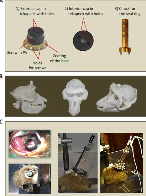
Fig.2. (A)Ontheleft,generalviewofthechronicrecordingchamberbuilttoaccessthepremotorcortex.Thebottomedgeinyellowcorrespondstothecoatingwith
hydroxyapatite.Inthemiddle,aninteriorcapensuredthesealingofthechronicrecordingchamberandisolatedtheduramater,whennoelectrophysiologicalrecording
sessiontakesplace.Therightmostpanelshowsthescrewdriverusedtomanipulatetheinternalcapinsidethechronicrecordingchamber(seesupplementaryvideosequence
#7http://www.unifr.ch/neuro/rouiller/research/multi/lanz/l7.html).(B)Viewofthe3Dprintreplicateofthemonkey’sskullinplastic(seetext;Mk-LI).(C)Ontheleft,the
chronicrecordingchamberisshownwhenitwasimplantedduringthesurgery(inMk-LI)andasitappearsseveralmonthsafterimplantation(inMk-JZ).Inthemiddleand
ontheright(Mk-JZ),thepicturesshowthesystemusedtofixtheheadofthemonkey,togetherwiththeadaptationonthechronicrecordingchamberofaNarishige®single
electrodedrivesystem(middle)oraNAN®multipleelectrodedrivingsystem(right).
2.5. Recordingchamber
In parallel to the head fixation device, a similar idea was employedtodesignnewchronicrecordingchambers.Withthese newchambers electrophysiological datacould bederived from behaving monkeys withtheirhead fixed.Theaim herewasto increase the animal’s comfort (again no dental or orthopedic cementtofixthechronicchamberontheskull,asrecentlyproposed byAdamsetal.,2011),whilereducingthedailycareofthe cham-berandinfectionrisks.Toensureoptimalanchoringofthechronic chamberonthescalp,itsshapewasadaptedtothe3Dreplicate (print)ofthecorrespondingmonkey’sskull.This3Dprintallowed definitionoftheexactpositionandtheshapeaswellasthebestfit ofthechronicrecordingchamberontheskull.
In the present study the chronic chamber has a cylindri-cal shape (Fig. 2 A) and is made of tekapeek- an industrial plastic with high temperature, chemical, electrical and radia-tion resistance (similar to metals such as titanium). Tekapeek has the advantage of guarding the option to perform subse-quent MRI investigations with minimal artifacts. Furthermore, tekapeek is lighter than metal, thus reducing the weight of the chamber placed onthe monkey’s skull. The millingof the chamber isillustratedin thesupplementaryvideo sequence #4 (http://www.unifr.ch/neuro/rouiller/research/multi/lanz/l4.html). Thechronicchamberiscomprisedofabase(28mmindiameter), whichadherestothebone,withacylinderontop(24mmin diam-eterand9mminheight)givingaccesstotheduraandofferingthe possibilitytofixanelectrode drivingsystem(Fig.2C).Thebase
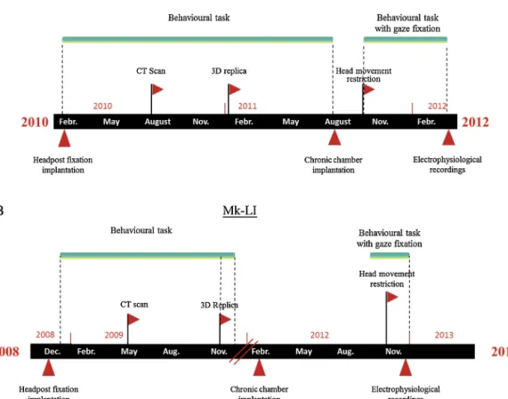
Fig.3.Timelineoftheimplantationprocedures,behavioralandelectrophysiologicalrecordingsthetwosubjectsunderwentduringtheoverallexperimentalprotocol.
oftherecordingchamberwascoatedwithHA(Fig.2A1),aswas donefortheheadfixationdevice.Briefly,thebaseoftherecording chamber was coated with a titanium bond layer between the substrateandtheHAcoating.Thetitaniumwasveryreactiveand bondedverywelltodifferentmaterials(formoredetail,see“Head fixationdevice”sectionabove).
Theexact positionof thechronicchamber ontheskull was defined by superimposing a MRI scan of the monkey’s brain (providinganatomicalpositionofsulciandcorticalgyrifor exam-ple)withthecorrespondingCTscan(providinganaccuratereplica oftheskullsurface)performedinthesameanimal.Thentheshape andcontourofitsbasewaspreciselyadjustedtothebonesurface, asitappearedonthe3Dprintoftheskull.Thebaseofthechronic chamberiscomprisedof7holesusedtopositionfixationscrews (Fig.2A1).Thecylinderwaslargeratthebottomthanatthetop formingashoulderonwhichtheelectrodedrivingsystemrested (Fig.2C).Theinternaldiameterofthecylinderwas21mm,which correspondedtothesizeofthegridguidingtheelectrodesheldby theelectrodedrivingsystem.
When the animal was not in the experimental set-up, the chronicrecording chamber was covered witha cap. Instead of usinga standard cap placed ontop of the cylinder,a capwas designedwhichcouldbeplaced insidethecylinder.The advan-tage hereis that thesize of theimplant is reduced. Toensure appropriatesealing,thecylinderisclosedfirstwithasealingring (Fig. 2A2), which is screwedinto thecylinder using anad-hoc screwdriver(Fig.2A3).Thesealingringwasusedmainlytocover aroundpieceofsilasticplacedontopofthedura.Thesealingring is5.2mmthickwithadiameterof20mm,andacentralopeningof 4.7mm(Fig.2A2).Thecentralopeningpreventsapressureincrease onthesilasticjointwhenthesealingringisscrewed/unscrewed. Toallowthescrewing/unscrewingofthesealingringtwosmall
3mmholesweremade.Finally,anexternalcapispositionedon topof thecylinder(Fig.2A1), ensuringhermeticclosure ofthe chamber.Theexternal capisa7.8mmthickdiskwitha diame-terof20mm,comprisedoftwoholesonthetop,andathreaded holeontheside.Theholeonthesideneedstobealignedwith a corresponding holein the side wallof the cylinder,in order toclosethechronicchamberbymeansofaheadlesspolyamide screw (M5×3). The cap can be removed by meansof a mod-ified crowbar. A comprehensive view of the chronic recording chamberwasavailableinthesupplementaryvideosequence#5 (http://www.unifr.ch/neuro/rouiller/research/multi/lanz/l5.html). 2.6. Implantationsurgery
Firsttheanimalwassedatedbyanintramuscularinjectionofa mixtureofketamine(Ketanarkon®10mg/kgbodyweight),
ben-zodiazepine(Midazolam0.1mg/kg)andmethadone (0.2mg/kg). Thissedationallowedthepreparationofthemonkeyforthe sur-gicalintervention,involvingshavingtheskullandpreparationfor anintravenousinjectionofPropofol(diisopropylphénol),coupled withgasanesthesia(Sevoflurane;seebelow).Inthisstep,the anal-gesicsCarprofen(Rimadyl®,Pfizer,4mg/kg)andbroadspectrum
antibiotics(Albipen®,Intervet,30mg/kg)wereinjected
subcuta-neously.Inaddition,toreduceedema,dexamethasone(Decadron 0.3mg/kg,mixed1:1withsaline)wasinjectedi.m.Finally,an injec-tionofatropine(0.05mg/kgi.m.)wasgiventoreducebronchial secretion.
In the operating room,under sterile conditions, the animal wasintubated, allowing ventilation witha 50%/50%mixture of O2andair,containing2.5%Sevofluranetoensureanesthesia.This
wascomplementedbycontinuousi.v.perfusionofPropofol (Fre-senius0.1mg/kg/min).Atpotentiallypainfulstepsofthesurgery
(e.g.craniotomy),ani.v.flowofopioid(Fentanyl0.1g/kg/min) wasused.Inaddition,duringtheentiresurgery,theanimalreceived acontinuousi.v.perfusionwithlactate-ringeratarateof5ml/kg/h. Duringtheentiresurgicalprocedure,physiologicalparameters werecontinuouslymonitored(e.g.bodytemperature,O2
satura-tion,heartrate,respirationrate,exhaledCO2).Atthebeginning
oftheprocedure,theskinoftheheadwascleanedanddisinfected withaniodizedsolution(BetadineorPovidone-iodine).Anincision oftheskinalongtheskullmidlineofabout10cmwasmadewith ascalpel.Themuscleswerethenreclinedfromtheskulltoexpose thebonesurface.Theheadfixationdevicewasthenpositionedon theskullatitsforeseenposition(asrostralaspossible:seeFig.1B). Atthisstep,ifnecessary,inordertoensureaperfect fitonthe skullsurfaceandtakingadvantageoftheflexibilityofthetitanium, thefootplatesoftheheadfixationdevicecouldbeslightlyadjusted withsteriletools.Beforeanchoringtheheadfixationdeviceonthe skull,thebone(periosteum)wasabradedextensively.The inser-tionofthescrewswasdoneaccordingtotheprotocolbyAdams etal.(2007).Namelythefirstscrewtobeimplantedwassituated ontheleft-handsideofthelegofthearch;thesecondwasplaced onthelast holeoftherightposteriorleg.For insertionof each screw,apowerdrillwasusedtomake,smallpilotholesofasmaller diameter.Furthermore,toavoidbonedamageresultingfroma tem-peratureincrease,aflowofsaline(0.9%)fromalargesyringewas usedforcoolingduringthedrilling.Anincreaseintemperature couldcausebonesofteningneartheholes.Thescrewswere care-fullyinsertedbyhand,withoutexertingtoomuchpressure,inorder tominimizerisksofcrackingordamagingthebone.Thelengthof screwswaschosenaccordingtotheCTscanperformedafewweeks beforetheoperationandthe3Dprintingoftheskull(seeabove).
Once the head fixation device was anchored to the skull, first the muscles then skin were stitched to the midline. A small opening was left on the posterior part of the cylinder of thehead fixation deviceto actas a natural catheter, allow-ing for possibleleakage of secretions, and toinject antibiotics, if needed. Several months later, a similar surgical procedure was conducted to anchor the chronic recording chamber on thesurface ofthe skull (seesupplementary video sequence#6 http://www.unifr.ch/neuro/rouiller/research/multi/lanz/l6.html andFig.2C-topleftpanel).
2.7. Dailycarefortheheadfixationdevice
Aftertheimplantationoftheheadfixationdevice,thewounds and scars werecleansed daily withan antiseptic iodized solu-tion(Betadine ® 500ml, Mundipharma Medical Company).The
animalwasexaminedforpossibleinflammationandappearance ofinfectionaswellasobservationofgeneralbehavior.An anti-inflammatory and antibacterial cream (Panalog ® ad US. Vet.,
Novartis)wasappliedaroundtheimplant.Furthermorethe ani-malreceivedantibioticsandanalgesicforatleast10days.Thebig advantagewiththistypeofimplantwasthatafterabout1month, dailycarewasnolongernecessary.However,abi-weekly clean-ingwithBetadinewasdone,aswellasadailyroutinecheckofthe animal.
2.8. Dailycareforthechronicrecordingchamber
Thechronicrecordingchamber’sdailycarerequiredmoretime and was more frequent than that of the head fixation device. Justaftersurgerythewoundsandscarswerecleanseddailywith anantisepticiodizedsolution(Betadine®500ml).Duringthe
fol-lowing ten days, the animal received injections of antibiotics (Albipen®)aswellasanalgesics(Rimadyl®).Attheendofevery
dailycare,acream(Panalog®15ml,Novartis)wasappliedaround
thechronicchamber.Similartoheadfixationdevice,thistreatment
was pursued during one month post-implantation. Afterwards cleaningtheexternalpartofthechronicrecordingchamberwasno longerperformedonadailybasis,butasuperficialcleaningwith Betadine®wasrequiredabouttwiceaweek.
The inside of the chronic recording chamber required a lot moreattentionandcare(seesupplementaryvideosequence #7 http://www.unifr.ch/neuro/rouiller/research/multi/lanz/l7.html). Thechamberwascleansedatleastonceeverythreedaysusing sterilized material (gaze band, surgical forceps, silastic joints, two different caps). During therecording period, cleaning was performedateveryrecordingsession. Theinsideof thechronic chamberwascleanedusingBetadine.Beforeclosingthechamber, thesilasticjoint(Siliconesheeting) (LP500-9,Manufacturedby LPI)wascoatedwithalayerofantibioticcream(FucithalmicVet®,
3mg/g; Dechra). Once the daily recording sessions had begun, theduramaterwasscrapedonceaweek,inordertolimittissue growthanddurathickening,whichwouldblockthepenetration ofrecordingelectrodes.
2.9. Timeline
Toclarifythetimecourseofeventsthetwosubjectswereto follow duringthe experimentalprotocol,a timeline was estab-lishedanddisplayed,inFig.3.Thistimelineindicatesthestarting pointoftheprocedures(headpostfixationimplantation,CTscan, 3D replica, chronic chamber implantation) and the behavioral tasks/first electrophysiological recordings accomplished. At the timeofthepublicationtheprotocolswerestillon-going.
3. Results
3.1. Headfixationdevice
Toensureahighandstableboneintegrationoftheheadfixation device,themonkeyswereimplantedveryearly,wellbeforethe behavioraltrainingwasstarted.Mk-LIwasimplantedonNovember 12th2008,whereasMk-JZwasimplantedonFebruary19th2010. AsshowninFig.1D,afterrespectively1month(1D1)and3months (1D2)post-implantation,onlythecylinderof theheadfixation devicewasvisibleontherostralpartofthemonkey’shead.The skinretractedandadoptedapositionfittingthecircumferenceof thecylinderoftheheadfixationdevice.Therewasnoinfectionor inflammationaroundtheimplantandthereforeonlyminimalcare wasnecessaryat2weeksintervalsintheformofcleaningaround theimplantwithBetadine®.
Afteraperiodofbehavioraltraining,themonkeysunderwent theimplantationofachronicrecordingchamber.Thissurgery pro-videdtheopportunitytochecktheintegrationoftheheadfixation devicetothebone,asitappearsafter1yearinMk-JZandafter3.5 yearsinMk-LI(Fig.1C).Theimplantwasperfectlyadaptedtothe topographyoftheskull,asexpectedafterthefinaladjustmentof thefootplatesduringthefirstsurgery.Theosseousintegrationwas notthesameforbothanimals,whichisinlinewiththedifferent timeintervals.Inthefirstmonkey(Mk-JZ;Fig.1C1),afteroneyear, itwasobservedthatanosseouslayerhadsettledalongthe foot-plateedge.Inthesecondanimal(Mk-LI;Fig.1C2),afterthreeand ahalfyears,therewasaclearbonegrowthoverthefootplateand inbetweentwoadjacenttitaniumscrews.Fromabehavioralpoint ofview,theanimalpresentednodiscomfortinrelationtothehead fixationdeviceduringthebehavioraltraining.Notethat,duringthe entiretrainingperiod,theheadoftheanimalwasnotfixedinthe experimentalset-up,toavoidmechanicalconstraints,whichcould disruptbonegrowtharoundthefootplates.Theuseofthehead fix-ationdevicetofixthemonkey’sheadusingarigidarmanchored totheexperimentalset-upisshowninFig.2C.
Fig.4.Picturesshowingthebuildingupofanotherchronicrecordingchambertoaccessadifferentarea(thalamus)aftercompletingtheinvestigationsinthepremotor
cortex(seetext).Ontheleft,thepictureshowsasimulationofthenewchronicrecordingchamberbeforeitwasfabricated.Ontherightthepictureshowstherealchamber
intekapeekandplacedonthe3Dreplicateprintingofthemonkey’sskull(Mk-LI).
3.2. Recordingchamber
The chronic recording chamber, illustrated in Fig. 2, was implanted in both monkeys (August 23rd 2011 in Mk-JZ and February9th2012inMk-LI),withtheaimtoaccessthepremotor cortexintherighthemisphere(Fig.2C).Astheshapeofthechronic chamberwaswelladaptedtothe3Dprintofthecorresponding monkey’s skull (Fig. 4) the implantation during surgery was straightforward and thefinal position easy to achieve. Indeed, onlythetargetedpositionontheskullprovidedaperfectmatch between the base of the chronic chamber and the contour of theskull.Apreliminarypositioningofthechronicchamberwas performedtodeterminetheregionof thebonetoberemoved. The bone was then marked with a sterile pencil around the circumferenceoftheinsideofthechronicchamber.Thecontourof thecylindricalpieceofbonewascautiouslyremovedusingadrill, exposingtheduramater.Thiswasimmediatelyphotographedin ordertoregister thepositionofblood vessels tobeavoided in subsequentelectrodepenetrations.Thepositioningofthechronic recordingchamberontheskullimmediatelyafterscrewfixation isshownforMK-LIinFig.2C -topleftpanel.Depending onthe monkey’ssizeandthepositionofthechronicchamber,apartof thetemporalmusclehadtoberemoved,especiallyifthechronic chamberwaslocatedlaterally.Firstthemusclesweresuturedand thentheskin,allaroundthechronicrecordingchamber.Complete scarringtookplaceovera fewweeks(Fig.2C).Theskinaround the chronic chamber (see supplementary video sequence #7 http://www.unifr.ch/neuro/rouiller/research/multi/lanz/l7.html) required only minimal, bi-weekly, cleansing with Betadine®,
as was the case for the head fixation device. As illustrated in Fig.2C,thechronicrecordingchamberallowedfixationofdifferent electrodedrivingsystemsliketheNarishige® system(Narishige
Internationallimited,Japan,Fig.2C)andtheNAN®system(NAN
Instrument,LTD,Israel,Fig.2C).
Thepresentapproacheliminatestheuseofdentalacrylicresin ororthopediccement,andgivestheoptiontoremoveachronic chamberatalatertimepointwhenelectrophysiological investiga-tionsinthecorrespondingbrainregionarecomplete.Inthisfashion anotherbrainregioncouldbetargetedforasubsequentstep.Itis truethatachamberfixedwithacryliccementcouldpossiblybe removed,buttheunderlyingbonemightbeinbadconditionand certainlymoretraumatized(softbone,presenceofinfection,bone thicknessnotsuitableforre-implantation,etc.),ascomparedto thepresentapproach.Suchastrategywouldallowtheuseofthe sametrainedmonkeytoextendtheinvestigationstoadditional brainregions.Inthepresent case,oncetheelectrophysiological recordingsarecompletedinthepremotorcortex,thefirstchronic
recordingchamberimplantedwillberemovedandreplacedbya secondone,designedtoreachthethalamus(Fig.4).Thesecond chamberisbasicallythesameasthefirstone:cylindricalshaped withabottomedgelargeenoughtoensureaperfectsealwiththe bone.Inaddition,theedgewillbeextendedlaterallyandrostrally inordertocovertheskullareawherethefirstchamberhadbeen implanted(Fig.4).Aroundprotrusionofthetekapeekwillbemade tofitperfectlyintotheboneholedrilledforthefirstchamber.This secondchamber willbepositionedmore caudallyand nearthe midlinetoallowverticalpenetrationstoreachthethalamus.
4. Discussion
Thepresentreportprovidesevidencethatboththehead fixa-tiondeviceandthechronicrecordingchambercanbeimplanted onamonkey’sskullforalongperiod(4yearsfortheheadfixation device),withouttheadverseeventsobservedinthepastwith den-talresinororthopediccement,intermsofinflammation,infection orrejection.Todemonstratethebenefitofthepresentapproach, acomplementaryretrospectiveanalysis(Table1)wasmadefrom datacollectedinourlaboratoryonsubjectswithheadpostfixation fixedwithdifferentcements(dentalresinororthopediccement). Thesedatademonstratethattheuseofcementisassociatedwith amuchshorterdurationofimplantsandanincreaseininfections and/orlossofthedevice,whencomparedwiththetwomonkeys includedinthepresentstudy.Thereforethisnewprotocol guar-antees anexcellent osseous-integrationofthe implants. Thisis dueto:(1)acoatingoftheimplantswithHA(Figs.1and2),thus potentiatingtheintegrationofferedbythetitaniumitselfand(2) a3Dprint replicateoftheskullof theliving animalforprecise designandadjustmentstoindividualskullshape.The3Dprintstep representsacrucialimprovementinallowingproductionoftruly custom-fittedimplantsthatperfectlyfitthemonkey’sskull.The longevityoftheimplantsdemonstratedhereontwomonkeys(up to4yearsfortheheadfixationdevice)issubstantialprogressand shouldbecompatiblewithvariousbehavioraland electrophysio-logicalprotocolsconductedinnon-humanprimates.Asillustrated inthesupplementaryvideomaterial,thetwoimplantsarewell tol-eratedbythemonkeys,evenduringthecleaningprocedures.Due totheeliminationofdentalacrylicresinororthopediccement,the implantshavealimitedmassabovethemonkey’shead,reducing theprobabilitythattheanimalhitsitsheadagainstobstaclesinits environment(e.g.enclosure).Anotheradvantageofavoiding den-talresinororthopediccementisaconsiderabletimegain,about onehour,duringsurgicalimplantationoftheheadfixationdevice orthechronicrecordingchamber.Inaddition,surgeryundertaken toanchortheimplantsismucheasier,asthedifficultprocessof
Table1
Retrospectivesurveyofheadpostimplantsfixedtotheskullwithdentalresinororthopediccementin11monkeys,involvedinelectrophysiologicalprotocols(1994–2006).
Thetablelistsforeachmonkeythedurationofimplantation(beforethedevicehadtoberemovedorwaslost),theoccurrenceofinfectionsandthedeviceslosses.
Subject(n=11) Implantationduration(months) Infections Lossofdevice
MK-1 10 MK-2 12 MK-3 7 X(2x) MK-4 6 MK-5 11 X MK-6 3 MK-7 12 X X(2x) MK-8 6 MK-9 8 MK-10 6 X(2x) MK-11 9 X X Average 8 18% 45%
skillfulapplication,insuccessivesmallamounts,ofdentalresinor orthopediccement,isskipped.Thusthepresentworksupportsthe ideaofeliminationofdentalacrylicresinororthopediccement,as previouslysuggestedbyAdamsetal.(2007,2011)orMcAndrew etal.(2012).
Anothervaluablecontributionofthepresentstudyconsistedin confirmingthesuitabilityofthe“K-shape”baseofthehead fixa-tiondevice,whichallowedtheeliminationofdentalacrylicresin ororthopediccement,asinitiallyproposedbyAdamsetal.(2007). Thelongevityoftheimplanteddevicewasobservedtobeatleast 17monthsbytheseaforementionedauthors.Thiswasextended to3yearsand8 monthsin amorerecent report(Adamsetal., 2011).Inthepresentstudy,thefollowupismorethan4years. Noincidentswereseen,despitethefactthatit wasusedtofix theheadoftwostrongadultmalemacaquemonkeys(8kgbody weight)inbehavioraland electrophysiologicalexperiments.The cleananddiscreteappearanceoftheheadfixationonthemonkey’s headisalsoconfirmedinthepresentstudy(seeFig.6ofAdams etal.,2007;Fig.1Dinthepresentreport).Experienceshowsthatit iscrucialtoimplanttheheadfixationdeviceearlyenoughinthe protocol,sothattheosseous-integrationcanoccurduringseveral months,beforethemonkey’sheadisfixedintheset-up.Atleast3 monthsisrecommended,butlongermaybesafer.Thisrelatively longdelaycanbeanticipatedtosomeextent,astheheadfixation devicecanbesuccessfullyimplantedinjuvenilemonkeys(Adams etal.,2007).Inthepresentstudy,incontrasttotheworkofAdams etal.(2007),afurtherimprovementwasintroducedintheform ofcoatingtheheadfixationdevicewithHAinordertoenhance theosseous-integration.AlthoughtitaniumiscompatiblewithMRI investigationsinspiteofsomeartifacts,onecouldalsoenvisage,in thefuture,buildingtheheadfixationdevicefromtekapeek,asthe chronicrecordingchamber,tominimizeartifacts.The“K-shape” headfixationdevice(Adamsetal.,2007,2011;presentstudy)is clearlylessbulkyandlessuncomfortableforthemonkeysthanthe headringapproach.(Isodaetal.,2005).
Thedesignofacustom-fittedchronicrecordingchambertoa liv-ingmonkeyisnew,assuchanattemptwasonlyrecentlyreported (McAndrewetal.,2012).Animportantdifferencehoweveristhat theseauthorsimportedtheCTreconstructionofthemonkey’sskull intoa3DCAD(computer-aideddesign)programwheretheimplant wasdesignedatthetargetlocationontheskull.Thepresentstudy went onestepfurther. Theskull derived fromtheCT scanwas printed outin 3D(Fig.2Band Fig.4)in orderto obtainatrue replicateofthelivingmonkey’sskull,similartowhatwouldhave beenobtainedifthemonkeyhadbeensacrificed.Thereplicateof themonkey’sskullcanbeusedtotestanddetermine,withhigh precision,severaloptionsconcerningsizeandpositionofchronic recordingchambers,toreachagivenbrainregion.Itisalso possi-bletoeasilycheckforpossibleconflictsbetweenseveralchronic chamberstobeimplantedeithersimultaneouslyoroneafterthe
other.Whentheapproachandthefinalpositionhavebeenchosen, thechronicrecordingchambercanbefabricatedbythe machin-istusingthecontourwhichwasdeterminedbymeansofthe3D skullreplicateprint.Intheirreport(McAndrewetal.,2012),the authorsmentionedthatthelongevityofthedeviceremainedto beseen,especiallyafterreportingthattheimplantbecameloose after6months,requiringre-implantation.Furthermore,duetoa gapbetweenthechronicchamber’sedgeandtheskull,therewas skinandhairgrowthintotheimplant(McAndrewetal.,2012). Suchundesiredeventdidnotoccurinthepresentstudy,evenafter ayearfollowingimplantation ofthechronicrecordingchamber (Mk-JZ).Itisverylikelythataperfect sealwasobtaineddue to the3Dprint replicateoftheskull.Usingthis3Dprint,the rela-tivelylargeandflexibleedgeatthebottomofthechambercould beformedtoadhereverytightlytotheskullwiththeuseof tita-niumscrews.Asaresult,pressureexertedbythescrewsallaround theimplantproducedaperfectseal(Fig.2).Asevidencedbythe cleaningprocedure,theinsideofthechronicrecordingchamber istotallyimpermeabletofluidwithrespecttotheoutside.Thisis animportantrequirementtominimizerisksofinfectioninsidethe chronicchamber.Thepresentstudydiffersinseveralaspectsfrom therecentreportofAdamsetal.(2011).First,intheirreport,Adams etal.(2011)usedanacrylicfreetitaniumchronicrecording cham-berwhereasherethechamberwasmachinedfromTekapeek.The latterpresentingtheadvantagetobelighterandtoreduce arti-factsinMRIinvestigations.Furthermore,Adamsetal.(2011)used HAasapastetosealthechambertotheskullduringthesurgery; bycontrast,inthepresentstudy,HAwascoatedontotheimplant itselfbeforeimplantation.Anotherimportantdifferenceisthatthe titaniumchamberusedbyAdamsetal.(2011)wascomprisedof 5–6feet,eachwithoneholetoinsertascrewtoanchorthe cham-bertotheskull.Inourcase(Fig.2),thefeetwerereplacedbya continuousthinedgeallaroundthebaseofthechamber(Fig.2) whichincludedtheholestoinsertthescrews(n=7).When tight-ened,thescrewpressureensuredaperfectmatchofthechamber’s baseontotheskullsurfaceduetooftherelativeflexibilityofthe Tekapeekanditsthincontinuousedge.Thepresentstudy demon-stratesthatTekapeekcanreplacetitaniumtofabricateimplants (atleastthechronicrecordingchamber,butpossiblyalsothehead fixationdevice)andexhibitsgoodosseous-integrationproperties duetothehydroxyapatitecoating.TheuseofTekapeekisespecially favorableforMRIinvestigation,asartifactsareminimized.Along thisline,afurtherimprovementmayconsistinreplacingtitanium screwsbyceramicscrews.
Thestepsnewlyintroducedinthepresentstudy,suchas coat-ingtheimplantswithHAandthe3Dprintreplicateoftheskull, increasesthecostoftheexperiments,butremainsaworthwhile investment consideringthelongevityof theimplants, the ethi-calandcommercialvaluesofthenon-humanprimates,aswellas thelongtimeperiodinvestedinthistypeofexperiments–mainly
trainingthemonkeystoperformdifficultbehavioraltasks.Indeed, therejectionofanimplant(headfixationdeviceand/orchronic recording chamber) by thebone is dramatic in any case, as it canruinalongandexpensivebehavioraland electrophysiologi-calexperiment.Itisextremelydifficulttore-implantasecondtime atthesamelocation.
Insummary,thepresent studycontributestorefiningrecent proposedtechniques(Adamsetal.,2007,2011;McAndrewetal., 2012)tooptimizingtheanchoringofaheadfixationdeviceanda chronicrecordingchamberontheskullofmacaquemonkeys.Inour opinionitshouldincludeacoatingwithHAanda3Dprintreplicate oftheskull.Assuch,thepresentstudyiswellinlinewiththe3Rs ini-tiativetoimprovetheconditionsoflaboratoryanimals.Indeed,the longevityoftheimplantsaswellastheminimaldiscomfortoffered bycustom-fittedimplantscontributetosubstantiallyreducingthe stressforthemonkeys,decreasingtherisksofinfectionand reduc-ingthenumberofanimalsusedinthis typeofexperiment.The possibilitytoremoveandreplaceimplantsisaverypositiveaspect becausethesameanimalcanbeusedoveralongertimeperiodand allowsinvestigationofremotebrainregions(e.g.premotorcortex andthalamus).
Conflictofinterest
HAisprovidedbyMedicoatAG(Switzerland).
Acknowledgments
Theauthors thankJoseph Corpataux,Laurent Bossy, Jacques Maillardfortheirhighlyvaluabledailycollaborationforthecare ofthe monkeysin theanimal facility, especially before, during and after the various interventions. Our thanks goalso to the “Ateliers Clément S.A.” for the fabrication of the head fixation deviceandtotheFribourg’sEngineeringschoolforthe3Dprint replicateofthemonkey’sskull.TheauthorsthankJaneFavrefor proofreading.
AppendixA. Supplementarydata
Supplementary data associated with this article can be found, in the online version, at http://dx.doi.org/10.1016/ j.jneumeth.2013.07.015.
References
Adams DL, Economides JR, Jocson CM,Horton JC. Abiocompatible titanium
headpost for stabilizing behaving monkeys. Journal of Neurophysiology 2007;98:993–1001.
AdamsDL,EconomidesJR,JocsonCM,ParkerJM,HortonJC.Awatertight
acrylic-freetitaniumrecordingchamberforelectrophysiologyinbehavingmonkeys. JournalofNeurophysiology2011;106:1581–90.
AlbrektssonT,Albrektsson B.Osseointegrationof boneimplants: Areview of
analternativemode offixation.ActaOrthopaedicaScandinavica1987;58: 567–77.
AugatP,ClaesL,HanselmannK-F,SugerG,FleischmannW.Increaseofstability
inexternalfracturefixationbyhydroxyapatite-coatedbonescrews.Journalof Biomaterials1995;6:99–104.
BetelakKF,MargiottiEA,WohlfordME,SuzukiDA.Theuseoftitaniumimplants
and prosthodontictechniques in thepreparation of non-humanprimates forlong-termneuronalrecordingstudies.JournalofNeuroscienceMethods 2001;112:9–20.
BrånemarkPI,AdellR,BreineU,HanssonBO,LindströmJ,OhlssonA.Intra-osseous
anchorageofdentalprostheses.I.Experimentalstudies.ScandinavianJournalof PlasticandReconstructive1969;3:81–100.
ChurchlandMM,Lisberger SG.Apparentmotionproducesmultiple deficitsin
visuallyguidedsmoothpursuiteyemovementsofmonkeys.Journalof Neu-rophysiology2000;84:216–35.
CookSD,ThomasKA,DeltonJE,VolkmanTK,WhitecloudTS,KeyJF.Hydroxylapatite
coatingofporousimplantsimprovesboneingrowthandinterfaceattachment strength.JournalofBiomedicalMaterialsResearch1992;26:989–1001.
DurifC,JouffraisC,RouillerEM.Single-unitresponsesintheauditorycortexof
monkeysperformingaconditionalacousticomotortask.ExperimentalBrain Research2003;153:614–27.
Fuchs AF, Luschei ES. Firingpatterns of abducens neurons of alert monkeys
in relationship to horizontal eye movement. Journal of Neurophysiology 1970;33:382–92.
GuoK,LiCY.Eyeposition-dependentactivationofneuronesinstriatecortexof macaque.Neuroreport1997;8:1405–9.
HarrisWH,SledgeCB.Totalhipandtotalkneereplacement.NewEnglandJournal
ofMedicine1990;323:801–7.
IsodaM,TsutsuiK-I,KatsuyamaN,NaganumaT,SaitoN,FurusawaY,etal.Design
ofaheadfixationdeviceforexperimentsinbehavingmonkeys.Journalof Neu-roscienceMethods2005;141:277–82.
JaffeWL,MorrisHB,NesslerJP,NaughtonM,ShenJ.Hydroxylapatite-coated
acetab-ularshellswitharcdepositedtitaniumsurfacerougheninganddualradius design.BulletinoftheNYUHospitalforJointDiseases2007;65:257–62.
KaeserM,WyssAF,BashirS,HamadjidaA,LiuY,BlochJ,etal.Effectsof
unilat-eralmotorcortexlesiononipsilesionalhand’sreachandgraspperformance inmonkeys:relationshipwithrecoveryinthecontralesionalhand.Journalof Neurophysiology2010;103:1630–45.
KaeserM,BrunetJ-F,WyssA,Belhaj-SaifA,LiuY,HamadjidaA,etal.
Autolo-gousadultcorticalcelltransplantationenhancesfunctionalrecoveryfollowing unilaterallesion ofmotorcortexinprimates: apilotstudy.Neurosurgery 2011;68:1405–16,Discussion1416-7.
KermadiI,LiuY,TempiniA,RouillerEM.Effectsofreversibleinactivationofthe
sup-plementarymotorarea(SMA)onunimanualgraspandbimanualpullandgrasp performanceinmonkeys.SomatosensoryandMotorResearch1997;14:268–80.
KermadiI,LiuY,TempiniA,CalciatiE,RouillerEM.Neuronalactivityinthe
pri-matesupplementarymotorareaandtheprimarymotorcortexinrelationto spatio-temporalbimanualcoordination.SomatosensoryandMotorResearch 1998;15:287–308.
LisbergerSG,Westbrook LE. Propertiesof visualinputsthat initiate
horizon-tal smoothpursuit eye movements in monkeys. Journal of Neuroscience 1985;5:1662–73.
LiuY,RouillerEM.Mechanismsofrecoveryofdexterityfollowingunilaterallesion
ofthesensorimotorcortexinadultmonkeys.ExperimentalBrainResearch 1999;128:149–59.
McAndrewRM,VanGilderJLL,NaufelSN,TillerySIH.Individualizedrecording
cham-bersfornon-humanprimateneurophysiology.JournalofNeuroscienceMethods 2012;207:86–90.
PeetersR,SimoneL,NelissenK,Fabbri-DestroM,VanduffelW,RizzolattiG,etal.
Therepresentationoftooluseinhumansandmonkeys:commonanduniquely humanfeatures.JournalofNeuroscience2009;29:11523–39.
RubodeRezendeML,JohanssonCB.Quantitativebonetissueresponseto
com-merciallypuretitaniumimplants.JournalofMaterialsScience:Materialsin Medicine1993;4:233–9.
SchmidlinE,KaeserM,GindratAD,SavidanJ,ChatagnyP,BadoudS,etal.Behavioral
assessmentofmanualdexterityinnon-humanprimates.JournalofVisualized Experiments2011.
Weatherallreport.Theuseofnon-human primatesinresearch; 2006,http://
royalsociety.org/The-Weatherall-report-on-the-use-of-non-human-primates-in-research/