HAL Id: hal-03009496
https://hal.archives-ouvertes.fr/hal-03009496
Submitted on 20 Nov 2020
HAL is a multi-disciplinary open access
archive for the deposit and dissemination of
sci-entific research documents, whether they are
pub-lished or not. The documents may come from
teaching and research institutions in France or
abroad, or from public or private research centers.
L’archive ouverte pluridisciplinaire HAL, est
destinée au dépôt et à la diffusion de documents
scientifiques de niveau recherche, publiés ou non,
émanant des établissements d’enseignement et de
recherche français ou étrangers, des laboratoires
publics ou privés.
in Drosophila melanogaster
Parvathy Venugopal, Hugo Veyssière, Jean-Louis Couderc, Graziella Richard,
Caroline Vachias, Vincent Mirouse
To cite this version:
Parvathy Venugopal, Hugo Veyssière, Jean-Louis Couderc, Graziella Richard, Caroline Vachias, et al..
Multiple functions of the scaffold protein Discs large 5 in the control of growth, cell polarity and cell
adhesion in Drosophila melanogaster. BMC Developmental Biology, BioMed Central, 2020, 20 (1),
�10.1186/s12861-020-00218-0�. �hal-03009496�
R E S E A R C H A R T I C L E
Open Access
Multiple functions of the scaffold protein
Discs large 5 in the control of growth, cell
polarity and cell adhesion in Drosophila
melanogaster
Parvathy Venugopal
1,2, Hugo Veyssière
1,3, Jean-Louis Couderc
1, Graziella Richard
1, Caroline Vachias
1and
Vincent Mirouse
1*Abstract
Background: Scaffold proteins support a variety of key processes during animal development. Mutant mouse for the MAGUK protein Discs large 5 (Dlg5) presents a general growth impairment and moderate morphogenetic defects. Results: Here, we generated null mutants for Drosophila Dlg5 and show that it owns similar functions in growth and epithelial architecture. Dlg5 is required for growth at a cell autonomous level in several tissues and at the organism level, affecting cell size and proliferation. Our results are consistent with Dlg5 modulating hippo pathway in the wing disc, including the impact on cell size, a defect that is reproduced by the loss of yorkie. However, other observations indicate that Dlg5 regulates growth by at least another way that may involve Myc protein but nor PI3K neither TOR pathways. Moreover, epithelia cells mutant for Dlg5 also show a reduction of apical domain determinants, though not sufficient to induce a complete loss of cell polarity. Dlg5 is also essential, in the same cells, for the presence at Adherens junctions of N-Cadherin, but not E-Cadherin. Genetic analyses indicate that junction and polarity defects are independent.
Conclusions: Together our data show that Dlg5 own several conserved functions that are independent of each other in regulating growth, cell polarity and cell adhesion. Moreover, they reveal a differential regulation of E-cadherin and N-cadherin apical localization.
Keywords: Drosophila, MAGUK, Yorkie, Hippo, Myc, Polarity, Adhesion, Growth Background
The accurate development of an organ or an organism requires a robust coordination size and shape control, both at the cell and tissue scales. Among the protein classes involved in such processes, many of them are scaffold proteins. These proteins are devoid of catalytic activity but contain multiple domains of protein-protein
interaction [1]. They allow the formation of complexes that are determinant, for instance, for cell polarity, cell adhesion or that are used as a platform for various sig-naling events.
Membrane-associated guanylate kinase (MAGUK) proteins are typical examples of scaffold proteins [2]. MAGUK domain is a structural unit formed by SH3 do-main next to a non-catalytically active guanylate kinase domain. These domains are usually flanked by one or several PDZ domains and potentially other protein-protein interaction domains. Some MAGUK domains
© The Author(s). 2020 Open Access This article is licensed under a Creative Commons Attribution 4.0 International License, which permits use, sharing, adaptation, distribution and reproduction in any medium or format, as long as you give appropriate credit to the original author(s) and the source, provide a link to the Creative Commons licence, and indicate if changes were made. The images or other third party material in this article are included in the article's Creative Commons licence, unless indicated otherwise in a credit line to the material. If material is not included in the article's Creative Commons licence and your intended use is not permitted by statutory regulation or exceeds the permitted use, you will need to obtain permission directly from the copyright holder. To view a copy of this licence, visithttp://creativecommons.org/licenses/by/4.0/. The Creative Commons Public Domain Dedication waiver (http://creativecommons.org/publicdomain/zero/1.0/) applies to the data made available in this article, unless otherwise stated in a credit line to the data.
* Correspondence:vincent.mirouse@uca.fr
1iGReD (Institute of Genetics, Reproduction and Development), Université
Clermont Auvergne, UMR CNRS 6293 - INSERM U1103, Faculté de Médecine, 28 Place Henri-Dunant, 63000 Clermont-Ferrand, France
Full list of author information is available at the end of the article Venugopal et al. BMC Developmental Biology (2020) 20:10 https://doi.org/10.1186/s12861-020-00218-0
recognize phosphopeptides, whereas some others work in cooperation with the adjacent PDZ domain, reinfor-cing the affinity of the latter for a specific partner [3, 4]. MAGUK proteins also emerge as important modulators of phase separation in cells [5,6].
Discs Large (Dlg) is a MAGUK protein that was iden-tified in Drosophila for its function in epithelial polarity as a determinant of the lateral domain and the neoplas-tic effect of its mutation [7–9]. Four paralogs of fly Dlg, Dlg1 to Dlg4, are found in mammals. A more divergent member of the family, Dlg5, is also found in fly and mammals with a conserved architecture: a coiled-coil domain, 4 PDZ domains and a MAGUK domain. Dlg5 studies in mammals emphasized a function in epithelial morphogenesis, the knock-out mouse showing mild defects of adherens junction and epithelial polarity in the kidney, the lung and the brain [10,11]. Dlg5 is also required for N-Cadherin (N-Cad) delivery to the mem-brane during synaptogenesis [12]. A report in Drosophila using partial loss of function conditions in follicle cells also described moderate defect in the recruitment of ap-ical determinants and junctional proteins [13]. This re-port suggested that Dlg5 acts mainly by a regulation of the apical determinant crumbs (crb). However, it is un-clear whether the effect on polarity determinants and adherens junction are causally linked our whether they reflect independent functions of Dlg5 protein. Dros-ophila Dlg5 is also required for the proper collective cell migration of the border cells [14, 15]. Beside these morphogenetic defects, new born Dlg5 mice are considerably smaller than their wild-type littermates, suggesting an involvement in growth control [10]. Interestingly, Dlg5 has been functionally linked to the hippo pathway both in mammals and in flies, where it interacts and regulates negatively the MAST/hippo kinase [16]. However, whether such a hippo regula-tion could account for all the growth defects associ-ated with the loss of Dlg5 is not known. Morever, Dlg5 was also identified as a positive regulator of the Target of Rapamycin complex 1 (TORC1) pathway in an in vitro RNAi screen [17].
Here, we identified Drosophila Dlg5 in an RNAi screen for genes linked to follicular epithelium devel-opment and we generated null mutants. These mu-tants allowed us to show that this gene is involved in the control of growth, both at the cellular and sys-temic levels. Our results suggest that Dlg5 regulates growth by at least two independent mechanisms. We also confirmed a moderate epithelial polarity defect and show a very strong and specific effect on N-Cad expression whereas E-Cadherin (E-Cad) is not af-fected. Importantly, we show that polarity defects and Adherens junction defects reflect independent func-tions of Dlg5.
Results
The loss of Dlg5 alters cell autonomously follicle cell growth
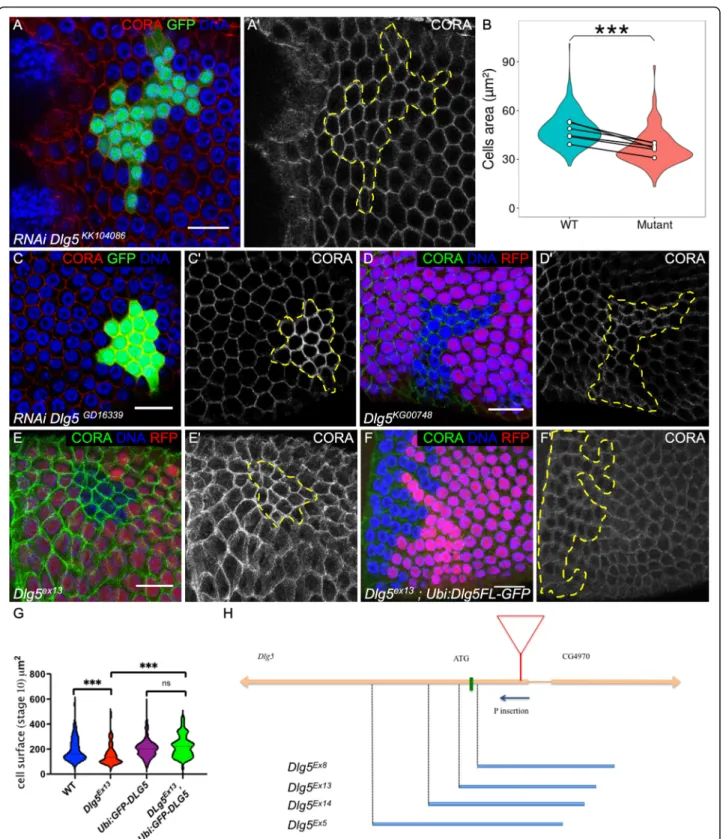
We performed a reverse genetics screen to identify new genes involved in Drosophila follicular epithelium devel-opment, a tissue used as a generic model for various as-pects of epithelium biology [18,19]. Follicle cells form a monolayer epithelium surrounding germline cyst with the apical domain facing the germline. Follicle undergoes a rapid growth through 14 developmental stages, with a 1000-fold volume increase. Follicle cell growth is associ-ated with proliferation until stage 6, then follicle cells become endoreplicative and larger. During the screen, we noticed that clones expressing RNAi against Dlg5 were small and the cells appeared also smaller than wild-type cells, especially after stage 6 (Fig.1a). This de-fect was quantified at stages 9-10A, showing an average reduction of 33% of the cell surface (Fig. 1b). A similar defect was observed with a different RNAi line (Fig.1c). A P-element insertion in the 5’UTR of Dlg5 was avail-able. This insertion was lethal and homozygous mitotic clones for this mutant also give small follicular cells (Fig.
1d). However, the defect obtained with this mutant ap-peared more variable than with the RNAi lines, suggest-ing that it may correspond to a hypomorphic mutant. We generated P-element excisions and most of them re-stored the viability of the stock indicating that its lethal-ity was associated with this insertion in Dlg5 gene. We also obtained several lethal imprecise excisions, Dlg5 Ex5,-Ex8,Ex13,Ex14
all, except Ex8, deleting the start codon and part of the coding sequence (Fig.1h). However, they also deleted part of the neighboring annotated gene (CG4970). This gene is only expressed in testis and is very poorly conserved, with no known domains and no ortholog in other insect species. Trans-heterozygous be-tween a Minos element inserted in the coding sequence of this gene (MimicMI02472) and the deletions that we generated complement perfectly in terms of viability and fertility, indicating no essential function of CG4970. We also obtained rescue of Dlg5ex13mutation lethality using a transgene with Dlg5 coding sequence under ubiquitin promoter and fused to GFP added in N-terminal (Ubi: GFP-Dlg5). Thus, we assumed that Dlg5ex13 could be considered as a bona fide null mutant. Mitotic clones for this allele contain cells with a reduced size (Fig. 1e,g), a defect also rescued by Ubi:GFP-Dlg5 (Fig.1f,g), confirm-ing the cell autonomous role of Dlg5 in follicle cell growth.
Dlg5 has a general function in growth
We next wondered whether Dlg5 function in cell growth could apply to other tissues. We induced Dlg5 knock-down specifically in the wing disc pouch, which corre-sponds to the future cells of the fly wing, using Nubbin:
Fig. 1 Dlg5 is required for follicle cell growth. a and c follicle cell clones marked by the GFP and expressing RNAi against Dlg5 using a KK10486 and c GD16339 lines and stained with Cora (red and a’ and c′). b violin plot of the quantification of follicle cell size expressing Dlg5KK10486RNAi clones (mutant) compared to wildtype (WT) surrounding cells on 6 stage 9 follicles. Mean values (white dots) are paired for each follicle. (P-value *** < 0,001.) d,e,f mutant follicle cell clone, marked by the absence of RFP, of D) Dlg5KG00748, e Dlg5Ex13and f Dlg5EX13rescued by a Ubi:Dlg5-GFP transgene. g violin plot of the quantification of follicle cell size of the indicated genotypes (n > 60 cells from at least 5 independent clones) h scheme of Dlg5 locus with the position of the P element KG00748 (red triangle) and the different imprecise excisions that we obtained (blue bars). For all pictures scale bar is 10μm
Gal4 [20]. It led to a dramatic reduction of the wing size (Fig. 2a-b, d). We also induced mitotic clones for Dlg5Ex13
in the wing disc and quantified several parame-ters. Of notice, the mutant cells tend to form a row of cells rather to extend the clone in all directions, a defect reminiscent of what has been recently described for other mutants generating small cells [21] (Fig.2c). Mu-tant clones were systematically smaller than their twin and contained fewer cells (Fig. 2 E,G). Moreover, the mutant cells had, in average, a size reduced by 40% (Fig.
2f). DCP-1 staining did not reveal apoptotic cells in Dlg5 mutant clones suggesting that the lower cell number per clone correspond to a growth and proliferation decrease (Fig.2h).
Finally, we looked at a transheterozygous combination of Dlg5 null alleles. First instar larvae hatch, are able to crawl around and are still alive 48 h after egg deposition. However, their growth is strongly impaired, indicating a systemic requirement for Dlg5 (Fig. 2j-k). Thus, altogether these results show that Dlg5 owns a general
growth function, as its mammal counterpart, and that this function is performed in a cell-autonomous manner. We aimed to define how Dgl5 modulates growth. It has been reported that Dlg5 modulated hippo pathway in the wing. However, this pathway is usually described as controlling of cell proliferation rather than cell growth. The Hippo signaling pathway regulates cell pro-liferation by inactivating Yorkie (Yki), the Drosophila Homolog of YAP. We therefore induced yki RNAi, and, as expected, its loss of function markedly reduces wing size and the estimated number of cells in the whole wing (Fig. 3a-d). Importantly, we also noticed a reduction of cell size in the same range than what has been observed with the loss of Dlg5 (Fig. 3e). Thus the hippo pathway also modulates cell size and could therefore explain Dlg5 contribution in this tissue. Yki is known to be required for normal follicle cell proliferation [22]. We induced null mutant clones and checked for cell size defects at stages 9–10. Comparing the cell surface indicates a mod-erate but significant effect of yki (Fig. 3f-g). Together,
Fig. 2 General requirement of Dlg5 for growth. a control wing and b Dlg5KK10486RNAi expressing wing with Nubbin:Gal4. The area of these wings is quantified in (d). c Dlg5Ex13mutant clone in the wing disc, marked by the absence of RFP, is smaller than the wild-type twin clone. Mutant clone has an elongated shape. Scale bar 50μm. e cell number f mean cell size and g total size quantification in Dlg5Ex13and twin clones in the wing disc of third instar larvae (n = 20 for each genotype). h and h′ DCP1 staining does not reveal apoptosis in Dlg5Ex13mutant clones (outlined in yellow) i control (Dlg5 heterozygous) third instar larva and j Dlg5 mutant larva of the same age at 25 °C. k quantification of larva size (n > 4, *** p-value < 0.001)
Fig. 3 (See legend on next page.)
these data clearly establish a role for yki in the control of cell size. However, this defect appears not stronger than the one induced by the loss of Dlg5 by RNAi (Fig. 1b). Moreover, inhibition of hippo pathway has not been de-scribed as affecting systemic growth, leading to the hy-pothesis that Dlg5 may modulate growth by at least another means. Looking for other potential growth ac-tors affected by the loss of Dlg5, we noticed that this gene was picked-up in a RNAi screen for TORC1 path-way regulators in S2 cells as affecting the level of S6K protein [17]. However, S6K level was unchanged in fol-licular cells or wing disc mutant cells for Dlg5ex13 (Fig.
3h,l). Moreover, phosphorylation level of S6 were not af-fected in Dlg5 mutant follicle cells, indicating that Dlg5 does not modulate S6K activity and more generally the Tor pathway in this tissue (Fig. 3i,l). We also, checked Insulin/PI3K pathway activity, which when affected, give similar defect both at the cellular and the systemic levels, but we did not observe any alteration of Phospho-Akt in Dlg5 mutant cells (Fig. 3j,l). Thus, how Dlg5 influences growth in these cells remains to determine. Nonetheless, we noticed a reduction of Myc expression in Dlg5 mu-tant follicle cells, suggesting that it is required for the ef-ficient signaling of one of the multiple pathways controlling Myc levels (Fig. 3k,l) [23]. Importantly, Myc levels were never affected in mutant follicle cells for yki, demonstrating that Dlg5 effect is independent of Hippo pathway (Fig. 3m). However, similar reduction was not detected in wing disc Dlg5 mutant cells (Fig. 3n), indi-cating that Myc regulation cannot account for Dlg5 ef-fect on growth in all tissues.
Dlg5 is required for the localization of apical polarity determinants
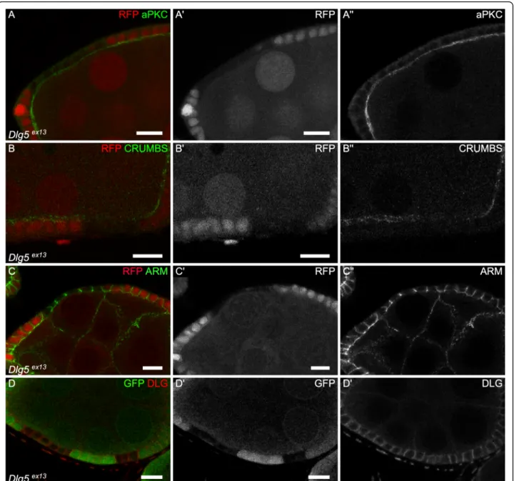
The fact that Dlg5 regulates growth both in mammals and in fly prompted us to check for an epithelial polarity phenotype, since such a defect has also been observed in Dlg5 mutant mouse. These defects have been detected for instance in the kidney or the lung, where a partial mislocalization of the apical determinant aPKC, a key component of the apical PAR complex, has been ob-served [10, 11]. Moreover, knock-down of Dlg5 in fol-licle cells also give similar phenotypes [13]. In follicular cells mutant for null mutant Dlg5Ex13 we saw a semi-penetrant reduction of the apical level of aPKC, the
apical domain of these cells being inwards, at the contact with the germline (12/19 clones) (Fig. 4a). The level of the apical determinant Crumbs (Crb) is also reduced (Fig. 4b). However, this apical reduction of apical deter-minants was never associated with an extension of lat-eral markers, such as Coracle (Cora), to the apical domain or to a mispositioning of the adherens junctions. Nonetheless, we pointed out that Cora was often upreg-ulated in the mutant cells (21/33 clones) (Fig. 1c,d), a defect never observed with another septate junction marker such as Dlg (n = 9) (Fig.4d). Moreover, we never spotted multi-layers or round mutant cells. Also, mutant cells tend to flatten, a defect more often observed in young follicles (Fig. 4c). Thus, although it is not suffi-cient to induce a complete loss of cell polarity in this tis-sue, Dlg5 null mutation can affect apical polarity determinants and cell morphology.
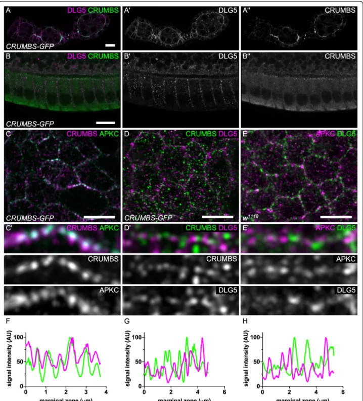
To characterize endogenous Dlg5 localization we gen-erated an antibody against the third and fourth PDZ do-mains. In follicle cells, Dlg5 antibody reveals a dotty pattern, similarly to what has been described in mam-mals (Fig.5) [10]. This signal is specific because the anti-body gives no signal in Dlg5 mutant follicle cells (Fig. 6g). These dots were observed both inside the cell and at the cell cortex. Moreover, Dlg5 pattern was dy-namic depending on the stages. During early stages (2– 8) Dlg5 localizes at apical and lateral membranes at stage 1, and appears therefore in apical sooner than Crb (Fig. 5a). Then the apical localization progressively de-creases, and is barely detectable at stage 9 (Fig.2b). Con-sequently, at later stages this cortical localization is restricted to the lateral domain. Because Dlg5 is present apically as key apical determinants such as aPKC and Crb and affect their localization, we compared their localization in the apical plain of the follicle cells with higher resolution using Airyscan. We observed that aPKC and Crb are usually colocalized, especially at the marginal zone, an area of cell–cell contact apical to the adherens junctions (Fig.5c,f). This observation comes as confirmation that these proteins cooperate to define the apical domain [24]. In contrast, no evident colocalization is observed between Dlg5 and those two proteins and their localization even tend to be exclusive in the mar-ginal zone, indicating that Dlg5 is not stably associated with these apical determinants (Fig.5d,e,g,h).
(See figure on previous page.)
Fig. 3 Hippo modulation is not sufficient to explain Dlg5 impact on growth. a control wing and b ykiGD40497RNAi expressing wing with Nubbin:Gal4. c, d, e) quantification of c wing aera) d cell number and e cell size for the indicated genotypes (control n = 9, Dlg5 RNAi n = 16, yki RNAi n = 11). f mutant follicle cell clone for ykiB5, marked by the absence of GFP at stage 9 and stained for Cora. g Quantification of follicle cell size in yki clones compared to wildtype (WT) or yki heterozygous surrounding cells on stage 9 follicles (n = 10). h-k) staining for (h) S6K, i phospho-S6 (pS6) j Phospho-AKT (pAKT) and k Myc in Dlg5ex13mutant clones in follicle. l Relative quantification of fluorescence for the indicated markers in Dlg5ex13clones compared to WT surroundings cells (n > 4). m dMyc staining in follicle cell mutant clones for ykiB5. o dMyc staining in wing disc mutant clones for Dlg5ex13. For all pictures scale bar is 10μm. (** p-value < 0.01, **** p p-value < 0.0001)
Dlg5 is required for N-cadherin localization independently of its effect on cell polarity
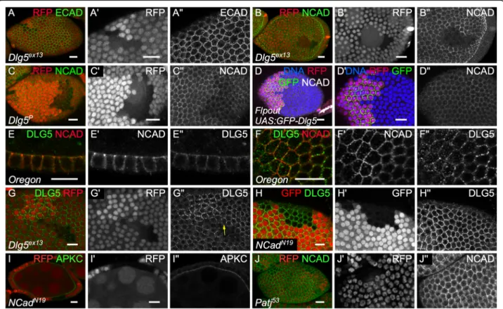
Looking at the Adherens junction, we found that E-Cad level was not affected (n = 18) (Fig.6a). Follicle cells also expressed N-Cad, which is integrated in adherens junc-tion, and Dlg5 has been functionally and molecularly linked to this cadherin in mammals [10, 12, 25]. We therefore looked at N-Cad and spotted an extremely strong and fully penetrant reduction in Dlg5 mutant cells (n > 20) (Fig. 6b). This effect correlates with the strength of Dlg5 loss of function because the reduction is weaker in hypomorphic conditions (Fig.6c). However, Dlg5 overexpression does not increase N-Cad levels at
cell contacts and has no visible impact on cell size (Fig.
6d). Thus, Dlg5 loss of function has a dramatic effect on N-Cad, but not on E-Cad, in the same cell type and at the same developmental stages. It indicates therefore a very specific effect on N-Cad membrane delivery or stability.
We therefore looked at a potential colocalization be-tween N-Cad and Dlg5. Both are present mainly at the cell periphery. However, at the adherens junction plain of the cells Dlg5 is mainly found as medioapical dots whereas N-Cad surrounds the cells (Fig.6f). Just above, less N-cad is observed whereas Dlg5 becomes more enriched at the cortex. As a result, only a weak
Fig. 4 Dlg5 moderately impacts epithelial polarity in follicle cells. a-d follicle cell mutant clones for Dlg5ex13and stained for a) aPKC, b) Crb, c) Arm and d) Dlg. For all pictures scale bar is 10μm
colocalization between the two proteins is observed with N-Cad being globally more apical than Dlg5 (Fig. 6f). Thus, although Dlg5 has a very strong and specific im-pact on N-Cad, their potential association is likely
transitory. We also noticed that when we induced Dlg5 mutant cell clones, Dlg5 disappears from the cell cortex even at the boundary with wild-type cells (Fig.6g). Clas-sical interpretation for such observation is that Dlg5 is
Fig. 5 Dlg5 is localized apically but is not colocalized with aPKC and Crb. a-b Dlg5 immunostaining on Crb-GFP knock-in follicles on a) early stages b) stage 10A follicle cells (c-d) Airyscan images of the apical plan of Crb-GFP knock-in follicle cells stained for c) aPKC D) Dlg5. e Airyscan images of the apical plan of WT follicle cells stained for aPKC and Dlg5. c’d’e) Zoom-in of the marginal zone of c, d, e. f, g, h) fluorescence intensity profile to the marginal zone shown in c′, d’ and e’. For all pictures scale bar is 10 μm
associated with proteins performing homophilic interac-tions between cells and that it is required for their localization, explaining its absence on the wild-type cell side. However, Dlg5 localization is not affected in N-Cad mutant clones (Fig. 6h). Thus, Dlg5 is probably associ-ated with another protein performing homophilic inter-actions and that is mainly localized at the lateral domain of the follicle cells.
The loss of Dlg5 affects both apical and adherens junc-tion proteins. Since there is important cross-talks be-tween the protein complexes acting at these two sites, we wondered whether those two defects were linked [26–28]. First, the loss of N-Cad had no effect on aPKC apical level, in agreement with the previous proposition that N-Cad and E-Cad are redundant in the follicle cells to maintain adherens junction and epithelial polarity (Fig. 6i) [25]. Thus, Dlg5 impact on N-Cad does not ex-plain the loss of apical proteins. Second, the reduction of apical determinant Crb and aPKC is much less expres-sive and penetrant than the loss of N-Cad and it is therefore unlikely the cause of such a defect. Patj muta-tion, a component of the Crb complex, leads to a very
similar mild effect to Dlg5 mutation on aPKC and Crb apical levels and on cell morphology [29]. However, patj mutant cells show a normal level of N-Cad (Fig. 6j). Thus, the reduction of apical determinants and of N-Cad observed in Dlg5 mutant cells correspond to two in-dependent functions of the protein.
Discussion
Altogether, our data show that Dlg5 owns several inde-pendent functions in Drosophila, suggesting that its scaf-fold abilities are used in different contexts. Of notice, these functions are somehow opposite to Dlg ones, both on growth and cell polarity, confirming that, despite the same naming, these two MAGUK proteins are unrelated.
First, Dlg5 requirement for growth is really strong, as revealed by the defect of the homozygous mutant larvae and its loss in the wing. It has been recently proposed that Dlg5 is involved in the hippo pathway, via a physical interaction and an inhibition of Hippo kinase by a mech-anism that remains to be elucidated [16]. In fly, the main argument for such a regulation is a reduction of the
Fig. 6 Dlg5 is essential for N-cadherin independently of its effect on polarity. a, b c mutant follicle cell clones, marked by the absence of RFP, for (a, b) Dlg5EX13c) Dlg5KG00748and stained for a)E-Cad and b)c) N-Cad. d flip-out clone overexpressing GFP-Dlg5 and stained for N Cad. e-f staining on WT follicles of N-cad and Dlg5 . f all the cells are not exactly in the same plane and Ncad is enriched more apically than Dlg5. g Dlg5EX1 mutant follicle cell clones, marked by the absence of RFP and stained for Dlg5. Note the absence of Dlg5 in WT cells at the contact of Dlg5 mutant cells (arrow). h, i NcadN19mutant follicle cell clones, marked by the absence of RFP and stained for h) Dlg5 and i) aPKC. j) Patj53mutant follicle cell clones, marked by the absence of RFP and stained for NCad. For all pictures scale bar is 10μm
Hippo reporter Expanded-lacZ in a Dlg5 loss of function in the wing disc. Consistently with this observation, yki loss of function induces a reduction of cell size both in the wing and in follicle cells. To our knowledge, this is the first report of a role of this gene in cell size control, which is actually accountable for about 15% of yki im-pact on tissue size. Interestingly, Hippo has been shown to be modulated by TORC1 or InR/PI3K pathways in different contexts [22, 30, 31]. Our results suggest that hippo modulation by these pathways may participate to explain their impact on cell size. Alternatively, these re-sults could suggest that hippo pathway could conversely modulate these pathways. However, several lines of evi-dence suggest that the regulation of hippo is not suffi-cient to explain Dlg5 impact on growth. First, in the wing disc, we did not see an effect of Dlg5 null mutation on Myc, a well-established target of the Hippo pathway in this tissue [32]. Moreover, to the difference of yki mu-tant cells, we did not find evidence that Dlg5 mumu-tant ones are eliminated by cell competition from the clones [33,34]. Second, in the follicle cells, the situation looks at the opposite because Myc is modulated by Dlg5 but not by yki. Moreover, the effect of null mutation for yki on follicle cell size appears weaker than the one of Dlg5 hypomorphic conditions induced by RNAi. Finally, we observed a strong requirement of Dlg5 for larval growth, whereas such an effect of yki loss of function has never been reported.
Dlg5 mutant defects are reminiscent of strong mutants for cell growth such as the ones of key components of the TORC1 pathway, with a decrease of cell size and cell proliferation but no induction of apoptosis, even in clonal analysis that can reveal cell competition. However, we did not confirm in vivo the proposed link between Dlg5 and S6K stability or TORC1 activity [17]. More-over, Myc expression is independent of TORC1 in fol-licle cells (Vachias, unpublished) whereas it is influenced by this complex in the wing disc [35], a contrary effect to the one of Dlg5. Thus, Dlg5 impact on growth is probably independent of TORC1. Similarly, misregula-tion of InR/PI3K pathway, which impacts cell size and proliferation, has not been observed in Dlg5 mutant cells. Thus, by which alternative pathway Dlg5 acts on cell growth and tissue size remains to be elucidated.
Our data using null mutants confirm the moderate im-pact of Dlg5 on apical-basal polarity observed in fly and mouse [10, 11, 13]. In follicle cells, these defects are really similar to the ones that we previously observed with Patj mutation, with both a semi-penetrant loss of apical determinants and a same cell shape defect in fol-licle cells [29]. This correspondence is in agreement with the proposition that Dlg5 regulates Crb complex [13]. Whereas Crb is essential to maintain follicle cell polarity, this complex is dispensable for the apical basal polarity
of the wing cells, as it seems to be the case for Dlg5 be-cause we observed no morphologic defect in this tissue [25, 36]. Interestingly, mouse Crb3 knock-out leads to cell polarity alteration in the kidney and the lung, which are also the epithelia known to be affected in Dlg5 mu-tant mouse, suggesting that the relationship between Dlg5 and Crb complex is conserved throughout evolu-tion [37]. However, high resolution imaging of follicle cell apical domain reveals no colocalization between Dlg5 and Crb, suggesting that the effect of Dlg5 on Crb is indirect or relies on a very transient interaction. More-over, apical localization of Dlg5 appears sooner than the one of Crb in young follicles suggesting different dynam-ics for the two proteins. Nonetheless, Crb is also a modulator of the Hippo pathway and it might be inter-esting in the future to explore the relationship between Dlg5, Crb and Hippo [38–42].
We also observed an extremely strong effect of Dlg5 null mutants on N-Cad localization at the membrane. Dlg5 does not affect E-Cad localization in the same cells and at the same stage, indicating a very specific effect. Although the specificity of the effect on N-Cad versus E-Cad was not established, available data in mammals also denote an impact of Dlg5 on N-Cad delivery associated with a physical interaction between the two proteins [10,
12]. Thus, the specific effect of Dlg5 on N-Cad is likely a conserved feature. E-Cad is usually associated with the acquisition of a stable epithelium architecture whereas N-Cad is more linked to collective cell migration and epithelium-mesenchyme transition [43, 44]. However, despite thousands of articles depicting E-cad versus N-cad expression, the molecular differences underlying these peculiarities are still an open question, relevant for developmental cell biology and cancer. The specific rela-tionship between Dlg5 and N-Cad might provide a nice entry point to understand these differences.
Conclusions
Together our data show that Dlg5 own several conserved functions that are independent of each other in regulat-ing growth, cell polarity and cell adhesion. Its effect on growth is likely pleiotropic, potentially acting on Hippo pathway but not only. Moreover, we revealed an effect of yki on cell size. Finally, this work reveals a differential regulation of E-cadherin and N-cadherin localization.
Methods
Molecular biology and antibody production
For transgenesis, Dlg5 coding sequence was amplified by PCR and cloned in phase in pUBi:GFP-Nterm-Gateway-AttB vector [45]. Transgenes were generated at AttP3-B landing site. Antibody were raised in rabbit against a fragment of Dlg5 corresponding to amino acids 1260 to 1600 fused to GST (Eurogentec).
Genetics
Fly were raised on wheat flour (8%) yeast extract (8%), Agar (1.1%) with antifungal and antibiotics. Dlg5 mu-tants were generated by imprecise excision of P {SUP-orP}KG00748. Excisions that were lethal when crossed with a deficiency covering Dlg5 locus were analyzed at the molecular level. Dlg5Ex13 contains a 1.5 kb deletion going from upstream the transcription start to 200 bp downstream of the translational start. The detailed geno-types, temperature and heat-shock conditions are given in supplemental Table S1.
Immunostaining and imaging
Dissection and immunostaining were performed as de-scribed previously [46], adding CaCl21 mM during fix-ation, excepted for Crb staining, which requires a specific fixation [25]. Primary antibodies used are DE-Cad (1/100, DHSB #DCAD2) and N-DE-Cad (1/100, DHSB, #DN-Ex), Cora (1/200, DHSB, #C615.16), cleaved DCP-1 (1/1000, Cell Signaling, #9578), S6K (1/2000, [47]), pS6K (1/400, [48], pAKT (1/500, Cell signaling, #4054),, dMyc (1/500, SantaCruz BioTechnology #), Crb (1/50, DHSB, #Cq4), aPKC (1/500, Santa Cruz Biotechnology, #C-20G), Dlg (1/200, DHSB, #4F3), Dlg5 (1/ 500, this study). Images were taken using a Leica SP5 confocal micro-scope or a Zeiss 800 Airyscan. Wing images were ac-quired on a Zeiss Axio Scan Z1.
Cell segmentation and size quantification were per-formed on Fiji. For wing disc clones, total size of the clone and cell number were determined and cell size was inferred from these values. Comparison were per-formed with twin WT cells. For adult wings, Fijiwings was used to determine wing size and cell density in the same posterior region on all images [49]. Cell size and total cell numbers were inferred from these values. In follicles, all the cells were segmented based on Cora staining using Tissue Analyzer [50] and comparison was realized with WT or heterozygous cells in the vicinity of the mutant (or RNAi) cells. Fluorescent signal was mea-sures in the whole cell surface excepted for pAKT for which only signal at junctions between mutant cells was compare to the signal between WT cells.
All the statistical analyses were performed on Prism using t-test. Figures were assembled with ScientiFig [51].
Supplementary information
Supplementary information accompanies this paper athttps://doi.org/10. 1186/s12861-020-00218-0.
Additional file 1.
Abbreviations
Dlg:Discs lage; Dlg5: Discs large 5; Tor: Target or Rapamycin; E-Cad: Epithelial-Cadherin; N-E-Cad: Neuronal-Cadherin; MAGUK: Membrane Associated Guanylate Kinase; PDZ: Post-synaptic density protein 95 Discs
large and Zona occludens 1; aPKC: Atypical protein kinase C; crb: Crumbs; GFP: Green Fluorescent Protein; DCP-1: Drosophila Caspase 1; S6K: S6 kinase; yki: yorkie; Patj: Pals1-Associated Tight Junction; InR: Insulin Receptor; PI3K: Phosphatidylinositol 3-kinase
Acknowledgments
We thank A Teleman, I Palacios, L Luo and Y Hong for stocks and reagents. We also thank the confocal imaging facility of Clermont-Ferrand (CLIC) and Pierre Pouchin for his help with statistical analyses.
Fundings
This work was funded by the Association pour la Recherche contre le Cancer (ARC) and the Auvergne-Rhône-Alpes Region. This research was also fi-nanced by the French government IDEX-ISITE initiative 16-IDEX-0001 (CAP 20–25). The funding bodies played no role in the design of the study or the collection, analysis, and interpretation of data, or in writing the manuscript.
Authors’ contributions
PV and VM designed the project. PV, JLC, HV, GR, CV and VM performed the experiments. PV, JLC, HV, CV and VM interpreted the data. CV prepared the figures and VM wrote the article. All authors have read and approved the manuscript.
Availability of data and materials
New materials (mutants, antibodies) will be shared upon request. The datasets used and/or analysed during the current study available from the corresponding author on reasonable request.
Ethics approval and consent to participate Not applicable
Consent for publication Not applicable
Competing interests
The authors declare that they have no competing interests.
Author details 1
iGReD (Institute of Genetics, Reproduction and Development), Université Clermont Auvergne, UMR CNRS 6293 - INSERM U1103, Faculté de Médecine, 28 Place Henri-Dunant, 63000 Clermont-Ferrand, France.2present address : School of Biotechnology, Amrita Vishwa Vidyapeetham, Kollam, Kerala 690525, India.3present address : University Clermont Auvergne, INSERM U1240, Centre de Lutte Contre le Cancer Jean PERRIN, 58 rue Montalembert, 63011 Clermont-Ferrand, France.
Received: 24 February 2020 Accepted: 28 April 2020
References
1. Flores-Benitez D, Knust E. Dynamics of epithelial cell polarity in Drosophila: how to regulate the regulators? Curr Opin Cell Biol. 2016;42:13–21. 2. Zhu J, Shang Y, Zhang M. Mechanistic basis of MAGUK-organized
complexes in synaptic development and signalling. Nat Rev Neurosci. 2016; 17:209–23.
3. Li Y, Wei Z, Yan Y, Wan Q, Du Q, Zhang M. Structure of crumbs tail in complex with the PALS1 PDZ-SH3-GK tandem reveals a highly specific assembly mechanism for the apical crumbs complex. Proc Natl Acad Sci U S A. 2014;111:17444–9.
4. Zhu J, Shang Y, Xia C, Wang W, Wen W, Zhang M. Guanylate kinase domains of the MAGUK family scaffold proteins as specific phospho-protein-binding modules. EMBO J. 2011;30:4986–97.
5. Beutel O, Maraspini R, Pombo-García K, Martin-Lemaitre C, Honigmann A. Phase Separation of Zonula Occludens Proteins Drives Formation of Tight Junctions. Cell. 2019;179:923–36 e11.
6. Schwayer C, Shamipour S, Pranjic-Ferscha K, Schauer A, Balda M, Tada M, Matter K, Heisenberg CP. Mechanosensation of Tight Junctions Depends on ZO-1 Phase Separation and Flow. Cell. 2019;179:937–52 e18.
7. Abbott LA, Natzle JE. Epithelial polarity and cell separation in the neoplastic l(1)dlg-1 mutant of Drosophila. Mech Dev. 1992;37:43–56.
8. Bilder D, Li M, Perrimon N. Cooperative regulation of cell polarity and growth by Drosophila tumor suppressors. Science. 2000;289:113–6. 9. Woods DF, Hough C, Peel D, Callaini G, Bryant PJ. Dlg protein is required for
junction structure, cell polarity, and proliferation control in Drosophila epithelia. J Cell Biol. 1996;134:1469–82.
10. Nechiporuk T, Fernandez TE, Vasioukhin V. Failure of epithelial tube maintenance causes hydrocephalus and renal cysts in Dlg5−/− mice. Dev Cell. 2007;13:338–50.
11. Nechiporuk T, Klezovitch O, Nguyen L, Vasioukhin V. Dlg5 maintains apical aPKC and regulates progenitor differentiation during lung morphogenesis. Dev Biol. 2013;377:375–84.
12. Wang SH, Celic I, Choi SY, Riccomagno M, Wang Q, Sun LO, Mitchell SP, Vasioukhin V, Huganir RL, Kolodkin AL. Dlg5 regulates dendritic spine formation and synaptogenesis by controlling subcellular N-cadherin localization. J Neurosci. 2014;34:12745–61.
13. Luo J, Wang H, Kang D, Guo X, Wan P, Wang D, Chen J. Dlg5 maintains apical polarity by promoting membrane localization of crumbs during Drosophila oogenesis. Sci Rep. 2016;6:26553.
14. Aranjuez G, Kudlaty E, Longworth MS, McDonald JA. On the role of PDZ domain-encoding genes in Drosophila border cell migration. G3 (Bethesda). 2012;2:1379–91.
15. Luo J, Zhou P, Guo X, Wang D, Chen J. The polarity protein Dlg5 regulates collective cell migration during Drosophila oogenesis. PLoS One. 2019;14: e0226061.
16. Kwan J, Sczaniecka A, Heidary Arash E, Nguyen L, Chen CC, Ratkovic S, Klezovitch O, Attisano L, McNeill H, Emili A, Vasioukhin V. DLG5 connects cell polarity and hippo signaling protein networks by linking PAR-1 with MST1/2. Genes Dev. 2016;30:2696–709.
17. Lindquist RA, Ottina KA, Wheeler DB, Hsu PP, Thoreen CC, Guertin DA, Ali SM, Sengupta S, Shaul YD, Lamprecht MR, Madden KL, Papallo AR, Jones TR, Sabatini DM, Carpenter AE. Genome-scale RNAi on living-cell microarrays identifies novel regulators of Drosophila melanogaster TORC1-S6K pathway signaling. Genome Res. 2011;21:433–46.
18. Horne-Badovinac S, Bilder D. Mass transit: epithelial morphogenesis in the Drosophila egg chamber. Dev Dyn. 2005;232:559–74.
19. Bastock R, St Johnston D. Drosophila oogenesis. Curr Biol. 2008;18:R1082–7. 20. Smith BN, Ghazanfari AM, Bohm RA, Welch WP, Zhang B, Masly JP. A
Flippase-Mediated GAL80/GAL4 Intersectional Resource for Dissecting Appendage Development in Drosophila. G3 (Bethesda). 2015;5:2105–12. 21. Ramanathan SP, Krajnc M, Gibson MC. Cell-Size Pleomorphism Drives
Aberrant Clone Dispersal in Proliferating Epithelia. Dev Cell. 2019;51:49–61 e4.
22. Borreguero-Muñoz N, Fletcher GC, Aguilar-Aragon M, Elbediwy A, Vincent-Mistiaen ZI, Thompson BJ. The hippo pathway integrates PI3K-Akt signals with mechanical and polarity cues to control tissue growth. PLoS Biol. 2019; 17:e3000509.
23. Vincent JP, Fletcher AG, Baena-Lopez LA. Mechanisms and mechanics of cell competition in epithelia. Nat Rev Mol Cell Biol. 2013;14:581–91.
24. Morais-de-Sá E, Mirouse V, St Johnston D. aPKC phosphorylation of bazooka defines the apical/lateral border in Drosophila epithelial cells. Cell. 2010;141: 509–23.
25. Tanentzapf G, Smith C, McGlade J, Tepass U. Apical, lateral, and basal polarization cues contribute to the development of the follicular epithelium during Drosophila oogenesis. J Cell Biol. 2000;151:891–904.
26. Coopman P, Djiane A. Adherens junction and E-cadherin complex regulation by epithelial polarity. Cell Mol Life Sci. 2016;73:3535–53. 27. St Johnston D, Ahringer J. Cell polarity in eggs and epithelia: parallels and
diversity. Cell. 2010;141:757–74.
28. Tepass U. The apical polarity protein network in Drosophila epithelial cells: regulation of polarity, junctions, morphogenesis, cell growth, and survival. Annu Rev Cell Dev Biol. 2012;28:655–85.
29. Pénalva C, Mirouse V. Tissue-specific function of Patj in regulating the crumbs complex and epithelial polarity. Development. 2012;139:4549–54. 30. Parker J, Struhl G. Scaling the Drosophila wing: TOR-dependent target gene access by the hippo pathway transducer Yorkie. PLoS Biol. 2015;13:e1002274. 31. Straßburger K, Tiebe M, Pinna F, Breuhahn K, Teleman AA. Insulin/IGF
signaling drives cell proliferation in part via Yorkie/YAP. Dev Biol. 2012;367: 187–96.
32. Neto-Silva RM, de Beco S, Johnston LA. Evidence for a growth-stabilizing regulatory feedback mechanism between Myc and Yorkie, the Drosophila homolog of yap. Dev Cell. 2010;19:507–20.
33. Huang J, Wu S, Barrera J, Matthews K, Pan D. The hippo signaling pathway coordinately regulates cell proliferation and apoptosis by inactivating Yorkie, the Drosophila homolog of YAP. Cell. 2005;122:421–34.
34. Thompson BJ, Cohen SM. The hippo pathway regulates the bantam microRNA to control cell proliferation and apoptosis in Drosophila. Cell. 2006;126:767–74.
35. Parisi F, Riccardo S, Daniel M, Saqcena M, Kundu N, Pession A, Grifoni D, Stocker H, Tabak E, Bellosta P. Drosophila insulin and target of rapamycin (TOR) pathways regulate GSK3 beta activity to control Myc stability and determine Myc expression in vivo. BMC Biol. 2011;9:65.
36. Salis P, Payre F, Valenti P, Bazellieres E, Le Bivic A, Mottola G. Crumbs, Moesin and Yurt regulate junctional stability and dynamics for a proper morphogenesis of the Drosophila pupal wing epithelium. Sci Rep. 2017;7: 16778.
37. Whiteman EL, Fan S, Harder JL, Walton KD, Liu CJ, Soofi A, Fogg VC, Hershenson MB, Dressler GR, Deutsch GH, Gumucio DL, Margolis B. Crumbs3 is essential for proper epithelial development and viability. Mol Cell Biol. 2014;34:43–56.
38. Grusche FA, Richardson HE, Harvey KF. Upstream regulation of the hippo size control pathway. Curr Biol. 2010;20:R574–82.
39. Grzeschik NA, Parsons LM, Allott ML, Harvey KF, Richardson HE. Lgl, aPKC, and crumbs regulate the Salvador/warts/hippo pathway through two distinct mechanisms. Curr Biol. 2010;20:573–81.
40. Ling C, Zheng Y, Yin F, Yu J, Huang J, Hong Y, Wu S, Pan D. The apical transmembrane protein crumbs functions as a tumor suppressor that regulates hippo signaling by binding to expanded. Proc Natl Acad Sci U S A. 2010;107:10532–7.
41. Robinson BS, Huang J, Hong Y, Moberg KH. Crumbs regulates Salvador/ warts/hippo signaling in Drosophila via the FERM-domain protein expanded. Curr Biol. 2010;20:582–90.
42. Varelas X, Samavarchi-Tehrani P, Narimatsu M, Weiss A, Cockburn K, Larsen BG, Rossant J, Wrana JL. The crumbs complex couples cell density sensing to hippo-dependent control of the TGF-β-SMAD pathway. Dev Cell. 2010;19: 831–44.
43. Pieters T, van Roy F. Role of cell-cell adhesion complexes in embryonic stem cell biology. J Cell Sci. 2014;127:2603–13.
44. Loh CY, Chai JY, Tang TF, Wong WF, Sethi G, Shanmugam MK, Chong PP, Looi CY. The E-cadherin and N-cadherin switch in
epithelial-to-Mesenchymal transition: signaling, therapeutic implications, and challenges. Cells. 2019;8.
45. Couderc JL, Richard G, Vachias C, Mirouse V. Drosophila LKB1 is required for the assembly of the polarized actin structure that allows spermatid individualization. PLoS One. 2017;12:e0182279.
46. Vachias C, Fritsch C, Pouchin P, Bardot O, Mirouse V. Tight coordination of growth and differentiation between germline and soma provides robustness for drosophila egg development. Cell Rep. 2014;9:531–41. 47. Hahn K, Miranda M, Francis VA, Vendrell J, Zorzano A, Teleman AA. PP2A
regulatory subunit PP2A-B' counteracts S6K phosphorylation. Cell Metab. 2010;11:438–44.
48. Romero-Pozuelo J, Demetriades C, Schroeder P, Teleman AA. CycD/Cdk4 and Discontinuities in Dpp Signaling Activate TORC1 in the Drosophila Wing Disc. Dev Cell. 2017;42:376–87 e5.
49. Dobens AC, Dobens LL. FijiWings: an open source toolkit for
semiautomated morphometric analysis of insect wings. G3 (Bethesda). 2013; 3:1443–9.
50. Aigouy B, Umetsu D, Eaton S. Segmentation and quantitative analysis of epithelial tissues. Methods Mol Biol. 2016;1478:227–39.
51. Aigouy B, Mirouse V. ScientiFig: a tool to build publication-ready scientific figures. Nat Methods. 2013;10:1048.
Publisher’s Note
Springer Nature remains neutral with regard to jurisdictional claims in published maps and institutional affiliations.