HAL Id: hal-00379382
https://hal.archives-ouvertes.fr/hal-00379382
Submitted on 28 Apr 2009
HAL is a multi-disciplinary open access archive for the deposit and dissemination of sci-entific research documents, whether they are pub-lished or not. The documents may come from teaching and research institutions in France or abroad, or from public or private research centers.
L’archive ouverte pluridisciplinaire HAL, est destinée au dépôt et à la diffusion de documents scientifiques de niveau recherche, publiés ou non, émanant des établissements d’enseignement et de recherche français ou étrangers, des laboratoires publics ou privés.
Point mutations in BCL6 DNA-binding domain reveal
distinct roles for the six zinc fingers.
Xavier Mascle, Olivier Albagli, Claudie Lemercier
To cite this version:
Xavier Mascle, Olivier Albagli, Claudie Lemercier. Point mutations in BCL6 DNA-binding domain reveal distinct roles for the six zinc fingers.. Biochemical and Biophysical Research Communications, Elsevier, 2003, 300 (2), pp.391-6. �hal-00379382�
Biochemical and Biophysical Research Communications 300 (2003) 391-396.
Point mutations in BCL6 DNA-binding domain reveal distinct roles for
the six zinc fingers
Xavier Mascle
a, Olivier Albagli
, b, 1and Claudie Lemercier
, a,
1a INSERM U309, Institut Albert Bonniot, Domaine de la Merci, 38706, La Tronche Cedex, France b CNRS UPR 1983, 7 rue Guy Môquet, BP 8, 94801, Villejuif Cedex, France
Corresponding author. 1claudie.lemercier@ujf-grenoble.fr, Fax: +33-4-76-54-95-95 1. Albagli@vjf.cnrs.fr Fax: +33-1-49-58-33-81.
Abstract
The B-cell lymphoma 6 (BCL6) gene encodes a transcriptional repressor containing six C-terminal Kr üppel-like zinc fingers. The zinc finger (ZF) cluster is necessary and sufficient for interaction with both DNA and several proteins and for nuclear targeting. However, the functional specificity of the six ZFs in these cellular roles is unknown. To characterize this domain, we mutated individually each ZF of BCL6. Our results reveal that mutation of the two N-terminal ZFs does not impair cognate DNA-binding, cellular localization of the protein nor the transcriptional repression capacity of BCL6. By contrast, mutation of any of the remaining ZFs abolishes the binding of BCL6 to DNA in vitro and the transrepressive function of the protein in vivo. Finally, none of the six mutations affect the interaction between BCL6 and class II histone deacetylases. Thus our experiments demonstrate that BCL6 uses each of the four C-terminus ZFs for binding to a target sequence while the two amino terminal fingers are likely engaged in other unknown function(s).
Author Keywords:
BCL6; Zinc finger; DNA-binding; Repression; HDACThe BCL6 (LAZ3) gene was first isolated from recurrent breakpoint sites of chromosomal translocation in diffuse large B-cell lymphomas ([1] for review). BCL6 encodes a sequence specific transcriptional repressor containing a N-terminal POZ domain (Poxvirus and zinc finger) and six C-terminal C2H2 Krüppel-like zinc finger (ZF) motifs, and hence belongs to the POK family (POZ domain and Krüppel-like). The POZ domain of BCL6 possesses an autonomous transcriptional repressive function by recruiting various co-repressors (SMRT, NCoR, and BCoR proteins) and class I histone deacetylases (HDACs) [1]. Additionally, it mediates self-interaction and
oligomerization and is required for overexpressed BCL6 to accumulate in nuclear, spherical domains, termed BCL6 bodies, whose function(s) are not clear at present, despite their ability to associate with newly synthesized, BrdU-substituted, DNA [2].
The zinc finger (ZF) region (170 amino acids) appears to participate in multiple interactions. Indeed, this region is necessary and sufficient for BCL6 to bind to the consensus core sequence TTCCT(A/C)GAA ([1] and references therein) found in several BCL6 target genes including cyclin D2, MIP1α, CD69 [3], and MCP-1 [4]. Moreover, the ZF region mediates protein–protein interaction, notably with class II HDACs [5], with Jun transcription factors [6], and it contributes to BCL6/PLZF association [7]. In contrast to the POZ domain, and despite this functional importance, the ZF cluster has not been subjected to mutational analysis. Thus, it is not clear at present whether the entire ZF region is involved in DNA-binding and protein–protein interaction or whether particular finger(s) are required for these functions. In order to characterize the BCL6 ZF region, we have mutated individually the six ZFs and analyzed the effects of these mutations on BCL6 functions. Our data indicate that the six ZFs are not equivalent with regard to BCL6 cellular localization, DNA-binding ability, and capacity to repress transcription of target genes.
Materials and methods
Plasmids and PCR mutagenesis. The two cysteine residues of each ZF motif were targeted by PCR mutagenesis and replaced by glycines using the QuickChange Site-Directed Mutagenesis Kit (Stratagene) and pcDNA-BCL6 [5] as a template. An extra 16 amino acids derived from the pcDNA vector are present following the BCL6 ORF in all the mutants. Incorporation of the mutations was confirmed by automated DNA sequencing. The following mutants were constructed: pcDNA-BCL6 mut1 (C/G520-523, cysteine 520 and 523 replaced by glycine), mut2 (C/G548-551), mut3 (C/G576-579), mut4 (C/G604-607), mut5 (C/G 632-635), and mut6 (C/G660-663).
Cell culture, reporter gene assays, and Western blot. HeLa cells were grown and transfected as described previously [5] using Exgen500 (Euromedex). Typically, 0.5 µg pcDNA-BCL6 wt or mutants, 1 µg B6(9)-SV40-Luc (9 tandem copies of BCL6 binding site, a gift of Dr. Staudt [3]) and 200 ng pCMV-β (Clontech) were used in each transfection. Cells extracts were prepared 24 h post-transfection and processed for luciferase (Luciferase Assay System, Promega) and β-galactosidase (Luminescent β-gal detection, Clontech) assays. Luciferase units were normalized according to the β-galactosidase values. For Western blot analysis, total cell extracts were prepared by direct lysis of the cells in SDS–PAGE sample buffer, followed by sonication to reduce viscosity.
Immunoreactive proteins were detected by chemiluminescence (ECL+, Amersham Biosciences).
Immunofluorescence. HeLa cells grown in Lab-Tek chamber slide (Nalgen Nunc International) were transfected with 4 µg pcDNA-BCL6 wt or mutant plasmids with Exgen500. Twenty-four hours after transfection,
immunofluorescent detection was performed using BCL6 polyclonal antibody (C19, Santa Cruz) and anti-rabbit-Alexa 488 secondary antibody as described previously [2]. Nuclei were visualized with DAPI. Slides were observed with an Axioscop microscope (Zeiss) under the 100× objective. Representative microscopic images were processed with Quips Path Vysion/IPlab (Vysis) and Adobe Photoshop softwares.
Electrophoretic mobility shift assay. Electrophoretic mobility shift assay (EMSA) was performed essentially as described before [8] except that the gels were cast and electrophoresed in 0.5× TBE (44.5 mM Tris–HCl, pH 8.0, 44.5 mM boric acid, and 1 mM EDTA). The oligonucleotides used in this study were (5′ to 3′): B6BS:
GAAAATTCCTAGAAAGCATA [9], mutant B6BS: GAAAATTtCgAaAAAGCATA, cyclin D2:
GGCCATTTCCTAGAAAGCTGCATC [3], and MIP1α: CCAAACTCTTTCCTAGAAATGCTT [3]. Unlabeled in vitro-translated proteins were prepared with the TNT rabbit reticulocyte system (Promega).
In vitro protein–protein interaction assay. In vitro-translated proteins were produced from pcDNA3 plasmids using T7 RNA polymerase, [35S]methionine (ICN), and the TNT rabbit reticulocyte lysate system (Promega). GST and GST-HDAC5 123–292 [5] fusion proteins were produced in Escherichia coli BL21 upon isopropyl1thioβ -galactopyranoside induction and purified using glutathione–agarose beads (Amersham Biosciences). GST pull-down assays were performed as described [10].
Results
Point mutations in BCL6 ZF region
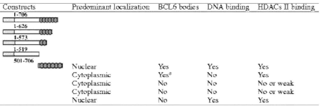
In a previous study characterizing the interactions between BCL6 and class II HDACs, several deletions within the BCL6 DNA-binding domain were created. Subcellular localization, presence of BCL6 bodies, and interaction with class II HDACs of the various truncated proteins are summarized in Table 1. In addition, we tested the capacity of these deletion mutants to interact with a BCL6 core DNA sequence [9]. Our data indicated that removal of the last two C-terminal fingers (BCL6 1–626) was sufficient to abolish DNA-binding ( Table 1). To further refine this analysis, we undertook a systematic mutagenesis of the individual ZF of BCL6.
Table 1. Characteristics of BCL6 deletion constructs
Data are summarized from [5] and from our unpublished data.
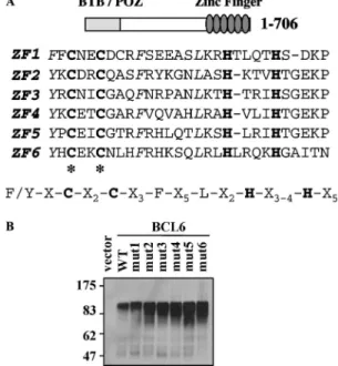
The BCL6 DNA-binding domain is composed of six C2H2 ZFs that fit the Krüppel-like consensus sequence [11]. The ZF forms a compact globular structure in which the two cysteines and the two histidines are tetrahedrally coordinated to a zinc ion. To disrupt the ZF architecture, we used PCR mutagenesis to replace the two cysteines of each finger with two glycine residues in the context of the full-length protein ( Fig. 1A). Mutants 1, 2, 3, 4, 5, and 6 corresponded to native BCL6 mutated in ZF1, 2, 3, 4, 5, and 6, respectively. The expression of wild type or mutated BCL6 proteins was verified after transient transfection of the corresponding plasmids into HeLa cells. Western blot analysis demonstrated that each of the mutant proteins (mut1 to mut6) was highly expressed in cells and similar in size to the wild type protein (Fig. 1B). The six cDNA plasmids containing a single mutated ZF were then used to address the functions of each ZF with regard to DNA binding, cellular localization, and transcriptional repression.
Fig. 1. (A) Sequence and alignment of the six ZFs of BCL6. The C2H2 backbone is in bold. The asterisk indicates the position of the cysteine to glycine mutations in each finger. (B) Mutant BCL6 proteins are expressed in cells. Plasmids encoding wild type BCL6 (wt) or mutants 1–6 were transiently transfected in HeLa cells. Total cell extracts were analyzed by Western blot with a polyclonal anti-BCL6 antibody (C19). Molecular weight markers are shown (kDa).
Mutation of ZF3–ZF6, but not ZF1 or ZF2, abrogates BCL6 transcriptional repression activity
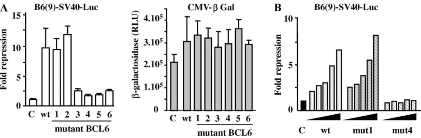
To investigate the effects of the mutations in the ZF domain, we first measured BCL6 transcriptional activity on a reporter gene. HeLa cells were transfected with a luciferase gene under the control of nine multimerized BCL6-binding sites upstream of the SV40 promoter (B6(9)-SV40-Luc) and expression vectors encoding wild type BCL6 or one of the various ZF mutants. A β-galactosidase reporter (pCMV-β) was used as an internal control for transfection efficiency and non-specific repression. No repression was observed with pCMV-β reporter, consistent with a lack of consensus BCL6-binding site in the CMV promoter (Fig. 2A). In the presence of wild type BCL6, we obtained a 10-fold reduction in luciferase activity, indicating that BCL6 efficiently represses transcription in this system (Fig. 2A). A similar transrepression was observed with mutants 1 and 2 (9.5- and 11.9-fold decrease, respectively) on B6(9)-SV40-Luc. Thus, these two proteins have the ability to bind to the reporter and to inhibit transcription to the same extent as the wild type protein. By contrast, BCL6 proteins carrying a mutation in any one of the remaining ZFs (mutants 3–6) exhibited a dramatic loss in their ability to repress transcription of the reporter (1.75- to 2.5-fold decrease). These results are not a reflection of low-level protein expression ( Fig. 1B). Moreover, transfection of HeLa cells with 25–500 ng expression plasmid encoding mutant 4 did not restore repressive activity, confirming that an intact ZF4 is critical to transrepressive function of BCL6. However, mutant 1 suppressed transcription from the reporter gene to levels equivalent to wild type protein (Fig. 2B). These results suggest that ZF3–ZF6 are required for the repressive activity of BCL6 while ZF1 and ZF2 do not participate in this event.
Fig. 2. Transcriptional activity of wild type and mutant BCL6 proteins. (A) Wild type and mutant 1–6 BCL6 plasmids were transfected in HeLa cells, with a luciferase reporter gene containing BCL6 binding sites (B6(9)-SV40-Luc) and pCMV-β plasmid to monitor transfection efficiency. Normalized luciferase values were expressed as fold repression over control (C: empty plasmid). Means ± SD of three independent transfections (left). β-galactosidase activities are not affected by BCL6 (right). (B) Increasing amounts (25, 50, 100, 250, and 500 ng) of plasmids encoding wt BCL6, mutant 1 or mutant 4 were transfected with B6(9)-SV40-luc and pCMV-β. DNA amount was kept constant with pcDNA empty vector. Data represent a typical experiment.
Cellular localization of over-expressed mutant BCL6
A possible explanation for the inability of mutants 3–6 to effectively eliminate transcription activity from the BCL6 reporter gene is an altered localization of the respective proteins. Using an antibody directed against BCL6, the compartmentalization of wild type and mutant proteins was assessed by immunofluorescent detection. Mutant 1, 2, 3, and 5 proteins exhibited a distribution undistinguishable from that of wild type BCL6: the proteins were predominantly in the nucleus and formed spherical nuclear bodies (Fig. 3 and data not shown). Interestingly, mutants 4 and 6 did not form the characteristic nuclear bodies but rather showed a discrete pattern of punctate staining (Fig. 3). Mutant 6 was also found consistently in the cytoplasm at a greater extent than the other proteins, suggesting that its nuclear localization signal was partially masked or disrupted. Our results argue that disruption of ZF4 or ZF6 leads to perturbed nuclear localization that may account for the failure of mutants 4 and 6 to inhibit transcription from BCL6 reporter genes. However, this explanation does not hold true for mutants 3 and 5 that exhibit a typical subcellular localization.
Fig. 3. Cellular localization of BCL6 mutant proteins. HeLa cells were transiently transfected with plasmids coding for wt and mutant BCL6. BCL6 protein was detected with anti-BCL6 C19 antibody and anti-rabbit Alexa 488 (FITC panel). Nuclei were counterstained with DAPI (DAPI panel). Mutants 1, 2, 3, and 5 are not shown as they display the same distribution as wild type BCL6.
Binding of mutant BCL6 to BCL6 DNA-binding sites
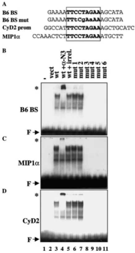
To further examine the functional roles of the ZFs, we tested the ability of the mutant proteins to interact with DNA. EMSA analysis was performed with unlabeled in vitro-translated proteins and a 32P-labeled-BCL6
consensus probe or with oligonucleotides whose sequences were found in the regulatory regions of cyclin D2 and
MIP1α (Fig. 4A). A specific binding of BCL6 was observed to the BCL6 consensus probe that could be further supershifted by an anti-BCL6 antibody, but not by a nonspecific antibody (Fig. 4B, lanes 3–5). No protein–DNA complexes were obtained with unprogrammed rabbit lysate or with a mutated probe (not shown). Under the same conditions, mutants 1 and 2 efficiently bound to the probe, whereas ZF mutants 3–6 were devoid of DNA binding activity ( Fig. 4B, lanes 6–11). Identical results were obtained with MIP1α (panel C) and cyclin D2 (panel D) probes that contained the BCL6 core consensus site with different flanking sequences. These DNA-binding assays indicate that mutation of ZF 3, 4, 5 or 6 results in a complete loss in the ability of BCL6 to interact with its cognate cis element.
Fig. 4. DNA-binding ability of mutant BCL6 proteins: EMSA analysis. (A) Sequence of the various DNA probes used in gel shift assay. The core BCL6 binding site is boxed. (B) Unlabeled in vitro-translated proteins were incubated with the 32P-labeled BCL6 consensus DNA probe. In some cases, the proteins were first incubated with 200 ng anti-BCL6 (N3, Santa Cruz, lane 4) or irrevelant antibody (anti-MyoD C20, Santa Cruz, lane 5).
Supershifts are indicated by an asterisk (*). Lane 1: probe alone; lane 2: probe with unprogrammed rabbit lysate. F: free probe. (C and D) Same experiment as in B using the MIP1α promoter (C) or the cyclin D2 (D) promoter probes.
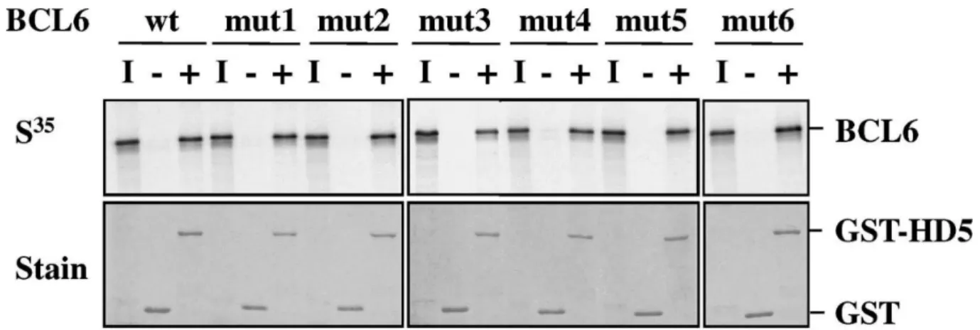
Interaction of mutant BCL6 with class II HDACs
We had previously demonstrated that BCL6 recruited class II HDACs through protein–protein interaction involving the ZF domain. Using deletion constructs in the ZF region, we showed that the last four ZFs (ZF3–ZF6) are required for the binding to HDAC5 and HDAC7 (Table 1). To determine the role of the individual ZFs as mediators of these interactions, we tested the ability of the ZF mutant proteins to form higher order complexes with class II HDACs. GST protein or GST-HDAC5 123–292, a fusion protein containing the HDAC5 region necessary for BCL6 interaction [5], was incubated with 35S-labeled BCL6. Bound proteins were analyzed by SDS–PAGE and
autoradiography. Data indicated that wild type and mutant BCL6 proteins interacted with GST-HDAC5 but not with GST alone (Fig. 5). No differences in binding affinity of HDAC5 for BCL6 or its mutant versions could be detected. Moreover, double immunodetection of full-length HDAC5 co-expressed with BCL6 in U2OS cells confirmed the fact that wild type and each of the six mutant proteins co-localized with HDAC5 (not shown). Our results suggest that BCL6 binding to HDAC5 does not rely on the integrity of a single ZF.
Fig. 5. Interaction of mutant BCL6 proteins with class II HDAC. GST pull-down assays were performed with GST (− lanes) or GST-HDAC5 123-292 (+ lanes) proteins and 35S-labeled BCL6 proteins synthesized in vitro. Protein:protein complexes were resolved by SDS-PAGE and visualized by autoradiography (top panel). Similar amounts of GST and GST-HDAC5 were used as shown by the Coomassie blue staining of the gels (lower panel). I: 10% input of 35S proteins.
Discussion
We show here by point mutation analysis that the six ZFs of BCL6 are not equivalent in DNA-binding activity. Similar findings were reported for the POK proteins ZF5 and Kaiso [12 and 13]. The described experiments differed from our work though, as the ZF region of these two proteins was not examined in the context of the full-length protein.
C2H2 zinc fingers are the most common DNA-binding motifs found in eukaryotic transcription factors. Our present knowledge of how ZFs bind specifically to DNA comes largely from several X-ray structures ([14] for review). In the protein–DNA complexes, the α-helix of each ZF binds in the major groove of the DNA. In the first structure analyzed, the three ZFs of Zif268 recognize successive, overlapping base pair quartets in the major groove covering a total of 10 bp, and each ZF makes a critical set of base contacts [14]. Crystal structure or NMR studies of BCL6 ZF domain bound to DNA are not available but information might be extrapolated from other published C2H2 ZF structures.
Based on the 3 bp per finger recognition theory, the binding site for BCL6 should cover at least 18 bp, assuming that the six fingers are all making contact with DNA. Several groups have isolated a consensus BCL6-binding site using a random oligonucleotide selection technique [1]. Although the importance of flanking sequence cannot be ignored, the highest affinity site has only a 9-bp core sequence, suggesting that all the ZFs are not used to make contact with DNA. This hypothesis would be in agreement with our EMSA data. Other lines of evidence also reveal the fact that in several proteins with multiple ZFs, not all of them make contact with DNA, as in the factors Gli [15] and TFIIIA [16], for example.
Beside the ZF backbone, the H–C link between adjacent fingers plays an active structural role in stabilization of the protein–DNA complex and in controlling the spacing of the ZF along the DNA. The most common linker has five residues and matches a consensus sequence TGEKP [14]. In the case of BCL6, a canonical linker is found
between fingers 2–6 ( Fig. 1A). The linker between ZF1 and ZF2 is slightly different as it is composed only of four residues (SDKP). This discrepancy maybe explains why ZFs 1 and 2 are not in the most favorable conformation to interact with DNA. Nevertheless, a report indicates a 96% evolutionary conservation between human, mouse, frog, and chicken BCL6 [1] within the entire ZF domain, including ZFs 1 and 2. This suggests that ZFs 1 and 2 are probably involved in other conserved biological function(s). First, it is possible that ZF1 and/or ZF2 are required to bind to DNA sequences not (or distantly) related to those we used in this study, like that identified in the IL5 gene [17]. In this regard, the 11 C2H2 ZF CTCF transcription factor uses different sets of ZFs to bind to different DNA sequences [18]. Another possibility is that ZF1 and ZF2 are involved in protein–protein interaction. In this case, BCL6 ZF region would parallel what has been proposed for other POK proteins. Indeed, it has been shown that the last seven ZFs of PLZF are necessary and sufficient for specific binding to DNA while the two first were reported to mediate protein–protein interaction with PML [19]. Moreover, it has been recently reported that the ZF1 of the POK protein Kaizo is dispensable for the binding to two unrelated DNA-target sequences, but it may be involved in the interaction between Kaizo and p120ctn [13]. Whether ZF1 and/or ZF2 of BCL6 are involved in protein–protein interaction, notably with JUN transcription factors, remains to be tested. Note however that our present results appear to exclude that their individual mutation affects the interaction between the ZF region of BCL6 and HDAC5. Finally, a third possibility would be that ZF1 and ZF2 are required for BCL6 to bind to endogenous, chromatinized target genes, a property that may become dispensable in our transient assays.
Acknowledgements
We thank Dr. L.M. Staudt (NCI, Bethesda) for the B6(9)-SV40-Luc reporter, L. Barki and S. Brown for help with microscopy and digital imaging, B. Peyrusse for assistance with figures, and Dr. J. Gaffé and Dr. S.E. Johnson (Pennsylvania State University) for their careful reading and critical comments on the manuscript. This work was supported by INSERM, CNRS, and l’Association pour la Recherche contre le Cancer (ARC).
References
1. A.L. Dent, F.H. Vasanwala and L.M. Toney , Regulation of gene expression by the proto-oncogene BCL-6. Crit. Rev. Oncol. Hematol.
41
(2002), pp. 1–9.2. O. Albagli, C. Lindon, S. Lantoine, S. Quief, E. Puvion, C. Pinset and F. Puvion-Dutilleul , DNA replication progresses on the periphery of nuclear cores formed by the BCL6 transcription factor. Mol. Cell. Biol.
20
(2000), pp. 8560–8570.3. A.L. Shaffer, X. Yu, Y. He, J. Boldrick, E.P. Chan and L.M. Staudt , BCL-6 represses genes that function in lymphocyte differentiation, inflammation, and cell cycle control. Immunity
13
(2000), pp. 199–212.4. L.M. Toney, G. Cattoretti, J.A. Graf, T. Merghoub, P.P. Pandolfi, R. Dalla-Favera, B.H. Ye and A.L. Dent , BCL-6 regulates chemokine gene transcription in macrophages. Nat. Immunol.
1
(2000), pp. 214–220.5. C. Lemercier, M.-P. Brocard, F. Puvion-Dutilleul, H.Y. Kao, O. Albagli and S. Khochbin , Class II histone deacetylases are directly recruited by BCL6 transcriptional repressor. J. Biol. Chem.
277
(2002), pp. 22045– 22052.6. F.H. Vasanwala, S. Kusam, L.M. Toney and A.L. Dent , Repression of AP-1 function: a mechanism for the regulation of Blimp-1 expression and B lymphocyte differentiation by the B cell lymphoma-6 protooncogene. J. Immunol.
169
(2002), pp. 1922–1929.7. P. Dhordain, O. Albagli, N. Honore, F. Guidez, D. Lantoine, M. Schmid, H.D. The, A. Zelent and M.H. Koken , Colocalization and heteromerization between the two human oncogene POZ/zinc finger proteins, LAZ3 (BCL6) and PLZF. Oncogene
19
(2000), pp. 6240–6250.8. C. Lemercier, R.Q. To, R.A. Carrasco and S.F. Konieczny , The basic helix–loop–helix transcription factor Mist1 functions as a transcriptional repressor of MyoD. EMBO J.
17
(1998), pp. 1412–1422.9. C.C. Chang, B.H. Ye, R.S. Chaganti and R. Dalla-Favera , BCL-6, a POZ/zinc-finger protein, is a sequence-specific transcriptional repressor. Proc. Natl. Acad. Sci. USA
93
(1996), pp. 6947–6952.10. C. Lemercier, A. Verdel, B. Galloo, S. Curtet, M.P. Brocard and S. Khochbin , mHDA1/HDAC5 histone deacetylase interacts with and represses MEF2A transcriptional activity. J. Biol. Chem.
275
(2000), pp. 15594– 15599.11. J. Turner and M. Crossley , Mammalian Krüppel-like transcription factors: more than just a pretty finger.
Trends Biochem. Sci.
24
(1999), pp. 236–240.12. T. Obata, A. Yanagidani, K. Yokoro, M. Numoto and S. Yamamoto , Analysis of the consensus binding sequence and the DNA-binding domain of ZF5. Biochem. Biophys. Res. Commun.
255
(1999), pp. 528–534.13. J.M. Daniel, C.M. Spring, H.C. Crawford, A.B. Reynolds and A. Baig , The p120(ctn)-binding partner Kaiso is a bi-modal DNA-binding protein that recognizes both a sequence-specific consensus and methylated CpG dinucleotides. Nucleic Acids Res.
30
(2002), pp. 2911–2919.14. S.A. Wolfe, L. Nekludova and C.O. Pabo , DNA recognition by Cys2His2 zinc finger proteins. Annu. Rev. Biophys. Biomol. Struct.
29
(2000), pp. 183–212.15. N.P. Pavletich and C.O. Pabo , Crystal structure of a five-finger GLI–DNA complex on zinc fingers. Science
261
(1993), pp. 1701–1707.16. R.T. Nolte, R.M. Conlin, S.C. Harrison and R.S. Brown , Differing roles for zinc finger in DNA recognition: structure of a six-finger transcription factor IIIA complex. Proc. Natl. Acad. Sci. USA
95
(1998), pp. 2938–2943.17. M. Arima, H. Toyama, H. Ichii, S. Kojima, S. Okada, M. Hatano, G. Cheng, M. Kubo, T. Fukuda and T. Tokuhisa , A putative silencer element in the IL-5 gene recognized by Bcl6. J. Immunol.
169
(2002), pp. 829–836.18. G.N. Filippova, S. Fagerlie, E.N. Klenova, C. Myers, Y. Dehner, G. Goodwin, P.E. Neiman, S.J. Collins and V.V. Lobanenkov , An exceptionally conserved transcriptional repressor, CTCF, employs different combinations of zinc fingers to bind diverged promoter sequence of avian and mammalian c-myc oncogenes. Mol. Cell. Biol.
16
(1996), pp. 2802–2813 |19. A. Melnick and J.D. Licht , Deconstructing a disease: RARα, its fusion partners, and their roles in the pathogenesis of acute promyelocytic leukemia. Blood