HAL Id: hal-00408485
https://hal.archives-ouvertes.fr/hal-00408485
Submitted on 4 Aug 2009
HAL is a multi-disciplinary open access
archive for the deposit and dissemination of
sci-entific research documents, whether they are
pub-lished or not. The documents may come from
teaching and research institutions in France or
abroad, or from public or private research centers.
L’archive ouverte pluridisciplinaire HAL, est
destinée au dépôt et à la diffusion de documents
scientifiques de niveau recherche, publiés ou non,
émanant des établissements d’enseignement et de
recherche français ou étrangers, des laboratoires
publics ou privés.
timing at the INK4a/ARF locus during senescence.
Hanane Agherbi, Anne Gaussmann-Wenger, Christophe Verthuy, Lionel
Chasson, Manuel Serrano, Malek Djabali
To cite this version:
Hanane Agherbi, Anne Gaussmann-Wenger, Christophe Verthuy, Lionel Chasson, Manuel Serrano,
et al.. Polycomb mediated epigenetic silencing and replication timing at the INK4a/ARF locus
during senescence.. PLoS ONE, Public Library of Science, 2009, 4 (5), pp.e5622.
�10.1371/jour-nal.pone.0005622�. �hal-00408485�
Polycomb Mediated Epigenetic Silencing and Replication
Timing at the
INK4a/ARF
Locus during Senescence
Hanane Agherbi1., Anne Gaussmann-Wenger1., Christophe Verthuy1, Lionel Chasson1, Manuel Serrano2, Malek Djabali1*
1 Centre d’immunologie INSERM-CNRS de Marseille Luminy, Marseille, France, 2 Spanish National Cancer Research Center (CNIO), Madrid, Spain
Abstract
Background: The INK4/ARF locus encodes three tumor suppressor genes (p15Ink4b, Arf and p16Ink4a) and is frequently
inactivated in a large number of human cancers. Mechanisms regulating INK4/ARF expression are not fully characterized.
Principal Findings: Here we show that in young proliferating embryonic fibroblasts (MEFs) the Polycomb Repressive Complex 2 (PRC2) member EZH2 together with PRC1 members BMI1 and M33 are strongly expressed and localized at the INK4/ARF regulatory domain (RD) identified as a DNA replication origin. When cells enter senescence the binding to RD of both PRC1 and PRC2 complexes is lost leading to a decreased level of histone H3K27 trimethylation (H3K27me3). This loss is accompanied with an increased expression of the histone demethylase Jmjd3 and with the recruitment of the MLL1 protein, and correlates with the expression of the Ink4a/Arf genes. Moreover, we show that the Polycomb protein BMI1 interacts with CDC6, an essential regulator of DNA replication in eukaryotic cells. Finally, we demonstrate that Polycomb proteins and associated epigenetic marks are crucial for the control of the replication timing of the INK4a/ARF locus during senescence.
Conclusions:We identified the replication licencing factor CDC6 as a new partner of the Polycomb group member BMI1. Our results suggest that in young cells Polycomb proteins are recruited to the INK4/ARF locus through CDC6 and the resulting silent locus is replicated during late S-phase. Upon senescence, Jmjd3 is overexpressed and the MLL1 protein is recruited to the locus provoking the dissociation of Polycomb from the INK4/ARF locus, its transcriptional activation and its replication during early S-phase. Together, these results provide a unified model that integrates replication, transcription and epigenetics at the INK4/ARF locus.
Citation: Agherbi H, Gaussmann-Wenger A, Verthuy C, Chasson L, Serrano M, et al. (2009) Polycomb Mediated Epigenetic Silencing and Replication Timing at the INK4a/ARF Locus during Senescence. PLoS ONE 4(5): e5622. doi:10.1371/journal.pone.0005622
Editor: Mikhail V. Blagosklonny, Roswell Park Cancer Institute, United States of America Received February 24, 2009; Accepted April 18, 2009; Published May 20, 2009
Copyright: ß 2009 Agherbi et al. This is an open-access article distributed under the terms of the Creative Commons Attribution License, which permits unrestricted use, distribution, and reproduction in any medium, provided the original author and source are credited.
Funding: This work was supported by grants from CNRS, ARC, and FRM. AH is funded by an ARC fellowship. AGW is funded by a Dr. Mildred Scheel fellowship of the Deutsche Krebshilfe. Histological analyses were performed on the mouse functional genomics platform RIO/Marseille-Nice Genopole with support from the INCA PROCAN program. The funders had no role in study design, data collection and analysis, decision to publish, or preparation of the manuscript. Competing Interests: The authors have declared that no competing interests exist.
* E-mail: djabali@ciml.univ-mrs.fr
.These authors contributed equally to this work.
Introduction
Cellular senescence is a fundamental cellular program that is activated after a finite number of cell divisions and operates to avoid further cell proliferation. In addition cellular senescence constitutes a tumor suppressor mechanism [1,2]. The tumor suppressor pathways, ARF/MDM2/p53 and p16INK4a/Rb, have been shown to play critical roles in the induction of cellular senescence [3]. Studies on Polycomb group genes (Pc-G) have demonstrated that beside their function in controlling the expression of Hox genes, Pc-G play a central role in cell proliferation through the repression of the INK4/ARF locus [4,5,6]. This locus encodes both p16INK4a, which prevents inactivation of the tumor suppressor RB, and p19ARF, which stabilizes the tumor suppressor p53 [7,8]. Evidence supporting the direct control of the cell cycle by Pc-G proteins in vertebrates came from studies on mouse Bmi1 mutants. Bmi1 was first identified as a proto-oncogene that cooperates with c-Myc to promote the generation of mouse B- and T-lymphomas [9]. Mice
lacking Bmi1 exhibit strong proliferative defects during lymphocyte development. In the absence of Bmi1, M33, or Phc2, primary embryonic fibroblasts (MEFs) are unable to progress into S phase, undergo premature senescence after only a few passages in culture and show an increased accumulation of the tumor suppressors p16INK4a, p19ARF and p15INK4b [4,10]. Generation of Bmi1/
Ink4a/Arf compound mutant mice have provided genetic evidence that at least part of these defects are due to activation of the INK4a/ARF locus [11].
Pc-G and Trx-G proteins function in distinct multiprotein complexes which control transcription by altering the structure of chromatin, organizing it into either a ‘‘closed’’ or an ‘‘open’’ conformation. The Trx-G protein MLL1 mediates lysine-directed histone methylation [12,13]. Methylation on lysine-4 of histone H3 (H3K4me2 and H3K4me3) is associated with a permissive and transcriptionally active state of the chromatin [14]. Pc-G proteins are transcriptional repressors that functionally can be separated into at least two different complexes: the initiation complex, Polycomb complex 2 (PRC2), which in humans consists of EZH2,
EED, and SUZ12; and the maintenance complex, PRC1, with the core proteins RNF2, HPC, and BMI1. Both PRC1 and PRC2 complex members have been linked to cell cycle control. EZH2 is the active component of PRC2 through its SET domain histone methyltransferase activity specific for Lys 27 (K27) of histone H3 and K26 of histone H1 [15,16]. It has been demonstrated that PRC2 is required for PRC1 binding to chromatin [17], presumably achieved through binding of HPC protein to H3K27me3 [15]. It has recently been shown that Polycomb proteins are bound to the INK4a/ARF locus and dissociated during senescence [18]. We have now extended this study and demonstrate that both Polycomb and the trithorax (Trx-G) member MLL1 are localized at this locus and importantly associated to the Regulatory Domain (RD) of the INK4/ARF locus identified as a DNA replication origin and as a global transcriptional regulator of the entire locus [19,20]. Also, we demonstrate that BMI1 interacts with the licencing factor CDC6. Finally, we show that the late timing of replication of the INK4a/ ARF locus in young proliferating MEFs shifts to an early replication timing in senescent and notably in PRC2 mutant MEFs. Together our results demonstrate that MLL1 and Polycomb group genes directly control the INK4a/ARF locus through epigenetic chromatin modifications and that the loss of repressive epigenetic marks both in senescent and Polycomb mutant cells leads to a shift of the replication timing of the INK4a/ ARF locus.
Materials and Methods
Mouse embryonic fibroblasts (MEFs)
Day-12p.c. wild type, M33 mutant and Bmi1 mutant embryos were mechanically dissociated into single cells and cultured in DMEM with 10% FCS and penicillin and streptomycin (Invitro-gen, Breda, The Netherlands). MEFs were passaged every two to three days and viable cells were counted by Trypan Blue (Invitrogen) exclusion. Senescent cells were detected using the beta-Galactosidase Staining Kit (BioVision) following the manu-facturer’s recommendations.
Quantification of mRNA levels by Q-PCR
For reverse transcriptase Q-PCR (Q-RT-PCR) analysis, total RNA was extracted from MEFs with the RNeasy Mini Kit (QIAGEN) according to the manufacturer’s protocol. cDNA was synthesized from 1 mg of total RNA using the QuantiTect Reverse Transcription Kit (QIAGEN). Q-PCR amplification of either mouse Ink4a transcripts or Arf was performed with the SYBR Green PCR Master Mix Kit (Applied Biosystems) in a 7500 Real-Time PCR System From Applied Biosystems. Primers used are given in Table 1. Q-RT-PCR analysis of Gapdh mRNA expression was performed as a positive control and for normalization.
Retroviral transduction of embryonic fibroblasts (MEFs)
Ecotropic virus producer cells (Phoenix) were transfected with pMSCV-Myc-cdc6 or the empty vector (mock) using Lipofectami-neTM 2000 Reagent (Invitrogen). Retroviral supernatant was collected after 24 h. For transduction wild type and Bmi2/2MEFs were plated in 6-well plates. Cells were incubated with the viral supernatant and Polybrene (4 mg/ml) for 30 min before centrifu-gation (2000 rpm, 32uC, 60 min). After 12 h incubation (10% CO2,
37uC) cells were washed and incubated with fresh medium.
Immunocytochemistry
MEFs (wild type and BMI2/2) were transduced with pMSCV-Myc-cdc6 or the empty vector (mock) at passage P3. 72 h after
transduction, cells were plated on glass coverslips, fixed in 4% paraformaldehyde and permeabilized with cold PBS containing 0.2% Triton X-100. After blocking, the cells were incubated with primary antibodies (mouse anti-Cdc6 (sc-9964; Santa Cruz Biotechnology), rabbit anti-p16INK4a (sc-1207; Santa Cruz Bio-technology), washed with secondary antibodies, stained with DAPI, and mounted before viewing.
Chromatin immunoprecipitation assays
ChIP were performed and analyzed essentially as described [16]. The antibodies used were rabbit anti-H3acetyl (06-599, Upstate Biotechnology), rabbit anti-H3K27me3 (07-449; Upstate Biotechnology), rabbit anti-M33 (kind gift of Dr. Higashinaka-gawa, T) [21], rabbit anti-H3K4Me3 (05-745; Upstate Biotech-nology), rabbit anti-EZH2 (07-689; Upstate BiotechBiotech-nology), goat anti-BMI1 (sc-8906; Santa Cruz Biotechnology), anti-MLL (05-765; Upstate Biotechnology). For normal ChIP, the immunopre-cipitated DNA was quantified by real-time Q-PCR (Applied Biosystems) and normalized with the 1/5 input. The sequences of the PCR primers are listed in Table 1.
Coimmunoprecipitations
NIH3T3 cells were transiently transfected with pCG-HAcdc6 using LipofectamineTM 2000 Reagent (Invitrogen) following the manufacturer’s instructions. 72 h after transfection CoIP were performed using Nuclear Complex Co-IP Kit (active motif) with mouse anti-HA (2367; Cell Signaling). Thymocytes were isolated from 5 to 7 weeks old mice and protein extraction and CoIP were performed using Nuclear Complex Co-IP Kit (active motif) with mouse anti-BMI1 (05-637; Upstate Biotechnology). Western blotting procedure and reagents for western blot analysis were previously described [4]. Antibodies used were mouse anti-CDC6 (sc-9964; Santa Cruz Biotechnology), goat anti-BMI1 (sc-8906; Santa Cruz Biotechnology).
Table 1. Sequences of Primers used for ChIP and gene expression analysis Primer Sequence 59-39 RDINK4a/ARF fwd TTCCTATTTCGCTGTAGCAAC RDINK4a/ARF rev AACTAACCAGGCCTCCTCCCA Exon 1b fwd AGGTGCCTCAACGCCGAAG Exon 1b rev CTGGTCCAGGATTCCGGTGCGG Exon 2 fwd ATGGGCAACGTTCACGTAGCAGC Exon 2 rev AGCGGTACACAAAGACCACCCA p19ARF
fwd TCGCAGGTTCTTGGTCACTGT p19ARF
rev TGCTACGTGA ACGTTGCCCA p16INK4afwd GTGTGCATGACGTGCGGG p16INK4a
rev GCAGTTCGAATCTGCACCGTAG Ezh2 fwd AGGATACAGCCTGTGCACATCATGA Ezh2 rev GATCCAGAACTTCATCCCCCATAT Bmi-1 fwd CCAGCAAGTATTGTCCTATTTGTGA Bmi-1 rev TTGAAGAGTTTTATCTGACCCTTATGTT UTX fwd GCAAGTGCAGATACATGGTGTTC UTX rev GCCTAGATCCATCCAGGCTGC JMJD3 fwd GGAAGCCACAGCTACAGGAGC JMJD3 rev CCCAATAGTGCTCATGTACCGC doi:10.1371/journal.pone.0005622.t001
Replication timing analysis
BrdU labeling, fixation in cold 70% ethanol, cell cycle fractionation by flow cytometry and isolation of BrdU-labeled DNA by immunoprecipitation were carried out as previously described [22].
Results and Discussion
Polycomb expression and cellular senescence in mouse embryonic fibroblasts
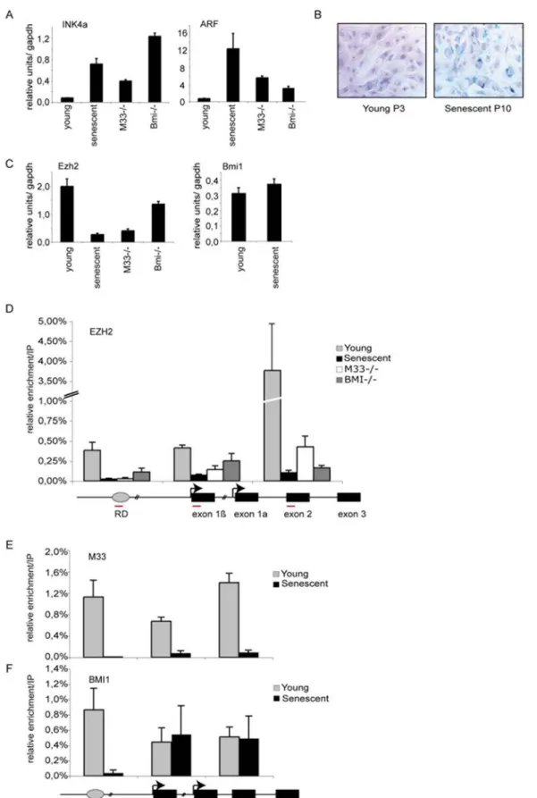
To study cellular senescence we have used serial passaging of mouse embryonic fibroblasts (MEFs) cultured from 12-day-old C57BL/6 embryos. As already described, MEFs were able to undergo a limited number of population doublings (P12) before they senesced. Senescence was assessed by monitoring the endogenous B-galactosidase (B-gal) activity at pH 6 (Fig. 1B). At this stage (P10), cells overexpress both p16Ink4a and p19Arf. As expected knock out cells derived from PRC1 members Bmi1 and M33 overexpress both p16Ink4aand p19Arfas soon as passage P3 [4] (Fig. 1A). Since EZH2 is required for Polycomb silencing we measured differences in expression levels of Ezh2 in young and senescent MEFs by Q-RT-PCR (Fig. 1C). Protein levels of EZH2 (not shown) in MEFs correlated with RNA levels; Ezh2 is more abundant in early passage (P3) than in senescent MEFs (P10) (Fig. 1C). In PRC1 mutant cells (M33, Bmi1) the expression level of Ezh2 is strongly diminished. Interestingly, expression level of the PRC1 member Bmi1 is not modified in senescent cells as compared to young proliferating cells (Fig. 1C) [18].
Polycomb proteins are localized at the RDINK4a/ARF
The RDINK4a/ARFhas been identified as a putative DNA replication origin that assembles a multiprotein complex contain-ing CDC6 and that coincides with a conserved non codcontain-ing DNA element identified as a transcriptional regulatory element [20]. Whether PRC1 and PRC2 members bind directly toRDINK4/ARF had not been tested yet. In order to assess this question, we performed ChIP assays to examine whether EZH2 directly binds this transcriptional regulatory element. Oligonucleotide primers were designed in order to examine in young proliferating MEFs the distribution of EZH2 at the RD element and along the INK4a/ ARF locus. We found that EZH2 is bound to the first exon of ARF, exon 1b, and with a maximum peak to the shared exon of INK4a and Arf, exon 2, in early-passage MEFs (Fig. 1D). Interestingly, we found that both EZH2 and PRC1 members BMI1 and M33 are also localized at the RD origin of replication in young proliferating MEFs. In contrast, in senescent cells most of the bound EZH2 and M33 protein was lost at all the examined sites along the INK4a/ ARF locus (Fig. 1D and E). A significant part of BMI1 is still retained at both exon 1b (p19ARF) and at the shared exon 2 upon senescence (Fig. 1F) however interestingly, BMI1 completely disappeared from RD (Fig. 1F). Since EZH2 and M33 are lost from the entire locus in senescent cells, these results suggest that BMI1 may bind to some parts of the locus (exon 1b and exon 2) in a manner that is independent of EZH2 and M33, but its binding to RD appears dependent on those two proteins. It has recently been shown that the BMI1 protein was dissociated from the locus at senescence [18]. While we do not explain this difference, the detected BMI1 protein bound at the locus correlates well with the fact that the expression level of Bmi1 is not modified in senescent cells. However, it was demonstrated using genome wide analysis that Polycomb domains can be segregated in two classes: the first occupied by both PRC2 and PRC1 (PRC1-positive) and the second specifically bound by PRC2 (PRC2-only) [23,24].
Several experiments indicate that H3K27 methylation by E(z) has a critical function in the establishment of transcriptional repression of PRC2 target genes. It has been demonstrated that the PRC2 complex containing E(z)/EZH2 is an active enzyme capable of methylating the histone H3 tail at lysine-9 (K9) and more importantly at K27 [15,25]. In E(z) Drosophila mutants the loss of functional E(z) induces a loss of Pc-G protein binding to polytene chromosomes [26]. Methylation of histone H3K27 by E(z) protein is strictly required for maintenance of HOX gene silencing in Drosophila [27]. Drosophila Polycomb (Pc or M33 in mouse), a core subunit of PRC1, selectively binds to histone H3 tail peptide trimethylated at K27, suggesting that H3K27me3 may contribute to targeting of PRC1 to HOX genes [28,29]. We therefore examined the status of methylation of histone H3 at the RD element and at the INK4a/ARF locus in young, senescent and Polycomb mutant cells. As shown in figure 2, H3K27me3 marks are lost from the RD element in senescent cells and from the shared exon 2 in senescent, Bmi12/2and M33 mutant MEFs. The loss of the H3K27 repressive mark correlates with the higher transcription of Arf and Ink4a in senescent and Polycomb mutant cells (Fig. 1A). Histone acetylation is generally viewed as a central switch that allows exchange between permissive and repressive chromatin domains in terms of transcriptional competence. It was shown that the levels of p19Arfare strongly upregulated in murine cells treated with histone deacetylase inhibitors (HDACis) [30]. Yet, examination of acetylation of histone H3(K9,K14) in actively cycling and senescent cells shows low level of H3 acetylation at the INK4a/ARF locus (Fig. 2). In contrast, we observe a strong enrichment of acetylated H3 at the Arf promoter in M33 and Bmi1 knockout MEFs (Fig. 2) similar to the effects observed after treatment with HDACis [30].
MLL1 is recruited to the INK4a/ARF locus during senescence
In Drosophila and in mouse the activity of Trx-G/MLL complexes is required to prevent Pc-G-mediated silencing of transcribed Hox genes [31,32,33,34]. MLL1 protein complexes catalyze the trimethylation of H3K4, which is generally associated with active transcription [35]. Accordingly, H3K4me3-modified nucleosomes are specifically enriched at the promoters of active genes [36]. We therefore monitored the binding of the MLL1 protein and the associated H3K4me3 positive transcriptional mark at the RD element and at the INK4a/ARF locus in MEFs during senescence and in Polycomb mutant cells. MLL1 was bound to the RD element and to both exon 1b and p16INK4a/p19ARFshared exon 2 in young cells (Fig. 3A). However, in both senescent and Polycomb mutant cells we observe a strong enrichment of MLL1 binding at the locus demonstrating that MLL1 participates to the transcription of Arf and Ink4a. Surprisingly, we did not observe a similar increase of the H3K4 methyl mark during senescence and in mutant cells (Fig. 3A). This positive mark is equally present in young, senescent or Polycomb mutant cells at the INK4a/ARF locus. Patterns of methylation at lysine 4 and 27 of histone H3 have been associated with gene activation and repression that are developmentally regulated and are thought to elicit the coordina-tion of lineage specific gene expression programs [37]. Interest-ingly, in ES cells, the Polycomb Hox target gene promoters often display both H3K4me3 and H3K27me3 marks, and such regions, containing both repressing and activating chromatin modifica-tions, were referred to as ‘‘bivalent domains’’ [38]. In stem cells, these bivalent domains may keep selected genes ‘‘poised’’ for activation. H3K27me3 is a rather stable modification, which could be progressively lost in the absence of PRC2, along with cell divisions [39]. However, studies in ES cells indicated that changes
Figure 1. Analysis of Polycomb EZH2, M33 and BMI1 binding at theINK4a/ARFregion. A) qPCR analysis of p16INK4aand p19ARFin young (P3),
senescent (P10) and in Bmi1 and M33 mutant MEFs. B) B-galactosidase staining to detect senescent cells at passage 3 (P3) and passage 10 (P10). C) qPCR analysis of the mRNA levels of Polycomb EZH2 and Bmi1 in the indicated cells. D–F) Schematic diagram of the INK4a/ARF locus: amplified regions that were tested in ChIP experiments are indicated by red bars (sequences are given in Table 1). Wild type P3 (young), P10–12 (senescent), M332/2 (P4) and Bmi2/2 (P4) MEFs were subjected to ChIP assays using anti EZH2, BMI1 and M33 antibodies. DNA enrichment was calculated as described in Materials and Methods. Bars represent the mean+/2s.d. of quantifications from two to four separate immunoprecipitations analyzed in triplicate.
in chromatin associated with Hox gene activation are likely to occur promptly, and involving an appropriate demethylase activity. We therefore monitored the expression of both Jmjd3 and Utx H3K27 histone demethylases. As shown in figure 3B expression of Utx is not modified in young, senescent or M33 mutant cells. However, transcription of Jmjd3 is significantly induced in senescent MEFs. These results strongly suggest that the upregulation of Jmjd3 (Fig. 3B) and the downregulation of Ezh2 (Fig. 1C) are critical determinants of the transcriptional activation of the INK4/ARF locus during senescence. It has been shown that UTX can interact with components of the MLL2 complex [40,41]. This physical association between enzymes removing the H3K27me3 repressive mark, on the one hand, with protein complexes promoting the deposition of the active H3K4me3 mark, on the other hand, suggests that both activities are required for a rapid and stringent response of target genes. MLL1 is cleaved by Taspase1, generating an N-terminal and a C-terminal fragment, which can heterodimerize in vitro [42,43]. In Drosophila it was demonstrated that TRX-N is present at thousand genomic sites, where no Pc-G binding can be observed. However, it was shown that TRX-C is strongly bound at Pc-G binding sites [44]. These results suggest the C-terminal part of TRX is specifically linked to Pc-G function. It was suggested that Pc-G proteins might repress transcription by anchoring the C-terminal portion of TRX
at Polycomb response elements (PREs/TREs) or that constitutive TRX-C binding at PREs/TREs might allow Pc-G target genes to switch their state upon transcriptional induction [44].
Physical and Functional Association of BMI1 and CDC6 in replication control
The identification of a DNA replication origin (RD) adjacent to INK4b [19,20] and its high degree of sequence conservation led to ask whether this domain might contribute to the regulation of transcription. Interestingly, overexpressing and loading CDC6 to the RD element, results in the transcriptional repression of all three genes in the INK4b–ARF–INK4a locus [45] leading to increased foci formation and enhanced transformation by oncogenic RAS. Importantly, silencing is accompanied by the recruitment of histone deacetylases and increased methylation of histone H3 on lysine 9 (H3K9), which are hallmarks of heterochromatin. We have demonstrated that both components of the PRC2 and PRC1 complex are localized at the RD element. This prompted us to search for possible physical interactions between the replication complex containing CDC6 and the components of Polycomb complexes. Co-Immunoprecipitation experiments using an antibody against BMI1 demonstrated that CDC6 is associated in a complex with BMI1 (Fig. 4B). Moreover using thymocytes we show that the CDC6-BMI1 interaction
Figure 2. Loss of EZH2 binding and H3K27me3 methylation at theINK4a/ARFlocus during senescence. P3 proliferating and P10–12 senescent MEFs were subjected to ChIP assays using the indicated antibodies.
doi:10.1371/journal.pone.0005622.g002
occurs in wild type non-transfected cells (Fig. 4C) indicating that the CDC6-BMI1 interaction is not due to the forced expression of CDC6 in transfected MEFs. In order to test if BMI1 is required for Ink4a/Arf repression mediated by CDC6, we transfected CDC6
in Bmi1 knock out and wild type MEFs. As already described [20], the forced expression of Cdc6 in wild type MEFs decreased the protein levels of ARF and INK4a (Fig. 4A,D). However, overexpression of Cdc6 in mutant Bmi1 cells failed to mediate
Figure 3. Recruitment of MLL1 at theINK4a/ARFlocus during senescence. A) ChIP analysis of the RD element, p19ARFexon 1b and p16/19 shared exon 2 using MLL-c antibody and H3K4me3 antibody. B) qPCR analysis of the mRNA levels of UTX and JMJD3 histone demethylase in the indicated cells.
Arf or Ink4a repression (Fig. 4A,D) demonstrating that CDC6 induced repression of the INK4a/ARF locus is dependent on Polycomb function.
Next we asked whether the localization of Polycomb proteins at an origin of replication together with the replication machinery [19] could affect replication of the locus. To investigate whether the
Figure 4. BMI1 interacts with CDC6 and is required for CDC6 repressing function. A) Western Blot analysis of Myc-CDC6 Wild type and Bmi1 mutant transduced cells. The antibodies used are indicated. GAPDH antibody is used as a loading control. Ab) quantitative PCR experiment showing p16INK4a and p19ARF expression in Myc-CDC6 transduced Bmi1 mutant cells. B) CDC6 interacts specifically with BMI1: HA immunoprecipitated proteins extracted from HA-CDC6 transfected cells were separated by SDS-PAGE and immunoblotted with a BMI1 antibody. C) anti-Bmi1 immunoprecipitated proteins extracted from wild type thymocytes and immunoblotted with a CDC6 antibody. D) BMI1 is required for INK4a CDC6 mediated repression. Wild type MEFs transfected with Myc-CDC6 were immunostained with a specific antibody against p16INK4a(red)
and CDC6 (green) middle panel. Untransfected cells are shown on the upper panel. Bmi1 mutant cells transfected with Myc-CDC6 (green) express high level of p16 (red) (Bottom panel).
doi:10.1371/journal.pone.0005622.g004
induction of senescence could result in a modification of the replication timing of the INK4a/ARF locus we examined young proliferating (P3), pre-senescent (P7) and Polycomb M33 mutant (P3) MEFs. The replication timing was assessed using a PCR based approach [22,46]. Non-synchronized cells were pulse labeled with 59Bromodeoxyuridine (BrdU), stained with propidium iodide (PI) and sorted according to DNA content by flow cytometry (Fig. 5). Newly synthesized DNA was isolated by immuno-precipitation with anti-BrdU antibody. As shown in figure 5 the exon 1b (p19ARF) is late replicating in young cells which do not express the Arf or Ink4a
genes; whereas this region becomes early replicating in pre-senescent and in M33 mutant cells when the Arf or Ink4a genes are expressed. In higher eukaryotes, it has been observed that the time of replication and transcriptional activity are often correlated; genes which are late replicating are not expressed while transcriptionally active regions are early replicating [47]. In addition, when the transcriptional stage of a gene switches from an active to inactive state, replication timing shifts from early- to late replicating. Importantly, it has recently been demonstrated that histone modifications at an origin of replication serve as a binary switch
Figure 5.INK4a/ARFtiming of replication. PCR based analysis of replication timing of the INK4a/ARF locus (exon 1b). BrdU pulse labeled cells were stained for DNA content with propidium iodide and sorted by flow cytometry into 5 cell cycle fractions (G1, S1, S2, S3 and G2M) according to DNA content. The Gbe D. melanogaster gene provides a control for recovery of BrdU-labeled DNA.
for controlling the timing of replication of the Beta-globin locus in human [48]. This replication switch is also observed in human diseases such as the fragile X syndrome. FMR1 silencing by the CGG expansion was shown to be mainly attributed to epigenetic regulated transcriptional silencing [49]. The Fmr1 gene normally transcribed is replicated early whereas it becomes silent and late replicating in patients [50,51]. In yeast it was recently shown that Swi6, an S. pombe counterpart of heterochromatin protein 1 (HP1), is required for early replication of the pericentromeric region and the mat locus [52]. In our study we show that in proliferating MEFs the INK4a/ARF locus is silent and late replicating whereas in Polycomb mutant the locus tends to be early replicating and expressed. It has recently been demonstrated in the Encode project that the H3K27me3 mark shows a positive correlation with late
replication of large DNA segments [53]. We have demonstrated in senescent and Polycomb mutant cells that the ‘‘bivalent’’ domain at the INK4a/ARF locus (H3K27me3 and H3K4me3) is resolved and the locus remains only enriched in H3K4me3 positive marks correlating with the recruitment of MLL1 protein. Jmjd3 overex-pression in senescent cells could indicate that this histone demethylase participates in removing the H3K27 marks at the INK4A/ARF locus. The epigenetic modifications could be respon-sible for the observed replication-timing shift at senescence (Fig. 6). Together, our results demonstrate that MLL1 and Polycomb group genes directly control the INK4a/ARF locus through chromatin epigenetic modifications and that the loss of the repressive epigenetic marks both in senescent and Polycomb mutant cells at an origin of replication leads to a shift of the replication timing of the locus.
Figure 6. Model for Pc-G and MLL1 proteins in regulation of cellular senescence at theINK4a/ARFlocus. (A) In young proliferating cells, the PRC2 complex is bound at RD and at the INK4a/ARF locus and maintains the levels of H3K27me3. This allows the association of M33 and BMI1-containing PRC1 complex and repression of the INK4a/ARF genes. (B) In senescent or Polycomb mutant cells binding of EZH2 is lost, leading to the disruption of the PRC2 complex, the loss of H3K27me3 and to the recruitment of the MLL1 protein. We propose a model in which Polycomb/MLL1 and JMJD3 epigenetic modifications at the RD element impact the replication timing and the expression of the locus. Moreover, in senescent cells BMI1 binding is specifically lost at the RD element.
doi:10.1371/journal.pone.0005622.g006
Acknowledgments
We thank Philippe Naquet and Pierre Golstein for critical reading of the manuscript and for helpful discussions.
Author Contributions
Conceived and designed the experiments: HA AGW MD. Performed the experiments: HA AGW CV LC MD. Analyzed the data: HA AGW MD. Contributed reagents/materials/analysis tools: MS. Wrote the paper: HA AGW MS MD.
References
1. Campisi J (2000) Cancer, aging and cellular senescence. In Vivo 14: 183–188. 2. Dimri GP, Campisi J (1994) Molecular and cell biology of replicative senescence.
Cold Spring Harb Symp Quant Biol 59: 67–73.
3. Sherr CJ, DePinho RA (2000) Cellular senescence: mitotic clock or culture shock? Cell 102: 407–410.
4. Core N, Joly F, Boned A, Djabali M (2004) Disruption of E2F signaling suppresses the INK4a-induced proliferative defect in M33-deficient mice. Oncogene 23: 7660–7668.
5. Sasaki M, Ikeda H, Sato Y, Nakanuma Y (2006) Decreased expression of Bmi1 is closely associated with cellular senescence in small bile ducts in primary biliary cirrhosis. Am J Pathol 169: 831–845.
6. Gil J, Peters G (2006) Regulation of the INK4b-ARF-INK4a tumour suppressor locus: all for one or one for all. Nat Rev Mol Cell Biol 7: 667–677. 7. Sherr CJ (2001) The INK4a/ARF network in tumour suppression. Nat Rev Mol
Cell Biol 2: 731–737.
8. Sherr CJ, Bertwistle D, W DENB, Kuo ML, Sugimoto M, et al. (2005) p53-Dependent and -independent functions of the Arf tumor suppressor. Cold Spring Harb Symp Quant Biol 70: 129–137.
9. van der Lugt NM, Domen J, Linders K, van Roon M, Robanus-Maandag E, et al. (1994) Posterior transformation, neurological abnormalities, and severe hematopoietic defects in mice with a targeted deletion of the bmi1 proto-oncogene. Genes Dev 8: 757–769.
10. Core N, Bel S, Gaunt SJ, Aurrand-Lions M, Pearce J, et al. (1997) Altered cellular proliferation and mesoderm patterning in Polycomb-M33-deficient mice. Development 124: 721–729.
11. Jacobs JJ, Kieboom K, Marino S, DePinho RA, van Lohuizen M (1999) The oncogene and Polycomb-group gene bmi1 regulates cell proliferation and senescence through the ink4a locus. Nature 397: 164–168.
12. Terranova R, Agherbi H, Boned A, Meresse S, Djabali M (2006) Histone and DNA methylation defects at Hox genes in mice expressing a SET domain-truncated form of Mll. Proc Natl Acad Sci U S A 103: 6629–6634. 13. Dou Y, Milne TA, Ruthenburg AJ, Lee S, Lee JW, et al. (2006) Regulation of
MLL1 H3K4 methyltransferase activity by its core components. Nat Struct Mol Biol 13: 713–719.
14. Ruthenburg AJ, Li H, Patel DJ, Allis CD (2007) Multivalent engagement of chromatin modifications by linked binding modules. Nat Rev Mol Cell Biol 8: 983–994.
15. Cao R, Zhang Y (2004) The functions of E(Z)/EZH2-mediated methylation of lysine 27 in histone H3. Curr Opin Genet Dev 14: 155–164.
16. Kuzmichev A, Jenuwein T, Tempst P, Reinberg D (2004) Different EZH2-containing complexes target methylation of histone H1 or nucleosomal histone H3. Mol Cell 14: 183–193.
17. Hernandez-Munoz I, Taghavi P, Kuijl C, Neefjes J, van Lohuizen M (2005) Association of BMI1 with polycomb bodies is dynamic and requires PRC2/ EZH2 and the maintenance DNA methyltransferase DNMT1. Mol Cell Biol 25: 11047–11058.
18. Bracken AP, Kleine-Kohlbrecher D, Dietrich N, Pasini D, Gargiulo G, et al. (2007) The Polycomb group proteins bind throughout the INK4A-ARF locus and are disassociated in senescent cells. Genes Dev 21: 525–530.
19. Gonzalez S, Serrano M (2006) A new mechanism of inactivation of the INK4/ ARF locus. Cell Cycle 5: 1382–1384.
20. Gonzalez S, Klatt P, Delgado S, Conde E, Lopez-Rios F, et al. (2006) Oncogenic activity of Cdc6 through repression of the INK4/ARF locus. Nature 440: 702–706.
21. Katoh-Fukui Y, Tsuchiya R, Shiroishi T, Nakahara Y, Hashimoto N, et al. (1998) Male-to-female sex reversal in M33 mutant mice. Nature 393: 688–692. 22. Azuara V (2006) Profiling of DNA replication timing in unsynchronized cell
populations. Nat Protoc 1: 2171–2177.
23. Ku M, Koche RP, Rheinbay E, Mendenhall EM, Endoh M, et al. (2008) Genomewide analysis of PRC1 and PRC2 occupancy identifies two classes of bivalent domains. PLoS Genet 4: e1000242.
24. Schoeftner S, Sengupta AK, Kubicek S, Mechtler K, Spahn L, et al. (2006) Recruitment of PRC1 function at the initiation of X inactivation independent of PRC2 and silencing. Embo J 25: 3110–3122.
25. Cao R, Wang L, Wang H, Xia L, Erdjument-Bromage H, et al. (2002) Role of histone H3 lysine 27 methylation in Polycomb-group silencing. Science 298: 1039–1043.
26. Czermin B, Melfi R, McCabe D, Seitz V, Imhof A, et al. (2002) Drosophila enhancer of Zeste/ESC complexes have a histone H3 methyltransferase activity that marks chromosomal Polycomb sites. Cell 111: 185–196.
27. Muller J, Hart CM, Francis NJ, Vargas ML, Sengupta A, et al. (2002) Histone methyltransferase activity of a Drosophila Polycomb group repressor complex. Cell 111: 197–208.
28. Klymenko T, Papp B, Fischle W, Kocher T, Schelder M, et al. (2006) A Polycomb group protein complex with sequence-specific DNA-binding and selective methyl-lysine-binding activities. Genes Dev 20: 1110–1122. 29. Fischle W, Wang Y, Jacobs SA, Kim Y, Allis CD, et al. (2003) Molecular basis
for the discrimination of repressive methyl-lysine marks in histone H3 by Polycomb and HP1 chromodomains. Genes Dev 17: 1870–1881.
30. Matheu A, Klatt P, Serrano M (2005) Regulation of the INK4a/ARF locus by histone deacetylase inhibitors. J Biol Chem 280: 42433–42441.
31. Klymenko T, Muller J (2004) The histone methyltransferases Trithorax and Ash1 prevent transcriptional silencing by Polycomb group proteins. EMBO Rep 5: 373–377.
32. Hanson RD, Hess JL, Yu BD, Ernst P, van Lohuizen M, et al. (1999) Mammalian Trithorax and polycomb-group homologues are antagonistic regulators of homeotic development. Proc Natl Acad Sci U S A 96: 14372–14377.
33. Yu BD, Hanson RD, Hess JL, Horning SE, Korsmeyer SJ (1998) MLL, a mammalian trithorax-group gene, functions as a transcriptional maintenance factor in morphogenesis. Proc Natl Acad Sci U S A 95: 10632–10636. 34. Yu BD, Hess JL, Horning SE, Brown GA, Korsmeyer SJ (1995) Altered Hox
expression and segmental identity in Mll-mutant mice. Nature 378: 505–508. 35. Schuettengruber B, Chourrout D, Vervoort M, Leblanc B, Cavalli G (2007)
Genome regulation by polycomb and trithorax proteins. Cell 128: 735–745. 36. Barski A, Cuddapah S, Cui K, Roh TY, Schones DE, et al. (2007)
High-resolution profiling of histone methylations in the human genome. Cell 129: 823–837.
37. Pietersen AM, van Lohuizen M (2008) Stem cell regulation by polycomb repressors: postponing commitment. Curr Opin Cell Biol 20: 201–207. 38. Bernstein BE, Mikkelsen TS, Xie X, Kamal M, Huebert DJ, et al. (2006) A
bivalent chromatin structure marks key developmental genes in embryonic stem cells. Cell 125: 315–326.
39. Ringrose L, Paro R (2007) Polycomb/Trithorax response elements and epigenetic memory of cell identity. Development 134: 223–232.
40. Lan F, Bayliss PE, Rinn JL, Whetstine JR, Wang JK, et al. (2007) A histone H3 lysine 27 demethylase regulates animal posterior development. Nature. 41. Agger K, Cloos PA, Christensen J, Pasini D, Rose S, et al. (2007) UTX and
JMJD3 are histone H3K27 demethylases involved in HOX gene regulation and development. Nature.
42. Takeda S, Chen DY, Westergard TD, Fisher JK, Rubens JA, et al. (2006) Proteolysis of MLL family proteins is essential for Taspase1-orchestrated cell cycle progression. Genes Dev 20: 2397–2409.
43. Hsieh JJ, Cheng EH, Korsmeyer SJ (2003) Taspase1: a threonine aspartase required for cleavage of MLL and proper HOX gene expression. Cell 115: 293–303.
44. Schuettengruber B, Ganapathi M, Leblanc B, Portoso M, Jaschek R, et al. (2009) Functional anatomy of polycomb and trithorax chromatin landscapes in Drosophila embryos. PLoS Biol 7: e13.
45. Gonzalez AG, Naldi A, Sanchez L, Thieffry D, Chaouiya C (2006) GINsim: a software suite for the qualitative modelling, simulation and analysis of regulatory networks. Biosystems 84: 91–100.
46. Perry P, Sauer S, Billon N, Richardson WD, Spivakov M, et al. (2004) A dynamic switch in the replication timing of key regulator genes in embryonic stem cells upon neural induction. Cell Cycle 3: 1645–1650.
47. Gilbert DM (2002) Replication timing and transcriptional control: beyond cause and effect. Curr Opin Cell Biol 14: 377–383.
48. Goren A, Tabib A, Hecht M, Cedar H (2008) DNA replication timing of the human beta-globin domain is controlled by histone modification at the origin. Genes Dev 22: 1319–1324.
49. Coffee B, Zhang F, Ceman S, Warren ST, Reines D (2002) Histone modifications depict an aberrantly heterochromatinized FMR1 gene in fragile x syndrome. Am J Hum Genet 71: 923–932.
50. Hansen RS, Canfield TK, Fjeld AD, Mumm S, Laird CD, et al. (1997) A variable domain of delayed replication in FRAXA fragile X chromosomes: X inactivation-like spread of late replication. Proc Natl Acad Sci U S A 94: 4587–4592.
51. Hansen RS, Canfield TK, Lamb MM, Gartler SM, Laird CD (1993) Association of fragile X syndrome with delayed replication of the FMR1 gene. Cell 73: 1403–1409.
52. Hayashi MT, Takahashi TS, Nakagawa T, Nakayama JI, Masukata H (2009) The heterochromatin protein Swi6/HP1 activates replication origins at the pericentromeric region and silent mating-type locus. Nat Cell Biol.
53. Consorsium E (2007) Identification and analysis of functional elements in 1% of the human genome by the ENCODE pilot project. Nature 447: 799–816.